Embed Size (px)
Citation preview
Eur. J. Immunol. 1991. 21: 1315-1317 Characterization of the mouse interleukin 5 receptor 1315
Short paper
Rend DevosO, Joel Vandekerckhove+, Antonius RolikA, Geert PlaetinckO, Josd Van der HeydenO, Walter Fie& and Jan TavernierO
Roche Research Gento, Laboratory of Genetics+, Laboratory of Molecular Biologyo, State University of Gent, Gent and Basel Institute for Immunologya, Basel
Amino acid sequence analysis of a mouse interleukin 5 receptor protein reveals homology with a mouse interleukin 3 receptor protein
A polypeptide chain for the mouse interleukin 5 receptor (IL5R) was purified from detergent-lysed B13 cells, a mouse IL5-dependent pre-B cell line. Purification was by a single immunoaffinity chromatographic step using an anti-mouse IL 5R monoclonal antibody, R52. Internal amino acid sequence was obtained from four trypsin-generated peptides. All peptides were found to be present in the published amino acid sequence of a mouse IL3R and the mouse IL3R-like protein deduced from the cDNA.This indicates that the mouse IL5R and the mouse IL3R have a homologous polypeptide in common and suggests that the specificity of these lymphokine receptors is mainly generated by association with another ligand-specific polypeptide chain.
1 Introduction
Besides its in vitro biological activity on murine B lympho- cytes [ 11, mouse IL 5 acts as a differentiation factor for cells of the eosinophil lineage [2]. Binding sites of high and low affinity for mouse IL5 have been observed on murine pre-B cell lines [3], while cross-linking studies revealed both a 60-kDa and a 140-kDa membrane polypeptide as candidate components of the mouse IL5R [4]. R52, an mAb reactive with the mouse IL5R has been described by Rolink et al. [5]. This antibody partially suppressed the mouse IL 5 and mouse IL3-promoted proliferation of B13 cells, a murine pre-B cell line expressing functional receptors for both IL 5 and IL 3. R52 also blocks the binding of radiolabeled mouse IL 5 to B13 cells and immunoprecipitates a doublet protein of 130-140 kDa from B13 cell lysates. mAb reacting with a 60-kDa membrane protein and which competitively inhibit the binding of mouse IL 5 to mouse IL 5-responding pre-B cells were also isolated ([6]; Van der Heyden et al., in preparation), confirming the two-chain model of the mouse IL 5R. In the present study, we show by direct sequencing of the protein purified by R52 immunoaffinity chromatogra- phy that this component of the mouse IL 5R is identical to a recently cloned mouse IL3R [7] or to the mouse IL3R-like protein [8].
2 Materials and methods
2.1 Biotinylation of BW cells and immunoprecipitation
Surface biotinylation of B13 cells, preparation of cell lysates and immunoprecipitation was according to Cole et al. [9]. In short, lysates were treated with the mAb R52 and the immune complex precipitated by addition of rabbit anti-rat IgG (Jackson Immuno Research Labs., West Grove, PA)-coupled protein A-Sepharose CL-4B (Phar- macia, LKB Biotechnology AB, Uppsala, Sweden). After SDS-PAGE and electrotransfer, biotinylated proteins were
[I 91171
Correspondence: RenC Devos, Roche Research Gent, Jozef Plateaustraat 22, B-9000 Gent, Belgium
0 VCH Verlagsgesellschaft mbH, D-6940 Weinheim, 1991
visualized with streptavidin-horseradish peroxidase (Amersham Int., Amersham, GB).
2.2 Large scale preparation of cell lysates
B13 cells were grown in large spinner flasks in Iscove’s modified Dulbecco’s medium (Gibco Laboratories, Grand Island, NY) containing 5% FCS, 2 mM L-glutamine, 50 pg/ml gentamycin, and 100 U/ml mouse rIL5 [lo], to a density of 2 x 10⁶ cells/ml. Cells from 10 I cultures were concentrated by centrifugation, washed with PBS and lysed in 200 ml PBS containing 1% Triton X-100 and a cocktail of protease inhibitors (1 mM PMSF, 10 mM benzamidine-HCI, 100 U/ml aprotinin). After 10 min on ice, the lysate was centrifuged for 10 min at lo00 x g and cleared by ultracen- trifugation (l00OOO x g) for 90 min at 4°C. The SN was diluted with NaCl to a final concentration of 0.5 M, and used for purification of the mouse IL5R.
2.3 Partial purification of the mouse ILSR, partial trypsin digestion and Edman degradation
Rat anti-mouse IL5R mAb, R52, was covalently bound to protein G-Sepharose 4 Fast Flow (Pharmacia) according to Schneider et al. [ l l] , at a concentration of 5 mg/ml gel.Two hundred milliliters lysate of B13 cells was passed at 4°C over 2 ml protein G-Sepharose 4 Fast Flow followed by 2 ml R52-linked protein G-Sepharose 4 Fast Flow, both packed in a 1-cm diameter column. The flowthrough was then reloaded on both columns. The gel was washed extensively (100 ml) with a buffer containing 50 mM Tris- HCl (pH 8.2), 1 mM EDTA, 0.5 M NaCl, 0.5% NP40, followed by 10 mlO.1%0 NP40. Next, the retained proteins were eluted in 4 ml50 mM diethylamine (pH 11) containing 0.1% NP40, neutralized by addition of 1 M NaH2P04 and concentrated by lyophilization. The purity was assessed by SDS-PAGE and Coomassie staining of 2.5% of the eluate. About 20 pg of partially purified mouse IL5R was sub- jected to electrophoresis on a 7.5% SDS-gel. After transfer to an Immobilon-P membrane (Millipore Corp., Bedford, MA), the 130-kDa protein band was excised and in situ digested with trypsin. Peptides were separated on a C4-
OO14-2980/91/0505-13 15$3 S O + .25/0
1316 R. Devos, J Vandekerckhove, A. Rolink et al. Eur. J. Immunol. 1991. 21: 1315-1317
reversed-phase column and subjected to sequence analysis using a 470A-type gas-phase sequenator equipped with an on-line 120A-type PTH-amino acid analyzer (Applied Biosystems, Foster City, CA).
2.4 Binding assay
Mouse rIL3 (E. cofi) was obtained from Dr. Y. Furuichi (Nippon Roche Research Center, Kamakura, Japan), while mouse rIL 5 was purified from recombinant baculovirus- infected Sf9 cells as described [ 101. lZI-labeled mouse IL 3 and mouse IL5 were prepared with Iodogen (Pierce Chemical Co., Rockford, IL). Binding was performed essentially as described elsewhere [ 121.
3.2 Amino acid sequence analysis of the mouse ILSR
Table 1 shows the amino acid sequence obtained from four peptides generated by trypsin digestion of the 130-kDa protein band obtained after SDS-PAGE. Peptide 8 and 12 were found to completely match, while peptide 6 and 13 almost completely matched the amino acid sequence published for a mouse IL3R [7]. It remains to be seen whether these differences are due to sequencing errors or to gene polymorphism. This mouse IL 3R protein alone binds mouse IL3 with low affinity. A mouse IL3R-like polypep- tide chain very homologous to the mouse IL3R, and also belonging to the cytokine receptor family, has recently been
A B 3 Results and discussion
3.1 Characterization of mouse IL SR-specific polypeptides recognized by mAb RS2 and 200 - purification of the mouse ILSR
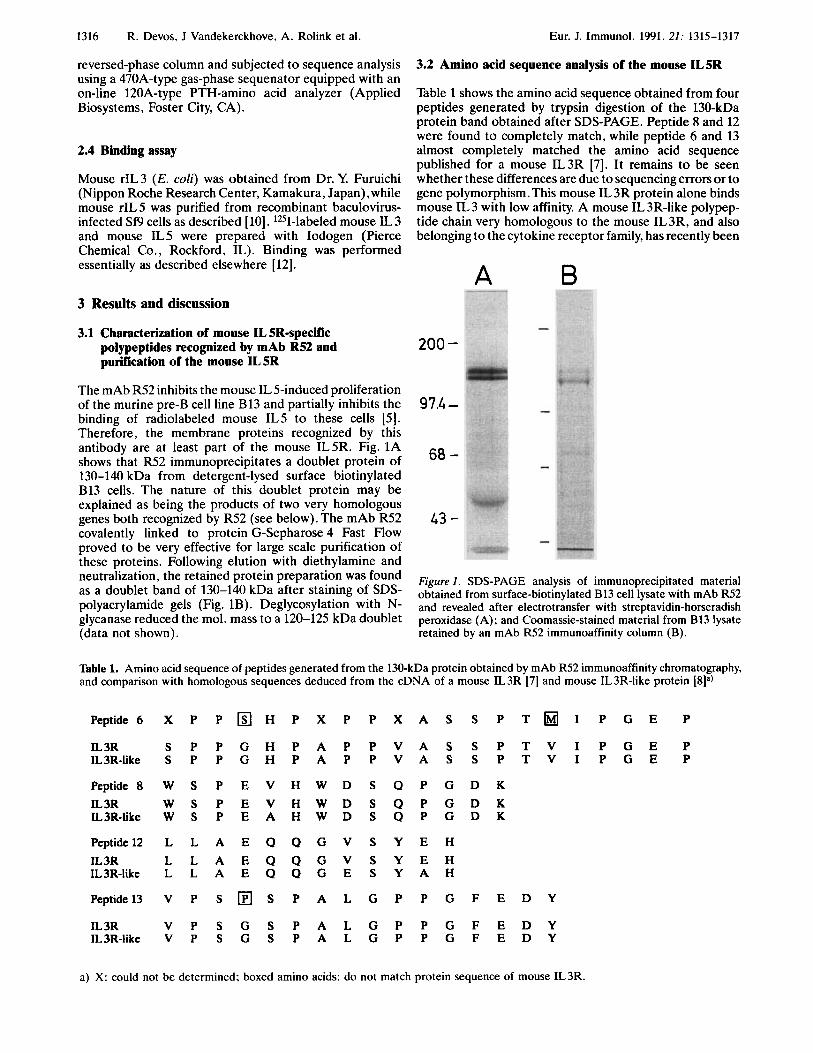
The mAb R52 inhibits the mouse IL 5-induced proliferation of the murine pre-B cell line B13 and partially inhibits the binding of radiolabeled mouse IL5 to these cells [ 5 ] . Therefore, the membrane proteins recognized by this antibody are at least part of the mouse IL5R. Fig. 1A shows that R52 immunoprecipitates a doublet protein of 130-140 kDa from detergent-lysed surface biotinylated B13 cells. The nature of this doublet protein may be explained as being the products of two very homologous genes both recognized by R52 (see below). The mAb R52 covalently linked to protein G-Sepharose 4 Fast Flow proved to be very effective for large scale purification of these proteins. Following elution with diethylamine and neutralization, the retained protein preparation was found as a doublet band of 130-140 kDa after staining of SDS- polyacrylamide gels (Fig. 1B). Deglycosylation with N- glycanase reduced the mol. mass to a 120-125 kDa doublet (data not shown).
97.4 -
68 -
43 -
Figure I. SDS-PAGE analysis of immunoprecipitated material obtained from surface-biotinylated B13 cell lysate with mAb R52 and revealed after electrotransfer with streptavidin-horseradish peroxidase (A); and Coomassie-stained material from B13 lysate retained by an mAb R52 immunoaffinity column (B).
Table 1. Amino acid sequence of peptides generated from the 130-kDa protein obtained by mAb R52 immunoaffinity chromatography, and comparison with homologous sequences deduced from the cDNA of a mouse IL3R [7] and mouse IL3R-like protein [8Ia)
Peptide6 X P P O H P X P P X A S S P T H I P G E P
IL 3R S P P G H P A P P V A S S P T V I P G E P IL3R-like S P P G H P A P P V A S S P T V I P G E P
Peptide8 W S P E V H W D S Q P G D K IL 3R W S P E V H W D S Q P G D K IL3R-like W S P E A H W D S Q P G D K
Peptide12 L L A E Q Q G V S Y E H IL 3R L L A E Q Q G V S Y E H IL3R-like L L A E Q Q G E S Y A H
Peptide13 V P S S P A L G P P G F E D Y
IL 3R V P S G S P A L G P P G F E D Y IL3R-like V P S G S P A L G P P G F E D Y
a) X: could not be determined: boxed amino acids: do not match protein sequence of mouse IL3R.

Eur. J. Immunol. 1991. 21: 1315-1317 Characterization of the mouse interleukin 5 receptor 1317
IL 5 also use two very homologous gene products as part of their receptor. All mouse IL5-dependent pre-B cell lines described sofar can also respond to mouse IL 3 [ 13, 141 .The establishment of mouse IL 5-dependent cells from mouse IL 3-dependent progenitors seem to require a BM stromal cell-dependent stage. Studies to evaluate how this cell contact regulates the expression of both lymphokine recep- tors will be important to determine the role of IL3 and IL5 in both myelopoiesis and B cell development.
Table 2. Binding of radiolabeled mouse IL 5 and radiolabeled mouse IL3 to B13 cells cannot be competed by each othera)
lzI-labeled ligand Competitor cpm
IL 5
IL 3
- 6292 IL5 458 IL 3 5 575 - n 726
n.5 28 733 IL3 1 107
Cells (3 X 1s) and 3.5 n M of mouse lZ5I-IL5 or mouse 1251-IL3 were incubated for 1 h at room temperature in 0.3 ml binding medium with 0.3 p~ of competitor as indicated.The fraction of label bound to the cells was determined as described [12].
described [8].This protein does not bind mouse IL3 and is 18 amino acids longer at its carboxyl terminus. Both the mouse IL3R and the mouse IL3R-like protein are recog- nized by an mAb anti-Aic2, which was used for screening an expression cDNA library [7,8]. As the mAb R52 inhibits both the mouse IL 3-induced and the mouse IL 5-induced proliferation of B13 cells [ 5 ] , it is conceivable that R52 also recognizes both these proteins, and therefore that the 140-kDa protein in the R52 affinity-purified material would correspond to the mouse IL3R-like chain (Van der Heyden et al., in preparation).The ligand for this cytokine receptor in unknown. Our results, however, suggest that this protein might be part of the mouse IL5R complex and has to associate with a 60-kDa component in order to generate a high-affinity mouse IL5R. Both the mouse IL3R and the mouse IL3R-like protein are present on mouse IL3- dependent cell lines [8]. These cell lines however do not bind mouse IL5, and the 60-kDa mouse IL5R protein can only be found on cell lines which also respond to mouse IL 5 (data not shown). Our finding indicates that the mouse IL 3R and/or the mouse IL 3R-like protein are components of the mouse IL5R. However, binding of radiolabeled mouse IL3 or radiolabeled mouse IL5 on B13 cells is specific and cannot be competed by each other (Table 2). This suggests that the specificity of the mouse IL 5R and the mouse IL3R would be mainly determined by a second ligand-specific protein.
Most probably the mouse IL3R gene and the mouse IL3R-like gene arose by a recent gene duplication. It will be interesting to find out whether human IL3 and human
We thank Ina Facht?, Tania Tuypens, Rita Bauden and Annick Verhee for their technical assistance. We are grateful to Dr. Yasuhiro Furuichi for the generous gift of recombinant mouse IL3.
Received December 3,1990; in revised form January 23, 1991.
4 References
1 Takatsu, K.,Tominaga, A., Harada, N., Mita, S., Matsumoto, M., Takahashi, T., Kikuchi,Y. and Yamaguchi, N., Immunol. Rev. 1988. 102: 107.
2 Sanderson, C. J., lnt. J. Cell Cloning 1990. 8: 147 (Suppl. 1). 3 Mita, S., Harada, N., Naomi, S., Hitoshi,Y, Sakamoto, K.,
Akagi, M.,Tominaga, A. and Takatsu, K., J. Exp. Med. 1988. 168: 863.
4 Mita, S., Tominaga, A., Hitoshi,Y., Sakamoto, K., Honjo, T., Akagi, M., Kikuchi,Y.,Yamaguchi, N. and Takatsu, K., Proc. Natl. Acad. Sci. USA 1989. 86: 2311.
5 Rolink, A. G., Melchers, E and Palacios, R., J. Exp. Med. 1989. 169: 1693.
6 Hitoshi, Y.,Yamaguchi, N., Mita, S., Sonoda, E., Takaki, S., Tominaga, A. and Takatsu, K., J. Immunol. 1990. 144: 4218.
7 Itoh, N.,Yonehara, S., Schreurs, J., Gorman, D. M., Maruya- ma, K., Ishii, A.,Yahara, I., Arai, K. and Miyajima, A., Science 1990. 247: 324.
8 Gorman, D. M., Itoh, N., Kitamura,T., Schreurs, J.,Yonehara, S. ,Yahara, I., Arai, K. and Miyajima, A., Proc. Natl. Acad. Sci. USA 1990.87: 5459.
9 Cole, S. R., Ashman, L. K. andEy, F! L., Mol. Immunol. 1987. 24: 699.
10 Tavernier, J., Devos, R.,Van der Heyden, J., Hauquier, G., Bauden, R., FachC, I., Kawashima, E.,Vandekerckhove, J., Contreras, R. and Fiers,W., DNA 1989. 8: 491.
11 Schneider, C., Newman, R. A., Sutherland, D. R., Asser, U. and Greaves, M. E, J. Biol. Chem. 1982. 257: 10766.
12 Plaetinck, G., Van der Heyden, J., Tavernier, J., Fache, I . , Tuypens,T., Fischkoff, S., Fiers,W. and Devos, R., J. Exp. Med. 1990. 172: 683.
13 Tominaga, A., Mita, S., Kikuchi,Y., Hitoshi,Y. and Takatsu, K., Growth Factors 1989. I : 135.
14 Tohyama, K., Lee, K. H.,Tashiro, K., Kinashi,T. and Honjo, T., EMBO J. 1990. 9: 1823.


![Automation of [ F]fluoroacetaldehyde synthesis ... · Automation of [18F]fluoroacetaldehydesynthesis:applicationtoarecombinanthuman interleukin-1 receptor antagonist (rhIL-1RA)†](https://img.pdfslide.net/doc/110x75/6011fd57f1072037ad7b66b8/automation-of-ffluoroacetaldehyde-synthesis-automation-of-18ffluoroacetaldehydesynthesisapplicationtoarecombinanthuman.jpg)