Embed Size (px)
Citation preview
Ammonium-Induced Impairment of Axonal Growth Is Preventedthrough Glial Creatine
Olivier Braissant,1* Hugues Henry,1* Anne-Marie Villard,1 Marie-Gabrielle Zurich,2 Marc Loup,1Barbara Eilers,1 Gianni Parlascino,1 Edouard Matter,1 Olivier Boulat,1 Paul Honegger,2 andClaude Bachmann1
1Clinical Chemistry Laboratory, University Hospital, CH-1011 Lausanne, Switzerland, and 2Institute of Physiology,University of Lausanne, CH-1005 Lausanne, Switzerland
Hyperammonemia in neonates and infants affects brain devel-opment and causes mental retardation. We report that ammo-nium impaired cholinergic axonal growth and altered localizationand phosphorylation of intermediate neurofilament protein in ratreaggregated brain cell primary cultures. This effect was restrictedto the phase of early maturation but did not occur after synapto-genesis. Exposure to NH4Cl decreased intracellular creatine,phosphocreatine, and ADP. We demonstrate that creatine cotreat-
ment protected axons from ammonium toxic effects, although thisdid not restore high-energy phosphates. The protection by crea-tine was glial cell-dependent. Our findings suggest that the meansto efficiently sustain CNS creatine concentration in hyperammone-mic neonates and infants should be assessed to prevent impair-ment of axonogenesis and irreversible brain damage.
Key words: hyperammonemia; axon; creatine; glia; neurofila-ment; phosphorylation
Liver failure and genetic defects affecting the urea cycle lead tohyperammonemia with reversible and irreversible neurologicaldamage that might be life-threatening (Podolsky and Isselbacher,1998; Brusilow and Horwich, 2001). Symptoms of irreversibledamage include cognitive impairment, seizures, and cerebralpalsy (Flint Beal and Martin, 1998). They mainly occur in cases ofprolonged hyperammonemic crises, or when blood ammoniumreaches levels between 180 and 500 �M, or both during the first 2years of life (Msall et al., 1984; Uchino et al., 1998; Bachmann,2002, 2003). In the few reported brain autopsies of pediatricpatients, Alzheimer’s type II astrocytes, perivascular spongiosis,and cystic necrosis at the junction of cortical gray and whitematter have been observed (Harding et al., 1984; Filloux et al.,1986; Dolman et al., 1988). The mechanisms leading to irrevers-ible alterations are not understood.
We have shown previously that mimicking hyperammonemiaby adding ammonium in a rat reaggregating brain cell culturemodel causes a reduction in aggregate size, which could be causedby a deficiency or developmental delay of cellular processes inregions where axons are prevalent (Braissant et al., 1999b). Thereis substantial evidence that creatine (Cr) and the Cr/phosphocre-atine (PCr)/creatine kinase (CK) system are involved in neuronalgrowth cone activity and axonal elongation (Wang et al., 1998). Inaddition, the CNS is the main affected target in infants with acreatine deficiency syndrome attributable to guanidinoacetatemethyltransferase, arginine:glycine amidinotransferase, or Crtransporter deficiencies. Such patients exhibit delayed psychomo-tor development or bilateral myelination delay (Stockler et al.,
1994; Schulze et al., 1997; Item et al., 2001; Salomons et al., 2001).We and others have also shown that the synthesis of arginine, themain precursor of creatine, is altered by hyperammonemia (Raoet al., 1995; Braissant et al., 1999b). Together, these findingssuggest that hyperammonemia impairs Cr metabolism, transportin the CNS, or both. Cr is used by the CNS Cr/PCr/CK system tobuffer and supply ATP, particularly to neurons (Hemmer andWallimann, 1993). CNS, at least in the rat, seems dependent onits own Cr synthesis (Braissant et al., 2001).
The aim of the present work was to test whether axons wereaffected by hyperammonemia and whether this could be causedby a Cr loss or decrease in brain cells. We used reaggregated braincell cultures prepared from the telencephalon of rat embryos,which provide a three-dimensional network of neurons and glialcells that progressively acquire a tissue-specific pattern resem-bling that of the brain (Honegger et al., 1979; Honegger andMonnet-Tschudi, 2001). We analyzed the axonal expression ofthe intermediate neurofilament protein (NF-M, 160 kDa) anddetermined the intracellular levels of Cr, PCr, ATP, ADP, andAMP under NH4Cl exposure. We identified the neurons im-paired in axonal growth by ammonium exposure. Finally, wetested the protective effect of Cr on the growth of axons exposedto NH4Cl, and the influence of the brain cell developmental stageon the axonal vulnerability to ammonium.
MATERIALS AND METHODSReaggregated brain cell cultures. Reaggregated brain cell primary cultureswere prepared from mechanically dissociated telencephalon of 16 d ratembryos as described previously (Honegger and Monnet-Tschudi, 2001).Cultures were grown in serum-free, chemically defined medium consist-ing of DMEM with high glucose (25 mM) supplemented with insulin (0.8�M), triiodothyronine (30 nM), hydrocortisone-21-phosphate (20 nM),transferrin (1 �g/ml), biotin (4 �M), vitamin B12 (1 �M), linoleate (10�M), lipoic acid (1 �M), L-carnitine (10 �M), and trace elements (Honeg-ger and Monnet-Tschudi, 2001). Gentamycin sulfate (25 �g/ml) was usedas an antibiotic. The cultures were maintained under constant gyratoryagitation (80 rpm) at 37°C and in an atmosphere of 10% CO2 and 90%humidified air.
Received June 19, 2002; revised Aug. 15, 2002; accepted Aug. 27, 2002.This work was supported by Swiss National Science Foundation Grant 31-
63892.00. We thank Dr. Marianna Giarre for critical reading of this manuscript.*O.B. and H.H. contributed equally to this work.Correspondence should be addressed to Olivier Braissant, Clinical Chemistry
Laboratory, University Hospital, CH-1011 Lausanne, Switzerland. E-mail:[email protected] © 2002 Society for Neuroscience 0270-6474/02/229810-11$15.00/0
The Journal of Neuroscience, November 15, 2002, 22(22):9810–9820
Two different kinds of aggregate cultures were grown: (1) regularmixed cell aggregates, developing with neurons and glial cells; and (2)neuron-enriched aggregates, which were obtained by the treatment of theregular mixed cell cultures at days 1 and 2 with cytosine arabinoside (0.4�M) to eliminate the proliferating glioblasts (Honegger and Monnet-Tschudi, 2001). Previous work has shown that the neuron-enrichedcultures contain �90% neurons, �8% astrocytes and very few, if any,oligodendrocytes (Honegger and Pardo, 1999). Neuron-enriched culturesreceived conditioned medium taken from mixed cell (neuron–glia) sistercultures and diluted 1:1 with fresh medium.
Aggregates were grown for 13 or 28 d. Aggregates harvested at 13 dwere treated from day 5 onward with NH4Cl, Cr, or both, with mediumreplenishment at days 8 and 11 (5 ml replaced of 8 ml total). Aggregatesharvested at 28 d were treated from day 20 onward with NH4Cl, Cr, orboth, with medium replenishment at days 22, 24, and 26 (5 ml replacedof 8 ml total). On the day of harvest, culture media were recovered,immediately centrifuged to remove cell debris, and frozen in liquidnitrogen. Aggregate pellets were washed three times with ice-cold PBSand embedded for histology or frozen in liquid nitrogen. Until analysis,culture media and aggregate pellets were kept at �80°C.
Histology and immunohistochemistry. Aggregates were embedded intissue-freezing medium (Jung, Nussloch, Germany), frozen in liquidnitrogen-cooled isopentane, and kept at �80°C until used. For histolog-ical staining, 16-�m-thick cryosections were prepared, postfixed 1 hr in4% paraformaldehyde (PFA) in PBS, and stained by hematoxylin col-oration or Bielschowsky’s silver impregnation (Cox, 1977). NN-18(Chemicon International, Temecula, CA; Shaw et al., 1986; Harris et al.,1991) and RMO-44 (Zymed Laboratories, San Francisco, CA) (Lee etal., 1987) monoclonal antibodies were used for immunohistochemistryagainst NF-M. Axonal growth cones were detected with a monoclonalantibody directed against growth cone-associated protein 43 (GAP43;Chemicon International). Astrocytes and oligodendrocytes were labeledusing monoclonal antibodies directed against glial fibrillary acidic pro-tein (GFAP; Chemicon International) and myelin basic protein (MBP;Boehringer Ingelheim). Cholinergic, GABAergic, and glutamatergicneurons were characterized by using monoclonal antibodies against cho-line acetyltransferase (ChAT; Oncogene, Cambridge, MA), glutamicacid decarboxylase (GAD, 65/67 kDa; Santa Cruz Biotechnology, SantaCruz, CA), and neuronal excitatory amino acid transporter 3 (EAAT3;Santa Cruz Biotechnology), respectively (Furuta et al., 1997; Pardo andHonegger, 1999; He et al., 2000). For immunohistochemistry againstNF-M, GAP43, GFAP, GAD, and EAAT3, cryosections (16 �M) werefixed for 1 hr in 4% PFA and PBS at room temperature, washed in PBS(three times for 5 min), and permeabilized for 5 min in 0.1% sodiumcitrate and 0.1% Triton X-100. For immunohistochemistry againstChAT, cryosections (16 �M) were fixed for 1 min in 5% acetic acid and95% ethanol at 4°C and washed in PBS (three times for 5 min). Forimmunohistochemistry against MBP, cryosections (16 �M) were fixed for1 hr in 4% PFA and PBS at room temperature, washed in PBS (threetimes for 5 min), dehydrated with increasing concentrations of ethanol(EtOH), delipidated in xylene, rehydrated with decreasing concentra-tions of EtOH, and finally washed in PBS. Sections were then processedfor immunohistochemistry using the Histostain-Plus kit according to themanufacturer’s protocol (Zymed). The primary antibody was diluted in1% bovine serum albumin in PBS and applied to sections. After washing,sections were incubated with a biotinylated anti-mouse IgG secondaryantibody followed by a streptavidin-peroxidase conjugate. Peroxidasestaining was performed for 10 min using aminoethyl carbazole and H2O2and stopped in distilled water. Sections were mounted under glycerol andobserved and photographed on an Olympus BX50 microscope equippedwith a DP-10 digital camera (Olympus Optical, Tokyo, Japan).
Protein dephosphorylation on cryosections. Cryosections (16 �m thick)were digested for 30 min (37°C) with alkaline phosphatase (200 �g/ml;Roche Molecular Biochemicals, Mannheim, Germany), in (in mM): 100Tris, 100 NaCl, and 50 MgCl2, pH 9.5. Sections were washed in PBS,fixed for 30 min in 4% PFA and PBS, and washed three times for 5 minin PBS. Immunohistochemistry against NF-M was then performed asdescribed above.
Quantitative Western blot analysis. Mixed cell and neuron-enrichedaggregates were homogenized in 10 mM Tris-HCl, pH 7.5, containing 4 Murea, 0.1% SDS, and protease inhibitors (Complete; Roche). Homoge-nates were centrifuged at 16,000 � g for 10 min, and supernatants wererecovered. Supernatant proteins were measured by the bicinchoninic acidassay (Pierce, Rockford, IL) and diluted at a final concentration of 1�g/�l in Laemmli sample buffer (Laemmli, 1970). Proteins were sepa-
rated by SDS-PAGE (9% total acrylamide). After transfer of the proteinsto polyvinylidene difluoride membranes (Immobilon; Millipore, Bed-ford, MA), blots were probed with NN-18 and RMO-44 anti-NF-M,anti-GAP43, and anti-GFAP monoclonal antibodies. Western blots wererevealed by chemiluminescence (ECL; Amersham Biosciences, Bucking-hamshire, UK). The radiographs (X-OMAT AR; Eastman Kodak, Roch-ester, NY) were scanned with an ImageScanner (Amersham Bio-sciences) and processed by image analysis (ImageMaster 1D; AmershamBiosciences).
Measure of intracellular Cr, PCr, ATP, ADP, and AMP. Frozen aggre-gate pellets were suspended in ice-cold 25 mM phosphate buffer, pH 7.5containing (in mM): 1 iodoacetate and 0.1 diadenosine pentaphosphate.Cells were extracted with 0.4 M perchloric acid and neutralized with 2 MK2CO3 followed by a centrifugation at 16,000 � g for 5 min. Superna-tants were assayed for adenine nucleotides (ATP, ADP, and AMP) andcreatine compounds (Cr and PCr) according to the isocratic reversedphase HPLC method of Seidl et al. (2000) using HPLC (Agilent) coupledwith a dual-absorbance detector. The absorbance at 214 nm (for Cr andPCr) and that at 254 nm (for ATP, ADP, and AMP) were monitoredsimultaneously. The acid precipitates of the aggregates were solubilizedin 0.1 M NaOH and 1% SDS, and their protein content was determinedby the bicinchoninic acid assay (Pierce). The concentrations of Cr, PCr,ATP, ADP, and AMP were expressed as nanomoles per milligram ofprotein.
Measure of NH4�, glucose, and lactate in culture media. Ammonium was
measured on a Cobas FARA II automate (Roche), using a UV enzy-matic ammonium kit (61025; Biomerieux). Glucose and lactate weremeasured on a Hitachi 917 automate (Roche), using the Glucose Ecoline100 kit (1.14891.0001; Merck, Darmstadt, Germany) and a lactate kit (1822 837; Roche).
RESULTSNet uptake of NH4
� and glucose and net release of lactate bymixed cell aggregate cultures exposed to NH4Cl are shown inTable 1. During the whole period of NH4Cl exposure, NH4
�
uptake by cultures was dose-dependent. Glucose uptake andlactate release showed highly significant increases at the maximaldose of 5 mM NH4Cl. The extent of metabolic changes observedwith increasing NH4Cl concentrations paralleled morphologicalchanges in the aggregates, which were absent or barely detectable
Table 1. NH4Cl dose-dependent uptake and release of ammonium,glucose, and lactate by mixed cell aggregate cultures
NH4Cl(mM)
Days 5–8 Days 8–11 Days 11–13
p p p
Ammonium
0 �26 � 1 �4 � 1 �2 � 1
1.0 �55 � 13 �0.001 �31 � 4 �0.001 �38 � 1 �0.001
2.5 �113 � 19 �0.001 �96 � 2 �0.001 �114 � 1 �0.001
5.0 �176 � 16 �0.001 �250 � 2 �0.001 �264 � 10 �0.001
Glucose
0 �399 � 19 �464 � 12 �756 � 4
1.0 �372 � 32 NS �389 � 22 �0.01 �656 � 48 �0.05
2.5 �525 � 32 �0.01 �468 � 7 NS �835 � 17 �0.01
5.0 �672 � 21 �0.001 �623 � 19 �0.001 �1026 � 7 �0.001
Lactate
0 137 � 5 166 � 6 685 � 26
1.0 152 � 18 NS 149 � 39 NS 549 � 21 �0.01
2.5 269 � 22 �0.001 153 � 22 NS 696 � 73 NS
5.0 431 � 34 �0.001 370 � 41 �0.001 1137 � 22 �0.001
Net uptake (net decrease in culture medium) and release (net increase in culturemedium) by aggregates, calculated from days 5–8, 8–11, and 11–13 are expressed asnanomoles per hour per milligram of protein. Measures were taken at days 5 (startof treatment), 8 (before and after medium change), 11 (before and after mediumchange), and 13 (end of treatment). Proteins were measured at day 13. Data aremeans � SD of three separate cultures. Statistical p values are shown (Student’s ttest) for comparison with controls (NH4Cl � 0 mM); NS, not significant.
Braissant et al. • Impaired Axonal Growth by Ammonium Prevented by Creatine J. Neurosci., November 15, 2002, 22(22):9810–9820 9811
at 1 and 2.5 mM NH4Cl (data not shown) but evident at 5 mM (seebelow). Therefore, morphological and biochemical effects of hy-perammonemia were further analyzed by treating aggregate cul-tures with 5 mM NH4Cl.
NH4Cl exposure impaired axonal growth in developingmixed cell aggregatesControl mixed cell aggregate cultures at day 13 presented acharacteristic distribution of cells, including a peripheral zonewith a low density of cell bodies (Fig. 1A,B, asterisk) in whichfibers were prevalent (Fig. 1C). These fibers were NF-M-positive,using the monoclonal anti-NF-M NN-18 antibody, which did notstain neuronal soma (Fig. 1D). Aggregates exposed to 5 mM
NH4Cl from days 5 to 13 showed more densely packed cell bodiesat their periphery (Fig. 1K,L), and the almost complete absenceof NF-M-positive fibers (Fig. 1M,N). Another monoclonal anti-NF-M antibody, RMO-44, localized NF-M in neuronal cell bod-ies but not in fibers (Fig. 1E). The somatic expression of NF-Mwas not altered by 5 mM NH4Cl exposure (Fig. 1O). Western blotanalysis of NF-M showed that NH4Cl exposure caused a drasticdecrease of NF-M (threefold using NN-18, Fig. 1U; and twofoldusing RMO-44, Fig. 1V).
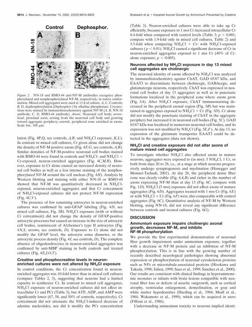
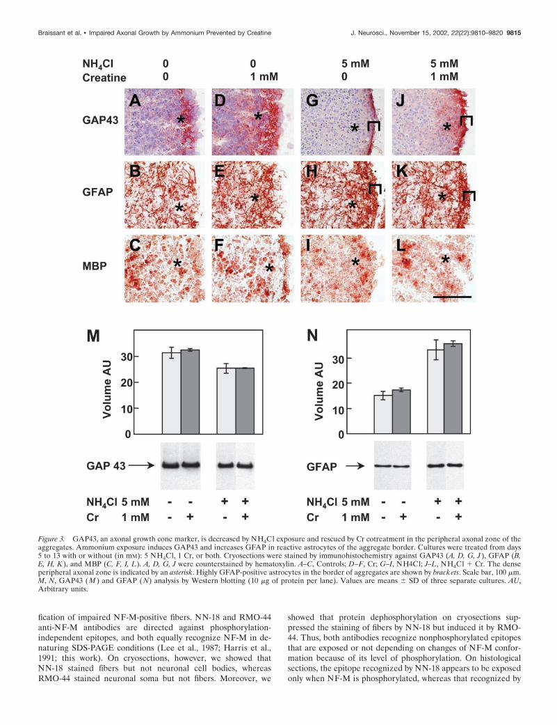
The specificity of NN-18 and RMO-44 antibodies for NF-Mrecognition was further characterized by dephosphorylation ofproteins in situ using alkaline phosphatase. Dephosphorylationabolished most of the NN-18 staining in fibers of the aggregateperiphery (Fig. 2A,B) and induced RMO-44 NF-M immunore-activity in distal fibers located at the aggregate periphery and inproximal fibers connecting neuronal cell bodies to the aggregateperiphery (Fig. 2C,D). This indicates that NN-18 predominantlyrecognized phosphorylated NF-M, whereas RMO-44 predomi-nantly recognized nonphosphorylated NF-M. Thus, the absenceof NN-18 anti-NF-M staining after NH4Cl exposure (Fig. 1N)suggests that ammonium exposure inhibited NF-M phosphoryla-tion. Fibers located at the aggregate periphery and containingphosphorylated NF-M were most probably axons, as suggested byanalyzing the expression of GAP43, an axonal growth conemarker. In control day 13 mixed cell aggregates, GAP43 washighly expressed in the same peripheral zone, exhibiting strongNF-M staining by NN-18 (Fig. 3A). As for NF-M, the GAP43staining was lost in this region after NH4Cl exposure (Fig. 3G).Interestingly, a strongly GAP43-immunoreactive zone appearedat the border of the aggregates (Fig. 3G, bracket). Westernblotting analysis of GAP43 after NH4Cl exposure showed a smalldecrease of the GAP43 signal (�20%) (Fig. 3M).
In day 13 mixed cell aggregates, glial cells were identified byimmunohistochemical staining for GFAP (specific for astrocytes)(Fig. 3B) and MBP (specific for oligodendrocytes) (Fig. 3C).NH4Cl exposure increased the number of GFAP-positive astro-cytic processes, particularly at the aggregate border (Fig. 3H,bracket), whereas no significant effect was found for oligodendro-cytes (Fig. 3I). Western blotting analysis of GFAP after NH4Clexposure showed a 2.2-fold increase of the GFAP signal (Fig. 3N).
NH4Cl exposure decreased intracellular Cr, PCr, andADP in developing mixed cell aggregatesTo test whether energy-rich phosphates, the Cr/PCr/CK system,or both were involved in ammonium-induced axonal growth im-pairment, we measured intracellular levels of Cr, PCr, ATP,ADP, and AMP in mixed cell aggregates (Table 2). NH4Clexposure reduced Cr, PCr, and ADP significantly (83, 75, and69% of controls, respectively), whereas no significant effect wasobserved for ATP and AMP content.
Creatine prevented NH4Cl-induced axonal growthimpairment in developing mixed cell aggregatesBecause Cr and PCr were decreased in NH4Cl-exposed aggre-gates, we examined whether Cr had a protective effect on axonalgrowth under NH4Cl exposure. By immunohistochemistry withthe NN-18 antibody, we found indeed that 1 mM Cr added toNH4Cl-exposed aggregates protected the peripheral axons, theirexpression of NF-M, and NF-M phosphorylation (Fig. 1P–S, seecontrols, A–D, NH4Cl exposure, K–N), whereas Cr given aloneincreased the density of NF-M-positive peripheral axons (Fig.1F–I). Immunohistochemistry with RMO-44 did not reveal anydifference in NF-M expression between aggregates treated withCr and controls (Fig. 1J,E) or between NH4Cl � Cr- and NH4Cl-exposed aggregates (Fig. 1O,T). Analysis of NF-M by Westernblotting showed that Cr cotreatment maintained NF-M at controllevels in mixed cell aggregates exposed to NH4Cl (NN-18) (Fig.1U) or showed partial protection (RMO-44, �25% comparedwith controls, �50% compared with NH4Cl exposure) (Fig. 1V),whereas Cr exposure alone did not affect the NF-M level com-pared with controls (Fig. 1U,V). A higher concentration of Cr (25mM) was also tested, which did not improve the protection ofaxonal growth under ammonium exposure compared with thatobtained with 1 mM Cr (data not shown).
GAP43 expression was partially protected in the peripheralNF-M-positive region of NH4Cl-exposed aggregates cotreatedwith Cr (Fig. 3J, asterisk, see control, A, NH4Cl exposure, G).However, the border of the aggregates was strongly positive forGAP43, as after exposure to NH4Cl alone (Fig. 3J,G, bracket).Compared with controls, Cr given alone increased the GAP43signal in the center of the aggregates (Fig. 3A,D). By Western blotanalysis, no difference was observed for GAP43 expression be-tween Cr-exposed cultures and controls or between NH4Cl � Cr-and NH4Cl-exposed cultures (Fig. 3M). Astrocytes and oligoden-drocytes presented similar patterns of expression for GFAP andMBP in cultures treated with Cr and in controls (Fig. 3, B,E,GFAP, C,F, MBP) as well as in NH4Cl � Cr- and NH4Cl-treatedaggregates (Fig. 3, H,K, GFAP, I,L, MBP).
No restoration of or increase in intracellular PCr, ATP, orADP could be observed in mixed cell aggregates cotreated with(in mM): 5 NH4Cl and 1 Cr compared with cultures exposed toNH4Cl only (Table 2), whereas intracellular Cr was increasedsignificantly (170% of controls). Cr was efficiently taken up bymixed cell aggregates exposed to 1 mM Cr only (190% of controls;p � 0.001); however, their PCr, ATP, and ADP content was notmodified. NH4Cl induced a significant decrease of intracellularCr in aggregates exposed to 1 mM Cr (75% of Cr-alone exposure;p � 0.005).
Creatine did not prevent the NH4Cl-induced axonalgrowth impairment in developing neuron-enrichedaggregatesUntreated neuron-enriched aggregate cultures also developed aperipheral zone with a low density of cell bodies but devoid of theglial cell lining found at the border of mixed cell aggregates (Figs.4A vs 1B; also compare staining for GFAP, Figs. 4D vs 3B, andMBP, Figs. 4E vs 3C). As in mixed cell cultures, the number ofaxons that developed in the aggregate periphery of day 13neuron-enriched aggregates (Fig. 4B) was decreased underNH4Cl exposure, as was NF-M phosphorylation (Fig. 4K,L). Incontrast to mixed cell cultures, however, Cr cotreatment ofneuron-enriched cultures did not prevent the NH4Cl-inducedaxonal growth impairment or the decrease of NF-M phosphory-
9812 J. Neurosci., November 15, 2002, 22(22):9810–9820 Braissant et al. • Impaired Axonal Growth by Ammonium Prevented by Creatine
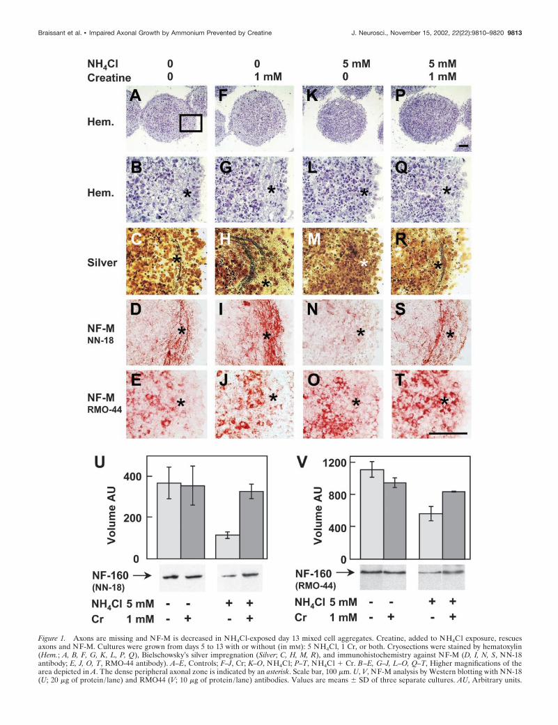
Figure 1. Axons are missing and NF-M is decreased in NH4Cl-exposed day 13 mixed cell aggregates. Creatine, added to NH4Cl exposure, rescuesaxons and NF-M. Cultures were grown from days 5 to 13 with or without (in mM): 5 NH4Cl, 1 Cr, or both. Cryosections were stained by hematoxylin(Hem.; A, B, F, G, K, L, P, Q), Bielschowsky’s silver impregnation (Silver; C, H, M, R), and immunohistochemistry against NF-M (D, I, N, S, NN-18antibody; E, J, O, T, RMO-44 antibody). A–E, Controls; F–J, Cr; K–O, NH4Cl; P–T, NH4Cl � Cr. B–E, G–J, L–O, Q–T, Higher magnifications of thearea depicted in A. The dense peripheral axonal zone is indicated by an asterisk. Scale bar, 100 �m. U, V, NF-M analysis by Western blotting with NN-18(U; 20 �g of protein/ lane) and RMO44 (V; 10 �g of protein/ lane) antibodies. Values are means � SD of three separate cultures. AU, Arbitrary units.
Braissant et al. • Impaired Axonal Growth by Ammonium Prevented by Creatine J. Neurosci., November 15, 2002, 22(22):9810–9820 9813
lation (Fig. 4P,Q, see controls, A,B, and NH4Cl exposure, K,L).In contrast to mixed cell cultures, Cr given alone did not changethe density of NF-M positive axons (Fig. 4F,G, see controls, A,B).Similar densities of NF-M-positive neuronal cell bodies stainedwith RMO-44 were found in controls and NH4Cl- and NH4Cl �Cr-exposed, neuron-enriched aggregates (Fig. 4C,M,R). How-ever, exposure to Cr alone caused an increased density of neuro-nal cell bodies as well as a less intense staining of the nonphos-phorylated NF-M around the cell nucleus (Fig. 4H). Analysis byWestern blotting and staining with either NN-18 or RMO-44showed that NF-M was quantitatively decreased in NH4Cl-exposed, neuron-enriched aggregates and that Cr cotreatmentof NH4Cl-exposed cultures did not protect NF-M expression(Fig. 4U,V).
The presence of few remaining astrocytes in neuron-enrichedcultures was confirmed by anti-GFAP labeling (Fig. 4D; seemixed cell cultures, Fig. 3B). NH4Cl exposure (with or withoutCr cotreatment) did not change the density of GFAP-positiveastrocytic processes but caused an increase in the size of astrocytecell bodies, reminiscent of Alzheimer’s type II astrocytes (Fig.4N,S, arrows, see controls, D). Exposure to Cr alone did notmodify the GFAP level, the astrocyte soma diameter, or theastrocytic process density (Fig. 4 I, see controls, D). The completeabsence of oligodendrocytes in neuron-enriched aggregates wasconfirmed by anti-MBP staining in both controls and treatedcultures (Fig. 4E,J,O,T).
Creatine and phosphocreatine levels in neuron-enriched cultures were not altered by NH4Cl exposureIn control conditions, the Cr concentration found in neuron-enriched aggregates was 10-fold lower than in mixed cell cultures(compare Tables 2, 3), suggesting that neurons had a limitedcapacity to synthesize Cr. In contrast to mixed cell aggregates,NH4Cl exposure of neuron-enriched cultures did not affect in-tracellular Cr and PCr (Table 3), but ATP, ADP, and AMP weresignificantly lower (67, 58, and 50% of controls, respectively). Crcotreatment did not attenuate the NH4Cl-induced decrease ofadenine nucleotides, nor did it modify the PCr concentration
(Table 3). Neuron-enriched cultures were able to take up Crefficiently, because exposure to 1 mM Cr increased intracellular Cr8.4-fold when compared with control levels (Table 3; p � 0.001;compare with 1.9-fold only in mixed cell cultures, Table 2) and3.5-fold when comparing NH4Cl � Cr- with NH4Cl-exposedcultures ( p � 0.01). NH4Cl caused a significant decrease of Cr inneuron-enriched aggregates exposed to 1 mM Cr (40% of Cr-alone exposure; p � 0.005).
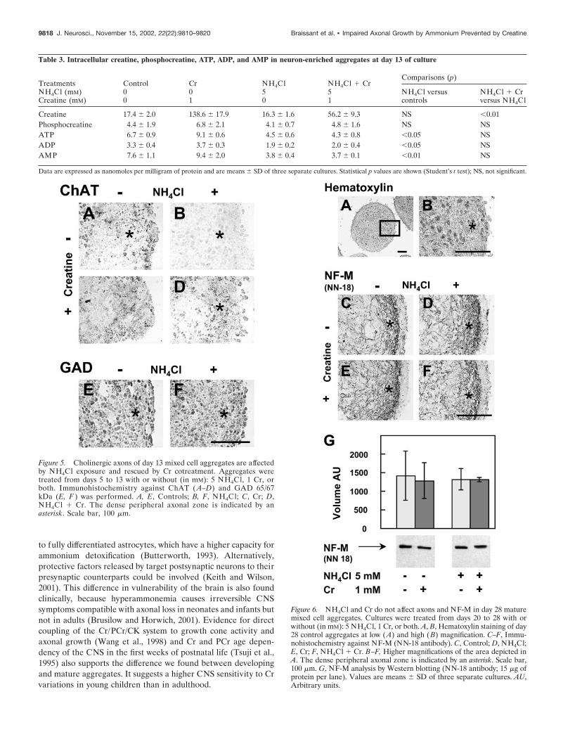
Neurons affected by NH4Cl exposure in day 13 mixedcell aggregates are cholinergicThe neuronal identity of axons affected by NH4Cl was analyzedby immunohistochemistry against ChAT, GAD 65/67 kDa, andEAAT3 to discriminate between cholinergic, GABAergic, andglutamatergic neurons, respectively. ChAT was expressed in neu-ronal cell bodies of day 13 aggregates as well as in punctuateformations localized in the peripheral zone where axons grow(Fig. 5A). After NH4Cl exposure, ChAT immunostaining de-creased in the peripheral axonal region (Fig. 5B) but was main-tained in aggregates exposed to NH4Cl � Cr (Fig. 5D). Cr alonedid not modify the punctuate staining of ChAT in the aggregateperiphery but increased it in neuronal cell bodies (Fig. 5C). GAD65/67 kDa was localized in numerous neuronal cell bodies, and itsexpression was not modified by NH4Cl (Fig. 5E,F). At day 13, noexpression of the glutamate transporter EAAT3 could be de-tected in the aggregates (data not shown).
NH4Cl and creatine exposure did not alter axons ofmature mixed cell aggregatesTo investigate whether NH4Cl also affected axons in matureneurons, aggregates were exposed to (in mM): 5 NH4Cl, 1 Cr, orboth from days 20 to 28, i.e., at a stage at which neurons progres-sively undergo synaptogenesis and myelination (Honegger andMonnet-Tschudi, 2001). At day 28, the peripheral dense fiberzone was clearly visible (Fig. 6A,B) and richer in the number ofaxons expressing NF-M than at day 13 (Fig. 6C; compare withFig. 1D). NH4Cl (5 mM) exposure did not affect axons of matureaggregates (Fig. 6D). Aggregates treated with 1 mM Cr (Fig. 6E)or with NH4Cl � Cr (Fig. 6F) were also comparable with controlaggregates (Fig. 6C). Quantitative analysis of NF-M by Westernblotting, using NN-18, did not reveal any significant differencebetween controls and treated cultures (Fig. 6G).
DISCUSSIONAmmonium exposure impairs cholinergic axonalgrowth, decreases NF-M, and inhibitsNF-M phosphorylationWe provide the first experimental demonstration of neuronalfiber growth impairment under ammonium exposure, togetherwith a decrease in NF-M protein and an inhibition of NF-Mphosphorylation. This is in line with the growing number ofrecently described neurological pathologies showing abnormalexpression or phosphorylation of neuronal cytoskeleton proteinssuch as NFs or microtubule-associated proteins (Hirokawa andTakeda, 1998; Julien, 1999; Saez et al., 1999; Sanchez et al., 2000).Our results are consistent with clinical findings in hyperammone-mic neonates or infants with brain lesions compatible with neu-ronal fiber loss or defects of neurite outgrowth, such as corticalatrophy, ventricular enlargement, demyelination, or gray andwhite matter hypodensities (Harding et al., 1984; Msall et al.,1984; Wakamoto et al., 1999), which can be acquired in utero(Filloux et al., 1986).
Understanding ammonium toxicity to neurons implied identi-
Figure 2. NN-18 and RMO-44 anti-NF-M antibodies recognize phos-phorylated and nonphosphorylated NF-M, respectively, in native confor-mation. Mixed cell aggregates were used at 13 d of culture. A, C, Controls;B, D, dephosphorylation (Dephosphor.) by alkaline phosphatase. Cryosec-tions were stained by immunohistochemistry against NF-M (A, B, NN-18antibody; C, D, RMO-44 antibody). Arrow, Neuronal cell body; arrow-head, proximal axon, arising from the neuronal cell body and growingtoward aggregate periphery; asterisk, peripheral zone enriched in axons.Scale bar, 100 �m.
9814 J. Neurosci., November 15, 2002, 22(22):9810–9820 Braissant et al. • Impaired Axonal Growth by Ammonium Prevented by Creatine
fication of impaired NF-M-positive fibers. NN-18 and RMO-44anti-NF-M antibodies are directed against phosphorylation-independent epitopes, and both equally recognize NF-M in de-naturing SDS-PAGE conditions (Lee et al., 1987; Harris et al.,1991; this work). On cryosections, however, we showed thatNN-18 stained fibers but not neuronal cell bodies, whereasRMO-44 stained neuronal soma but not fibers. Moreover, we
showed that protein dephosphorylation on cryosections sup-pressed the staining of fibers by NN-18 but induced it by RMO-44. Thus, both antibodies recognize nonphosphorylated epitopesthat are exposed or not depending on changes of NF-M confor-mation because of its level of phosphorylation. On histologicalsections, the epitope recognized by NN-18 appears to be exposedonly when NF-M is phosphorylated, whereas that recognized by
Figure 3. GAP43, an axonal growth cone marker, is decreased by NH4Cl exposure and rescued by Cr cotreatment in the peripheral axonal zone of theaggregates. Ammonium exposure induces GAP43 and increases GFAP in reactive astrocytes of the aggregate border. Cultures were treated from days5 to 13 with or without (in mM): 5 NH4Cl, 1 Cr, or both. Cryosections were stained by immunohistochemistry against GAP43 (A, D, G, J ), GFAP (B,E, H, K ), and MBP (C, F, I, L). A, D, G, J were counterstained by hematoxylin. A–C, Controls; D–F, Cr; G–I, NH4Cl; J–L, NH4Cl � Cr. The denseperipheral axonal zone is indicated by an asterisk. Highly GFAP-positive astrocytes in the border of aggregates are shown by brackets. Scale bar, 100 �m.M, N, GAP43 (M ) and GFAP ( N) analysis by Western blotting (10 �g of protein per lane). Values are means � SD of three separate cultures. AU,Arbitrary units.
Braissant et al. • Impaired Axonal Growth by Ammonium Prevented by Creatine J. Neurosci., November 15, 2002, 22(22):9810–9820 9815
RMO-44 seems exposed only when NF-M is nonphosphorylated.Accordingly, fibers at aggregate periphery preferentially containphosphorylated NF-M, whereas neuronal soma essentially con-tain nonphosphorylated NF-M. Our findings may explain whyNN-18 does not stain neuronal soma except in cases of hyper-phosphorylated NF-M accumulation (Harris et al., 1991; Nguyenet al., 2001) and why RMO-44 preferentially labels neuronal soma(Lee et al., 1987).
NFs are distributed in neuronal soma, dendrites, and axons.Nonphosphorylated NFs are predominantly found in the soma-todendritic compartment, whereas phosphorylated NFs are en-riched in axons (Rosenstein and Krum, 1996; Brown, 1998; Ulfiget al., 1998). Moreover, GAP43 and ChAT showed a localizationsimilar to that of NN-18-stained NF-M and reacted like it underexposure to NH4Cl, Cr, or both. GAP43 is found in growth conesof axons exclusively (Goslin et al., 1988), and ChAT is recognizedas a presynaptic axonal marker (Phelps et al., 1985). Together,these data suggest that NN-18 stained axons specifically.
Cholinergic, GABAergic, and glutamatergic neurons differen-tiate in the brain aggregate culture system (Pardo and Honegger,1999; Honegger and Monnet-Tschudi, 2001). Data presented heresuggest that early in development, axons impaired by NH4Clbelong to cholinergic neurons. This does not exclude the possi-bility that ammonium could also impair axonal growth of gluta-matergic and GABAergic neurons later in development, becauseglutamatergic neurotransmission and GAD activity are altered byhyperammonemia (Albrecht, 1998; Braissant et al., 1999b). Ourfindings are in line with data obtained in the spf mouse, a modelof hyperammonemia caused by ornithine transcarbamylase defi-ciency, which shows cholinergic neuronal loss in the cerebralcortex (Ratnakumari et al., 1994). Lack of elongation of cholin-ergic axons during cortical development may provide a basis forunderstanding some of the severe cognitive defects caused byhyperammonemia.
NH4Cl-induced inhibition of axonal growth depends onintracellular CrNH4Cl exposure of day 13 mixed cell aggregates impaired axonalgrowth and decreased intracellular levels of Cr, PCr, and ADP.Cr cotreatment under NH4Cl exposure protected axonal growthbut neither restored nor increased PCr, ATP, and ADP levels.This suggests that under NH4Cl exposure, the rescue of axonalgrowth by Cr does not depend on high-energy phosphates, whichremain lowered under NH4Cl � Cr exposure and are known todecrease in the hyperammonemic CNS (Ratnakumari et al.,1992). We cannot exclude, however, the possibility that concen-tration changes in specific cell types or in subcellular compart-ments (e.g., mitochondria) remain undetected. Intracellular Crwas decreased by 60% in neuron-enriched but only by 25% in
mixed cell aggregates exposed to NH4Cl � Cr compared with Crexposure, suggesting that NH4Cl exposure reduced the capacityto accumulate Cr in neurons preferentially.
Glial dependency of axonal protection by creatineunder NH4Cl exposureCr added to the culture medium was sufficient to protect axonalgrowth in NH4Cl-exposed mixed cell aggregates but not inneuron-enriched cultures, suggesting that the mechanism of axonprotection by Cr depends on glial cells, be it astrocytes or oligo-dendrocytes. In the case of an astrocyte-dependent mechanism,the very few astrocytes still present in neuron-enriched culturesmight suffice for supporting axonal growth in control conditionsbut not for protecting axons exposed to NH4Cl. The absence ofoligodendrocytes in neuron-enriched cultures might also suggestan oligodendrocyte-dependent mechanism. Because Cr is takenup by neuron-enriched cultures without protecting axons exposedto NH4Cl, it is unlikely that Cr per se is the sole glia-derivedaxonal growth-promoting factor. Our findings preferentially sup-port the hypothesis that a glial factor is needed, which is modifiedthrough Cr in glial cells (e.g., by the Cr/PCr/CK system), released,and used by neurons to promote axonal growth. This would stillallow axonal growth in control neuron-enriched cultures, becausethey are grown in a culture medium conditioned by mixed cellcultures (Honegger and Monnet-Tschudi, 2001). Because we haveshown that axons do not grow in neuron-enriched aggregatesexposed to ammonium, it also implies that NH4Cl has a directneuronal inhibitory effect on axonal growth that cannot be pre-vented in the absence of glial cells but is counteracted by Crcotreatment in mixed cell aggregates.
The glial mechanism of axonal growth protection might involveprotein phosphorylation, which is directly linked to cell content inhigh-energy phosphates and the Cr/PCr/CK system and altered inglial cells under ammonium exposure (Neary et al., 1987; Schliesset al., 2002) and was proposed as a main signaling pathway inaxon–glia inter-relationships (Witt and Brady, 2000). Cr-dependent modification of glial protein phosphorylation couldsupport axonal growth, which per se is regulated through phos-phorylation of axonal cytoskeleton proteins (e.g., NFs) underdirect influence of oligodendrocytes (de Waegh et al., 1992). Sucha mechanism is supported by our data, namely, the NH4Cl-induced inhibition of NF-M phosphorylation that is counteractedby a Cr cotreatment.
NH4Cl exposure does not alter axonal morphology inmature aggregatesWe have shown that ammonium impaired axons in developingaggregates but did not affect them in mature cultures. The ab-sence of an NH4Cl effect on mature axons could be attributable
Table 2. Intracellular creatine, phosphocreatine, ATP, ADP, and AMP in mixed cell aggregates at day 13 of culture
TreatmentsNH4Cl (mM)Creatine (mM)
Control00
Cr01
NH4Cl50
NH4Cl � Cr51
Comparisons (p)
NH4Cl versuscontrols
NH4Cl � Crversus NH4Cl
Creatine 178.6 � 5.8 340.2 � 3.5 147.7 � 4.1 255.5 � 25.6 �0.01 �0.01Phosphocreatine 13.0 � 0.4 12.1 � 0.3 9.7 � 0.8 9.8 � 0.5 �0.05 NSATP 5.5 � 0.7 7.0 � 0.9 4.5 � 2.1 3.1 � 0.2 NS NSADP 39.3 � 2.3 37.6 � 2.9 27.2 � 1.5 29.1 � 3.8 �0.01 NSAMP 17.2 � 0.4 18.8 � 1.4 19.6 � 7.3 15.3 � 4.2 NS NS
Data are expressed as nanomoles per milligram of protein and are means � SD of three separate cultures. Statistical p values are shown (Student’s t test); NS, not significant.
9816 J. Neurosci., November 15, 2002, 22(22):9810–9820 Braissant et al. • Impaired Axonal Growth by Ammonium Prevented by Creatine
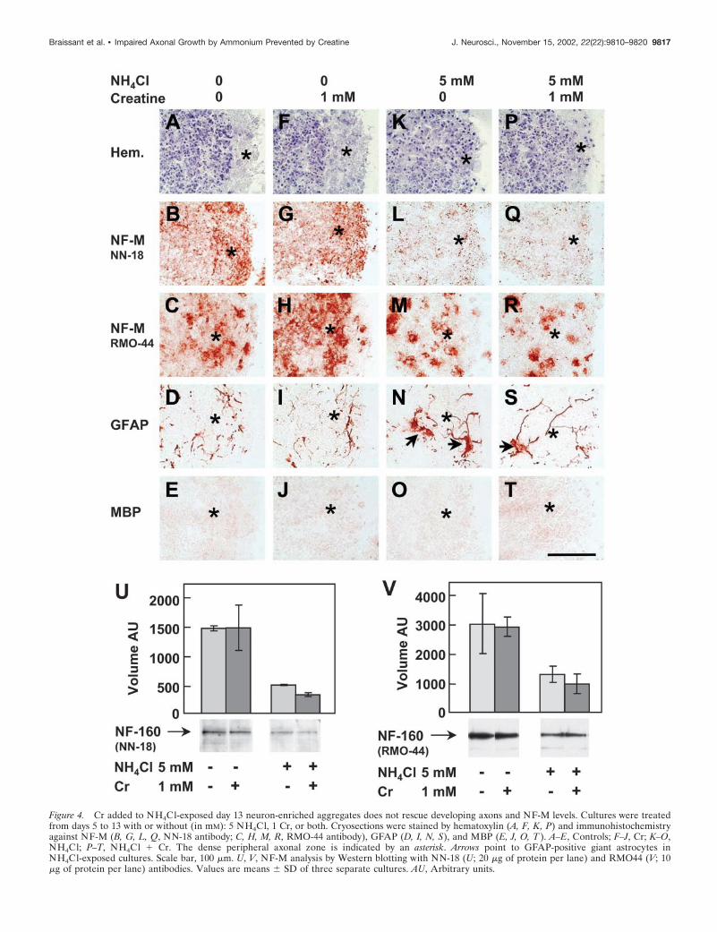
Figure 4. Cr added to NH4Cl-exposed day 13 neuron-enriched aggregates does not rescue developing axons and NF-M levels. Cultures were treatedfrom days 5 to 13 with or without (in mM): 5 NH4Cl, 1 Cr, or both. Cryosections were stained by hematoxylin (A, F, K, P) and immunohistochemistryagainst NF-M (B, G, L, Q, NN-18 antibody; C, H, M, R, RMO-44 antibody), GFAP (D, I, N, S), and MBP (E, J, O, T ). A–E, Controls; F–J, Cr; K–O,NH4Cl; P–T, NH4Cl � Cr. The dense peripheral axonal zone is indicated by an asterisk. Arrows point to GFAP-positive giant astrocytes inNH4Cl-exposed cultures. Scale bar, 100 �m. U, V, NF-M analysis by Western blotting with NN-18 (U; 20 �g of protein per lane) and RMO44 (V; 10�g of protein per lane) antibodies. Values are means � SD of three separate cultures. AU, Arbitrary units.
Braissant et al. • Impaired Axonal Growth by Ammonium Prevented by Creatine J. Neurosci., November 15, 2002, 22(22):9810–9820 9817
to fully differentiated astrocytes, which have a higher capacity forammonium detoxification (Butterworth, 1993). Alternatively,protective factors released by target postsynaptic neurons to theirpresynaptic counterparts could be involved (Keith and Wilson,2001). This difference in vulnerability of the brain is also foundclinically, because hyperammonemia causes irreversible CNSsymptoms compatible with axonal loss in neonates and infants butnot in adults (Brusilow and Horwich, 2001). Evidence for directcoupling of the Cr/PCr/CK system to growth cone activity andaxonal growth (Wang et al., 1998) and Cr and PCr age depen-dency of the CNS in the first weeks of postnatal life (Tsuji et al.,1995) also supports the difference we found between developingand mature aggregates. It suggests a higher CNS sensitivity to Crvariations in young children than in adulthood.
Figure 5. Cholinergic axons of day 13 mixed cell aggregates are affectedby NH4Cl exposure and rescued by Cr cotreatment. Aggregates weretreated from days 5 to 13 with or without (in mM): 5 NH4Cl, 1 Cr, orboth. Immunohistochemistry against ChAT ( A–D) and GAD 65/67kDa (E, F ) was performed. A, E, Controls; B, F, NH4Cl; C, Cr; D,NH4Cl � Cr. The dense peripheral axonal zone is indicated by anasterisk. Scale bar, 100 �m.
Figure 6. NH4Cl and Cr do not affect axons and NF-M in day 28 maturemixed cell aggregates. Cultures were treated from days 20 to 28 with orwithout (in mM): 5 NH4Cl, 1 Cr, or both. A, B, Hematoxylin staining of day28 control aggregates at low (A) and high (B) magnification. C–F, Immu-nohistochemistry against NF-M (NN-18 antibody). C, Control; D, NH4Cl;E, Cr; F, NH4Cl � Cr. B–F, Higher magnifications of the area depicted inA. The dense peripheral axonal zone is indicated by an asterisk. Scale bar,100 �m. G, NF-M analysis by Western blotting (NN-18 antibody; 15 �g ofprotein per lane). Values are means � SD of three separate cultures. AU,Arbitrary units.
Table 3. Intracellular creatine, phosphocreatine, ATP, ADP, and AMP in neuron-enriched aggregates at day 13 of culture
TreatmentsNH4Cl (mM)Creatine (mM)
Control00
Cr01
NH4Cl50
NH4Cl � Cr51
Comparisons (p)
NH4Cl versuscontrols
NH4Cl � Crversus NH4Cl
Creatine 17.4 � 2.0 138.6 � 17.9 16.3 � 1.6 56.2 � 9.3 NS �0.01Phosphocreatine 4.4 � 1.9 6.8 � 2.1 4.1 � 0.7 4.8 � 1.6 NS NSATP 6.7 � 0.9 9.1 � 0.6 4.5 � 0.6 4.3 � 0.8 �0.05 NSADP 3.3 � 0.4 3.7 � 0.3 1.9 � 0.2 2.0 � 0.4 �0.05 NSAMP 7.6 � 1.1 9.4 � 2.0 3.8 � 0.4 3.7 � 0.1 �0.01 NS
Data are expressed as nanomoles per milligram of protein and are means � SD of three separate cultures. Statistical p values are shown (Student’s t test); NS, not significant.
9818 J. Neurosci., November 15, 2002, 22(22):9810–9820 Braissant et al. • Impaired Axonal Growth by Ammonium Prevented by Creatine
The brain cell aggregate culture system as a model tostudy CNS hyperammonemiaAxonal growth was altered in developing brain cell aggregates by5 mM NH4Cl exposure. This concentration mimicked the in vivoextracellular ammonium level found in the brain of experimentalhyperammonemic rats, which was measured as high as 5 mM
(Swain et al., 1992). For human hyperammonemic patients, dataare lacking on extracellular brain ammonium concentration.However, serum levels of ammonium leading to irreversible dam-age to the developing CNS can peak as high as 2 mM, usually afterchronic hyperammonemia in the range of 200 �M (Butterworth,1998; Bachmann, 2002). Brain cell aggregate cultures fulfilled therequirements that irreversible ammonium toxicity to CNS shouldbe studied in models mimicking brain complexity (Butterworth,1998; Braissant et al., 1999a,b) but devoid of confusing variablesattributable to secondary effects of hyperammonemia found inanimal studies (Bachmann, 1992). Our findings are in agreementwith data showing that hyperammonemia stimulates brain glyco-lysis with release of lactate into CSF and causes ammoniumdetoxification through astrocytic glutamine synthesis, which canlead to gliosis and occurrence of Alzheimer’s type II astrocytes(Butterworth, 1998). Moreover, GAP43, an exclusive marker ofaxonal growth cones in physiological conditions, was induced inthe GFAP-positive border of NH4Cl-exposed aggregates, as inreactive astrocytes (Sensenbrenner et al., 1997).
ConclusionsWe have shown that glial Cr can protect axonal growth underammonium exposure. This work exemplifies the importance ofneuron–glial interactions in the pathophysiology of hyperam-monemia. Future work will aim at identifying those factors mod-ified through glial Cr that support axonal growth and are im-paired by hyperammonemia and at understanding changes inbrain Cr metabolism and transport under ammonium exposure.This study also suggests that one should assess means for sustain-ing the CNS Cr level of hyperammonemic neonates and infants toprevent irreversible brain damage caused by impairment ofaxonogenesis.
REFERENCESAlbrecht J (1998) Roles of neuroactive amino acids in ammonium neu-
rotoxicity. J Neurosci Res 51:133–138.Bachmann C (1992) Ornithine carbamoyl transferase deficiency: find-
ings, models and problems. J Inherit Metab Dis 15:578–591.Bachmann C (2002) Mechanisms of hyperammonemia. Clin Chem Lab
Med 40:653–662.Bachmann C (2003) Outcome and survival of patients with urea cycle
disorders (UCD). Eur J Pediatr, in press.Braissant O, Gotoh T, Loup M, Mori M, Bachmann C (1999a)
L-Arginine uptake, the citrulline-NO cycle and arginase II in the ratbrain: an in situ hybridization study. Brain Res Mol Brain Res70:231–241.
Braissant O, Honegger P, Loup M, Iwase K, Takiguchi M, Bachmann C(1999b) Hyperammonemia: regulation of argininosuccinate synthetaseand argininosuccinate lyase genes in aggregating cell cultures of fetalrat brain. Neurosci Lett 266:89–92.
Braissant O, Henry H, Loup M, Eilers B, Bachmann C (2001) Endoge-nous synthesis and transport of creatine in the rat brain: an in situhybridization study. Brain Res Mol Brain Res 86:193–201.
Brown A (1998) Contiguous phosphorylated and non-phosphorylateddomains along axonal neurofilaments. J Cell Sci 111:455–467.
Brusilow SW, Horwich AL (2001) Urea cycles enzymes. In: The meta-bolic and molecular bases of inherited disease (Scriver CR, BeaudetAL, Sly WS, Valle D, eds), pp 1909–1963. New-York: McGraw-Hill.
Butterworth RF (1993) Portal-systemic encephalopathy: a disorder ofneuron-astrocytic metabolic trafficking. Dev Neurosci 15:313–319.
Butterworth RF (1998) Effects of hyperammonaemia on brain function.J Inherit Metab Dis 21 [Suppl 1]:6–20.
Cox G (1977) Neuropathological techniques. In: Theory and practice of
histological techniques (Bandcroft JD, Stevens A, eds), pp 249–273.Edinburgh: Churchill Livingstone.
de Waegh SM, Lee VM, Brady ST (1992) Local modulation of neuro-filament phosphorylation, axonal caliber, and slow axonal transport bymyelinating Schwann cells. Cell 68:451–463.
Dolman CL, Clasen RA, Dorovini-Zis K (1988) Severe cerebral dam-age in ornithine transcarbamylase deficiency. Clin Neuropathol7:10–15.
Filloux F, Townsend JJ, Leonard C (1986) Ornithine transcarbamylasedeficiency: neuropathologic changes acquired in utero. J Pediatr108:942–945.
Flint Beal M, Martin JB (1998) Major complications of cirrhosis. In:Harrison’s principles of internal medicine (Fauci AS, Braunwald E,Isselbacher KJ, Wilson JD, Martin JB, Kasper DL, Hauser SL, LongoDL, eds), pp 2451–2457. New-York: McGraw-Hill.
Furuta A, Martin LJ, Lin CL, Dykes-Hoberg M, Rothstein JD (1997)Cellular and synaptic localization of the neuronal glutamate transport-ers excitatory amino acid transporter 3 and 4. Neuroscience81:1031–1042.
Goslin K, Schreyer DJ, Skene JH, Banker G (1988) Development ofneuronal polarity: GAP-43 distinguishes axonal from dendritic growthcones. Nature 336:672–674.
Harding BN, Leonard JV, Erdohazi M (1984) Ornithine carbamoyltransferase deficiency: a neuropathological study. Eur J Pediatr141:215–220.
Harris J, Ayyub C, Shaw G (1991) A molecular dissection of the car-boxyterminal tails of the major neurofilament subunits NF-M andNF-H. J Neurosci Res 30:47–62.
He Y, Janssen WG, Rothstein JD, Morrison JH (2000) Differentialsynaptic localization of the glutamate transporter EAAC1 and gluta-mate receptor subunit GluR2 in the rat hippocampus. J Comp Neurol418:255–269.
Hemmer W, Wallimann T (1993) Functional aspects of creatine kinasein brain. Dev Neurosci 15:249–260.
Hirokawa N, Takeda S (1998) Gene targeting studies begin to reveal thefunction of neurofilament proteins. J Cell Biol 143:1–4.
Honegger P, Monnet-Tschudi F (2001) Aggregating neural cell culture.In: Protocols for neural cell culture (Fedoroff S, Richardson A, eds), pp199–218. Totowa, NJ: Humana.
Honegger P, Pardo B (1999) Separate neuronal and glial Na �, K �-ATPase isoforms regulate glucose utilization in response to membranedepolarization and elevated extracellular potassium. J Cereb BloodFlow Metab 19:1051–1059.
Honegger P, Lenoir D, Favrod P (1979) Growth and differentiation ofaggregating fetal brain cells in a serum-free defined medium. Nature282:305–308.
Item CB, Stockler-Ipsiroglu S, Stromberger C, Muhl A, Alessandri MG,Bianchi MC, Tosetti M, Fornai F, Cioni G (2001) Arginine:glycineamidinotransferase deficiency: the third inborn error of creatine me-tabolism in humans. Am J Hum Genet 69:1127–1133.
Julien JP (1999) Neurofilament functions in health and disease. CurOpinion Neurobiol 9:554–560.
Keith CH, Wilson MT (2001) Factors controlling axonal and dendriticarbors. Int Rev Cytol 205:77–147.
Laemmli UK (1970) Cleavage of structural proteins during the assemblyof the head of bacteriophage T4. Nature 227:680–685.
Lee VM, Carden MJ, Schlaepfer WW, Trojanowski JQ (1987) Mono-clonal antibodies distinguish several differentially phosphorylatedstates of the two largest rat neurofilament subunits (NF-H and NF-M)and demonstrate their existence in the normal nervous system of adultrats. J Neurosci 7:3474–3488.
Msall M, Batshaw ML, Suss R, Brusilow SW, Mellits ED (1984) Neuro-logic outcome in children with inborn errors of urea synthesis: outcomeof urea-cycle enzymopathies. N Engl J Med 310:1500–1505.
Neary JT, Norenberg LO, Gutierrez MP, Norenberg MD (1987) Hy-perammonemia causes altered protein phosphorylation in astrocytes.Brain Res 437:161–164.
Nguyen MD, Lariviere RC, Julien JP (2001) Deregulation of Cdk5 in amouse model of ALS: toxicity alleviated by perikaryal neurofilamentinclusions. Neuron 30:135–147.
Pardo B, Honegger P (1999) Selective neurodegeneration induced inrotation-mediated aggregate cell cultures by a transient switch to sta-tionary culture conditions: a potential model to study ischemia-relatedpathogenic mechanisms. Brain Res 818:84–95.
Phelps PE, Houser CR, Vaughn JE (1985) Immunocytochemical local-ization of choline acetyltransferase within the rat neostriatum: a cor-related light and electron microscopic study of cholinergic neurons andsynapses. J Comp Neurol 238:286–307.
Podolsky DK, Isselbacher KJ (1998) Major complications of cirrhosis.In: Harrison’s principles of internal medicine (Fauci AS, Braunwald E,Isselbacher KJ, Wilson JD, Martin JB, Kasper DL, Hauser SL, LongoDL, eds), pp 1710–1717. New-York: McGraw-Hill.
Rao VL, Audet RM, Butterworth RF (1995) Increased nitric oxidesynthase activities and L-[ 3H]arginine uptake in brain following porta-caval anastomosis. J Neurochem 65:677–678.
Braissant et al. • Impaired Axonal Growth by Ammonium Prevented by Creatine J. Neurosci., November 15, 2002, 22(22):9810–9820 9819
Ratnakumari L, Qureshi IA, Butterworth RF (1992) Effects of congen-ital hyperammonemia on the cerebral and hepatic levels of the inter-mediates of energy metabolism in spf mice. Biochem Biophys ResCommun 184:746–751.
Ratnakumari L, Qureshi IA, Butterworth RF (1994) Evidence for cho-linergic neuronal loss in brain in congenital ornithine transcarbamylasedeficiency. Neurosci Lett 178:63–65.
Rosenstein JM, Krum JM (1996) Cytoskeletal protein immunoexpres-sion in fetal neural grafts: distribution of phosphorylated and nonphos-phorylated neurofilament protein and microtubule-associated protein 2(MAP-2). Cell Transpl 5:233–241.
Saez R, Llansola M, Felipo V (1999) Chronic exposure to ammoniumalters pathways modulating phosphorylation of microtubule-associatedprotein 2 in cerebellar neurons in culture. J Neurochem 73:2555–2562.
Salomons GS, van Dooren SJ, Verhoeven NM, Cecil KM, Ball WS,Degrauw TJ, Jakobs C (2001) X-linked creatine-transporter gene(SLC6A8) defect: a new creatine-deficiency syndrome. Am J HumGenet 68:1497–1500.
Sanchez C, Diaz-Nido J, Avila J (2000) Phosphorylation of microtubule-associated protein 2 (MAP2) and its relevance for the regulation of theneuronal cytoskeleton function. Prog Neurobiol 61:133–168.
Schliess F, Gorg B, Fischer R, Desjardins P, Bidmon HJ, Herrmann A,Butterworth RF, Zilles K, Haussinger D (2002) Ammonia inducesMK-801-sensitive nitration and phosphorylation of protein tyrosineresidues in rat astrocytes. FASEB J 16:224–248.
Schulze A, Hess T, Wevers R, Mayatepek E, Bachert P, Marescau B,Knopp MV, De Deyn PP, Bremer HJ, Rating D (1997) Creatinedeficiency syndrome caused by guanidinoacetate methyltransferase de-ficiency: diagnostic tools for a new inborn error of metabolism. J Pe-diatr 131:626–631.
Seidl R, Stockler-Ipsiroglu S, Rolinski B, Kohlhauser C, Herkner KR,
Lubec B, Lubec G (2000) Energy metabolism in graded perinatalasphyxia of the rat. Life Sci 67:421–435.
Sensenbrenner M, Lucas M, Deloulme JC (1997) Expression of twoneuronal markers, growth-associated protein 43 and neuron-specificenolase, in rat glial cells. J Mol Med 75:653–663.
Shaw G, Osborn M, Weber K (1986) Reactivity of a panel of neurofila-ment antibodies on phosphorylated and dephosphorylated neurofila-ments. Eur J Cell Biol 42:1–9.
Stockler S, Holzbach U, Hanefeld F, Marquardt I, Helms G, Requart M,Hanicke W, Frahm J (1994) Creatine deficiency in the brain: a new,treatable inborn error of metabolism. Pediatr Res 36:409–413.
Swain M, Butterworth RF, Blei AT (1992) Ammonium and relatedamino acids in the pathogenesis of brain edema in acute ischemic liverfailure in rats. Hepatology 15:449–453.
Tsuji M, Allred E, Jensen F, Holtzman D (1995) Phosphocreatine andATP regulation in the hypoxic developing rat brain. Brain Res DevBrain Res 85:192–200.
Uchino T, Endo F, Matsuda I (1998) Neurodevelopmental outcome oflong-term therapy of urea cycle disorders in Japan. J Inherit Metab Dis21 [Suppl 1]:151–159.
Ulfig N, Nickel J, Bohl J (1998) Monoclonal antibodies SMI 311 andSMI 312 as tools to investigate the maturation of nerve cells and axonalpatterns in human fetal brain. Cell Tissue Res 291:433–443.
Wakamoto H, Manabe K, Kobayashi H, Hayashi M (1999) Subclinicalportal-systemic encephalopathy in a child with congenital absence ofthe portal vein. Brain Dev 21:425–428.
Wang YE, Esbensen P, Bentley D (1998) Arginine kinase expressionand localization in growth cone migration. J Neurosci 18:987–998.
Witt A, Brady ST (2000) Unwrapping new layers of complexity in axon/glial relationships. Glia 29:112–117.
9820 J. Neurosci., November 15, 2002, 22(22):9810–9820 Braissant et al. • Impaired Axonal Growth by Ammonium Prevented by Creatine