Embed Size (px)
Citation preview
AN ANALYSIS OF TWITCH AND TONUS FIBERS IN THE HATCHING MUSCLE
WALTER J. BOCK
Department of Biological Sciences Columbia University
and
Department of Ornithology American Museum of Natural History New York, New York 10027
AND ROBERT S. HIKIDA
Department of Zoology University of Illinois Urbana, Illinois 61801
The hatching of a bird from its hard-shelled egg belongs to that most interesting class of biological actions that an individual animal must do only once during its life, but upon which its survival depends. As expected, birds possess several attributes that insure success- ful hatching. These include an egg tooth on the tip of the beak and a unique pattern of de- velopment of the so-called hatching muscle, the M. complexus ( = M. cucullaris of some au- thors; see George and Berger 1966:272-273). Recently Fisher ( 1958) refocused attention on the structure, development, and biological role of the hatching muscle in birds. Fisher studied the developmental changes in size, amount of lymph, and fiber type in the hatching muscle of the chicken and described this muscle in grebes (1961), gulls (1962), and ducks (1966). J. Fisher (1962) provided a similar description of coots. This work led to a series of studies on the development of this muscle and the role that it has in hatching (Brandstetter 1960; Brandstetter et ~2. 1962; George and Iype 1963; Watterson et al. 1964; Smail 1964, 1965; and additional unpublished works by Watter- son and his associates).
lymph and interstitial fluid in the muscle, which pushes the fibers apart. The rise in the relative number of relaxed fibers prior to hatching was interpreted as an indication of the increased ability of the hatching muscle to contract vigorously at the time of hatching.
One of the most interesting findings re- ported by Fisher is the change in proportions of “contracted” and “relaxed” muscle fibers. Densely staining fibers were interpreted as contracted fibers while lighter staining fibers were described as relaxed fibers. Decrease in the relative number of contracted fibers was regarded by Fisher as an indication of a more advanced stage in the development of the muscle. Moreover, Fisher found that the num- ber of both contracted fibers and relaxed fibers per unit cross-sectional area decreased after the fifteenth day of incubation; this re- duction is due, in part, to the increase of
A major problem lies in the distinction be- tween the two fiber types described by Fisher and in the interpretation of the darkly staining (= contracted) fibers. Fisher did not elab- orate on the exact criteria used in ascertain- ing that the darkly staining fibers are truly contracted (and shortened) fibers. No gen- eral criteria are known to us by which to establish whether vertebrate striated muscle fibers observed in cross-section with light microscopy are or are not contracted and shortened. Although Fisher did not state the magnifications of his photographs, they appear to be about 100-300 X. Contracted and short- ened fibers may be distinguished in cross-sec- tions from relaxed fibers at normal resting length only with high-magnification electron microscopy and then only if the section passes through one of the several favorable levels of the sarcomere. Even electron microscopy can- not always distinguish between relaxed fibers and isometrically contracting fibers, or estab- lish the amount of shortening in an isotoni- cally contracting fiber when fibers are viewed in cross-section. Although it seems doubtful that the densely staining fibers are actually contracted and shortened fibers, no doubt exists that two distinct fiber types are present in the developing M. complexus and that their numbers change during incubation,
A second problem mentioned by Fisher is that the hatching muscle becomes filled with fluid (presumably lymph and interstitial fluid) just before hatching. The entire muscle be-
El11 The Condor, 70:211-222, 1968
212 WALTER J. BOCK AND ROBERT S. HIKIDA
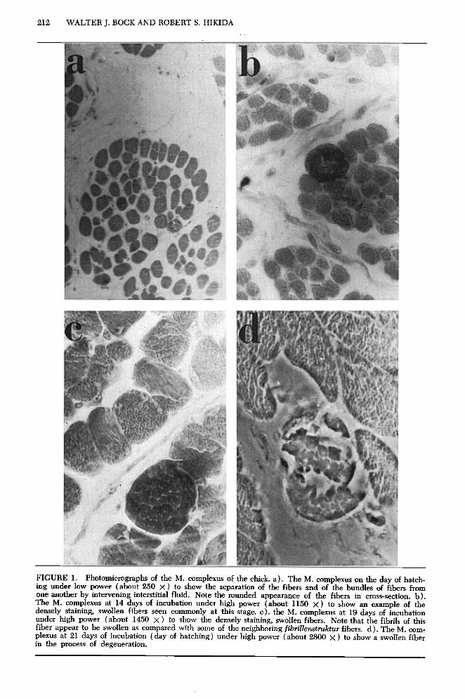
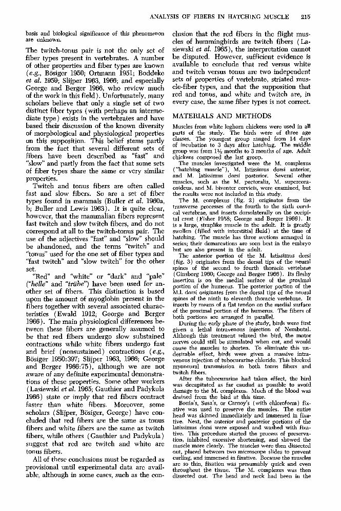
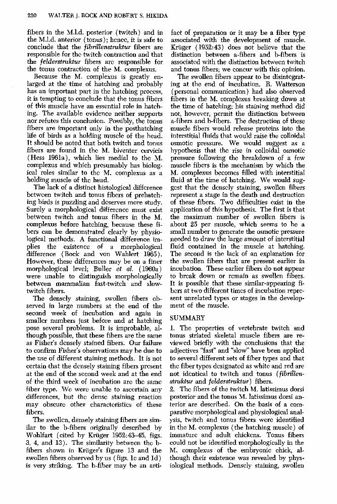
FIGURE 1. Photomicrographs of the M. complexus of the chick. a). The M. complexus on the day of hatch- ing under low power (about 250 x ) to show the separation of the fibers and of the bundles of fibers from one another by intervening interstitial fluid. Note the rounded appearance of the fibers in cross-section. b). The M. complexus at 14 days of incubation under high power (about 1150 X) to show an example of the densely staining, swollen fibers seen commonly at this stage. c). the M. complexus at 19 days of incubation under high power (about 1450 X) to show the densely staining, swollen fibers. Note that the fibrils of this fiber appear to be swollen as compared with some of the neighboring fibdenstdtur fibers. d). The M. com- plexus at 21 days of incubation (day of hatching) under high power (about 2800 x ) to show a swollen fiber in the process of degeneration.
ANALYSIS OF FIBERS IN HATCHING MUSCLE 213
comes turgid, and the individual fibers are separated from one another (fig. 1) . Pohlman (1919:103) stated that the turgid muscle was “physiologically incapacitated to undergo pro- nounced muscular contraction,” while Fisher ( 1958) and George and Iype ( 1963) believed that the hatching muscle provided the muscu- lar force for pipping the egg. Unfortunately, all of these workers argued from indirect evi- dence. Bock and Abbott (unpublished data) undertook an investigation of the mechanical properties of the hatching muscle. They ascer- tained by electrical stimulation of the hatch- ing muscle in situ that this muscle was able to contract and shorten even when fully turgid with interstitial fluid. Moreover, they showed by both electrical and acetylcholine stimula- tion that the hatching muscle at the time of hatching and in the adult chicken contained both fast (twitch) and slow (tonus) relaxing striated skeletal muscle fibers.
On the basis of the evidence just reported, we undertook a histological investigation of the M. complexus, hoping to determine whether or not the lightly and densely stain- ing fibers reported by Fisher are actually twitch and tonus fibers or the progenitors of these fibers.
Two separate problems had to be solved. The first was to determine whether two fiber types can be distinguished morphologically in the M. complexus. If two histologically separable fiber types are present, the second problem was to correlate the morphologically determined types with the physiologically de- termined types (based upon the unpublished data of Bock and Abbott). The second prob- lem could not be solved directly because, as was discovered, the two morphologically de- termined fiber types were mixed throughout the hatching muscle and could not be sepa- rated easily for direct physiological investiga- tion. Hence, it was necessary to compare the morphologically determined fiber types in the M. complexus with fibers in known twitch (fast) and tonus (slow) muscles. If morpho- logical agreement was found between the fibers in the twitch and tonus muscles with the fiber types in the M. complexus, then we can assume with assurance that these morphologi- cally determined fiber types are responsible for the observed twitch and tonus contractions in the M. complexus.
The M. latissimus dorsi posterior and the M. latissimus dorsi anterior (for a description of these muscles see George and Berger 1966: 288-94) were chosen as examples of pure twitch and pure tonus fibered avian muscles (indeed, the M.1.d. anterior may be the only
pure tonus fibered muscle in vertebrates) and were used as the basis for all histological com- parisons in this study. Moreover, it was nec- essary to study twitch and tonus fibers in frog muscles and to compare the histological pic- ture of these fibers with the avian fiber types. This comparison was necessary because much of the previous research on twitch and tonus fibers has been done on frog muscles; consid- erably less interest has developed in the his- tology of avian fast and slow muscles (Kruger 1952; Hess 1961a). Bock and Abbott (unpub- lished data) have undertaken a comparative study of the mechanical properties of the M. latissimus dorsi anterior and posterior in con- nection with their investigation of the M. complexus.
TWITCH AND TONUS FIBERS
A brief review of the properties of twitch and tonus fibers is required because these fiber types are little known to ornithologists and anatomists and, more importantly, because of the widespread confusion between twitch and tonus fibers and other sets of fiber types found in vertebrates (Bock 1967). The greatest prob- lem in studies of twitch and tonus fibers is the correlation of morphological properties with physiological properties. Indeed, most investi- gators in this field have been hindered by this problem and have stressed its importance in their papers. The terms fibriZZen&uktur (= twitch) and felderstruktur ( = tonus) fibers have been used in morphological studies (Kruger 1952) while the terms twitch and tonus fibers have been employed when dis- cussing physiological properties; the latter terms will be used as the general names for these fiber types. The pioneering work on the correlation of fibrillewtruktur and felder- struktur fibers with physiological properties was done by Kruger ( 1952), who advocated the concept of twitch ( = phasic) versus tonus (= holding) muscle fiber types in vertebrates. However, tonus fibers are not the only type of holding muscle fiber in vertebrates.
Although considerable work has been done on the properties of twitch and tonus fibers since the publication of Kruger’s monograph (Kruger 1950,1951, 1952; Ortmann 1951; Kuf- fler and Vaughan-Williams 1953a, 1953b; Teigs 1953; Burke and Ginsborg 1956; Kruger and Gunther 1956a, 195613, 1958; Gray 1957, 1958; Ginsborg 1960; Haggqvist 1960; Hess 1960, 1961a, 1961b, 1962a, 1962b, 1963, 1965; Ginsborg and Mackay 1961; Peachey, 1961; Hlggqvist and Lindberg 1962; Kaplan and Cahn 1962; Peachey and Huxley 1962; Feng, et al. 1963; Hess and Pilar 1963; Abbott and
214 WALTER J. BOCK AND ROBERT S. HIKIDA
Brady 1964; Page 1965; Hoyle et al. 1966; Pilar and Hess 1966), large gaps still exist in our knowledge of these muscle fibers. Future studies, especially extensive comparative in- vestigations, may modify much of what we believe today. A major unsolved problem is the correlation between the known properties of twitch and tonus fibers and those of other sets of muscle fiber types.
The morphological differences between twitch and tonus fibers may be distinguished only on the basis of histology and fine struc- ture (electron miscroscopy ); these fibers can- not be separated on the basis of gross struc- ture. Indeed, a major difficulty still exists because twitch and tonus fibers may be sep- arated histologically only with difficulty and then only at high magnification (500 x is the minimum). Even at the highest obtainable magnifications with the light microscope, it is frequently impossible to distinguish with cer- tainty between twitch and tonus fibers.
In histological cross-section, fibrillenatruk- tur fibers show very regular punctate fibrils evenly distributed throughout the sarcoplasm. Nuclei are found only or mainly at the pe- riphery of the fiber just under the sarcolemma. In longitudinal sections, many prominent longitudinal striations are present (Hess 1960); however, the significance of this feature re- mains unknown. Felderstmktur fibers are characterized in histological cross-section by either large irregularly shaped pseudofibrils” ( = “fibrils”) or by a diffuse “mushy” sarco- plasm without the appearance of any distinct pseudofibrils (Kruger 1952; Gray 1958; Hess 1960,196la). The difference between the two fel&rstruktur appearances is due to fixation and not to any basic difference between two kinds of feldemtruktur fibers. Good fixation results in the diffuse mushy sarcoplasm with- out distinct pseudofibrils, while poorer fixa- tion results in the appearance of large irregu- lar pseudofibrils. The pseudofibrils are fixa- tion artifacts, and quite useful ones. Nuclei are frequently found in the center of felder- struktur fibers. These fibers do not show the longitudinal striations of fibrillenstruktur fi- bers. Fibrillenstruktur fibers have a slightly larger diameter, on the average, than do fel- derststruktur fibers (Gray 1958), but the dif- ference in average diameters is so small and
* Fibrils are defined as a group of filaments sur- rounded by the sarcotubular system. A greatly re- duced or absent sarcotubular system means that the filaments are not arranged into fibrils. Upon fixation, groups of filaments clump together, giving rise to a feature similar to fibrils. These clumps of filaments in feldertiruktur fibers have been called fibrils, but we prefer to call them pseudofibrils.
the amount of overlap is so large that diameter is useless as a distinction between the fiber types.
In the fine structure, fibrillenstruktztr fibers have a well-developed sarcotubular system with triads pres- ent at every sarcomere, sharply delimited fibrils (myo- fibrils), an M-band, a thinner, straight Z-band with well-ordered thin filaments attached to it, a larger number of mitochondria, and lipid droplets. Felah- struktur fibers have a greatly reduced sarcotubular system with the triad system greatly reduced or ab- sent, no fibrils, no M-band with thin connections be- tween the thick filaments, a thicker, amorphic, irregu- lar Z-band with the thin filaments attached to it irregularly, and a smaller number of mitochondria and no lipid droplets.
Fibrillenatruktur fibers have one (usually) large en plaque end-plate of large-fibered nerves with junctional folds of the muscular plasma membrane, while feldewtruktur fibers have numerous small en gmppe end-plates of small-fibered nerves with an almost complete absence of junctional folds of the muscular plasma membrane.
FibriUenstruktur fibers contain a variable amount of heart lactic dehydrogenase ( H-LDH ), varying from 1 to 25 per cent certainly, and perhaps to 50 per cent. Felderstruktur fibers (of the M. latissimus dorsi anterior) contain 99 per cent H-LDH (Kaplan and Cahn 1962). The upper limit of H-LDH in fibrilhstruktur fibers cannot be determined at this time because some of the muscles reported may be muscles with mixed fibers. The M. latissimus dorsi posterior with 23 per cent H-LDH has the highest amount of this monomer for a pure twitch-fibered muscle.
The two most important known physiological dif- ferences between twitch and tonus fibers are the speed of contraction and relaxation and the propaga- tion ability of the membrane. Twitch fibers exhibit the well-known twitch-a rapid rise of tension (about 25 milliseconds), following stimulation and a rapid drop of tension following cessation of stimulation. Fusion of twitches into a tetany requires a high rate of stimulation, generally 20 or more pulses per sec- ond. The sarcolemma of twitch fibers propagates action potentials along the length of the fiber. And twitch fibers do not maintain a contracture when ex- posed to depolarizing agents such as acetycholine and potassium chloride or to direct-current stimulation. Tonus fibers do not respond to a single electrical stimulation with the characteristic all-or-none twitch, but have a slow increase of tension with continued stimulation; attainment of maximum tension requires several seconds. Following cessation of stimulation, tonus fibers lose tension slowly, again in a matter of seconds or minutes. Fusion into a tetany occurs with a low rate of stimulation, about 2-5 pulses per second. The sarcolemma of tonus fibers cannot propogate ac- tion potentials, and tonus fibers respond to depolariz- ing agents such as acetylcholine and potassium chlo- ride or to direct current with a prolonged contracture. Contraction following acetylcholine stimulation is the standard test for the presence of tonus fibers. The characteristics of the mechanical properties of tonus fibers are still unknown in detail.
In contrast to the atrophy seen in twitch fibers following denervation, tonus fibers hypertrophy fol- lowing denervation (Feng et al. 1963; and personal observations confirming Eeng’s report). The tonus fibers hypertrophy following denervation in both the pure tonus M. latissimus dorsi anterior and in mixed muscles such as the M. biventer cervicis. The causal
ANALYSIS OF FIBERS IN HATCHING MUSCLE 215
basis and biological significance of this phenomenon are unknown.
The twitch-tonus pair is not the only set of fiber types present in vertebrates. A number of other properties and fiber types are known (e.g., BBsiger 1950; Ortmann 1951; Boddeke et al. 1959; Slijper 1963, 1966; and especially George and Berger 1966, who review much of the work in this field). Unfortunately, many scholars believe that only a single set of two distinct fiber types (with perhaps an interme- diate type) exists in the vertebrates and have based their discussion of the known diversity of morphological and physiological properties on this supposition. This belief stems partly from the fact that several different sets of fibers have been described as “fast” and “slow” and partly from the fact that some sets of fiber types share the same or very similar properties.
Twitch and tonus fibers are often called fast and slow fibers. So are a set of fiber types found in mammals (Buller et al. 196Oa, b; Buller and Lewis 1963). It is quite clear, however, that the mammalian fibers represent fast twitch and slow twitch fibers, and do not correspond at all to the twitch-tonus pair. The use of the adjectives “fast” and ‘%.low” should be abandoned, and the terms “twitch” and “tonus” used for the one set of fiber types and “fast twitch” and “slow twitch” for the other set.
“Red” and “white” or “dark” and “pale” (“belle” and “ttibe”) have been used for an- other set of fibers. This distinction is based upon the amount of myoglobin present in the fibers together with several associated charac- teristics (Ewald 1912; George and Berger 1966). The main physiological differences be- tween these fibers are generally assumed to be that red fibers undergo slow substained contractions while white fibers undergo fast and brief (nonsustained) contractions (e.g., Bosiger 1950:397; Slijper 1963, 1966; George and Berger 1966:75), although we are not aware of any definite experimental demonstra- tions of these properties. Some other workers (Lasiewski et al. 1965; Gauthier and Padykula 1966) state or imply that red fibers contract faster than white fibers. Moreover, some scholars (Slijper, Bosiger, George) have con- cluded that red fibers are the same as tonus fibers and white fibers are the same as twitch fibers, while others (Gauthier and Padykula) suggest that red are twitch and white are tonus fibers.
All of these conclusions must be regarded as provisional until experimental data are avail- able, although in some cases, such as the con-
elusion that the red fibers in the flight mus- cles of hummingbirds are twitch fibers (La- siewski et al. 1965), the interpretation cannot be disputed. However, sufficient evidence is available to conclude that red versus white and twitch versus tonus are two independent sets of properties of vertebrate, striated mus- cle-fiber types, and that the supposition that red and tonus, and white and twitch are, in every case, the same fiber types is not correct.
MATERIALS AND METHODS
Muscles from white leghorn chickens were used in all parts of the study. The birds were of three age classes. The youngest group ranged from 14 days of incubation to 3 days after hatching. The middle group was from 1% months to 3 months of age. Adult chickens composed the last group.
The muscles investigated were the M. complexus (“hatching muscle”), M. latissimus dorsi anterior, and M. latissimus dorsi posterior. Several other muscles, such as the M. pectoralis, M. supercora- coideus, and M. biventer cervicis, were examined, but the results were not included in this study.
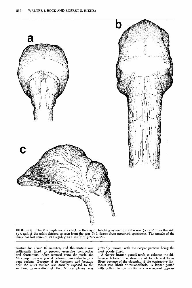
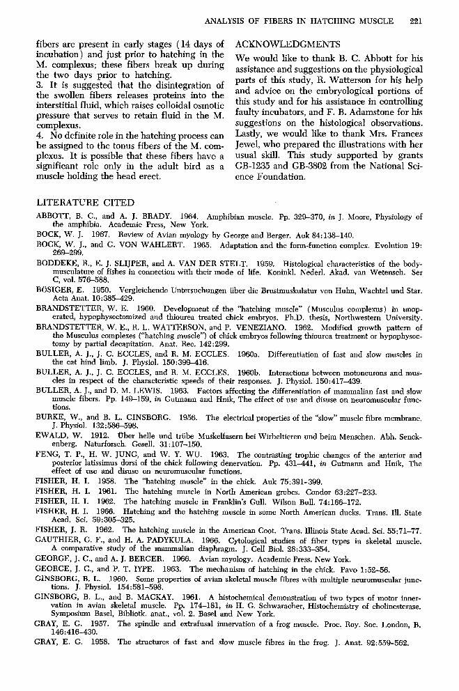
The M. complexus (fig. 2) originates from the transverse processes of the fourth to the sixth cervi- cal vertebrae, and inserts dorsolaterally on the occipi- tal crest f Fisher 1958: George and Beraer 1966 ). It is a large; straplike muscle in the adult- It is greatly swollen (filled with interstitial fluid) at the time of hatching. The muscle has three sections arranged in series; their demarcations are seen best in the embryo but are also present in the adult.
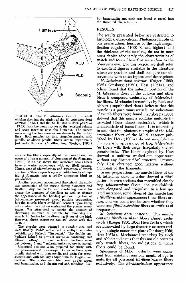
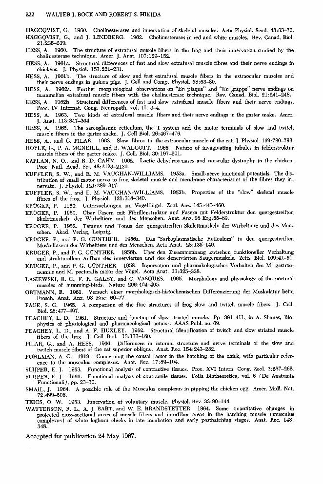
The anterior portion of the M. latissimus dorsi (fig. 3) originates from the dorsal tius of the neural .- - spines of the second to fourth thoracic vertebrae I Ginsbora 1966: George and Bereer 1966). Its fleshy insertion Is on ‘the medial surf&e of the proximal portion of the humerus. The posterior portion of the M.l. dorsi originates from the dorsal tips of the neural spines of the ninth to eleventh thoracic vertebrae. It inserts by means of a flat tendon on the medial surface of the proximal portion of the humerus. The fibers of both portions are arranged in parallel.
During the early phase of the study, birds were first given a lethal intravenous injection of Nembutal. Although this treatment relaxed the bird, the motor nerves could still be stimulated when cut, and would cause the muscles to shorten. To eliminate this un- desirable effect, birds were given a massive intra- venous injection of tubocurarine chloride. This blocked myoneural transmission in both tonus fibers and twitch fibers.
After the tubocurarine had taken effect, the bird was decapitated as far caudad as possible to avoid damage to the M. complexus. Much of the blood was drained from the bird at this time.
Bouin’s, Susa’s, or Camoy’s (with chloroform) fix- ative was used to preserve the muscles. The entire head was skinned immediately and immersed in fixa- tive. Next, the anterior and posterior portions of the latissimus dorsi were exposed and washed with fixa- tive. This procedure started the process of preserva- tion, inhibited excessive shortening, and showed the muscle more clearly. The muscles were then dissected out, placed between two microscope slides to prevent curling, and immersed in fixative. Because the muscles are so thin, fixation was presumably quick and even throughout the tissue. The M. complexus was then dissected out. The head and neck had been in the
216 WALTER J. BOCK AND ROBERT S. HIKIDA
a
FIGURE 2. The M. complexus of a chick on the day of hatching as seen from the rear (a) and from the side ( c), and of the adult chicken as seen from the rear (b ), drawn from preserved specimens. The muscle of the chick has lost some of its turgidity as a result of preservation.
fixative for about 10 minutes, and the muscle was probably uneven, with the deeper portions being the sufficiently fixed to prevent excessive contraction most poorly fixed. and shortening. After removal from the neck, the A shorter fixation period tends to enhance the dif- M. complexus was placed between two slides to pre- ference between the structure of twitch and tonus vent curling. Because of its thickness and because fibers because of the clumping of the contractive fila- only the outer surface was initially exposed to the ments into fibrils or pseudofibrils. A longer period solution, preservation of the M. complexus was with better fixation results in a washed-out appear-
ANALYSIS OF FIBERS IN HATCHING MUSCLE 217
FIGURE 3. The M. latissimus dorsi of the adult chicken showing the origins of the M. latissimus dorsi anterior (ALD) and the M. latissimus dorsi posterior ( PLD ) from the neural spines of the vertebral column and their insertion onto the humerus. The nerves innervating the two muscles are shown by the broken lines. Both muscles are thin, straplike muscles with parallel or almost parallel fibers and lie superficially just under the skin. (Modified from Ginsborg 1960.)
ante of the fibers, especially of the tonus fibers, be- cause of a lesser amount of clumping of the filaments. Hess (1961a) has shown that well-fixed tonus fibers have a washy appearance with no fibril pattern. Hence, ease of identification and separation of twitch and tonus fibers depends upon an artifact-the clump- ing of filaments into a solidly appearing fibril or pseudofibril.
Another problem encountered throughout the study was contraction of the muscle during dissection and fixation. Any contraction and shortening would in- crease the diameter of the fiber as well as change the appearance of the banding pattern. Injection of tubocurarine prevented much possible contraction, but the muscle fibers could still contract upon being cut or when the fixation contracted the plasma mem- brane. We attempted to restrict the amount of shortening as much as possible by immersing the muscle in fixative before dissecting it out of the bird. However, slight shortening can still occur with this method.
The muscles were trimmed to suitable size and were usually double embedded in methyl benzoate- celloidin and Fisher’s “Tissuemat.” The double em- bedding helped to prevent excessive hardening and allowed cutting of thinner sections. Sections were cut between 5 and 7 microns unless otherwise stated.
Unstained sections were prepared for study with the phase-contrast microscope. Other sections were usually stained with hematoxylin and eosin for cross- sections and with Mallory’s triple stain for longitudinal sections. Other stains were tried, such as fast green and hematoxylin, and alizarin red and toluidine blue,
but hematoxylin and eosin was found to reveal best the structural characteristics.
RESULTS
The results presented below are restricted to histological observations. Photomicrographs of our preparations, because of the high magni- fication required (1000 x and higher) and the thickness of the sections, do not in most cases depict adequately the characteristics of twitch and tonus fibers that were clear to the observer’s eye. For this reason, we shall refer to excellent figures available in the literature whenever possible and shall compare our ob- servations with these figures and descriptions.
M. lutissimus dorsi anterior. Kruger (1950, 1952) Ginsborg ( 1960), Hess ( 1961a), and others found that the anterior portion of the M. latissimus dorsi of the chicken and other birds is composed exclusively of felderstruk- tur fibers. Mechanical recordings by Bock and Abbott (unpublished data) indicate that this muscle is a pure tonus muscle; no indications of twitch fibers were found. Ginsborg (1960) showed that this muscle contains multiple in- nervated fibers almost exclusively, which is characteristic of tonus fibers. It is interesting to note that the photomicrographs of the feld- erstruktur fibers of the M.1.d. anterior pub- lished by Hess (1961a) do not resemble the characteristic appearance of frog felderstruk- tur fibers with their large, irregularly shaped pseudofibrils. The fibers depicted by Hess showed a uniform washed-out appearance without any distinct fibril structure. Presum- ably Hess obtained good fixation without clumping of the filaments.
In our preparations, the muscle fibers of the M. latissimus dorsi anterior showed a fibril pattern in cross-sections that resembled closely frog felderstruktur fibers; the pseudofibrils were elongated and irregular. In a few iso- lated instances, some fibers of this muscle had a fibrillenstruktur appearance; these fibers are rare, and we could not be sure whether they were true fibrillenstruktur fibers or artifacts of preparation.
M. lathsimus dorsi posterior. This muscle contains fibrillenstruktur fibers almost exclu- sively (Kruger 1950, 1952; Hess 1961a), which are innervated by large-diameter neurons end- ing in a single motor end-plate (Ginsborg 1960; Hess 1961a). Mechanical recording by Bock and Abbott indicates that this muscle contains only twitch fibers; no indications of tonus fibers could be found.
Specimens of M.1.d. posterior were exam- ined from chickens from one month of age to maturity; all possessed fibrilhstruktur fibers exclusively. The fibrillenstruktur appearance
218 WALTER J. BOCK AND ROBERT S. HIKIDA
seen in this muscle is identical with that seen in frog’s fibrillenstruktur fibers.
Our observations on the histological struc- ture of the fibrillenstruktur fibers of the M.1.d. posterior and the felderstruktur fibers of the M.1.d. anterior agree completely with the his- tological observations reported in the litera- ture, and these morphological attributes are correlated with the physiological characteris- tics of twitch and tonus contraction. These observations will form the basis for identifica- tion of fibrillenstruktur and felderstruktur fibers in the M. complexus.
The M. complexus of immature and adult chickens. In young and adult chickens at least two types of fibers were present in the M. complexus. A majority of the fibers have the classical fibrillenstruktur appearance as seen in the frog’s twitch fibers and in the M.1.d. posterior of the chicken. The second type shows the irregular pattern of fibrils seen in the frog’s felderstruktur fibers (Kruger 1952; Gray 1958) and in the M.1.d. anterior of the chicken. Some of the fibers had a larger, more-rounded pattern of fibrils, but still had an irregular appearance. They resembled most closely the “area1 pattern” of felderstruktur fibers described by Gray ( 1958).
The ratio of tonus to twitch fibers was ob- tained by counting the number of felderstruk- tur and fibrillenstruktur fibers seen in the field of the microscope at 750 X magnification for phase-contrast observations and at 600 x mag- nification for bright-field observations. When possible, at least five fields per cross-section were counted and the results averaged. The tonus fibers appeared to be distributed uni- formly throughout the muscle; no concentra- tions, such as those in the tonus bundle of the frog’s iliofibularis muscle, were found. The average percentage of tonus fibers in a sample of 19 adult chickens is 15.3 per cent, with a standard deviation of 2.7, showing that there are many more twitch fibers than tonus fibers in the M. complexus. These results are in rough agreement with the findings of Bock and Abbott (unpublished data) that less than 25 per cent of the total tension developed by the M. complexus is contributed by the tonus fibers.
The diameters of the fibrillenstruktur and felderstruktur fibers were compared. This comparison proved to be fairly difficult owing to the great variety in the size of the fibers in the different regions of the muscle. Another problem was that perfect cross-sections are re- quired to obtain accurate comparisons of the fiber diameters. Moreover, the problem of contraction and shortening during fixation
could not be eliminated, and some fibers may have been compared in different states of contraction. We decided to measure the great- est diameter of adjacent fibrillen- and felder- struktur fibers for comparisons of fiber diam- eters. A Leitz substage micrometer was used in conjunction with an American Optical reti- cule in the ocular lens to obtain the diameter of the fibers. Five to 10 pairs of adjacent fibers per cross-section were measured and the results averaged; 15 muscles from adult chickens were measured. The diameters ranged from 29 to 75 microns (average 46.9) for fibrillenstruktur fibers, and 23 to 70 mi- crons (average 41.4) for felderstruktur fibers. The main distribution of diameter was be- tween 35 to 55 microns for the fibrillenstmk- tur fibers and between 30 to 50 microns for felderstruktur fibers. Felderstruktur fibers tend to be slightly smaller than the adjacent fibrillewtruktur fibers; the average ratio for felder- to fibrillenstruktur diameter was 0.89, with a range from 0.79 to 1.03. Most of the ratios fell between 0.85 and 0.93. Hence, it was found that the diameters of twitch and tonus fibers overlap greatly although tonus fibers tend to be slightly smaller than twitch fibers. These results are in good agreement with Gray’s (1957) measurements of fiber diameters in the extensor muscle of the frog’s fourth toe. Gray found that tonus fibers had diameters ranging from 10 to 80 microns with a peak distribution at 40-50 microns. Twitch- fiber diameters ranged from 30 to 120 microns with a peak at 60-70 microns. Although Gray found a greater range in fiber size, the tonus fibers were still smaller than the twitch fibers, with a large overlap. The difference in fiber diameter is so slight and the range of overlap is so broad that fibrillenstruktur and felder- struktur fibers cannot be distinguished on this basis.
The M. complexus of embyonic chicks. The small fiber diameter in the embryonic M. complexus makes their study quite formidable. Hess (1961a:222) commented that it was dif- ficult to see differences in cross-sections be- tween fibers of the M.1.d. anterior and M.1.d. posterior in chicks between hatching and seven days of age. We experienced the same difficulty and were unable to distinguish with certainty twitch and tonus fibers in embryonic chicks.
Comparisons were made first between mus- cles that were allowed to contract and shorten to approximately 75 per cent of their resting length before fixation and muscles that were fixed at resting length with great care to pre- vent contraction. No noticeable differences
ANALYSIS OF FIBERS IN HATCHING MUSCLE 219
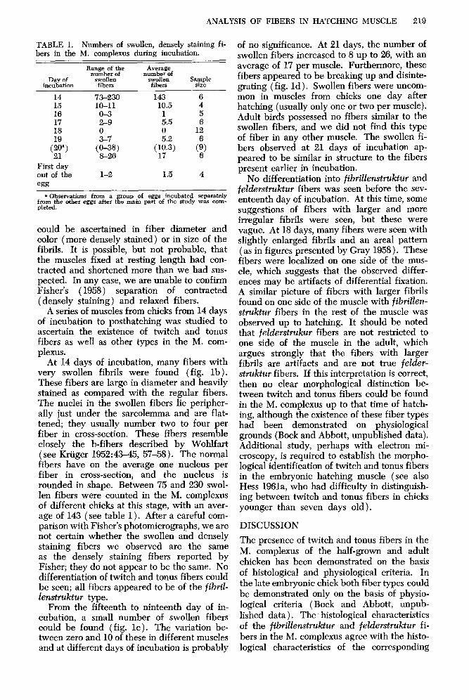
TABLE 1. Numbers of swollen, densely staining fi- bers in the M. complexus during incubation.
“;ig&c ;; Average number of
Day of swollen SWOllen Sample incubation fibers fibers size
14 73-230 143 6 15 10-11 10.5 4 16 O-3 17 2-9 :.5
5 6
18 0 12
cp, SJ
z.2
($3’ ci,
First day out of the l-2 1.5 4
egg
a Observations fmm a group of eggs incubated separately from the other eggs after the main part of the study was corn-- pleted.
could be ascertained in fiber diameter and color (more densely stained) or in size of the fibrils. It is possible, but not probable, that the muscles fixed at resting length had con- tracted and shortened more than we had sus- pected. In any case, we are unable to confirm Fisher’s ( 1958) separation of contracted (densely staining) and relaxed fibers.
A series of muscles from chicks from 14 days of incubation to posthatching was studied to ascertain the existence of twitch and tonus fibers as well as other types in the M. com- plexus.
At 14 days of incubation, many fibers with very swollen fibrils were found (fig. lb). These fibers are large in diameter and heavily stained as compared with the regular fibers. The nuclei in the swollen fibers lie peripher- ally just under the sarcolemma and are flat- tened; they usually number two to four per fiber in cross-section. These fibers resemble closely the b-fibers described by Wohlfart (see Kriiger 1952:43-45, 57-58). The normal fibers have on the average one nucleus per fiber in cross-section, and the nucleus is rounded in shape. Between 75 and 230 swol- len fibers were counted in the M. complexus of different chicks at this stage, with an aver- age of 143 (see table 1). After a careful com- parison with Fisher’s photomicrographs, we are not certain whether the swollen and densely staining fibers we observed are the same as the densely staining fibers reported by Fisher; they do not appear to be the same. No differentiation of twitch and tonus fibers could be seen; all fibers appeared to be of the fibd- lenstruktur type.
From the fifteenth to ninteenth day of in- cubation, a small number of swollen fibers could be found (fig. lc). The variation be- tween zero and 10 of these in different muscles and at different days of incubation is probably
of no significance. At 21 days, the number of swollen fibers increased to 8 up to 26, with an average of 17 per muscle. Furthermore, these fibers appeared to be breaking up and disinte- grating (fig, Id), Swollen fibers were uncom- mon in muscles from chicks one day after hatching (usually only one or two per muscle). Adult birds possessed no fibers similar to the swollen fibers, and we did not find this type of fiber in any other muscle. The swollen fi- bers observed at 21 days of incubation ap- peared to be similar in structure to the fibers present earlier in incubation.
No differentiation into fibrillenstruktur and felderstruktur fibers was seen before the sev- enteenth day of incubation. At this time, some suggestions of fibers with larger and more irregular fib& were seen, but these were vague. At 18 days, many fibers were seen with slightly enlarged fibrils and an area1 pattern ( as in figures presented by Gray 1958). These fibers were localized on one side of the mus- cle, which suggests that the observed differ- ences may be artifacts of differential fixation. A similar picture of fibers with larger fibrils found on one side of the muscle with fibrillen- struktur fibers in the rest of the muscle was observed up to hatching. It should be noted that fel&rstrukur fibers are not restricted to one side of the muscle in the adult, which argues strongly that the fibers with larger fibrils are artifacts and are not true feZ&r- struktur fibers. If this interpretation is correct, then no clear morphological distinction be- tween twitch and tonus fibers could be found in the M. complexus up to that time of hatch- ing, although the existence of these fiber types had been demonstrated on physiological grounds (Bock and Abbott, unpublished data). Additional study, perhaps with electron mi- croscopy, is required to establish the morpho- logical identification of twitch and tonus fibers in the embryonic hatching muscle (see also Hess 1961a, who had difficulty in distinguish- ing between twitch and tonus fibers in chicks younger than seven days old).
DISCUSSION
The presence of twitch and tonus fibers in the M. complexus of the half-grown and adult chicken has been demonstrated on the basis of histological and physiological criteria. In the late embryonic chick both fiber types could be demonstrated only on the basis of physio- logical criteria (Bock and Abbott, unpub- lished data). The histological characteristics of the fibrillenstruktur and felderstruktur fi- bers in the M. complexus agree with the histo- logical characteristics of the corresponding
220 WALTER J. BOCK AND ROBERT S. HIKIDA
fibers in the M.1.d. posterior (twitch) and in the M.1.d. anterior (tonus); hence, it is safe to conclude that the fibrillenstruktur fibers are responsible for the twitch contraction and that the felderstruktur fibers are responsible for the tonus contraction of the M. complexus.
Because the M. complexus is greatly en- larged at the time of hatching and probably has an important part in the hatching process, it is tempting to conclude that the tonus fibers of this muscle have an essential role in hatch- ing. The available evidence neither supports nor refutes this conclusion. Possibly, the tonus fibers are important only in the posthatching life of birds as a holding muscle of the head. It should be noted that both twitch and tonus fibers are found in the M. biventer cervicis (Hess 1961a), which lies medial to the M. complexus and which presumably has biolog- ical roles similar to the M. complexus as a holding muscle of the head.
The lack of a distinct histological difference between twitch and tonus fibers of prehatch- ing birds is puzzling and deserves more study. Surely a morphological difference must exist between twitch and tonus fibers in the M. complexus before hatching, because these fi- bers can be demonstrated clearly by physio- logical methods. A functional difference im- plies the existence of a morphological difference (Bock and von Wahlert 1965). However, these differences may be on a finer morphological level; Buller et al. (196Oa) were unable to distinguish morphologically between mammalian fast-twitch and slow- twitch fibers.
The densely staining, swollen fibers ob- served in large numbers at the end of the second week of incubation and again in smaller numbers just before and at hatching pose several problems. It is improbable, al- though possible, that these fibers are the same as Fisher’s densely stained fibers. Our failure to confirm Fisher’s observations may be due to the use of different staining methods. It is not certain that the densely staining fibers present at the end of the second week and at the end of the third week of incubation are the same fiber type. V17e were unable to ascertain any differences, but the dense staining reaction may obscure other characteristics of these fibers.
The swollen, densely staining fibers are sim- ilar to the b-fibers originally described by Wohlfart (cited by Kruger 1952:4345, figs. 3, 4, and 13). The similarity between the b- fibers shown in Kruger’s figure 13 and the swollen fibers observed by us (figs. lc and Id) is very striking. The b-fiber may be an arti-
fact of preparation or it may be a fiber type associated with the development of muscle. Kruger (1952:43) does not believe that the distinction between a-fibers and b-fibers is associated with the distinction between twitch and tonus fibers; we concur with this opinion.
The swollen fibers appear to be disintegrat- ing at the end of incubation. R. Watterson (personal communication) had also observed fibers in the M. complexus breaking down at the time of hatching; his staining method did not, however, permit the distinction between a-fibers and b-fibers. The destruction of these muscle fibers would release proteins into the interstitial fluids that would raise the colloidal osmotic pressure. We would suggest as a hypothesis that the rise in colloidal osmotic pressure following the breakdown of a few muscle fibers is the mechanism by which the M. complexus becomes filled with interstitial fluid at the time of hatching. We would sug- gest that the densely staining, swollen fibers represent a stage in the death and. destruction of these fibers. Two difficulties exist in the application of this hypothesis. The first is that the maximum number of swollen fibers is about 25 per muscle, which seems to be a small number to generate the osmotic pressure needed to draw the large amount of interstitial fluid contained in the muscle at hatching. The second is the lack of an explanation for the swollen fibers that are present earlier in incubation. These earlier fibers do not appear to break down or remain as swollen fibers. It is possible that these similar-appearing fi- bers at two different times of incubation repre- sent unrelated types or stages in the develop- ment of the muscle.
SUMMARY
1. The properties of vertebrate twitch and tonus striated skeletal muscle fibers are re- viewed briefly with the conclusions that the adjectives “fast” and “slow” have been applied to several different sets of fiber types and that the fiber types designated as white and red are not identical to twitch and tonus (fibrillen- struktur and felderstruktur) fibers. 2. The fibers of the twitch M. latissimus dorsi posterior and the tonus M. latissimus dorsi an- terior are described. On the basis of a com- parative morphological and physiological anal- ysis, twitch and tonus fibers were identified in the M. complexus (the hatching muscle) of immature and adult chickens. Tonus fibers could not be identified morphologically in the M. complexus of the embryonic chick, al- though their existence was revealed by phys- iological methods. Densely staining, swollen
ANALYSIS OF FIBERS IN HATCHING MUSCLE 221
fibers are present in early stages (14 days of incubation) and just prior to hatching in the M. complexus; these fibers break up during the two days prior to hatching. 3. It is suggested that the disintegration of the swollen fibers releases proteins into the interstitial fluid, which raises colloidal osmotic pressure that serves to retain fluid in the M. complexus. 4. No definite role in the hatching process can be assigned to the tonus fibers of the M. com- plexus. It is possible that these fibers have a significant role only in the adult bird as a muscle holding the head erect,
LITERATURE CITED
ACKNOWLEDGMENTS
We would like to thank B. C. Abbott for his assistance and suggestions on the physiological parts of this study, R. Watterson for his help and advice on the embryological portions of this study and for his assistance in controlling faulty incubators, and F. B. Adamstone for his suggestions on the histological observations. Lastly, we would like to thank Mrs. Frances Jewel, who prepared the illustrations with her usual skill. This study supported by grants GB-1235 and GB-3802 from the National Sci- ence Foundation.
ABBOTT, B. C., and A. J. BRADY. 1964. Amphibian muscle, the amphibia. Academic Press, New York.
Pp. 329-370, in J. Moore, Physiology of
BOCK, W. J. 1967. Review of Avian myology by George and Berger. Auk 84:138-140.
BOCK, W. J., and G. VON WAHLERT. 1965. Adaptation and the form-function complex. Evolution 19: 269-299.
BODDEKE, R., E. J. SLIJPER, and A. VAN DER STELT. 1959. Histological characteristics of the body- musculature of fishes in connection with their mode of life. Koninkl. Nederl. Akad. van Wetensch. Ser C, vol. 576588.
BOSIGER, E. 1950. Vergleichende Untersuchungen iiber die Brustmuskulatur von Huhn, Wachtel und Star. Acta Anat. 10:385429.
BRANDSTETTER, W. E. 1960. Development of the “hatching muscle” (Musculus complexus) in unop- erated, hypophysectomized and thiourea treated chick embryos. Ph.D. thesis, Northwestern University.
BRANDSTETTER, W. E., R. L. WATTERSON, and P. VENEZIANO. 1962. Modified growth pattern of the Musculus complexes (“hatching muscle”) of chick embryos following thiourea treatment or hypophysec- tomy by partial decapitation. Anat. Rec. 142:299.
BULLER, A. J., J. C. ECCLES, and R. M. ECCLES. 1966a. Differentiation of fast and slow muscles in the cat hind limb. J. Physiol. 150:399-416.
BULLER, A. J.. J. C. ECCLES, and R. M. ECCLES. 1960b. Interactions between motoneurons and mus- cles in respect of the characteristic speeds of their responses. J. Physiol. 156:417439.
BULLER, A. J., and D. M. LEWIS. 1963. Factors affecting the differentiation of mammalian fast and slow muscle fibers. Pp. 149-159, in Gutmann and Hnik, The effect of use and disuse on neuromuscular func- tions.
BURKE, W., and B. L. GINSBORG. 1956. J. Physiol. 132:586-598.
The electrical properties of the “slow” muscle fibre membrane.
EWALD, W. 1912. Uber helle und triibe Muskelfasern bei Wirbeltieren und beim Menschen. Abh. Senck- enberg. Naturforsch. Gesell. 31: 107-150.
FENG, T. P., H. W. JUNG, and W. Y. WU. 1963. The contrasting trophic changes of the anterior and posterior latissimus dorsi of the chick following denervation. effect of use and disuse on neuromuscular functions.
Pp. 431441, in Gutmann and Hnik, The
FISHER, H. I. 1958. The “hatching muscle” in the chick. Auk 75:391-399.
FISHER, H. I. 1961. The hatching muscle in North American grebes. Condor 63:227-233.
FISHER, H. I. 1962. The hatching muscle in Franklin’s Gull. Wilson Bull. 74:16&172.
FISHER, H. I. 1966. Hatching and the hatching muscle in some North American ducks. Trans. Ill. State Acad. Sci. 59:305-325.
FISHER, J. R. 1962. The hatching muscle in the American Coot. Trans. Illinois State Acad. Sci. 55:71-77.
GAUTHIER, G. F., and H. A. PADYKULA. 1966. Cytological studies of fiber types in skeletal muscle. A comparative study of the mammalian diaphragm. J. Cell Biol. 28:333-354.
GEORGE, J. C., and A. J. BERGER. 1966. Avian myology. Academic Press. New York.
GEORGE, J. C., and P. T. IYPE. 1963. The mechanism of hatching in the chick. Pavo 1:52-56.
GINSBORG, B. L. 1960. Some properties of avian skeletal muscle fibres with multiple neuromuscular junc- tions. J. Physiol. 154:581-598.
GINSBORG, B. L., and B. MACKAY. 1961. A histochemical demonstration of two types of motor inner- vation in avian skeletal muscle. Pp. 174-181, in H. G. Schwaracher, Histochemistry of cholinesterase. Symposium Basel, Bibliotk. anat., vol. 2. Base1 and New York.
GRAY, E. G. 1957. The spindle and extrafusal innervation of a frog muscle. Proc. Roy. Sot. London, B. 146:416-430.
GRAY, E. G. 1958. The structures of fast and slow muscle fibres in the frog. J. Anat. 92:559-562.
222 WALTER J. BOCK AND ROBERT S. HIKIDA
HXGGQVIST, G. 1960. Cholinesterases and innervation of skeletal muscles. Acta Physiol. Scad. 48:63-70.
HXGGQVIST, G., and J. LINDBERG. 1962. Cholinesterases in red and white muscles. Rev. Canad. Biol. 21:235-239.
HESS, A. 1960. The structure of extrafusal muscle fibers in the frog and their innervation studied by the cholinesterase technique. Amer. J. Anat. 107: 129-152.
HESS, A. 1961a. Structural differences of fast and slow extrafusal muscle fibres and their nerve endings in chickens. J. Physiol. 157:221-231.
HESS, A. 1961b. The structure of slow and fast extrafusal muscle fibers in the extraocular muscles and their nerve endings in guinea pigs. J. Cell and Comp. Physiol. 58:63-80.
HESS, A. 196% Further morphological observations on “En plaque” and “En grappe” nerve endings on mammalian extrafusal muscle fibers with the cholinesterase technique. Rev. Canad. Biol. 21:241-248.
HESS, A. 1962b. Structural differences of fast and slow extrafusal muscle fibers and their nerve endings. Proc. IV Intemat. Cong. Neuropath. vol. II, 3-4.
HESS, A. 1963. Two kinds of extrafusal muscle fibers and their nerve endings in the garter snake. Amer. J. Anat. 113:347-364.
HESS, A. 1965. The sarcoplasmic reticulum, the T system and the motor terminals of slow and twitch muscle fibers in the garter snake. J. Cell Biol. 26:467-476.
HESS, A., and G. PILAR. 1963. Slow fibres in the extraocular muscle of the cat. J. Physiol. 169:780-798.
HOYLE, G., P. A. MCNEILL, and B. WALCOTT. 1966. Nature of invaginating tubules in felderstruktur muscle fibers of the garter snake. J. Cell. Biol. 30:197-201.
KAPLAN, N. O., and R. D. CAHN. 1962. Lactic dehydrogenases and muscular dystrophy in the chicken. Proc. Natl. Acad. Sci. 48:2123-2130.
KUFFLER, S. W., and E. M. VAUGHAN-WILLIAMS. 1953a. Small-nerve junctional potentials. The dis- tribution of small motor nerve to frog skeletal muscle and membrane characteristics of the fibres they in- nervate. J. Physiol. 121:289-317.
KUFFLER, S. W., and E. M. VAUGHAN-WILLIAMS. 1953b. Properties of the “slow” skeletal muscle fibres of the frog. J. Physiol. 121:318-340.
KRUGER, P. 1950. Untersuchungen am Vogelfliigel. Zool. Anz. 145:445-460.
KRUGER, P. 1951. Wber Fasem mit Fibrillenstruktur und Fasem mit Felderstruktur den quergestreiften Skeletmuskeln der Wirbeltiere und des Menschen. Anat. Anz. 98 Erg:m9.
KRUGER, P. 1952. Tetanus und Tonus der quergestreiften Skelettmuskeln der Wirbeltiere und des Men- schen. Akad. Verlag, Leipzig.
KRUGER, P., and P. G. GUNTHER. 1956a. Das “Sarkoplasmatische Reticulum” in den quergestreiften Muskelfasern der Wirbeltiere und des Menschen. Acta Anat. 28:135-149.
KRUGER, P., and P. G. GUNTHER. 1956b. Wb er d en Zusammenhang zwischen funktioneller Verhaltung und strukturellem Aufbau des innervierten und des denervierten Saugermuskels. Zeits. Biol. 109:41-61.
KRUGER, P., and P. G. GWNTHER. 1958. Innervation und pharmakologisches Verhalten des M. gastroc- nemius und M. pectoralis major der Vogel. Acta Anat. 33:325-338.
LASIEWSKI, R. C., F. R. GALEY, and C. VASQUES. 1965. Morphology and physiology of the pectoral muscles of humming-birds. Nature 206:404405.
ORTMANN, R. 1961. Versuch einer morphologisch-histochemischen Differenzierung der Muskulatur beim Frosch. Anat. Anz. 98 Erg: 69-77.
PAGE, S. G. 1965. A comparison of the fine structures of frog slow and twitch muscle fibers. J. Cell. Biol. 26:477-497.
PEACHEY, L. D. 1961. Structure and function of slow striated muscle. Pp. 391-411, in A. Shanes, Bio- physics of physiological and pharmacological actions. AAAS Publ. no. 69.
PEACHEY, L. D., and A. F. HUXLEY. 1962. Structural identification of twitch and slow striated muscle fibers of the frog. J. Cell Biol. 13:177-180.
PILAR, G., and A. HESS. 1966. Differences in internal structure and nerve terminals of the slow and twitch muscle fibers of the cat sunerior oblique. Anat. Rec. 154:243-252.
POHLMAN, A. G. 1919. Concerning the causal factor in the hatching of the chick, with particular refer- ence to the musculus complexus. Anat. Rec. 17:89-104.
SLIJPER, E. J. 1963. Functional analysis of contractive tissues. Proc. XVI Intern. Cong. Zool. 3:257-262.
SLIJPER, E. J. 1966. Functional analysis of contractile tissues. Folia Biotheoretica, vol. 6 (De Anatomia Functionah), pp. 23-30.
SMAIL, J. 1964. A possible role of the Musculus complexus in pipping the chicken egg. Amer. Midl. Nat. 72:499-506.
TEIGS, 0. W. 1953. Innervation of voluntary muscle. Physiol. Rev. 33:90-144.
WATTERSON, R. L., A. J. BART, and W. E. BRANDSTETI’ER. 1964. Some quantitative changes in projected cross-sectional areas of muscle fibers and interfiber areas in the hatching muscle (musculus complexus) of white leghorn chicks in late incubation and early posthatching stages. Anat. Rec. 148: 348.
Accepted for publication 24 May 1967.