Embed Size (px)
Citation preview
Tel +1 855 872 2013 www.KenericHealthcare.com | Follow us
8925 Sterling Street, Suite 100 Irving, TX 75063, USA
An Economical Treatment Protocol for Peristomal Skin Excoriation
JAMES MCLEAN, BSN, RN, CWOCN, WOUND AND OSTOMY SPECIALIST | KLARUS HOMECARE, FORT WORTH, TX
IntroductionApproximately 45% of stoma patients experience peristomal skin excoriation.1 Common causes include mechanical trauma, infectious dermatitis, and pyoderma gangrenosum. Pyoderma gangrenosum most often is associated with inflammatory bowel disease; presentation may begin with mild excoriation that rapidly develops into painful, pyogenic ulcers within hours or days. Regardless of the cause, the consequences of peristomal excoriation are troublesome for the patient and costly from a health-economic viewpoint. Early intervention and treatment of excoriated skin results in better outcomes for patients.2
The unique properties of the RTD® Wound Dressing provide an excellent care option, effectively and quickly healing peristomal skin excoriation. The RTD® Wound Dressing is a highly absorbent, ready-to-use polyurethane foam dressing available in 1/8-inch thickness. It is the only dressing on the market that contains 2 known organic active ingredients integrated into the polymer matrix — methylene blue (0.25mg/g) and gentian violet (0.25 mg/g) — plus a silver compound (silver zirconium phosphate [7 mg/g]). This dressing provides sustained antimicrobial protection and is effective against a broad spectrum of Gram-negative and Grampositive bacteria, yeast, and fungi. The 3 active ingredients, including silver, provide combined antimicrobial properties that have demonstrated effectiveness with common wound pathogens, such as Staphylococcus aureus, methicillin-resistant S. aureus (MRSA), Escherichia coli, Pseudomonas aeruginosa, and Bacillus subtilis.3 In addition, this dressing is hydrophilic and possesses absorptive properties that create an optimal environment for wound healing.
For use with an ostomy, the 1/8-inch thickness RTD® Wound Dressing can be easily cut to wound size or into the shape of a ring. If cut to form a ring, the outer diameter of the dressing should be trimmed to fit just inside the adhesive rim of the ostomy pouch. The inner diameter of the dressing should be trimmed to fit the stoma. The dressing can be left in place for up to 3 days.4 The following case study presents a cost-effective and beneficial treatment for peristomal skin excoriation using RTD® Wound Dressing.
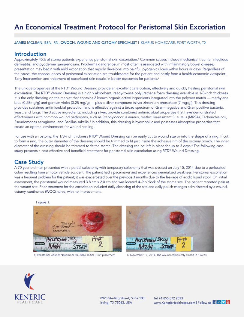
Case StudyA 70-year-old man presented with a partial colectomy with temporary colostomy that was created on July 15, 2014 due to a perforated colon resulting from a motor vehicle accident. The patient had a pacemaker and experienced generalized weakness. Peristomal excoriation was a frequent problem for this patient; it was exacerbated over the previous 3 months due to the leakage of acidic liquid stool. On initial assessment, the peristomal wound measured 3.8 cm x 2.0 cm and was located 4–9 o’clock of the stoma site. The patient reported pain at the wound site. Prior treatment for the excoriation included daily cleansing of the site and daily pouch changes administered by a wound, ostomy, continence (WOC) nurse, with no improvement.
a) Peristomal wound: November 10, 2014, Initial RTD® placement b) November 17, 2014, The wound completely closed in 1 week
Figure 1.
a b
Tel +1 855 872 2013 www.KenericHealthcare.com | Follow us
8925 Sterling Street, Suite 100 Irving, TX 75063, USA
An Economical Treatment Protocol for Peristomal Skin Excoriation
JAMES MCLEAN, BSN, RN, CWOCN, WOUND AND OSTOMY SPECIALIST | KLARUS HOMECARE, FORT WORTH, TX
The RTD® treatment protocol was initiated November 10, 2014. The treatment was provided by a WOC nurse as follows: RTD® Wound Dressing, 1/8-inch thickness, was cut to wound size and placed on the wound. A convex pouch with a wafer was attached using tube paste around the precut opening for the stoma. An ostomy belt was used to maintain a snug fit. At a follow-up visit the following day to evaluate the effectiveness of the treatment protocol, significant improvement, including a reduction in pain, was noted; subsequent follow-up visits occurred on November 14, 2014 and November 17, 2014. The wound was completely resolved on November 17, 2014 (see Figure 1a,b). RTD® was discontinued, and the patient continued with pouch changes every 3 days.
Before use of RTD®, treatment for this patient cost the home health agency an average of $405 per week (daily skilled nursing visits [SNVs] plus $90 pouch supplies). After treatment with RTD®, the cost of treatment for this patient averaged $114 per week (2 SNVs per week plus $24 pouch supplies).
ConclusionThe RTD® Wound Dressing provides an effective option for treating peristomal excoriation related to trauma, fungal and bacterial infection, and pyoderma gangrenosum. In this case, the wound resolved in 1 week. The patient reported less pain following the use of RTD® (likely due to the dressing’s gentian violet). In addition, a cost savings of 72% was achieved for the agency. Ultimately, a quicker healing time for peristomal wounds could help save a home health agency staff time and money on costly ostomy and wound care products. Managing peristomal excoriation quickly and effectively also results in a better quality of life for individuals living with an ostomy.
References1. Colwell J, Goldberg M, Carmel J. The state of the standard diversion. J Wound Ostomy Continence Nurs. 2001;28(1):6–17.2. Alvey B, Beck DE. Peristomal dermatology. Clin Colon Rectal Surg. 2008:21(1):41–44.3. Keneric Healthcare. Data on File 2006.4. Keneric Healthcare RTD® Wound Dressing Instructions for Use.
Tel +1 855 872 2013 www.KenericHealthcare.com | Follow us
8925 Sterling Street, Suite 100 Irving, TX 75063, USA
Incorporating a New Highly Absorbent Antimicrobial Polyurethane Foam* in a Multimodal High Risk Diabetic Wound Care Algorithm
DR. KAREN BROOKS, DPM & MR. MICHAEL OLDEN, H.T, OST, C-PED, PMAC, NAWCC OF PEDORTHIC CLINICS KERRVILLE DIVISION-SOUTH TEXAS VETERANS HEALTH CARE SYSTEMS
IntroductionRTD® Wound Care Dressing is a novel new, highly absorbent antimicrobial polyurethane foam dressing that has been integrated into our Diabetic Wound Algorithm. RTD® is recommended to protect the wound from bacterial trapping, as well as bacterial binding, thus preventing biofilmformation. It is a highly conformable and absorptive dressing designed for first line defense for exudate & infection control. It works extremely well with the use of biologics and prevents the active ingredients in the other modalities from being drawn away and does not appear to impact their effectiveness.
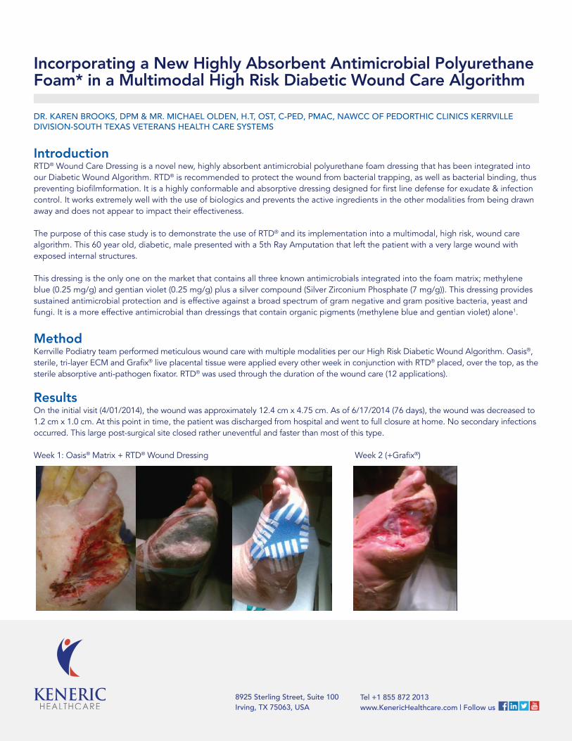
The purpose of this case study is to demonstrate the use of RTD® and its implementation into a multimodal, high risk, wound care algorithm. This 60 year old, diabetic, male presented with a 5th Ray Amputation that left the patient with a very large wound with exposed internal structures.
This dressing is the only one on the market that contains all three known antimicrobials integrated into the foam matrix; methylene blue (0.25 mg/g) and gentian violet (0.25 mg/g) plus a silver compound (Silver Zirconium Phosphate (7 mg/g)). This dressing provides sustained antimicrobial protection and is effective against a broad spectrum of gram negative and gram positive bacteria, yeast and fungi. It is a more effective antimicrobial than dressings that contain organic pigments (methylene blue and gentian violet) alone1.
MethodKerrville Podiatry team performed meticulous wound care with multiple modalities per our High Risk Diabetic Wound Algorithm. Oasis®, sterile, tri-layer ECM and Grafix® live placental tissue were applied every other week in conjunction with RTD® placed, over the top, as the sterile absorptive anti-pathogen fixator. RTD® was used through the duration of the wound care (12 applications).
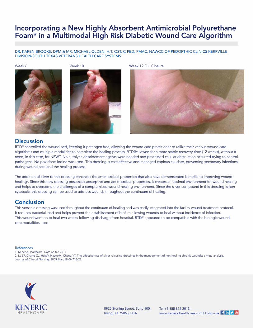
ResultsOn the initial visit (4/01/2014), the wound was approximately 12.4 cm x 4.75 cm. As of 6/17/2014 (76 days), the wound was decreased to 1.2 cm x 1.0 cm. At this point in time, the patient was discharged from hospital and went to full closure at home. No secondary infections occurred. This large post-surgical site closed rather uneventful and faster than most of this type.
Week 1: Oasis® Matrix + RTD® Wound Dressing Week 2 (+Grafix®)
Tel +1 855 872 2013 www.KenericHealthcare.com | Follow us
8925 Sterling Street, Suite 100 Irving, TX 75063, USA
Incorporating a New Highly Absorbent Antimicrobial Polyurethane Foam* in a Multimodal High Risk Diabetic Wound Care Algorithm
DR. KAREN BROOKS, DPM & MR. MICHAEL OLDEN, H.T, OST, C-PED, PMAC, NAWCC OF PEDORTHIC CLINICS KERRVILLE DIVISION-SOUTH TEXAS VETERANS HEALTH CARE SYSTEMS
Week 6 Week 10 Week 12 Full Closure
DiscussionRTD® controlled the wound bed, keeping it pathogen free, allowing the wound care practitioner to utilize their various wound care algorithms and multiple modalities to complete the healing process. RTD®allowed for a more stable recovery time (12 weeks), without a need, in this case, for NPWT. No autolytic debridement agents were needed and processed cellular destruction occurred trying to control pathogens. No povidone-Iodine was used. This dressing is cost effective and managed copious exudate, preventing secondary infections during wound care and the healing process.
The addition of silver to this dressing enhances the antimicrobial properties that also have demonstrated benefits to improving wound healing2. Since this new dressing possesses absorptive and antimicrobial properties, it creates an optimal environment for wound healing and helps to overcome the challenges of a compromised wound-healing environment. Since the silver compound in this dressing is non cytotoxic, this dressing can be used to address wounds throughout the continuum of healing.
ConclusionThis versatile dressing was used throughout the continuum of healing and was easily integrated into the facility wound treatment protocol. It reduces bacterial load and helps prevent the establishment of biofilm allowing wounds to heal without incidence of infection. This wound went on to heal two weeks following discharge from hospital. RTD® appeared to be compatible with the biologic wound care modalities used.
References1. Keneric Healthcare: Data on file 20142. Lo SF, Chang CJ, HuWY, HayterM, Chang YT. The effectiveness of silver-releasing dressings in the management of non-healing chronic wounds: a meta-analysis. Journal of Clinical Nursing. 2009 Mar; 18 (5):716-28.
Tel +1 855 872 2013 www.KenericHealthcare.com | Follow us
8925 Sterling Street, Suite 100 Irving, TX 75063, USA
Uses of a Novel New Absorbent Antimicrobial Polyurethane Foam Wound Dressing
DR. BELINDA MARCUS, MD, FACEP, CWS, MEDICAL DIRECTOR AND KATHY KAUFMAN, LPN, CHT, WOUND CARE NURSE, HYPERBARXS AT NORTHSIDE FORSYTH, ATLANTA, GA
IntroductionObjectives:1. Describe a new unique highly absorbent antimicrobial wound care dressing2. Identify the indications for use, versatility, and application of this new dressing3. Describe the outcomes of 5 clinical case studies
Chronic non-healing wounds represent a problem for clinicians. The more difficult it is for chronic wounds to heal, the greater the potential burden to patients, their families, and the healthcare system. Treating wounds is most challenging when they become chronic. The prevalence of chronic wounds increases with age and compounding medical conditions. An estimated 6 million patients in the U.S. have chronic wounds, representing an estimated annual $20 billion burden on the healthcare system1. It is therefore important to effectively address wound concerns early and help prevent non-healing chronic wounds.
The purpose of this case series is to demonstrate the effectiveness of a novel new highly absorbent polyurethane foam dressing for both chronic wounds and its efficacy as a first line therapy. Clinical case studies will be presented that demonstrate the versatility and functionality of RTD® Wound Care Dressing. This dressing is the only one on the market that contains known organic active ingredients integrated into the foam matrix; methylene blue (0.25 mg/g) and gentian violet (0.25 mg/g) plus a silver compound (Silver Zirconium Phosphate (7 mg/g)). This dressing provides sustained antimicrobial protection and is effective against a broad spectrum of gram negative and gram positive bacteria, yeast and fungi. It is a more effective antimicrobial than dressings that contain organic pigments (methylene blue and gentian violet) alone2.
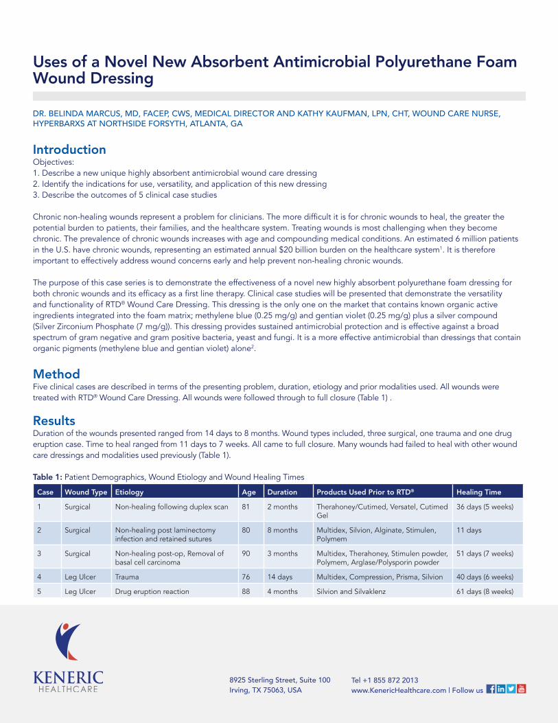
MethodFive clinical cases are described in terms of the presenting problem, duration, etiology and prior modalities used. All wounds were treated with RTD® Wound Care Dressing. All wounds were followed through to full closure (Table 1) .
ResultsDuration of the wounds presented ranged from 14 days to 8 months. Wound types included, three surgical, one trauma and one drug eruption case. Time to heal ranged from 11 days to 7 weeks. All came to full closure. Many wounds had failed to heal with other wound care dressings and modalities used previously (Table 1).
Table 1: Patient Demographics, Wound Etiology and Wound Healing Times
Case Wound Type Etiology Age Duration Products Used Prior to RTD® Healing Time
1 Surgical Non-healing following duplex scan 81 2 months Therahoney/Cutimed, Versatel, Cutimed Gel
36 days (5 weeks)
2 Surgical Non-healing post laminectomy infection and retained sutures
80 8 months Multidex, Silvion, Alginate, Stimulen, Polymem
11 days
3 Surgical Non-healing post-op, Removal of basal cell carcinoma
90 3 months Multidex, Therahoney, Stimulen powder, Polymem, Arglase/Polysporin powder
51 days (7 weeks)
4 Leg Ulcer Trauma 76 14 days Multidex, Compression, Prisma, Silvion 40 days (6 weeks)
5 Leg Ulcer Drug eruption reaction 88 4 months Silvion and Silvaklenz 61 days (8 weeks)
Tel +1 855 872 2013 www.KenericHealthcare.com | Follow us
8925 Sterling Street, Suite 100 Irving, TX 75063, USA
Uses of a Novel New Absorbent Antimicrobial Polyurethane Foam Wound Dressing
DR. BELINDA MARCUS, MD, FACEP, CWS, MEDICAL DIRECTOR AND KATHY KAUFMAN, LPN, CHT, WOUND CARE NURSE, HYPERBARXS AT NORTHSIDE FORSYTH, ATLANTA, GA
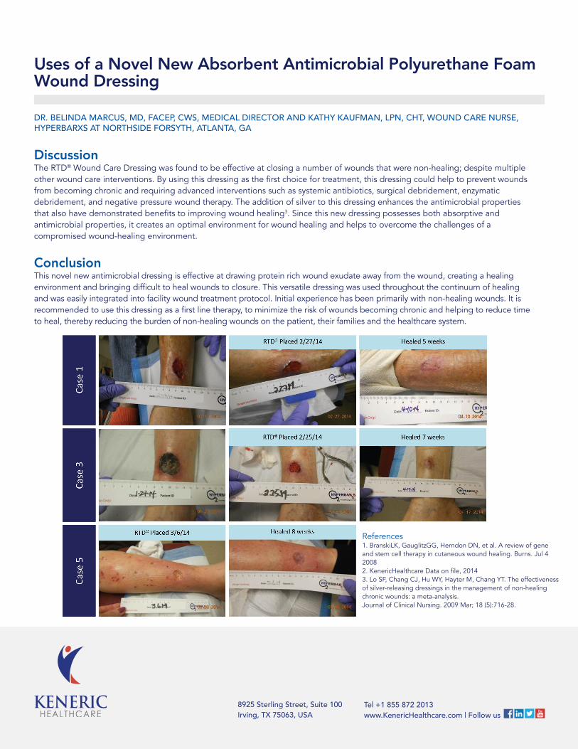
DiscussionThe RTD® Wound Care Dressing was found to be effective at closing a number of wounds that were non-healing; despite multiple other wound care interventions. By using this dressing as the first choice for treatment, this dressing could help to prevent wounds from becoming chronic and requiring advanced interventions such as systemic antibiotics, surgical debridement, enzymatic debridement, and negative pressure wound therapy. The addition of silver to this dressing enhances the antimicrobial properties that also have demonstrated benefits to improving wound healing3. Since this new dressing possesses both absorptive and antimicrobial properties, it creates an optimal environment for wound healing and helps to overcome the challenges of a compromised wound-healing environment.
ConclusionThis novel new antimicrobial dressing is effective at drawing protein rich wound exudate away from the wound, creating a healing environment and bringing difficult to heal wounds to closure. This versatile dressing was used throughout the continuum of healing and was easily integrated into facility wound treatment protocol. Initial experience has been primarily with non-healing wounds. It is recommended to use this dressing as a first line therapy, to minimize the risk of wounds becoming chronic and helping to reduce time to heal, thereby reducing the burden of non-healing wounds on the patient, their families and the healthcare system.
References1. BranskiLK, GauglitzGG, Herndon DN, et al. A review of gene and stem cell therapy in cutaneous wound healing. Burns. Jul 4 20082. KenericHealthcare Data on file, 20143. Lo SF, Chang CJ, Hu WY, Hayter M, Chang YT. The effectiveness of silver-releasing dressings in the management of non-healing chronic wounds: a meta-analysis. Journal of Clinical Nursing. 2009 Mar; 18 (5):716-28.
THE ADDITION OF SILVER TO METHYLENE BLUE AND GENTIAN VIOLET: ANTIMICROBIAL BENEFITS OF A POLYURETHANE FOAM WOUND CARE DRESSING DR. JEAN ACHTERBERG, DC AND MARIUSZ KNAP (KENERIC HEALTHCARE, IRVING TEXAS)
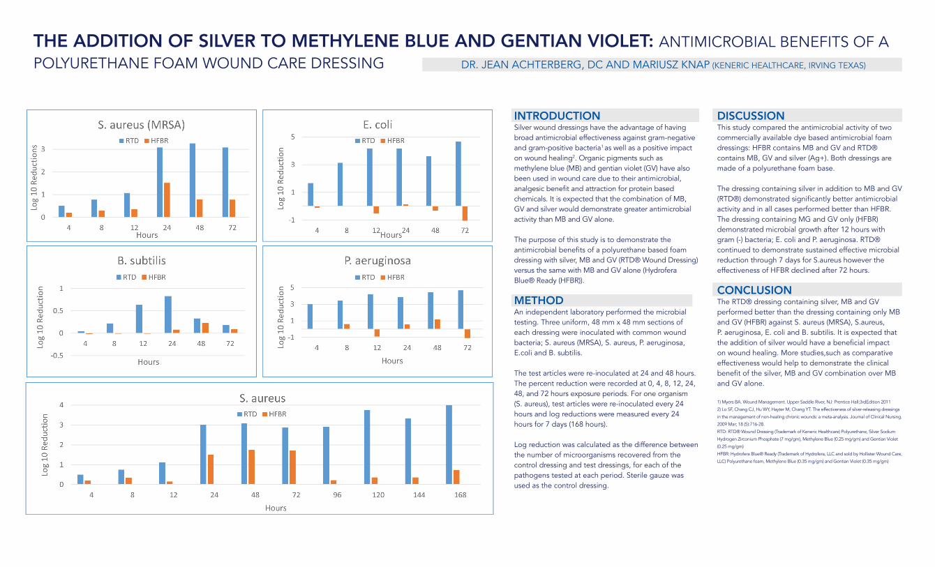
INTRODUCTIONSilver wound dressings have the advantage of having broad antimicrobial effectiveness against gram-negative and gram-positive bacteria1 as well as a positive impact on wound healing2. Organic pigments such as methylene blue (MB) and gentian violet (GV) have also been used in wound care due to their antimicrobial, analgesic benefit and attraction for protein based chemicals. It is expected that the combination of MB, GV and silver would demonstrate greater antimicrobial activity than MB and GV alone.
The purpose of this study is to demonstrate the antimicrobial benefits of a polyurethane based foam dressing with silver, MB and GV (RTD® Wound Dressing) versus the same with MB and GV alone (Hydrofera Blue® Ready (HFBR)).
METHODAn independent laboratory performed the microbial testing. Three uniform, 48 mm x 48 mm sections of each dressing were inoculated with common wound bacteria; S. aureus (MRSA), S. aureus, P. aeruginosa, E.coli and B. subtilis.
The test articles were re-inoculated at 24 and 48 hours. The percent reduction were recorded at 0, 4, 8, 12, 24, 48, and 72 hours exposure periods. For one organism (S. aureus), test articles were re-inoculated every 24 hours and log reductions were measured every 24 hours for 7 days (168 hours).
Log reduction was calculated as the difference between the number of microorganisms recovered from the control dressing and test dressings, for each of the pathogens tested at each period. Sterile gauze was used as the control dressing.
DISCUSSIONThis study compared the antimicrobial activity of two commercially available dye based antimicrobial foam dressings: HFBR contains MB and GV and RTD® contains MB, GV and silver (Ag+). Both dressings are made of a polyurethane foam base.
The dressing containing silver in addition to MB and GV (RTD®) demonstrated significantly better antimicrobial activity and in all cases performed better than HFBR. The dressing containing MG and GV only (HFBR) demonstrated microbial growth after 12 hours with gram (-) bacteria; E. coli and P. aeruginosa. RTD® continued to demonstrate sustained effective microbial reduction through 7 days for S.aureus however the effectiveness of HFBR declined after 72 hours.
CONCLUSIONThe RTD® dressing containing silver, MB and GV performed better than the dressing containing only MB and GV (HFBR) against S. aureus (MRSA), S.aureus, P. aeruginosa, E. coli and B. subtilis. It is expected that the addition of silver would have a beneficial impact on wound healing. More studies,such as comparative effectiveness would help to demonstrate the clinical benefit of the silver, MB and GV combination over MB and GV alone.
1) Myers BA. Wound Management. Upper Saddle River, NJ: Prentice Hall;3rdEdition 2011
2) Lo SF, Chang CJ, Hu WY, Hayter M, Chang YT. The effectiveness of silver-releasing dressings
in the management of non-healing chronic wounds: a meta-analysis. Journal of Clinical Nursing.
2009 Mar; 18 (5):716-28.
RTD: RTD® Wound Dressing (Trademark of Keneric Healthcare) Polyurethane, Silver Sodium
Hydrogen Zirconium Phosphate (7 mg/gm), Methylene Blue (0.25 mg/gm) and Gentian Violet
(0.25 mg/gm)
HFBR: Hydrofera Blue® Ready (Trademark of Hydrofera, LLC and sold by Hollister Wound Care,
LLC) Polyurethane foam, Methylene Blue (0.35 mg/gm) and Gentian Violet (0.35 mg/gm)
AN EFFECTIVE AND ECONOMICAL APPROACH TO RESOLVING SEVERE HYPERGRANULATION USING RTD® WOUND DRESSING: ANTIMICROBIAL POLYURETHANE FOAM WITH INTEGRATED METHYLENE BLUE, GENTIAN VIOLET AND SILVER SONIA VARGAS RN, WCC, COS-C (APRIL SKYYHOME HEALTH CARE, CORPUS CHRISTI, TEXAS), DR. JEAN ACHTERBERG DC, AND MARIUSZ KNAP
1) Vuolo, J, Hypergranulation: Exploring possible management options, British Journal of
Nursing, Vol 19, No. 6 SUPPL., pp. S4-S8 2010.
2) SilvaSorb® Antimicrobial Wound Gel, is a registered trademark of Medline Industries,
Mundelein, Illinois
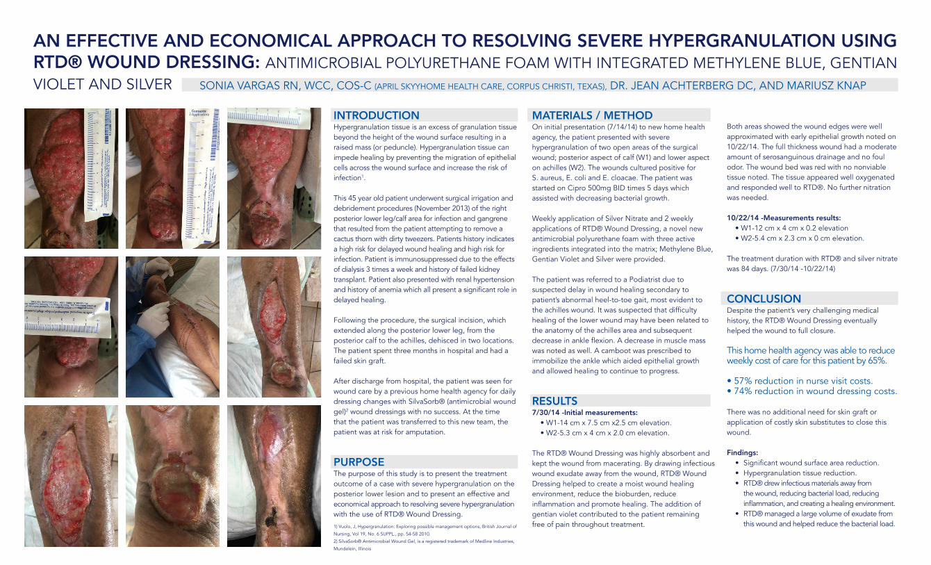
INTRODUCTIONHypergranulation tissue is an excess of granulation tissue beyond the height of the wound surface resulting in a raised mass (or peduncle). Hypergranulation tissue can impede healing by preventing the migration of epithelial cells across the wound surface and increase the risk of infection1.
This 45 year old patient underwent surgical irrigation and debridement procedures (November 2013) of the right posterior lower leg/calf area for infection and gangrene that resulted from the patient attempting to remove a cactus thorn with dirty tweezers. Patients history indicates a high risk for delayed wound healing and high risk for infection. Patient is immunosuppressed due to the effects of dialysis 3 times a week and history of failed kidney transplant. Patient also presented with renal hypertension and history of anemia which all present a significant role in delayed healing.
Following the procedure, the surgical incision, which extended along the posterior lower leg, from the posterior calf to the achilles, dehisced in two locations. The patient spent three months in hospital and had a failed skin graft.
After discharge from hospital, the patient was seen for wound care by a previous home health agency for daily dressing changes with SilvaSorb® (antimicrobial wound gel)2 wound dressings with no success. At the time that the patient was transferred to this new team, the patient was at risk for amputation.
PURPOSEThe purpose of this study is to present the treatment outcome of a case with severe hypergranulation on the posterior lower lesion and to present an effective and economical approach to resolving severe hypergranulation with the use of RTD® Wound Dressing.
MATERIALS / METHODOn initial presentation (7/14/14) to new home health agency, the patient presented with severe hypergranulation of two open areas of the surgical wound; posterior aspect of calf (W1) and lower aspect on achilles (W2). The wounds cultured positive for S. aureus, E. coli and E. cloacae. The patient was started on Cipro 500mg BID times 5 days which assisted with decreasing bacterial growth.
Weekly application of Silver Nitrate and 2 weekly applications of RTD® Wound Dressing, a novel new antimicrobial polyurethane foam with three active ingredients integrated into the matrix; Methylene Blue, Gentian Violet and Silver were provided.
The patient was referred to a Podiatrist due to suspected delay in wound healing secondary to patient’s abnormal heel-to-toe gait, most evident to the achilles wound. It was suspected that difficulty healing of the lower wound may have been related to the anatomy of the achilles area and subsequent decrease in ankle flexion. A decrease in muscle mass was noted as well. A camboot was prescribed to immobilize the ankle which aided epithelial growth and allowed healing to continue to progress.
RESULTS7/30/14 -Initial measurements:
• W1-14 cm x 7.5 cm x2.5 cm elevation.• W2-5.3 cm x 4 cm x 2.0 cm elevation.
The RTD® Wound Dressing was highly absorbent and kept the wound from macerating. By drawing infectious wound exudate away from the wound, RTD® Wound Dressing helped to create a moist wound healing environment, reduce the bioburden, reduce inflammation and promote healing. The addition of gentian violet contributed to the patient remaining free of pain throughout treatment.
Both areas showed the wound edges were well approximated with early epithelial growth noted on 10/22/14. The full thickness wound had a moderate amount of serosanguinous drainage and no foul odor. The wound bed was red with no nonviable tissue noted. The tissue appeared well oxygenated and responded well to RTD®. No further nitration was needed.
10/22/14 -Measurements results:• W1-12 cm x 4 cm x 0.2 elevation• W2-5.4 cm x 2.3 cm x 0 cm elevation.
The treatment duration with RTD® and silver nitrate was 84 days. (7/30/14 -10/22/14)
CONCLUSIONDespite the patient’s very challenging medical history, the RTD® Wound Dressing eventually helped the wound to full closure.
This home health agency was able to reduce weekly cost of care for this patient by 65%.
• 57% reduction in nurse visit costs.• 74% reduction in wound dressing costs.
There was no additional need for skin graft or application of costly skin substitutes to close this wound.
Findings:• Significant wound surface area reduction.• Hypergranulation tissue reduction.• RTD® drew infectious materials away from
the wound, reducing bacterial load, reducing inflammation, and creating a healing environment.
• RTD® managed a large volume of exudate from this wound and helped reduce the bacterial load.

Time (in min) exposed to light
Cha
nge
in F
luor
esce
nce
(at 5
25nm
)
25
20
15
10
5
0
0 20 40 60
A NOVAL POLYURETHINE FOAM WOUND CARE DRESSING WITH SILVER, METHYLENE BLUE AND GENTIAN VIOLET: CONFIRMING THE PRESENCE OF SINGLET OXYGEN GENERATION AND ANTIMICROBIAL ACTIVITYAUTHORS – DR. GORDON A WOOD, AL HENRY, AND MARIUSZ KNAP (KENERIC HEALTHCARE, IRVING TEXAS)
References:1 The RTD® Advanced Wound Care Dressing, RTDWP-052014.2) Myers BA. Wound Management. Upper Saddle River, NJ: Prentice Hall;3rd Edition 20113) Lo SF, Chang CJ, Hu WY, Hayter M, Chang YT. The effectiveness of silver-releasing dressings in the management of non-healing chronic wounds: a meta-analysis. Journal of Clinical Nursing. 2009 Mar; 18 (5):716-28.
INTRODUCTION
Silver wound dressings have the advantage of having broad antimicrobial effectiveness against gram-negative and gram-positive bacteria2 as well as a positive impact on wound healing3. Organic pigments such as methylene blue (MB) and gentian violet (GV) have also been used in wound care due to their antimicrobial, analgesic benefit and attraction for protein based chemicals. It is expected that the combination of MB, GV, Silver and the proprietary technology of integrating and tightly bounding these ingredients into a foam matrix will significantly increase the antimicrobial activity and production of Singlet Oxygen.
An independent laboratory has developed and performed assays to provide scientific evidence for the effectiveness of the RTD® dressing and to define its mechanism of action. Two Technical Objectives were proposed to characterize the RTD® Wound Care Dressing:
Objective 1: Designed to develop an analytical method to detect singlet oxygen generated by the formula.Objective 2: Monitor the antimicrobial activity of the wound dressing over time.
The results of these two objectives are reported below.
METHOD – CONFIRMING THE PRESENCE OF SINGLET OXYGEN GENERATED BY RTD® WOUND CARE DRESSING
The detection reagent, Sensor Green (Thermo Fisher Scientific, Inc.), emits at 525nm maxima (excitation maxima 504nm) in the presence of singlet oxygen, and does not respond to the other reactive oxygen species, such as hydroxyl radical or superoxide.
Test samples containing methylene blue alone, methylene blue with silver sodium hydrogen zirconium phosphate (Alphasan RC-2000), or a section of RTD® wound care dressing were added to deionized water (DI water) or 0.9%wt. sodium chloride solution with 1μM of Sensor Green.
The sample mixtures were placed in separate wells on a 96-well plate, and exposed to a halogen lamp (<50W/12V) at a 10 cm distance for up to 70 minutes,
at 10 minute intervals. At each 10-minute interval, the fluorescence spectrum (490-600nm emission/470nm excitation) of each sample was measured by a plate reader (Varioskan LUX, Thermo Fisher Scientific).
The fluorescence emission scans of methylene blue with Sensor Green and Sensor Green only (negative control) were overlaid and shown in Figure 1. The fluorescence of the methylene blue (with Sensor Green) increased after each 10-minute light exposure, whereas the fluorescence of the Sensor Green only sample remained relatively unchanged after light exposure. This confirms that singlet oxygen was generated by methylene blue when exposed to light, and that the assay is effective. For comparison, the change in fluorescence (at maxima 525nm) was plotted against time (in minutes). The change in fluorescence was calculated by: Change in fluorescence = F - F0 where F is the fluorescence measured and F0 is the fluorescence measured before light exposure (time=0 mins). Figure 2 presents the change in fluorescence over time for the RTD® Wound Dressing compared to that of a control foam (without antimicrobial active ingredients). The data suggest that singlet oxygen was generated by the RTD® Wound Dressing when exposed to light.
In order to measure the effect of silver sodium hydrogen zirconium phosphate on the singlet oxygen generation efficiency of methylene blue, 3.2μg/mL of methylene blue was mixed with 89.6μg/mL of silver sodium hydrogen zirconium phosphate (same weight ratio as in the RTD® Wound Dressing formula) and Sensor Green. The change in fluorescence of the mixture was plotted and shown in Figure 3.
The change in fluorescence of the methylene blue-silver complex mixture over time had a minor improvement from the methylene blue sample alone.
METHOD – ANTIMICROBIAL ACTIVITY OF THE RTD® WOUND CARE DRESSING
A previous report provided by an independent laboratory1 has shown broad spectrum antimicrobial activity of the RTD® Wound Dressing after 24+ hours of contact time.
The goal of our experiment is to measure the log reduction of selected bacteria at shorter contact times.
The assay was modeled after the standard Zone of Inhibition assay. Two cm by two cm cut-outs of the RTD® Wound Dressing were soaked in 0.9% sodium chloride solution, and placed on Luria-Bertani (LB) medium agar plated with 106, 105, 104 or 103 of bacteria. The surface of the RTD® Wound Dressing squares covering the agar plates were in contact with approximately 105, 104, 103 and 102 bacteria, depending on the total number of bacteria plated on the agar.
The wound dressing cut-outs were in contact with the agar surface for 10 minutes, 30 minutes, 1 hour, 5 hours and 24 hours at room temperature, then the agar plates were incubated at 37°C for 16 hours. A complete inhibition of growth of the bacteria where the RTD® cut-outs were in contact with the agar plate is considered to be a log10 reduction of ≥5, 4, 3 or 2, depending on the number of bacteria in contact of the RTD® cut-outs. A summary of the contact time required for log10 5, 4, 3 or 2 reductions of four selected bacteria that are relevant to human infections are shown in Table 1.
CONCLUSION
The proposed objectives were completed and below are the key findings:
1. A fluorescence-based singlet oxygen detection method was established.2. The generation of singlet oxygen by the RTD® Wound Dressing was confirmed.3. Shorter contact times required for a 5 log reduction by the RTD® Wound Dressing were measured for Staphylococcus aureus and Pseudomonas aeruginosa, compared to previously reported data1.
Log10 Reduction Staphylococcus aureus(ATCC 12600)
Enterococcus faecalis(ATCC 19433)
Pseudomonas aeruginosa (ATCC 10145)
Klebsiella pneumoniae (ATCC 49472)
≥ 5 1 hour >24 hours 30 mins 24 hours
≥ 4 10 mins >24 hours 10 mins 6 hours
≥ 3 10 mins 24 hours 10 mins 6 hours
≥ 2 10 mins 24 hours 10 mins 1 hour
Table 1. Contact time required for log reduction of four selected bacteria strains.
Wavelength (nm)
Fluo
resc
ence
14.000
12.000
10.000
8.000
6.000
4.000
2.000
0.000490 540 590
10 min
20 min
30 min
40 min
50 min
60 min
70 min
Wavelength (nm)Fl
uore
scen
ce
14.00012.00010.000
8.0006.0004.0002.0000.000
490 540 590
10 min
20 min
30 min
40 min
50 min
60 min
70 min
A. B.
Figure 1. (Above) Overlay of fluorescence spectra of (A) methylene blue with Sensor Green and (B) Sensor Green only in DI water, after each 10-minute light exposure. The emission fluorescence spectra were measured from 490nm to 600nm, with 470nm excitation.
Figure 2. (Left) Change in fluorescence (at maxima 525nm) of RTD® Dressing compared to a negative control foam. Each sample was incubated with Sensor Green in 0.9% sodium chloride solution.
Figure 3. (Below) Comparison of the change in fluorescence of methylene blue versus methylene blue + silver sodium zirconium phosphate in (A) water and(B) 0.9% sodium chloride solution.
Time (in min) exposed to light0 50
Cha
nge
in F
luor
esce
nce
(at 5
25nm
)
10
9
8
7
6
5
4
3
2
1
0
RTD® Dressing
control foam
Time (in min) exposed to light0 50
Cha
nge
in F
luor
esce
nce
(at 5
25nm
)
25
20
15
10
5
0
Methylene Blue
Methylene Blue + Silver complexA. B.
RTD: RTD® Wound Dressing (Trademark of Keneric Healthcare) Polyurethane, Silver Sodium Hydrogen Zirconium Phosphate (7 mg/gm), Methylene Blue (0.25 mg/gm) and Gentian Violet (0.25 mg/gm)