Embed Size (px)
Citation preview

AD-A242 809 AD
AN EXPERIMENTAL BRAIN MISSILE WOUND:ASCERTAINING PATHOPHYSIOLOGY AND EVALUATING TREATMENTS
TO LOWER MORTALITY AND MORBIDITY
ANNUAL/FINAL REPORT O TICNOVI 9 1991
MICHAEL E. CAREY
JUNE 4, 1991
Supported by
U.S. ARMY MEDICAL RESEARCH AND DEVELOPMENT COMMANDFort Detrick, Frederick, Maryland 21702-5012
Contract No. DAMD17-86-C-6098
Louisiana State University Medical Center1542 Tulane Avenue
New Orleans, Louisiana 70112
Approved for public releaseT distribution unlimited.
The findings in this report are not to be construed as anofficial Department of the Army position unless so designated
by other authorized documents
91-15850

June 4, 1991 Annual/Final 14 Apr 86 - 13 Apr 91
An Experimental Brain Missile Wound: Ascertaining DAMD17-86-C-6098
Pathophysiology and Evaluating Treatments to LowerMortality and Morbidity 62772A
35162772A874 AAMichael E. Carey
Louisiana State University Medical Center1542 Tulane AvenueNew Orleans, LA 70112-2822
U.S. Army Medical Research and Development CommandFort DetrickFrederick, MD 21702-5012
Annual report for April 14, 1990 - April 13, 1991
Approved for public release; distribution unlimited
Cerebral ischemia does not occur following a brain wounding provided that ICPis <60 mmHg. Rather focal hyperperfusion occurs.
Mechanical and chemical control of CBF are disturbed following brain woundingbut these disturbances have wide regional variations.
The missile-wounded brain may show severe CBF reductions with even a mild fallin MABP. No reflow follows cerebral ischemic occurring after frain wounding.
Brain wounding is associated with decreased brain stem and hypothalamicbiogenic amines especially epinephrine. This seems to be a part of ageneralized stress response.
Free radicals appear in the brain within minutes of brain injury.
Experimental brain wounding; Cerebral blood flow; Brainbiogenic amines; Free radicals after brain wounding;RA 2
Unclassified Unclassified Unclassified Unlimited


3SUMMARY
This brain wound project has shown the following:
1) a brain missile wound may kill by producing apnea. Thismissile-induced apnea may be reversed providedrespiratory support is provided. Drug therapy to reverseapnea should be sought.
2) mild vasogenic edema occurs with missile wounding butthis does not require specific treatment.
3) immediately following brain wounding extremely largeincreases in CSF prostaglandins occur. Thesebiologically active molecules may cause physiologiceffects at a distance from the missile wound.
4) a brain missile wound does not cause cerebral ischemiaprovided that the ICP is not > 60 mmHg. Rather thanischemia, a transient increased rCBF occurs particularlyabout the missile track
5) both mechanical and chemical CBF regulation areprofoundly affected following a brain missile wound butthe effect is not uniform throughout the brain; rathermarked regional variations occur.
6) even a slight drop in mean arterial blood pressure afterbrain wounding may cause a profound decrease in CBF.Furthermore, once a decreased CBF has occurred followingMABP reduction, restoration of MABP with blood may notimprove CBF: the missile-wounded brain is subject to NOREFLOW. It is imperative that blood pressure bemaintained in brain wounded soldiers.
7) brain wounding causes a change in hypothalamic and brainstem biogenic amines particularly a decrease inepinephrine. The so-called "Cushing" response appears tobe a variant of a generalized stress response. Anextreme disordering of biogenic amines in the brain stemdoes not appear to account for death following brainwounding.
8) GM1 ganglioside suggestively improved the extent andrapidity of neurologic recovery following brain missilewounding. Further tests with this drug are warranted. ° yet
9) free radicals are formed within minutes of traumatic ,brain injury. To combat their action our reserach .suggests that antioxidant therapy should be started t1 t,
within a few minutes of brain injury.
io

4
FOREWORD: In conducting the research described in this report,
the investigators adhered to the "Guide for the care an"' use oflaboratory animals", prepared by the committee on care and use oflaboratory animals of the Institute of Laboratory AnimalResources, National Research Council (DHEW Publication No. (NIH)78-23, Revised 1978).
The experiments reported in this report were performed by:
D. Torbati, Ph.D.J. Soblosky, Ph.D.D.F. Church, Ph.D.W.A. Pryor, Ph.D.D. Awasthi, M.D.H.C. McKowen, M.D.L. Rogers, M.D.J.B. Farrell, B.S.J.F. Davidson, B.S.
This manuscript was typed by:
Mrs. E.P. Hulbert

5TABLE OF CONTS
PAGE
Summary ............................................................ 3
Background ........................................................ 7
Method ............................................................. 7
Significant Findings .............................................. 8
A. Contract DAMD7-83-C-3145 (1983-1985) ..................... 8
1. Respiratory Dysfunction .............................. 8
2. Brain Edema .......................................... 9
3. CSF Prostaglandings .................................. 9
B. Contract DAMD17-86-C-6098 (1986-1991) ..................... 9
1. Regional CBF After Brain Wounding .................... 10
2. Mechanical Regulation of CBF after Brain Wounding .... 14
3. Chemical Regulation of CBF Following Brain Wounding..19
a. response to increasing arterial PCO2 . . . . . . . . . . . . . . 20
b. response to decreasing arterial PCO2 . . . . . . . . . . . . . . 24
c. response to decreasing arterial PO2.. .. .. ......... 28
d. response to increasing arterial P02.............. 31
e. response to simultaneously increased arterialPO2 and PCO2............................ ...... 33
f. summary ........................................... 37
4. Determining Why Cats Died Following Brain Wounding..38
5. Brain Biogenic Amines ............................... 41
6. Plasma Catecholamines ............................... 45
7. Effect of GM-1 Ganglioside on Recovery of Functionafter a Missile Wound to the Brain ................ 49
6
TABLE OF CONTENTS
PAGE
8. Generation of Free Radicals after Traumatic BrainInjury ............................................ 54
9. Directions for Future Missile Wound Research ........ 60
10. Epilogue ............................................. 61
11. References ........................................... 62
12. Appendix 1........................................... 76
13. Publication From This Research ...................... 77
14. Distribution List .................................... 81

7
This report summarizes the high points of research done undercontract DAMD17-86-C-6098 from April 1986 through November 1989when research funding was terminated through Congressional actionapparently instigated by the Honorable Robert Livingston (R-LA)acting at the behest of animal activists. Methodological detailsand data from all experiments have been provided in the followingyearly reports: DAMDl7-83-C-3145 of 31 December 1985; DAMDI7-86-C-6098 of 21 September 1987, 27 April 1989, 26 October 1990 and 9May 1991, (more than 500 pages). To provide continuity of thoughtour major findings from our 1983 to 1985 contract will also bepresented in this report.
BACKGROUND
Brain wounding accounts for almost half of all single-woundcombat deaths among soldiers. No essential change has occurred inthe post operative neurosurgical mortality of brain wounds fromWWII when it ranged from 11-14% to Vietnam where the postoperative mortality was 10-12% (17). Too few Americans receivedbrain wounds in Desert Storm to draw conclusions about currentmortality. In the civilian community more than 16,000 individualsdie each year from brain gunshot wounds (18). Thus, brainwounding is a severe problem for all segments of American society.
Virtually nothing is known about brain wounding from missilesin modern pathobiologic terms. Fewer than 25 papers have everbeen written concerning animal-based brain wound experiments wherecerebral and systemic reactions to a missile wound have beenstudied. At this point one sixth of these have come from ourlaboratory.
METHOD
In our first contract period from 1983 to 1985 (see reportDAMD17-83-C-3145, 31 December 1985) we refined the brain woundmodel of Gerber and Crockard (24,47) and developed our laboratorymodel simulating a non-lethal wound to the brain. This brainwound model has been used in all experiments discussed below.Three to 5 kg cats were anesthetized with i.p. and i.v.pentobarbital (35-40mg/kg) and, after appropriate surgical andother procedures, were placed in a stereotaxic frame. Thethoroughly anesthetized cats (feeling no pain) were then woundedin the right cerebral hemisphere through the intact skull with a2mm, 31.7 mg steel sphere fired at 240-390 m/sec. This produced abrain wound by a missile having either 0.9, 1.4 or 2.7 Joules (J).Steel spheres have been frequently employed in basic, experimentalballistic research (58,157). The lower missile energies weselected were designed to simulate those of a fraQment wound.Figure 1 shows the characteristic wound produced in the rightcerebral hemisphere.

8
ab.c. d.e.f. g. h.L
I i i I I I I I I I I I
15 10 5 015 10 5 015 10 5 0
mm mm mmFiqure 1: Drawings of coronal brain sections 14 mm caudal to theright frontal tip showing characteristic missile track locationsand sizes in nine cats (a to i). Only occasionally did missiletracks deviate from this region.
Statistics: Appropriate statistics were done in allexperiments. These are well defined in the individual yearlyreports in which these experiments are contained and in publishedpapers developed from these experiments.
SIGNIFICANT FINDINGS
A. Contract DAMD17-83-C-3145 (1983-1985)
1) Respiratory Dysfunction
We determined that post wounding respiratory dysfunctionaccounted for the majority of deaths following this righthemisphere brain missile wound and that the probability of a fatalapnea was missile-energy dependent being 14% with a 0.9 J missile,40% with 1.4 J missile and -70% with a 2.4 J missile.Importantly, the apnea resulting from brain wounding would oftenspontaneously resolve up to two hours after wounding if the animalwere provided respiratory support. The significant clinicalramification of this finding is that possibly soldiers withfragment wounds to their brain die not from an intrinsically fatalbrain wound p se but from missile wound-induced apnea whichmight reverse itself over time provided respiratory support weregiven (as by CPR). Of course, if the brain wound-induced apneacould reverse by itself (ie if the brain stem cardiorespiratorycenters were only transiently disturbed and not permanentlydestroyed by energy transfer from the missile) the possibilityalso exists that drug therapy might be found which would enhance

9
the recovery of temporarily deranged respirations thereby loweringthe mortality of soldiers with brain wounds. No work on thisconcept of trying to reverse trauma-induced apnea has been carriedout but it has civilian as well as military applications not onlyfor missile wounding but for closed head injury as well (18).
2) Brain Edema
In our initial work we also evaluated post wound brain edemaformation and found it to be vasogenic in nature, not too severe,and confined to the wounded hemisphere (19). The edema peaked 24-48 hours after wounding, then receded and resolved in a week. Theamount of edema formation was the same whether the missile had 0.9J or 1.4 Js of energy. We concluded that, clinically, afterfragment wounding, provided that brain hypoxia or ischemia did notoccur, the amount of brain edema to be expected was mild, selflimited and would not require specific treatment as by steroids.No information exists on the augmentation of missile-wound causedbrain edema by simultaneous hemor:hagic hypotension or hypoxia butthis would be worthy of study because it might directly affect howindividuals with brain wounds are handled. Soldiers in combat areparticularly prone to multiple wounds and hemorrhagic shockbecause of concomitant major vascular injury so the possibility ofshock occurring with a brain wound is real.
3) CSF Prostaglandins
Because brain injury is associated with prostaglandinformation and because prostaglandins themselves may causeadditional tissue, vascular, or neural effects, we evaluated theappearance of prostaglandins in the CSF after brain wounding.Within 5 minutes of wounding enormous amounts of multipleprostaglandins (Fl', Thromboxane, E2 and D2) were seen incisternal CSF. As CSF circulates widely throughout the brain,prostaglandins contained in CSF would be widely dispersed andthese biologically potent prostaglandins could, theoretically,alter neural or vascular function in the brain at distances quitefar removed from the actual wound site.
B. Contract DAMD17-86-C-6098 (1986-1991: experimentationterminated November 1989 by crder of Congress)
Clearly, normal brain function requires an adequatecirculation. We have been able to find only a few prior studiesinvestigating cerebral blood flow (CBF) after missile wounding butin none was reQional cerebral blood flow (rCBF) studied(25,33,92). Because of the importance of CBF for brain function,from 1986-1989 we undertook an exhaustive study of total andregional CBF and how its mechanical and chemical regulation mightbe affected by brain wounding. Microspheres and the referencesyringe withdrawal method were used to measure CBFs in allexperiments in this report.

10
1) Regional cerebral blood flow after brain wounding (DAND17-
86-C-6098, 21 September 1987)
Our initial experiments evaluated rCBF changes (-30 brainregions) including 3 concentric zones about the wound track,figure 2 A,B.
Pol PC4/ 0
Figure 2A: Brain dissection scheme and missile trajectories. Theheavier line depicts the usual missile trajectories. The lighterline indicates less frequent trajectories.
mmi i ... i ' i I i i '
S15 10 5 0 5 10 15
Figure 2B: The usual wound track measured 1-2 min in radius. The"inner core" extended 2.0-4.0 mmn outward from the center of themissile track, the "middle core" extended 4.0-5.5, and the "outercore" 5.5-7.0.

11
We observed no brain ischemia in any region caused by missilewounding whether the brain wound was created by a missile havingan energy of 0.9, 1.4 or 2.4 J. Considering all brain woundedanimals together, after wounding there was a general tendency fortotal and regional CBF to decrease but not to an ischemic level(<15ml/100g/min in the cat, 62). We attributed the generalized,mild CBF decrease to the increase in intracranial pressure (ICP)caused by missile wounding and a concomitant loss of CBFautoregulation.
In animals where the post wounding ICP remained -40 mmHg andtheir cerebral perfusion pressures (CPP) were greater than 75mmHg, no CBF reductions occurred. CCP= mean arterial bloodpressure (MABP) - ICP. CBF decreases after wounding were morelikely to occur when the post wounding ICP was 60 mmHg or greaterand the CPPs were consequently rc':.-ced to -50 mmHg owing tointracranial clots or brain swell, figure 3.
Control
SWithout Intracranial Hemorrhage
50 v With Intracranial Hemorrhage
40 -
E 30 36\Minute•0 p CFue :\ u
C.Ba w
107
C1 3060 90Minutes
*p(0.05 compared to control period and to cats without hemorrhage
Figure 3: Control unwounded cats had no change in whole brainCBF. Brain wounded cats with lower ICP elevations (-4OmmHg) alsomaintained CBF. If ICP rose to greater than 60 mmHg followingwounding significant CBF reductions were likely to occur.
12
In order to avoid the obfuscating effect of those animals withhigh post wounding ICPs and consequent generalized CBF reductionswe then analyzed regional CBF data from animals which had lowerpost wounding ICPs and which maintained CBF. By this means weascertained the effects of brain wounding alone on rCBF (asopposed to brain wounding plus elevated ICP).
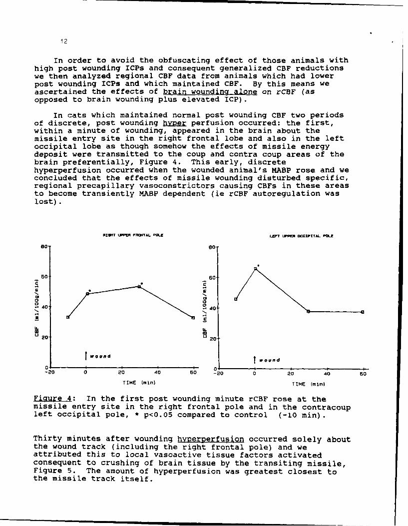
In cats which maintained normal post wounding CBF two periodsof discrete, post wounding hyper perfusion occurred: the first,within a minute of wounding, appeared in the brain about themissile entry site in the right frontal lobe and also in the leftoccipital lobe as though somehow the effects of missile energydeposit were transmitted to the coup and contra coup areas of thebrain preferentially, Figure 4. This early, discretehyperperfusion occurred when the wounded animal's MABP rose and weconcluded that the effects of missile wounding disturbed specific,regional precapillary vasoconstrictors causing CBFs in these areasto become transiently MABP dependent (ie rCBF autoregulation waslost).
RIU t RON TAL POL LEFT LPPE OCCIPITAL POL
50 90" /
40 - 40
1 A[00
W 20 20
wound wound0 I I I 0 !
-20 0 20 40 50 -20 0 20 40 60
TIME (min) TIME (min)
Figure 4: In the first post wounding minute rCBF rose at themissile entry site in the right frontal pole and in the contracoupleft occipital pole, * p<O.05 compared to control (-10 min).
Thirty minutes after wounding hyperperfusion occurred solely aboutthe wound track (including the right frontal pole) and weattributed this to local vasoactive tissue factors activatedconsequent to crushing of brain tissue by the transiting missile,Figure 5. The amount of hyperperfusion was greatest closest tothe missile track itself.

13
70-
m 50-
1/4
40-00/ 30-
m
In 20-
0- INNER0... MIDOLE
10 -0-- OUTER
0 I I i I I0 20 40 60 80
TIME (min)
Figure 5: Hyperperfusion occurred about the wound missile track30 minutes after wounding. The amount of hyperperfusion appearedsignificantly greater closer to the missile track than furtheraway from it. (* p<0.05 vs control; + p<0.05 vs middle; # p<0.05vs outer>
We concluded that if intracranial pressure were not elevated(as by an associated intracranial clot) and the CPP were above 60-70 mmHg, brain wounding would not be expected to be associatedwith cerebral ischemia. Any neurologic deficits observed afterwounding were the result of the mechanical effects of wounding andwere not caused by or potentiated by concomitant rCBF deficits.This knowledge is important because, intuitively, one might expectbrain wounding would cause some degree of ischemia and that drugsused to treat stroke might also be useful in treating brainwounds. Such is unlikely to be the case if the intended drug'smain purpose is to improve a reduced CBF as would be desirable instroke. Drugs with other properties must be sought to amelioratethe neurologic defects caused by brain missile wounding.

14
Comments on missile energy and observed pathobiological events
The effect of missile energy deposit upon the small andcompact brain stem producing apnea was unequivocally missile-energy dependent (p 2). Other physiologic changes which we havemeasured consequent to brain wounding were not correlated withmissile energy eg: the amount of brain edema formation, rise inrCBF in specific brain areas after wounding, and the level of CSFprostaglandin increase after missile injury. We hypothesize that:1) the lack of correlation of these physiologic variables withmissile energy was because of the extremely narrow energy windowswhich we had to use to produce a relatively non-fatal brain wound;0.9J to 1.4J. 2) Pathobiologically, the cerebral hemispheres aremuch larger than the brain stem and are able to absorb energy morereadily than the compact medulla. They, thus, reacted to theseclosely spaced missile energies as to a single energy.
2) Mechanical requlation of CBF after brain woundinq
Because we often saw a mild CBF reduction following brainwounding in the above experiments, suggesting a loss of CBFautoregulatory control, and because trauma and other CNS insultsare known to impair CBF autoregulation (26,93,94,96,107) weundertook a detailed study of rCBF autoregulation specificallyfollowing a brain missile wound. To our knowledge no such studyhad ever been undertaken.
Mechanical CBF regulation occurs when CBF is maintained in theface of a decreasing MABP (130). Classically, CBF remains normalas MABP is lowered by bleeding to -60 mmHg owing to relaxation ofcerebral resistance vessels. This phenomenon defines classicalCBF autoregulation. Below this level of hemorrhagic hypotensionCBF falls along with MABP: the brain can no longer regulate itsblood flow because precapillary arterioles can not dilate furtherto decrease resistance and maintain flow (Flow=Pressure/Resistance). CBF may also be attenuated by elevations ofICP which may reduce the CPP (107). Brain wounding affects thebrain stem,, increases ICP, and may reduce CPP(18,23,25,33,47,92). One may anticipate that brain wounding wouldadversely affect CBF autoregulation but the extent and degree ofsuch an impairment has never been investigated.

15
Hemorrhagic hypotension to lower MABP with the simultaneousmeasuring of CBF has been the standard means by which mechanicalCBF regulation has been tested (93,130). This time-honoredphysiological technique coincidently happens to mimic the clinicalsituation wherein a soldier might get a brain wound as well asanother wound (eg femoral artery) leading to significant bloodloss. Multipla fragment wounds are commonly seen in combat. Morethan 90% of wounded soldiers in VII Corps during Desert Storm wereso injured (unpublished data gathered by Dr. Carey). Militarily,it would be very important to know how the missile-wounded brainhandles its blood flow in the face of substantial blood loss.
In the experiments on mechanical CBF regulation we alsodecided to see whether the reinfusion of shed blood would improveany observed CBF derangements. This would mimic an exsanguinated,brain-wounded soldier receiving a blood transfusion to restoreMABP and hopefully CBF.
The non-wounded brain autoregulated CBF (i.e. maintained CBF)down to a mean MABP of -48mmHg because cerebrovascular resistanceappropriately and significantly decreased from 3.6 to 1.6resistance units. Reinfusion of blood did not alter CBF nor wasit associated with a rise in ICP, Figure 6.
50
40-R
30"0
0
o-
0-N
pO"
0 ! I I I 0 20 40 60 s0 100 120 140
MABP (mmHg)
Ficr.r : Mechanical CBF autoregulation was intact in unwoundedcats; CBF did not significantly change even when MABP was reducedto 48 mmHg forty five minutes after bleeding began. Once MABP wasreduced to 48 mmHg blood was reinfused over 30 minutes. BecauseICP was not elevated CPP was always greater than 40 mmHg.

16
We measured mechanical CBF regulation in 10 cats after brainwounding and found that CBF autoregulation was severely deranged;not only about the wound track but widely throughout the brain.Mean blood flows (thick line, Figure 7) showed a significant andinexorable fall from control levels with but slight or moderatedecreases in MABP.
50-- .... O M aintain ad
COF
0"00 All Animals
40" -- Didnot Maintain C
CBF A.. ............ ................
0..''"
....'.""
030"
120-
1
0 I I , I I0 20 40 60 80 100 120 140
MASP (mmHg)
Figurei: As a group (thick line) brain wounded cats showed adefect in mechrnical CBF regulation, wherein CBF appeared MABPdepender:. W. nin this group, however, two distinct post woundingCBF pattarns ere evident. Group A maintained flow while group Bdid not. To the right, C indicates the control CBF(s). Rindicates blood reinfusion, which took place from 45 to 75 minsafter wounding. Final CBF was at 90 min. On each line the firstpoint to the left of control is 5 min after wounding and thesecond is 20 min. CPPs (mmHg) were: control, 120; 5 min, 46; 20min, 23; 45 min, 12; 90 min, 27.

17
The clinical significance of this finding is that if an individualsustains a brain wound and also incurs blood loss so that MABPgoes down, even modestly, CBF may be dangerously reduced, even toischemic levels. Therefore, in brain-wounded individuals, MABPmust be closely monitored and assiduously maintained to preventirreparable cerebral ischemia.
Further analysis of the CBF-MABP responses in the 10 cats inour experiments revealed that 4 brain wounded cats were able tomaintain CBF despite hemorrhagic hypotension (A line, Figure 7)while 6 demonstrated an extremely precipitous fall in CBF witheven slight MABP decreases (B line, Figure 7). Cats able tomaintain flow had mean post wounding ICPs of -40 mmHg and CPPs of60 mmHg. Animals unable to maintain flow had mean post wounding
ICPs of -60 mmHg and CPPs ~40mmHg. We hypothesize that the groupA cats were able to vasodilate as MABP fell. They thus maintainedautoregulatory ability to a falling MABP. Group B brain-woundedanimals which had a large CBF fall to even slight MABP decreasesclearly had lost all autoregulatory control and we hypothesizethat their precapillary vasodilatory mechanisms were inoperative,possibly as a consequence of their higher ICPs and reduced CPPs.
Upon reinfusion of shed blood neither group of animalsexhibited an improvement in CBF. This failure is most easilycomprehended in the group B animals which became overtly ischemic.The "no-reflow" phenomenon (5,63) has been seen followingpercussion injury of the brain, cerebral ischemia, increased ICP,and hypovolemic shock (11,51,94,122,144) and has been attributedto several causes as cerebral edema compressing capillaries or tointravascular sludging (27,45). The failure of blood reinfusionto improve CBF in the group A animals which maintained CBF is moredifficult to understand because mechanisms presumably activated bycerebral ischemia would not have come into play (because theseanimals were not ischemic).
In both groups, whether CBF was maintained or not, the abilityto control ICP was lost. When blood reinfusion raised MABP ineither group increased systemic vascular pressure was transmittedto the intracranial space. This raised the ICP and drasticallyreduced the CPP. The reduced CPP might have accounted for thefailure of group A cats to improve flow. We hypothesize thatpossibly in group A animals an autoregulatory derangement waspresent that prevented dilated precapillary arterioles fromconstricting again in response to rising MABP. Consequently, thesystemic blood pressure was transmitted directly into the cerebralcapillary bed and thence into the brain parenchyma which greatlyincreased ICP.

18
Because brain wounding is associated with increased ICP weevaluated the effect of elevating ICP alone on CBF in otherwisenormal cats. ICP was increased by infusing mock CSF into thecisterna magna. During these experiments CPP was kept in the 30-35mmHg range. Under these circumstances CBF fell somewhat as ICPwas raised but with blood reinfusion systemic arterial pressurewas not transmitted intracranially and CBF was restored, Figure 8.
50.tlCP A lone
-"ODft-ICP +Wounding
40-
30-Q
~20-
C.) R10
010 20 40 60 80 100 120 140 160
MABP (mmHg)
Figure 8: The effect of increased ICP alone (thin line andcircles) or brain wounding with increased ICP (thick line andsquares) on CBF. Wounding severely disturbed mechanical CBFregulation while elevated ICP alone did not.

19
These experiments lead us to conclude that the autoregulatorydisturbances seen following brain wounding are not entirely as aresult of ICP increases because ICP increases alone up to levelsseen with brain wounding did not result in drastic CBFreductions. Furthermore, such an ICP increase by itself did notresult in an autoregulatory disturbance so severe that, uponreinfusion, systemic blood pressure was transmitted to theintracranial space reducing CPP.
The critical clinical point is that following brain wounding,once systemic hypotension has occurred and CBF has decreased,restoration of MABP by blood infusion is unlikely to improve CBFand reverse or prevent cerebral ischemia. This doubly emphasizesthe critical need for maintenance of MABP following brainwounding. This is not an idle or trivial point because one brainwounded American soldier seen by Dr. Carey in the recent DesertStorm operation had a BP of 70/0 when first seen at the 31st CASHSuperimposed widespread cerebral ischemia unrelieved byrestoration of MABP might have contributed to his poor neurologicoutcome.
3) Chemical regulation of CBF following brain wounding
Chemical regulation of CBF is defined as the ability of PaCO2and PaO to alter CBF. Carbon dioxide is a very potentvasodilitor of cerebral arterioles and normally, hypercapniaincreases CBF by diminishing cerebral vascular resistance (CVR).Hypocapnia causes cerebral vasoconstriction and decreases CBF(10,54,72,128). Arterial hypoxemia (PCO < 60 mmHg > 50 mmHg)increases CBF while arterial hyperoxygenition has been shown todecrease CBF about 10-15% (73,87,103). Increase CBF withhypercarbia and hypoxia (as is usually seen with respiratorydistress) may be viewed as protective to the brain becauseincreased CBF caused by the elevated PaCO and decreased PaOwould provide more oxygen per unit time t9 a brain threateneg byoxygen lack.
CO freely penetrates into the cerebral extracellular space,dissociates, and decreases the extracellular and perivascular pHwhich, in turn, affects the CVR (78,89,113). Decreasingextracellular pH leads to a delayed vasodilation of precapillaryarterioles while increasing pH causes vasoconstriction (84,131).Hypoxic cerebral vasodilitation is thought to be primarily relatedto an increase in adenosine followed by increased Kconcentrations (84).
Mild arterial hypercapnia of 50-60 mmHg and severe hypoxia ofPaO < 40 mmHg both increase the cerebral glycolytic rate, leadingto iissue lactoacidosis (112). This also, in turn, decreases pHand promotes vasodilitation. Thus, superimposed metabolic factorsalso may lead to cerebral vasodilation and add to the vasodilatoryeffect of an increased PCO 2.

20
Hypoventilation and/or transient apnea are common features oftraumatic brain injury, including brain missile wounding,(18,22,24,26,155). If chemical regulation of CBF is severelyimpaired by missile wounding, hypoventilation-induced hypercapniamay not be effective in increasing CBF and providing increasedoxygen to the brain. Loss of chemical CBF autoregulationfollowing missile wounding may, thus, be regarded as a seriousevent further degrading post-injury brain homeostasis.
The induction of hypocapnia is a preferred initial treatmentof patients with acutely increased intracranial pressure (ICP),(66,96,104,131). Clinical and experimental studies have shownthat acute and prolonged hyperventilation reduces trauma-inducedincreases in the ICP (10,66,96) by reducing CBF and intracranialblood volume (10).
The specific questions which these experiments on the chemicalregulation of CBF sought to answer were: 1- Is the response ofcerebral arterioles to changes in PaCO and PaO impaired after abrain missile wound? If so, is the impairment iocal orgeneralized? Does the missile-wounded brain respond tohypercapnia and hypoxia with a protective CBF increase? 2- Is thevasoconstrictive response of hypocapnia preserved after missilewounding so that a hypocapnia-induced reduction in CBF could lowerICP? 3- Would any vasoconstrictive effect of hypocapnia beenhanced after wounding? This might lead to excess CBF reductionsleading to ischemia if hyperventilation were employed.
In these experiments all cats were anesthetized withpentobarbital, paralyzed with pancuronium bromide, intubated, andplaced on a respirator so that various gas mixtures could begiven. All experiments were designed as a test-retest paradigmfor each animal. The cat's response in the unwounded state servedas a control for the wounded condition.
a) Response to increasing arterial PCO2
We evaluated the effect of raising arterial PCO from 29 to 54mmHg in normal cats. As expected, rCBFs significanily increasedin all 12 brain structures examined; total CBF rose from 34 to 72ml/lOOg/min.* Following the hypercapnic challenge mean CVRappropriately decreased from -4 to -2 resistance units allowing
* In addition to reporting the total CBF change in response to agiven arterial PCO , the alteration of any observed CBF changemay also be expres ed in terms of "reactivity" defined as theunit change in CBF per unit change in arterial PCO Totalreactivity to hypercapnea in these normal cat braiis averaged38/25 or 1.5 ml/100g/minute/mmHg (see appendix 1). Owing to thelarge increases in Pao2 employed to test the response tohyperoxia, > 280 mmHg, reactivity units to increased PaO 2 werenot calculated.

21
the CBF increase. After wounding, when arterial PCO was raisedfrom 32 to 56 mmHg, cats failed to show any changes in eitherregional or total CBF. No CVR changes occurred and total cerebralvascular reactivity was almost abolished being reduced to 0.01ml/100g/min/mmHg.
Brain tissue about the wound track* responded paradoxically tothe increased PCOg: its blood flow decreased from 48 to 28ml/100g/min correiponding to a reactivity of -0.83ml/lOOg/min/mmHg. This periwound flow decrease probably resultedfrom an actual local CVR increase in response to hypercarbia. Itwas unlikely to have resulted from a "steal" of blood flow fromnon-reactive vasculature about the wound track to more normallyreactive brain blood vessels because our results indicate avirtual lack of dilitation in response to increased PaCO anywherein the brain after wounding, Figures 9A to 9D, 10A to 10.
Impaired vascular reactivity to hypercapnia has been alsodemonstrated in cats subjected to fluid percussion injury andother experimentally induced cerebral trauma (6,94,141,161).Lewelt et al (94) studied the effect of hypercapnia oncerebrovascular reactivity in two separate groups of catssustaining either mild or severe fluid percussion injury.Severely traumatized cats had a greatly attenuated CBF in responseto hypercapnia of 54 mmHg, (about the same PaCO level obtained inour hypercapnic trials). In their experiments iCPs increased froma control of 12 mmHg to 44 mmHg immediately after severe injury.The increased ICP declined rapidly, however, and was at contrnllevels during post-percussion hypercapnic trial when cerebralvascular reactivity was noted to be impaired. Their mildlyconcussed cats showed no substantial increase in ICP at any timeafter injury but nevertheless demonstrated impaired CVR responsesto increased PaCO It thus appears unlikely that the impairedchemical responsei to hypercapnia noted by Lewelt et al (94) andby us were caused by transient increases ICP alone. In ourexperiments after brain wounding, however, a static ICP increaseof 34-39 mmHg occurred and the effect of such a long standing ICPincrease is unknown. In our unwounded cats an ICP increase below60 mmHg did not significantly affect mechanical CBF autoregulation(39,40,52,68,164), but the effect of sustained intracranialhypertension on chemical regulation of CBF is entirely unknown.
* In this report brain tissue about the wound track is oftencalled "periwound" tissue. In all CBF experiments control valuesfor "periwound" tissues indicate CBF in these tissues beforeinjury i.e. in brain destined to become periwound in locationafter missile passage. White matter taken from sites away fromthe wound track is called "distant" white matter.

22
A B1l1 O atter: welle offset:10etll Periwrei.d
too from Waed 100'
S80 80.
A I -...........W. O e
- 40 40
S0......................................-o2
28 30 35 40 48 So 55 60 25 30 35 40 45 50 55 soPaCOx (meN) PecOs (mmN)
Figures 9 A and B: Before wounding hypercapnia significantlyincreased rCBF in brain white matter. After wounding it did notincrease rCBF in either brain area. Periwound rCBF tended todecrease as PaCO2 increased (9B).
C D2.0- = Waite wetter?' 2.0- Wolfe Metter2.0T See ,
Distfo f freom wooedf Foalwoon "-
• 1.0 1.0 -
0.5 0.5
-0.5 -0.5t
U 05
. -0.9 -0.8
q a
-2.0 #*for* Wounding -2.0 After Wounding
Fiqures 9 C and D: Vascular reactivity in both brain areasbecame significantly less after wounding compared to theirprewound levels. The post wounding reactivity of the periwoundwhite was also significantly reduced compared to the postwounding reactivity of white matter distant from the wound(compare both bar graphs, 9D). This indicates a paradoxicalhypersensitivity of damaged brain to hypercapnia.

23
A a100 100' C raD
j 60 8Roor *eel
.- ... •0. ... ........•~ ~~~~~~~~~~~~~~~. ......................................
... ..
•201 20
0
- 01 6 0 0
2 30 35 40 45 50 55 60 25 30 35 4 4 5 55 s
Pecos (Ommne) Pecos (mm~g)
0iaures 10 A and B: Before wounding increasing arterial PaC 2significantly increased CBF in cerebral cortex and cerebellar
gray matter. After wounding this effect was abolished in thecerebral cortex. Furthermore, increasing PaCO2 tended todecrease cerebellar gray rCBF.
C D2.0 elle 2.0--
1 .
0 0.50,
. .0 .0C f "oe-
a -
N -
-o.5- Before Wounding -0. . After Wounding
Eiclures 10 C and D: After wounding vascular reactivity in bothcerebral cortex and cerebellar gray was significantly decreasedcompared to their prewound levels. The negative values indicate
slight vasoconstriction.mmC.mm m m mI m.mmm mmmI m

24
b) Response to decreasing arterial PCO2
In our experiments hyperventilating normal cats and reducingarterial PCO from 31 to 21 mmHg decreased total CBF from 31 to 26ml/100g/min f-18%). Total vascular reactivity was -0.50ml/100g/min/mmHg ( CBF = -5 ml/100g/min; PaCO = 10). Afterbrain wounding total CBF also significantly dec eased from 23.1 to20.5 ml/100g/min with normoxic hypocapnia. Vascular reactivitywas suggestively but insignificantly reduced to-0.26 ml/lOOg/min/mmHg.
Whilc total CBF showed a significant diminution in response tohypocapnia after wounding, marked regional variations occurred,Figures 11A to D; 12A to D.
1) Cerebellar and brain stem blood flow decreases normally seenwith hypocapnia were virtually abolished following brain wounding,Figures lIB. This total loss of vasoconstrictive ability afterwounding suggests that posterior circulation vessels have afundamentally different response to chemical stimuli mediatingvasoconstriction than do anterior circulation vessels.
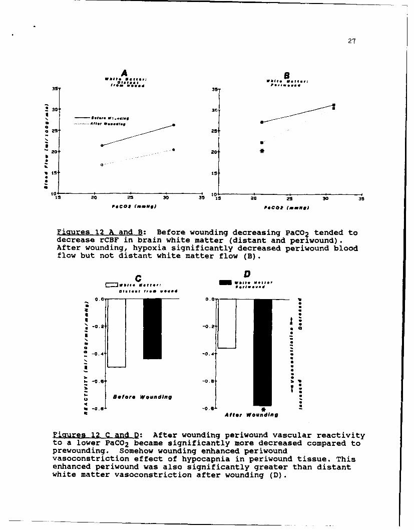
2) In contrast to the rest of the brain, damaged brain about thewound track exhibited an increased response to hypocapnia. Beforewounding, brain in the region of what was to become the missiletrack demonstrated a 14 to 18% rCBF decrease when arterial PCOwas reduced from 31 to 21 mmHg. Afterwards, a similar arteriaiPCO reduction caused periwound rCBF to decrease significantly by24 io 27%. Vascular reactivity about the wound track fell from-0.44 to -0.77 ml/100g/min/mmHg indicating enhancedvasoconstrict2on. This decreased reactivity(-0.77 ml/100g/min/mmHg) was also significant'y greater than thepost-wounding reactivity in the distant whitE matter0.35 ml/lOOg/min/mmHg). Reasons why periwour white matter wouldshow an increased reactivity to the lowered zO remain obscure.Since an increase in periwound vascular reactiviiy also occurredduring the post-wounding hypercapnic trials, it seems that bloodvessels in the periwound tissues do not react to chemical stimulias does the vasculature in the rest of the brain. Presumably, yetto be determined vasoactive factors from damaged brain diffuseinto surrounding tissues and alter the vascular response tochemical stimuli. It would appear that the greatest potential fordamage to the brain tissue because of impairment of chemicalregulation is greatest about the wound track because rCBF tends todecrease with either hypercapnia or hypocapnia.

25
3) After brain wounding the ICP rose to 53 mmHg andhyperventilation, wherein the arterial PCO was reduced from 32 to21 mmHg, failed to lower the ICP even thouih CBF fell from 31 to23 ml/100g/min. The failure of hyperventilation to lower ICP maypossibly be explained by the diminished post wounding cerebralvascular reactivity to decreasing PaCO After brain wounding itwould appear that cerebral blood vesseis cannot constrictsufficiently to decrease cerebral blood volume enough to lowerICP. The explanation does not consider any effect on CSFproduction or absorption. Clinically, data from brain-injuredchildren indicate that hyperventilation cannot control ICP levelsof greater than 60 mmHg (114). Possibly, the ICP of 53 mmHgobserved in our cats after wounding approached the level wherehyperventilation becomes ineffective. The limits to theeffectiveness of hyperventilation as an ICP-reducing therapyfollowing brain wounding should be investigated because if anelevated ICP has reached a level where hyperventilation isunlikely to be effective in lowering it, other means of reducingICP should be immediately employed (67).

26
A a45 C;, rA 45'Correz C .,eboUUU
40- 40
Q ............ A fter WoNNP ing
§35' 35
30- 30b
"25- 25 1I.......... -4
............................... 0
S20- 20-
15 1 5 o15 20 25 30 15 20 25 30
PeCos (mmHg) Pecos (RIuHgJ
Figures 11 A and B: Before wounding decreasing PaCO2 tended todecrease rCBF in cerebral cortex and cerebellum gray. Afterwounding, no reduction .n rC9F of cerebellum gray matter occurred(B).
C DBefore Wounding After Wounding
" 0.0 0.0.
C I CSCI U
I IO,,.F
-- 2 -0.2 A" 0N
30
S-0.4 -0.4
Figures 11Q-dfe ondin the reciiyofcrbla
vaoosrcton atal,()
S-0.6 ej -0.U "
=tue U1CadD fe onigteratvt fcrbla
Fgres 11 hcana D a Aft wouggding the rectivtybfecrebar
vasoconstriction at all (D).
27
Dieta0m11 wi e telr:39 41aw ooed 35- olweeso
S30"
Refer* WC-00160....... After, WOOROJOV
o25"2
2020.*
101 105
15 20 25 30 35 15 20 25 30 35Pecos (uMMe) Pecos (Name)
Fiatures 12 A and B: Before wounding decreasing PaCO2 tended todecrease rCBF in brain white matter (distant and periwound).After wounding, hypoxia significantly decreased periwound bloodflow but not distant white matter flow (B).
___ wooeMCl~
0.0 0.0.U
0.0Afe Wondn
*rwunig Soeo onigehneSeion
vaoosrcinefc8fhpcpi nprwudtsu.Ti
-0.nce -0.4n wsas igiiaty rae ha itnwhit mater asoonsticton aterwouning(D)

28
c) Reasonse to decreasing arterial PO2
In normal cats, breathing 10% 0 reduced arterial Po from 127mmHg to 54 mmHg. Under this circumitance total CBF significantlyincreased (35%) from 33 to 46 ml/lOOg/min. CVR tended to decreasebut not significantly. After wounding, all cats failed to showany increase in total or regional CBFs following hypoxia whichreduced arterial PO to 53 mmHg. Cortical tissue adjacent to thewound track showed i significant and paradoxical decrease in CBFfrom 34 to 17 ml/lOOg/min, Figures 13A to D; 14A to D.
Impaired cerebrovascular reactivity to hypoxia has beendemonstrated in cats subjected to fluid percussion injury andother experimentally induced cerebral trauma (6,94,141,160,161).In Lewelt et al's (94) study mentioned above (p 15) severely braintraumatized cats had a greatly attenuated CBF respcse to profoundhypoxemia while their ICPs were at control levels. The ICPs inmildly concussed cats were never substantially increased at anytime after injury yet these animals, too, demonstrated impairedvasodilitation to a decreased PaO . These results suggest thatimpaired cerebrovascular reactivity to hypoxia was not caused byan ICP increase alone. The mean levels of ICP and CPP observed inour wounded cats before hypoxic trial were 60 and 79 mmHgrespectively. They were further increased to 69 and 87 mmHg withhypoxia. These switic ICP levels observed in our experiments afterwounding were considerably above those observed by Lewelt et al(94) and conceivably these ICP elevations alone could haveimpaired the vascular reactivity to hypoxia in our cats. Cecil etal (20), however, demonstrated in hypoxic lambs that increases ofICP to about 60 mmHg did not affect cerebral vasodilation inresponse to severe hypoxemia of 30 mmHg. This suggests thatpossibly an ICP increase alone into the 60 mmF range may not besufficient to abolish rCBF responses to hypox
Our experiments have shown that after sus- ning a missilewound the feline brain is totally incapable or chemical CBFregulation to hypoxia or hypercapnia. If these results can beextrapolated to the human brain wound situation they stronglysuggest that the missile-wounded brain will not be able toincrease CBF should hypoxia-hypercapnia supervene from anyrespiratory embarassment. Not being thus able to be supplied withadditional oxygen by virtue of increased CBF the missile woundedbrain is under great jeopardy from any hypoxia. Such an event mustbe avoided at all costs.

29
A BWAItO MeIIn: w tl@ Matter:
front Wooffd
40 40,
@30' 30"0 U
a 0. 20.
* 10. - *fare WoooEU 10.-S............ After Woundief
0 25 50 75 100 125 15o 0 25 50 75 100 125 150POOR (mong) PO02 (m uN)J
Figures 13 A and B: Before wounding, hypoxia (PaO2 ~ 55 mmHg)significantly increased rCBF in brain white matter. Afterwounding, hypoxia did not increase rCBF in brain white matter,(distant or periwound).
C , D02 .2, Wolf, Matter: 0 a m or
Oitent from woued
a
- 0.1 0.0* 0
-0.0 -0.0
qforo Wounding After Wounding
Figures 13 C anld D: After wounding, vascular reartivity tohypoxia in both areas appeared greatly, but not significantlyattenuated. The negative reactivity of the periwound white
matter (D) suggests a paradoxical hypersensitivity of damagedbrain to hypoxia.

30
A BCo.eb,.I CoereqlIVM
, 70 70
,60 60
- o0 ...... ffers Woun~ding.
5 ( ............. 50
. . ... 40.
~ 3030,
*20T 20
0 12705 150 2 50 75 100 125 i0PaO2 (mmN) PeO (moH)
Figures 14 A and B: Before wounding hypoxia, (PaO 2 = 55 mmHg)significantly increased rCBF in cerebral cortex and cerebellargray. After wounding hypoxia did not increase rCBF in eitherbrain area.
C D0.3- 0.3
0. 0., aIIF.Befo e4 ndn Atr ondn
Gi* a
*D a
as a t
o a
0 .1 0.1 a
q Cello. a
BefreOounin 0.0BeoeWonigAfter Wounding a
S
Fioures 14 C and D: After wounding, vascular reactivity tohypoxia in both brain areas appeared greatly, but notsignificantly attenuated.

31
d) Reaonse t2 increasing arterial P02
In contrast to other investigations which have indicated amild CBF reduction with increased PaO , CBFs as evaluated in 9cats by us showed no change when the aO was increased to > 280mmHg for 10 min. Unexpectedly, this levil of hyperoxia caused asignificant CBF increase in brain stem and cerebellar blood flows(from 25 to 32 ml/lOOg/min and 33 to 48 ml/lOOg/min) againsuggesting that basivertebral vascular control to chemical stimulidiffers from that of the anterior circulation.
Owing to post wounding deaths only 5 of 9 wounded catscompleted both pre and post wound hyperoxic challenges. In theseanimals total CBF decreased slightly but significantly from 19.5to 17.0 ml/100g/min to a hyperoxic challenge after wounding.Since this response did not occur before wounding it may beconsidered abnormal. The brain stem and cerebellar flowincreases, so prominent in the normal brain when hyperoxia wasinduced, were totally absent after wounding, Figures 15A to D.

32
A gWage Betie: w1h10 veollO:
@,)1.e., Pge u °114
frem woweddO.
401
* 30s.30,
02
200120 0
S ................ .... ...... ....................0
u10" Before Wooding0* ....... Aler Wge~ddg
0 15 200 250 300o
100 150 200 250 300 100 150 20( e3P802 (~ WlW
C DCorearei Cerebe llm
20. C2r0e so
- 40' d
Iao030 30"
0 0 0 100 IS 200 ' 300S... ..... 0
IL
C I
100 150 200 250 300 100 150 200 as0 300
POR (owNs) P.O* (meNN)
Figures 15 A to D: Before wounding 10 minutes of hyperoxia(PaO2 > 280 mmHg) did not alter rCBF in white matter or cerebralcortex (A to C). The pre-wounding rCBF in cerebellum (D) wassuggestively injajg by hyperoxia. After wounding rCBF tendedto decrease in periwound white matter and the suggestive rise incerebellar blood flow was completely abolished.

33
e) Response to simultaneously increased arterial P02 and PCO 2
In applying either increased arterial PO or PCO to thenormal brain two different effects were noteA: hypeicapniaincreased total CBF while hyperoxia showed no effect. Followingwounding, hyperoxia significantly reduced total CBF. Hypercapniahad no effect. These two chemical stimuli seemed to have somewhatopposite effects and what was particularly disturbing was that thevasoconstrictor effect of hyperoxia appeared to be enhancedfollowing brain wounding (see d. above). This could have clinicalramifications because oxygen is often given to patients followingbrain injury and increasing PaO could theoretically lead tovasoconstriction and CBF reductions in brain areas that might havemarginal blood flows. We endeavored to see whether the postwounding enhanced vasoconstrictive effect of hyperoxia could beameliorated by the simultaneous administration of CO .Accordingly, we measured CBF in normal brains and miisile woundedbrains when Pao was increased to >400 mmHg and PaCO was elevatedto 54 mmHg. Whin the normal brain was exposed to thise arterialgas tensions total CBF rose significantly indicating that thevasodilatory effect of the elevated PaCO predominated. Whereasvirtually all rCBFs in brain-wounded animals tended to showinsignificant flow decreases when subject to hyperoxia this effectwas prevented after wounding when hypercapnia was induced alongwith hyperoxia. In fact rrBFs all tended to increase slightlysave the periwound tissue which still demonstrated a significantflow decrease from 27 to 19 ml/lOOg/min. Why all brain areastended to show a flow increase after wounding when subject tohyperoxia plus hypercapnia but showed no post wound flow increasewhen the PaCO alone was elevated is unknown. Cerebellar graytissue exhibiied a significant flow increase after wounding whenexposed to hypercapnia and hyperoxia suggesting that in thecerebellum after wounding the effect of 0 chemical controlpredominates over CO chemical control. We infer this becauseafter wounding increised PaCO tended to decrease cerebellar CBF;while both before and after wgunding increased Pao tended toincrease cerebellar CBF, Figures 16A to D; 17A to

34
A 100to00 wbt. welte: Oetter
- efore wevefo....... .. A te W ing
40 40-
•O... . ... 20 0
* 20
0
0 ,
- - - ----
as 30 35 40 45 50 55 25 30 35 40 45 50 55
PeCOz (m gN) Pect (Nome.)
Figures 16 A and B: Before wounding, increasing PaCO2 duringhyperoxia significantly increased the rCBF in white matterdistant from wound and periwound white matter. After wounding itdid not increase rCBF in distant white matter (A) while itsignificantly decreased rCBF in periwound white matter (B).
O.0T C 2.""" Matte,: M ettets from Wooed
1.5 O.S"
0 .0' 0-."
°M,,. 0. -0.5"
o Wounding Afte Wounding
Figures 16 C and D: After wounding the vascular reactivity tohyperoxia-hypercapnia was greatly attenuated in both brain areas,showing a significant vasoconstriction (D) in periwound whitematter. This pattern resembles that observed with hypercapniaalone indicating that the PaCO 2 effect predominates.

35
A BCeoroar: CorOab low
CONOa *iep
120 120"
to. 100"
...... - ter' WNAd101 *1 60-
S 40 - 40. .......... ......
ILa
20 20.
0 00
03
25 30 3404 o 5 5 0 3 O 4 o 5
PeosO (MMlMV) Pecos2 (fiMily)
Figgrs 17 A and B: Before wounding increasing PaCO2 and PaO2significantly increased rCBF in cerebral cortex and cerebellumgray. After wounding, hyperoxic hypercapnia increased cerebellarrCBF alone.
C D
Cerebral
Core
2.0
UV
COGo 6 t
0.0 0.0 0
&*for* Wounding After Wounding.
Fiuures 17 C and D: After wounding vascular reactiv ity toincreased Pa 2 and PaC 2 was greatly reduced in the cerebral
cortex and cerebellum but despite the reduced vasodilation a postwounding flow increase occurred in the cerebellum (D). Note that
R~st wounding reactivity remained positive with hyperoxia(indicating some vasodilation) but became negative withhypercapnia alone, (D) indicating mild vasoconstriction.
36
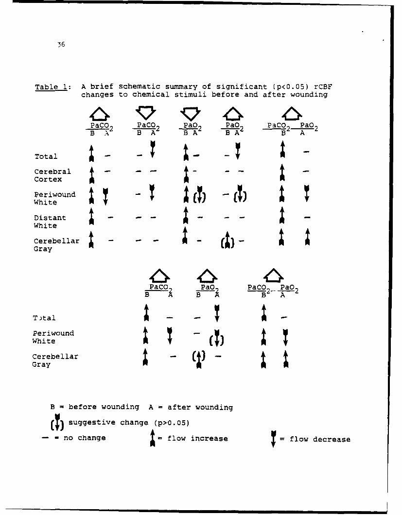
Table 1: A brief schematic summary of significant (p<0.05) rCBFchanges to chemical stimuli before and after wounding
PaCO2 PaCO2 PaO2 PaO2 PaCO PaO 2B A2 B -A B 2 "A
Total-CerebralCortex
White
DistantWhite
Cerebellar I - - a - a-a
PaC ,Pao PaCO2 PaOBB A
To)tala
PeriwoundWhite
CerebellarGray
B - before wounding A - after wounding
(T) suggestive change (p>O.05)
-- no change a = flow increase flow decrease

37
Possible brain stem effects consequent to wounding which couldalter the chemical regulation of CBF:
While local tissue factors, other mediators, or innatedifferences in neural control of vascular beds may be implicatedin these observed CBF changes to chemical stimuli following braininjury, brain stem dysfunction after wounding may be anotherfactor. Brain stem perturbation, manifested by cardiovascular andrespiratory changes, is seen with brain wounding (7,23,24,25,47)The locus coerceus or other brain vasoregulatory centers lyingjust below the floor of the IV ventricle and implicated inchemical CBF regulation (129) could be impaired by missile energytransfer and contribute to the observed loss of chemicalautoregulatory control following brain wounding.
f) Summary
These experiments reiterate what has been known for many yearsfrom closed head injury experiments: namely that after a brainmissile wound mechanical and chemical CBF regulation mechanismsare disturbed. Consequently, the missile wounded brain is atgreat risk to the effect of hemorrhage and hypoxia. Importantly,for military purposes these experiments denote the kind ofmechanical and chemical autoregulatory loss to be expectedspecifically after missile wounding. For instance 1) following abrain missile wound and hemorrhagic hypotension, marked CBFreductions may occur with only slight MABP reductions. Brainischemia, once having occurred from blood loss and MABP reductionmay not be reversed with blood reinfusion. IT IS IMPERATIVE THATBLOOD PRESSURE BE MAINTAINED IN THE BRAIN-WOUNDED INDIVIDUAL. 2)The loss of chemical CBF regulation following brain wounding isnot uniform but heterogeneous throughout the brain. Periwoundtissue appears profoundly affected and tissue about the woundtrack is apt ti show flow decreases to both hypercapnia andhypoxemia. 3) The basivertebral vascular system appears torespond to chemical CBF autoregulatory stimuli differently thanthe anterior circulation. 4) The effect of elevated PaCO 2 on CBFappears to predominate over that of an elevated PaO 2 both beforeand after wounding. To provide maximal brain blood flow andoxygenation possibly some thought should be given to administeringC02 with oxygen to patients with brain injury. This would avoidany vasoconstrictive effects of oxygen alone. Before any suchclinical application of this proposed therapy, however, furtherlaboratory experimentation is necessary.

58
4) Determining why cats died following brain wounding
Brain missile wounding may affect cardiac and respiratoryfunctions (18,24,25,26,92,121) even though the missile wound doesnot involve the brain stem directly. A missile wound of thecerebral hemisphere may even cause immediate death from apnea(4,18,26,47,61). A brain missile wound may also cause additionalintracranial phenomena as hematoma and brain swelling which mayraise ICP. These associated conditions, if moderate, may merelyaggravate mechanical damage to the brain caused by missilepassage. If severe, associated ICP elevations and CBF reductionsmay cause widespread cerebral ischemia or failure of brain energymetabolism. This chain of events may further depress medullaryfunction and cause additional cardiorespiratory abnormalities. Ifrespiratory and cardiac circulatory functions are severelyimpaired either primarily as a consequence of missile energydeposit itself or secondarily because of associated intracranialeffects, widespread "secondary" cerebral damage may occur whichmight greatly aggravate initial missile damage or lead to death.
In addition to stereotyped "brain stem" effects, brainwounding is associated with potentially life threatening systemicchanges as transient hypertension in cats and monkeys and delayedhypotension which occurs in monkeys (18,23,24,47). Very largeelevations in plasma catecholamines also occur after brainwounding. (J. S. Soblosky, see page ). Decreased cardiac output(CO) has also been observed after experimental brain wounding andit has been suggested that a post wounding reduction in COaccounts for CBF failure and death.
From the above it is clear that death from apnea following amissile wound could have been from a multiplicity of causes: 1-from primary brain stem damage caused by missile energy transfer;2- from secondary associated intracranial events such as increasedICP; 3- from systemic changes as CO decrease or 4- from acombination of several factors.
In order to determine just what factors did account for deathwe evaluated multiple physiologic variables in 15 spontaneouslybreathing, pentobarbital anesthetized cats before and up to 90 minafter brain wounding in the right cerebral hemisphere by a 1.4Jmissile. Physiological variables evaluated were: total and rCBFand CO (microsphere technique); arterial blood, pH, PO and PCO2;MABP, ICP, CPP, EKG, heart rate (HR) and EEG. Respiratgryfrequency (f), tidal volume (V ) and ventilation (V) were recordedduring each flow measurement a~d periodically throughout theexperiment. Four unwounded cats served as controls.
Unwounded cats showed no significant changes in anyphysiologic variables measured during a 100 min experimentalperiod. Four brain wounded cats survived a 90 min post-woundingperiod and these had only a transient brain stem effect includinga brief, 50% increase in MABP concurrently with a temporary, 50%reduction in respiratory frequency and heart rate. Non-survivors

39
(11/15) lived from 1 to 41 min after wounding and after woundinghad persistently unstable blood pressures, rates of breathing andheart rates possibly indicating persisting brain stem damage,Figure 18.
.9 NO-SURVIVORS
301
10
-g o s o Is zo an 30 - 0 * t . a 5 3Time (mini Time (minia ,,.. SURVIVORS
~Fioure 18: Survivors exhibited~stable medullary function
mmw manifested by rapid return toNO-SURVIVORS normal of respirator, blood
~pressure, and heart ratei mechanisms after wounding. Non-
survivors had persistently"T impaired respirations, bloodpressure, and heart rate.
4 0 3 10 1 0 29 30 as s g s 2
Time (mn)
Causes of death following an experimental brain missile wound:
In this feline model of brain wounding the immediatecardiorespiratory, brain stem effects of missile injury appearstereotyped and similar to that noted in many other speciessustaining brain injury, open or closed. No one or twophysiologic factors consistently appeared to account for theappearance of sustained apnea after wounding. Rather, fatal apNaappeared to result from an interplay of many factors including: -

40
loss of CBF autoregulation, coupled with CO decrease, leading to
cerebral ischemia (a- Table 2). 2- indirect damage to brain stem
respiratory centers from missile energy (b- Table 2); 2- loss of
CBF autoregulation, coupled with CO decrease, leading to cerebral
ischemia (b- Table 2); 3- ICP increase or CPP decrease affecting
respirations (c,d- Table 2); and 4- cardiac arrest, (e- Table 2).
Table 2: changes in Physiological Variables and Causes of Death
in 5 Non-surviving Cats After Brain Missile Wounding
CAT NUMBER AND TIME OF LAST MEASUREMENT (min)VARIABLES ---------------- ------ ---------- MEAN+SE
1(5) 2(20) 3(5) 4(5) 5(20)
PRESSURES (mmHg):
MABP 141 85 120 71 120 107+10
ICP 27 10 90 84 61 54+13
CPP 114 75 30 -13 59 5317
BLOOD FLOW(ml/100g/min):
TOTAL CBF 11 23 57 31 55 35+7
BRAIN STEM 8 23 44 23 32 26+5
CO (ml/min/kg) 67 91 53 144 149 101+16
RESPIRATORY PARAMETERS:
f/min 2 14 43 16 5 16+6
V (lit/min) 0.16 0.46 0.89 0.14 0.54 0.44+0.1
PaO 2 (mmHg) 40 - 47 56 72 54+5
PaCO2 (mmHg) 58 - 71 52 52 58+3
pH 7.17 - 7.05 7.14 7.20 7.14+0.02
NUMBER OFTRANSIENT APNEA 4 5 5 6 1 4.2+0.7
CAUSE AND TIME APNEA APNEA APNEA APNEA CARDIAC
OF DEATH (min) (8) (25) (8) (10) ARREST (26) -
---------------------------------------------ab c d

41
Basically, the cats which survived brain wounding appearedto maintain all aspects of physiologic function (HR, MABP, f,CBF, Co, V) while animals which died tended to exhibit multiplecardiorespiratory and other physiologic abnormalities. Factorsaccounting for the differences between these groups should besought because knowledge and manipulation of these criticalpathophysiologic events may decrease the mortality of brainwounds.
5) BRAIN BIOGENIC ANINES
Our brain injury experiments including wounding andincreasing intracranial pressure (ICP) were associated with brainstem effects consisting of systemic arterial hypertension pluschanges in heart rate and respirations. Alterations in bloodpressure have been correlated with changes in biogenic amines(norepinephrine, NE; epinephrine, EPI; dopamine, DA; serotonin,5-HT) in the nucleus tractus solitarius (12,76) rostral andcaudal ventrolateral medulla (14,71,127) locus coeruleus (13),hypothalamus (117,118,144) and dorsal raphe nucleus (35.43). Afew studies have determined that brain trauma alters biogenicamines in gross brain areas but none have studied post-traumaticchanges in biogenic amines in specific hypothalamic and brainstem nuclei which relate to observed post-traumatic brain stemeffects (36,64,75).
We examined hypothalamic and brain stem biogenic amines byhigh pressure liquid chromatography following a 2.4 J missilewound to the right cerebral hemisphere in our pentobarbitalanesthetized cats. Wounds of this energy produce profound brainstem effects and cause death 70% of the time from apnea (18). Wewished to know whether such a high energy wound induced extremelydisorganized changes in brain stem and hypothalamic biogenicamines which could account for the fatal effects of such a highenergy missile.
Because brain missile wounding is also associated withincreased ICP we also measured hypothalamic and brain stembiogenic amine changes after artificially increasing ICP (120-140mm Hg) by intracisternal infusion of mock CSF to see whether thepattern of biogenic amine changes associated with missilewounding was intrinsically different than biogenic amine changesassociated with increased ICP alone.
Both brain wounded cats and those with mock CSF infusions toincrease ICP had significant decreases (47-74%) in epinephrine(EPI) levels in the posterior hypothalamus, nucleus tractussolitarius, area AIC1, locus coeruleus and raphe nuclei. Brainwounded cats also had significant EPI reduction (31%) in theanterior hypothalamus as well. Cats whose ICP were raised bymock CSF infusion also had reduced EPI in the anteriorhypothalamus but these reductions did not reach significance(Fig. 19).

42
100 --- - - - - - - - - - - - - - - - - - - - - - - - - - - -
90"
80"
707
C 600N 50" * ***TR 40" *** **
L 30"
20"
0NTS ACI LC RAPHE AH PH
BRAIN AREA
Fig. 19. The effect of artificially-induced increase in ICP (openbars) and brain missile wounding (solid bars) on epinephrinelevels in the nucleus tractus solitarius (NTS), area AlCl, locuscoeruleus (LC), raphe nuclei, anterior hypothalamus (AH) andposterior hypothalamus (PH) 6 mins. after wounding and/orelicitation of the pressor response. Values (Mean ± SEM) areexpressed as percentage of control data. Artificially-inducedincreases in ICP n=6; brain wounding n=8. * p<.05, ** p<.Ol, **p<.001, ANOVA.

43
Brain wounding with attendant, brief intracranial pressuredifferences between supra- and infratentorial compartments mightbe expected to cause brain stem displacement, while mock CSFinfusions into the cisterna magna would not. In the latter casethe induced ICP rise would have occurred relatively much moreslowly and the resultant pressure increase would have beendistributed evenly within the cranium. Since hypothalamic andbrain stem EPI changes occurred with increased ICP alone (withoutbrain stem displacement) it would appear that the ICP increaseassociated with brain wounding was the adequate stimulus to causethe post-wounding biogenic amine changes. Possibly, brain stemdisplacement caused by wounding might have enhanced these changesbut brain stem distortion would not appear to be a primaryfactor.
Norepinephrine (NE) was decreased in the posteriorhypothalamus in response to wounding and ICP increase, but wasunaffected by either stimulus in the anterior hypothalamus, locuscoeruleus or raphe nuclei. The fact that both EPI and NEdecreased in the posterior hypothalamus may be of particularsignificance because the posterior hypothalamus supposedlymediates the vasopressor response and application of EPI or NE tothe posterior hypothalamus will increase MABP(70,117,119,126,148). We cannot be certain if the depletionsfound in our experiments were caused by increased EPI or NEutilization or decreased neuronal functioning. Likewise wecannot definitively say whether EPI and NE changes representcausative, vasopressor changes or represent attempts atvasopressor compensation.
The levels of NE and DA and its metabolites (DOPAC and HVA)did not show consistent changes paralleling EPI suggesting adissociation between the EPI and the NE and DA systems. Brainwounding significantly reduced NE, DA and HVA in the nucleustractus solitarius and area AICI. Increasing ICP alone decreasedNE, DA and HVA in these two areas but insignificantly. Again, itwould appear that mechanisms involved with wounding augmentedthese biogenic amine changes. Since DA is a precursor to NE innoradrenergic neurons and because the levels of DA in the nucleustractus solitarius and area AiC1 are small, the observed DAdecreases might have basically resulted from NE reductions.
Serotonin (5-HT) and its metabolite 5-HIAA as well as DAand HVA were significantly reduced (22% to 37%) in the raphenuclei in response to either brain wounding or increased ICPalone but these results are difficult to interpret relative tocardiovascular changes because discrete raphe nuclei were notsampled.
L___

44
The most basic effect of an increase in ICP is a massiveincrease in sympathetic nerve activity causing a sympatheticpressor response (16,44,77,100,116,140). We have demonstratedthat ICP elevations and the pressor response are also associatedwith biogenic amine changes (particularly EPI and NE) in thehypothalamus and brain stem.
Generalized activation of the sympathetic nervous system isalso the main component of the classic stress response alsoassociated with an MABP increase. Both foot shock andimmobilization stress have shown EPI and/or NE decreases in thehypothalamus, nucleus tractus solitarius, area AlCI and the locuscoeruleus (48,85,135-137,142). Thus, from the similarity ofpatterns of biogenic amine responses in the hypothalamus andbrain stem from either stress or increased ICP we hypothesizethat the response to increased ICP and brain wounding is avariant of a generalized stress response. Though the peripheralmanifestations of an ICP increase (brady- instead of tachycardiaand respiratory slowing instead of tachypnea) differ somewhatfrom somatic stress owing to direct brain stem stimulation ordistortion, the hypothalamic and brain stem biogenic aminechanges are remarkably similar as though the hypothalamus andbrain stem are "hard wired" to react to all stress, whether it befrom an increase in ICP or other source, in a specific,stereotypic fashion.
Despite the severe brain injury and/or large increases inICP in our experiments, all hypothalamic and biogenic amines werenot totally depleted. In so far as the monoaminergic system wasconcerned the pattern of a generalized stress response waspreserved indicating the basic integrity of this system remainedintact. Only selective, organized alterations occurred in theEPI system overall and the 5-HT system in the raphe nuclei. Thissuggests that a severe brain wound likely to cause early deathfrom apnea does not do so by totally disrupting the hypothalamicand brain stem biogenic amine system. If this system remainsbasically intact following severe brain injury, discretealterations in the biogenic amine system which in the future areshown to be physiologically important may be susceptible topharmacologic therapy.

45
6) PLASMA CATECHOLANINES
Plasma catecholamines (CAs) have been shown to be elevatedby brain trauma including experimental fluid percussion (134).The plasma CAs are of interest because high circulating levels ofplasma CAs may have deleterious effects on the cardiovascular andpulmonary systems, basal metabolic rate and neuronal function inthe CNS (21). Circulating CAs do not normally cross the blood-brain barrier (BBB) and enter the brain but a missile wound tothe brain does break down the BBB and plasma CAs may enter thebrain. Such abnormal entry could affect local microcirculationand directly affect brain neuronal function (98). Therefore,delineating the effect of missile wounds to the brain on plasmaCAs may be of importance because this systemic effect of brainwounding may, in turn, affect brain function particularly in thearea where the BBB is damaged.
An increase in intracranial pressure (ICP) alone, withoutbrain injury, has been shown to increase the levels of plasma CAs(50,133). Since a missile wound to the brain may cause dramaticelevations in ICP, adjunct experiments were performed todetermine the contributions, if any, of increased intracranialpressure (ICP) on the plasma CAs and the time course of the CAresponse. Additionally, in order to determine if thephysiological and biochemical responses to brain wounding may bea function of trajectory angle, in a few experiments our standardanterior to posterior (AP) trajectory was changed to a transversetrajectory.
We ascertained:A. A missile wound to the brain caused IMMEDIATE elevations
in plasma CAs, even though the ICP increase may not have beenenough to evoke a significant rise in MABP (Figs. 20 & 21). Thetime course of the plasma CA elevations suggest that this effectmay not be the result of merely a large increase in ICP. In ourmodel a 0.9 J brain wound which increases ICP by only 10-15 mm Hgcaused an immediate MABP rise and a large CA elevation. Bycontrast, ICP had to be raised in excess of 80 mm Hg by mock CSFinfusion to elicit a systemic pressor response and plasma CAincrease.
B. An increase in ICP alone, without injury, caused onlydelayed elevations in plasma CAs if the ICP were increased enoughto elicit a rise in MABP (Figs. 22 & 23). Even when an immediateincrease in MABP occurred the plasma CA elevations were stilldelayed. It thus appears that the initial MABP rise does notdepend upon plasma CAs.

46
17900-*
15000...
12500.
10000 - . 41d
7500/
5000
2500
01 3 5 to 20 30 60
TIME (MINS)
Fig. 20. Effect of brain missile wounding on plasma epinephrinelevels for 60 mins. after wounding at 0.9 J (square), 1.4 J(diamond), or 2.4 J (triangle). Controls (circles) weresurgically prepared but uninjured. Values are Mean _ SEM, n=3.* p<.05, paired t-test.
12000-
10000
N 8000 coNr
E 0.. 1° BOO0- I.4J
6000 -*-2. 41
9/
I 000
2000,. . . , .,t ..... . . .. .
0135 10 20 30 so
TIME (MINS)
Fig. 21. Effect of brain missile wounding on plasmanorepinephrine levels for 60 mins. after wounding at 0.9 J(square), 1.4 J (diamond), or 2.4 J (triangle). Controls(circles) were surgically prepared but uninjured. Values areMean ± SEM, n=3. * p<.05, paired t-test.
i i I IA

47
16000.
15000" 1
12000p i900/gooo..
I
600
3000.
01 3 5 o g0 30
TIME (WINS)
Fig. 22. Effect of brain missile wounding at 1.4 J (squares) andartificially-induced increases in ICP (circles) on plasmaepinephrine levels for 30 mins. after brain wounding and/orelicitation of the pressor response. Values are Mean ± SEM, n=3,* p<.05, ** p<.O1, paired t-test.
18000"
15000
12000
P
9 000U
16000.
3000 ............................................... ................................
00 1 3 5 10 20 30
TIME (WINS)
Fig. 23. Effect of brain missile wounding at 1.4 J (squares) andartificially-induced increases in ICP (circles) on plasmanorepinephrine levels for 30 mins. after brain wounding and/orelicitation of the pressor response. Values are Mean ± SEM, n=3,** p<.0l, paired t-test.

48
C. Four animals sustained a transverse wound and twoexhibited immediate rises in plasma CAs after wounding. Thosewhich showed immediate CA rises also had large increases in ICP(152 and 153 mm Hg) immediately after wounding and correspondingimmediate increases in MABP. The two cats which did not haveplasma CA increases had immediate post-wounding ICPs of 38 and 73mm Hg and they did not have a large MABP rise.
We infer that the transverse trajectory appeared to have adifferent effect on MABP and plasma CA responses than did the APtrajectory because only two of four transversely injured animalsresponded with elevations in MABP and plasma CAs. By contrastnine of nine AP injured animals responded with immediate plasmaCA increases even if they were wounded with as little as 0.9J andICP rose only to 18 mm Hg! This may mean that the peripheralCAs, local brain distortion or the direction of the forcesapplied to the brain stem are more important than ICP increase.Furthermore it may take more force from a side trajectory toeffect the same brain stem sympathetic response irrespective ofany ICP increase.
The origin of the immediate plasma CA elevations may be theresult of pressure forces acting on the brain stem (e.g.displacement) and activating medullary sensitive areas(34,60,150) as mildly suggested from the results using atransverse trajectory. It is also possible that brain areasinvolved in the sympathoadrenal response also partake in theresponse, but if they do, it probably is not caused by merelyincreasing ICP.
It is also important to note that ballistic literatureindicates that with missile transit there is an extremely rapid(msec) large pressure wave that occurs intracranially (58). Weare unable to reliably quantify either its duration or magnitudein our model but we know that it occurs. How this extremely rapidpressure fluctuation affects either brain or plasma CAs, if atall, is presently unknown and must await further experimentation.

49
7) EFFECT OF GH-1 GANGLIOSIDE ON RECOVERY OF FUNCTIONAFTER A MISSILE WOUND TO THE BRAIN
One of the purposes of the brain wound model which wedeveloped was to create a standard neurologic injury in order toevaluate whether specific drugs would improve the speed andextent of neurologic recovery. Much work has been and is beingdone using rat models (1 gm. brain) to test recovery but we feltand feel that it would be very useful to use a more complex catbrain (20 gm.) and to evaluate how drugs affected recoveryspecifically from missile wound injury.
Dr. Soblosky developed a very sophisticated catneurological/behavioral recovery paradigm. The first drug heevaluated was GM-i ganglioside.
Gangliosides are sialic acid-containing glycosphingolipidsfound in high concentrations in the CNS, especially in synapticmembranes. GM-1 ganglioside treatment has been demonstrated tosignificantly enhance behavioral and neurochemical recovery fromdiscrete neurotoxic and mechanical lesions(1,2,38,53,69,99,138,139,151-153). Moreover, the GM-1gangliosides can be given exogenously and do not possess anyknown toxicological effects (59). Therefore, the encouragingdata from the aforementioned discrete lesions experiments, theease in which treatment can be started and continued, and thelack of toxicological effects all made GM-I gangliosides an idealdrug to initially test in our brain wounding model. The presentstudy was undertaken in order to determine if GM-I gangliosidestreatment would affect behavioral recovery specifically from amissile wound of the brain.
A population of cats (both sexes) were matched according toweight into pairs to preclude weight as a factor in thebehavioral motor tests, especially beam balance performance.Each pair of cats was injured (0.9 J) then randomly assigned toeither the control group or drug treatment group. Control catsreceived saline (I.P) and drug-treated cats received GM-iganglioside (20 mg/kg, I.P.), beginning approximately 10 mins.after injury, then daily for the next 10 days. When an injectionday coincided with a test day, the injection was given one houror more prior to testing. The cats were tested by a "blind"rater, i.e. the rater did not know to which of two groups thecats were assigned to. Testing began third day post-injury, thenevery third day thereafter for 30 days; then weekly for 5 weeks(65 days post-injury). The cats were scored according to thecriteria previously described in detail in our Annual reportdated April 27, 1989.
Because of inadvertent pregnancies, three cats had to beeliminated from the GM-I ganglioside treatment group along withone atypically affected cat. This resulted in an n=7 for thecontrol group and only an n=3 for the GM-I ganglioside treatedgroup. Therefore, the results are not conclusive because the

50
total number of subjects (n) for each group is too small to applythe appropriate statistical evaluation at this time. The data,however, are encouraging. Beam balance performance appeared tobe enhanced in the GM-I ganglioside-treated cats (Fig. 24) andthe GM-i-treated cats ultimately appeared to have a betterrecovery with non-visual placing (days 37-65) (Fig. 25). Visualplacing performance appeared to be improved due to GM-iganglioside treatment (days 3-12) (Fig. 26).
The accelerated recovery in beam balance performance of GM-1-treated cats may be related to their initially better visualplacing performance or to their ultimately better non-visualplacing performance, or both.
Because further testing was precluded by U.S. RepresentativeBob Livingston's stopping our project we could not continue tothe point of achieving statistical significance. Nevertheless,the results are very encouraging and suggest that GM-Igangliosides may speed up and even enhance recovery of neuralfunctioning following a missile wound to the brain. Furthertesting is definitely warranted.

51
1 1 q~~ ~ .......... e ....
10
9
7--SC 6-0R - CONTROLR 5-- .. ..sM-1
E
2"
I
0 3 6 9 12 15 18 21 24 27 30 37 44 51 5 65
DAYS (POST-INJURY)
Fig. 24. Effect of GM-1 ganglioside treatment (20 mg/kg I.P., 10days) on the Beam Balance performance of cats injured at 0.9 J(circles). Control animals were similarly injured but receivedsaline. Values are Mean ± SEM, GM-1 n=3, control n=7.

52
° ' I ... ........ .........
7-
SC0 6_R ,
. SALINE
4, - , , 1: 1 I I I I
0 3 8 9 2 .18 21 24 27 30 37 44 51 58 5
DAYS (POST-INJURY)
Fig. 25. Effect of GM-I ganglioside treatment (20 mg/kg I.P., 10days) on Non-Visual Placing performance of cats injured at 0.9 J(circles). Control animals were similarly injured but receivedsaline. Values are Mean ± SEM, GM-1 n=3, control n=7.

j3
577
7 q o o .......... • .......... 0 ........ . .......... • ..........
6- i
S
0 .
R:
EU- SALINE
5--
4-0 3 6 9 12 15 18 21 24 27 30 37 44 51 58 65
DAYS (POST-INJURY)
Fig. 26. Effect of GM-I ganglioside treatment (20 mg/kg I.P., 10days) on Visual Placing performance of cats injured at 0.9 J(circles). Control animals were similarly injured but receivedsaline. Values are Mean ± SEM, GM-I n=3, control n=7.

54
8) Generation of Free Radicals After Traumatic Brain Injury
In recent years it has been hypothesized that secondary neuronaland cerebrovascular damage following traumatic brain injury may becaused by an increase in oxidative reactions initiated by free radicalformation (56,65,115,159). Free radicals after brain trauma may begenerated by: 1) the arachidonic acid cascade (37,79,83,159); 2)increased leakage of superoxide from mitochondrial electron transport(55,65,125) (Superoxide is the primordial species leading to oxidativedamage of membrane phospholipids, proteins and nucleic acids); 3)enhanced activities of xanthine oxidase (65,74,102); 4) autooxidationof catecholamines; and 5) activation of neutrophils (65,149). Theseoxidative reactions are presumably mediated by hydrogen peroxide andthe hydroxyl radical (generated by the Fenton reaction) (125). Lipidperoxidation initiated by the active oxygen species may be a majormechanism for damage in the lipid- rich tissues of the CNS (29,30,55).Antioxidants, such as ascorbate and alpha-tocopherol as well asenzymes, such as superoxide dismutase, can serve to protect the CNSfrom damage caused by free radical reactions (6,65,74,97,124)
We felt it very important to begin to examine the occurrence offree radicals in the brain after missile wounding. We wereparticularly interested in the time course of free radical generationbecause if free radicals were massively generated within seconds ofbrain wounding antioxidant therapy might be of no avail. Conversely,if free radicals were formed many minutes or hours later, intercedingwith antioxidants might be expected to help ameliorate neural damagecaused by free radicals.
Free radical reactions have been analyzed by several indirectmethods. Evidence of lipid peroxidation reflecting oxidative stressin injured tissue can be obtained by measuring breakdown products ofperoxidated poly-unsaturated fatty acids: the thiobarbituric acidreactions (7,28) or by increased production of malondialdehyde(65,162). Consumption of major antioxidants, alpha-tocopherol orascorbate, also provides indirect evidence of free radical reaction(8,9,109,120,147). The superoxide radical reduces nitrobluetetrazolium (NBT) ion to its insoluble blue form and this provides asimple, direct method of detecting superoxide radicals (80).
We elected to measure free radical formation after brain injuryas directly as possible and collaborated with Drs. William Pryor andDaniel Church from the Biodynamic Institute at LSU in Baton Rouge.These scientists are internationally known for their work in electronspin reasonance (ESR) spectroscopy and spin trapping which candirectly measure free radicals. This technique is complex, and beforeascertaining the occurrence of free radicals in missile-wounded catswe elected to work out the technique in rats sustaining brain damageby means of a 95 gm weight dropped 60 cm onto the right skullconvexity. All rats were anesthetized by ether and their scalpsremained intact.

55Spin trapping with ESR measures free radical species in picomole
concentrations. Superoxide, hydroxyl and lipid radicals are so shortlived, however, and are present at such low steady-stateconcentrations that for ESR detection they must be joined to or"trapped" by a non-radical organic compound (spin trap) which forms arelatively more stable and measurable radical called the "spin adduct"(free radical + spin trap) (86). In these studies we used lipidsoluble alpha-phenyl-N-t-butyl nitrone (PBN) as the spin trap. Thepeak heights of the free radical species detected by ESR (measured inarbitrary units) is taken as a measure of the amount of free radicalbeing produced.
In the experiments described below we used ether - anesthetizedmale Sprague-Dawley rats (250-300 g) which were infused i.v. beforebrain injury with 1 ml of either 0.1M PBN or saline. Twenty-nine ratswere then placed under a weight drop device for brain injury while 14served as uninjured controls. We used 5 experimental groups:
I- 7 PBN-infused control rats; decapitated 60' after infusionII- 11 PBN-infused injured rats; decapitated 60' after infusionIII- 7 saline-infused control rats; decapitated 60' after infusionIV- 6 saline-infused injured rats; decapitated 60' after infusionV- 12 PBN-infused injured rats; decapitated 5'after injury
After completion of the experiment and decapitation, brains wereremoved within one minute, processed appropriately, and analyzed byESR to determine the presence of any free radicals being formed.
We also measured tissue ascorbate concentrations by the ascorbateoxidase assay to provide an indirect measure of free radical formationin our injury paradigm.
No rats in groups 1 to IV sacrificed -60 minutes after injuryshowed evidence of any spin adducts for peroxy radicals. Ascorbylfree radical signals were, however, observed in all rats sacrificed at60 minutes whether injured or not, Figure 27. Four of 12 injured ratssacrificed 5 minutes after brain injury (Group V) showed a carboncentered PBN-spin adduct providing direct evidence of tissue lipidperoxidation at this early time point, Figure 28. Brain ascorbateconcentrations were significantly lower in brain injured rats than inuninjured controls, Table 3.
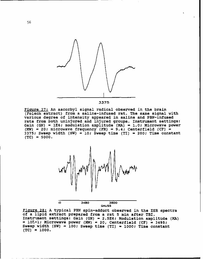
56
3375
Fimrure 27: An ascorbyl signal radical observed in the brain(Folsch extract) from a saline-infused rat. The same signal withvarious degree of intensity appeared in saline and PBN-infusedrats from both uninjured and injured groups. Instrument settings:Gain (GN) = 1E6; modulation amplitude (MA) = 1.0; Microwave power(MW) = 20; microwave frequency (FR) = 9.4; Centerfield (CF) =3375; Sweep width (SW) = 10; Sweep time (TI) = 200; Time constant(TC) = 5000.
;0 3480 3500GAUSS
Figure 28: A typical PBN spin-adduct observed in the ESR spectraof a lipid extract prepared from a rat 5 min after TBI.Instrnent settings: Gain (GN) = 2.5E6; Modulation amplitude (MA)= 10T-l; Microwave power (MW) = 20, Centerfield (CF) = 3495;Sweep width (SW) = 100; Sweep time (TI) - 1000; Time constant(TC) = 1000.

57
Table 3: Brain ascorbate concentration (AA), the ESR peak height (PH)of the ascorbyl signal and the ratio of the peak height andthe brain ascorbate concentration (PH/AA) in control andbrain injured rats
Values are mean + SE
GROUP BRAIN ASCORBATE PEAK HEIGHT OF OXIDATIVECONCENTRATION ASCORBYL SIGNAL STRESS INDEX(AA) (nM/g) (PH) PH/AA
UninjuredSaline & PBN 860 + 41 263 + 44 0.300 + 0.04Combined (n=9)
InjuredSaline & PBN 655 + 75* 317 + 21 0.506 + 0.05*
(n=6)
*Significant, p<0.05; uninjured rats were compared with injured ratsusing an unpaired t-test
The brain has a large pool of ascorbic acid approximately 99% ofwhich is in the reduced form and thus able to act as anantioxidant.(41,46) In these experiments ESR detected ONLY ascorbylradicals at 60 minutes in both control and brain injured rats. Weinfer from this that some oxidative stress occurred in all braintissues whether injured or not during animal sacrifice and tissueextraction. The brain ascorbate concentration, however, in injuredrats was significantly less than that seen in controls at at the sametime. This difference suggests oxidative stress. In another group(V) 4 out of 12 rats sacrificed 5 mins after trauma showed an ESRsignal indicating the presence of a carbon-centered radical. Thissuggests that, following brain injury, oxidative stress from freeradical formation occurred quite early.
Drs. Church and Pryor hypothesize that after traumatic braininjury tissue ascorbate is consumed and oxidized into an ascorbylradical as it acts as an antioxidant. The peak height (PH) of theascorbyl signal measured by ESR indicates the amount of ascorbatebeing changed into the ascorbyl. With oxidative stress the brainascorbate concentration (AA) should decrease and the ascorbyl PHincrease. Figure 22 suggests that, indeed, this happens in our model.

58
As mentioned above oxidative stress in tissues can be indicated bymeasuring either the ascorbyl signal by ESR or by determining tissueascorbic acid concentrations. By measuring both simultaneously,however, one gets a more accurate picture of why tissue ascorbate islow; if oxidative stress is truly occurring the appearance of anascorbyl radical indicates the consumption of ascorbate and itstransformation into ascorbyl. Thus depicting the kinetics ofascorbate utilization. We, therefore, feel it is useful to combinepeak height ascorbyl measurements with tissue ascorbate concentrationsto form a PH/AA ratio. This maneuver normalizes the peak ascorbylradical height for differing amounts of residual tissue ascorbate(units: signal height in arbitrary units/ tissue ascorbateconcentration). We designate this PH/AA ratio as an "oxidative stressindex." This ratio was significantly larger in our brain-injured ratsthan in controls, Table 3.
Although we demonstrated oxidative stress 60 minutes followinginjury, we did not show the generation of lipid peroxyl radicals (PBN-spin adduct signals) at this time point. Two major possibilities mayaccount for this: I- time course of free radical generation; 2- theoccurrence of seizures.
The time course of free radical generation is very critical forthe detection of free radical signals by ESR. We used a 60 minuteperiod in these experiments because superoxide production has beenreported for at least 1 hour after brain injury (65,80,81). In ourexperiments, however, this time period may have fallen outside theperiod of free radical generation and maintenance. PBN-trappedperoxyradicals, formed earlier, could have been metabolized orsubstituted ascorbate by 60 minutes following injury. This may wellhave been the case in our model because 4 of 12 rats in which ESRanalysis was done within 5 minutes of injury demonstrated the presenceof PBN spin adducts. The relatively small number of rats showingearly spin adducts was not a significant number, howver. It couldhave been because at the 5 min sacrifice time the formation of carbon-centered peroxy radicals could have been just starting increasing,peaking or decreasing. In future experiments the time course of freeradical generation after brain injury should clearly be delineatedstarting within minutes up to 2 hours after traumatic brain injury.
In another set of experiments peroxy radical signals whichdeveloped in the rat brain during exposure to hyperbaric oxygenationwere significantly reduced following convulsions (154). Reasons whyconvulsions should reduce free radical signals are unknown.Recurrent seizures were observed in three of our brain injured animalsafter withdrawal of anesthesia.

59
Conclusions
1) The present data indicate that there is a definite increasedoxidative stress in the brain after traumatic injury as determined byin vivo ESR spectroscopy and measurements of the endogenous ascorbateantioxidant.
2) While either peak height (PH) ascorbyl radical measurementsdetermined by ESR or residual tissue ascorbate levels (AA) may be usedto measure the oxidative stress we feel the PH/AA ratio provides asuperior measure.
3) Knowledge of the time course of free radical generationfollowing brain injury is critical in making treatment decisions. Iffree radicals are formed very early after brain injury (within secondsto minutes) then the successful initiation of any type of antioxidanttreatment would be difficult. If free radicals continue to be formedlater after injury (e.g. for several hours) then antioxidant agentsmay ameliorate the extent of oxidative damage which free radicals maycause. Although early generation of free radicals is suggested, ourresults do not conclusively prove early or late free radicalgeneration.
4) We feel it very important to begin to examine the time courseof oxidative stress by free radicals in the brain aftermissile wounding so that a rational approach for the most optimaltherapy of brain missile wounding may be provided.

60
DIRECTIONS FOR FUTURE MISSILE WOUND RESEARCH
1) Apnea occurring after missile injury may prove fatal. Thepathobiologic reasons for this apnea should be delineatedand therapy to reverse it sought.
2) Our experiments have clearly demonstrated that no reflow mayoccur in the missile-wounded brain. Future experimentsshould delineate the exact physiologic conditions which leadto post-wounding no reflow, its pathobiologic substrate, andthe treatment of post-wounding no reflow.
3) Experiments should answer whether, after wounding,hypercapnia-hyperoxia combined provides more benefit to thebrain than hyperoxia alone.
4) The time course of free radical generation after brainwounding should be delineated to provide optimal treatment.
5) Specific neurochemical effects of brain wounding must bedelineated to ensure the most rational future treatmentstrategies for this specific type of brain injury.
6) Drug testing should continue in order to find the best drugsmaximize the extent of neural recovery following missileinjury.

61
EPILOGUE
Dr. Michael E. Carey's research on experimental brainwounding has been conducted from 1983 to 1989 in an attempt tobring knowledge concerning the pathobiology of a brain missilewound into the 21st century. This must be done if more effectivetreatments for a brain wound are to be developed. When Dr.Carey's research began fewer than 25 papers on brain woundingusing experimental, anesthetized animals had ever been written,world wide. (For comparison from 1983 to 1989 about 3000 paperswere written concerning animal-based research for stroke).
Animal activists, with no concern for the American soldier orthe American people, acting through Representative RobertLivingston (RLa), have stopped this research through legislativeaction.
That the federal government should mandate that AmericanServicepersonnel fight for America's interests and, at the sametime, withhold research on the best way to treat their wounds isunconscionable.
The United States Army to its great credit has courageouslyattempted to improve wound treatment for the U.S. soldier throughbasic research on wounds. Animal activists on the other hand,have persistently disregarded the welfare of wounded Americansoldiers by stopping valuable wound research : 1) Brain woundresearch conducted by Dr. Michael E. Carey stopped in 1989; 2)bone and tendon wound research at the Letterman Army Institute ofResearch, stopped in 1989; 3) muscle wound research at theUniformed Services University of the Health Sciences inBesthesda, M.D., stopped in 1984. All 3 aforementioned researchprojects were stopped without any hearings to which the involvedresearchers, other scientists, Army medical representatives, oreven wounded Veterans could express their views. Only animalactivists were heard.
Will basic, advanced, wound research to help U.S. soldiersand civilians be possible in the future? Not if animal activistsand their benighted aims continue to prevail. only 2 hopesremain: 1) that enough Congresspeople awake to the perniciousactions of the animal activists and realize how they are workingdirectly against our servicepersonnel- those who give so much; 2)that the American people awaken to know how the animal activistsare trying to destroy the development of modern care for theirloved ones. One or both groups must take action if more advancedwound care is to develop.

62REFRENCES
1. Agnati, L.F., Fuxe, K., Benfenati, F., Zoli, M., Owman, C.,Diemers, N.H., Kahrstrom, J.K., Toffano, G., and Cimino, M.Effect of ganglioside GM1 on striatal glucose metabolism,blood flow, and protein phosphorylation of the rat. ActaPhysiol. Scand. 125: 43-53, 1985.
2. Agnati, L.F., Fuxe, K., Zoli, M., Davalli, P., Corti, A.,Zini, I., and Toffano, G. Effects of neurotoxic andmechanical lesions of the mesostriatal dopamine pathway onstriatal polyamine levels in the rat: Modulation by chronicGM1 ganglioside treatment. Neurosci. Lett. 61: 339-344,1985.
3. Allen AR: Surgery of experimental lesion of spinal cordequivalent to crush injury of fracture dislocation of spinalcolumn. JAMA 11:878-880, 1911.
4. Allen IV, Scott R, Tanner JA: Experimental high velocitymissile head injury. Injury 14, 183-193, 1982.
5. Ames A III, Wright RL, Kowada M, et al: Cerebral IschemiaII: the no-reflow phenomenon- Am J Pathol 52:437-453, 1968.
6. Andersen BJ, Unterberg AW, Clarke GD, et al: Effect ofposttraumatic hypoventilation on cerebral energy metabolism.J Neurosurg 68:601-607, 1988.
7. Anderson DK, Saunders, RD, Demediuk P, et al: Lipidhydrolysis and peroxidation in injured spinal cord: Partialprotection with methylprednisolone or Vitamin E andselenium. CNS Trauma 12:257-267, 1985.
8. Anderson DK, Mean ED: Iron-induced lipid peroxidation inspinal cord: Protection with mannitol andmethylprednisolone. J Free Rad Biol Med 1:59-64, 1985.
9. Anderson DK, Waters TR, Means ED: Pretreatment with alphatocopherol enhances neurologic recovery after experimentalspinal cord compression injury. J Neurotrauma 5:61-67, 1988.
10. Artru AA: Reduction of cerebrospinal fluid pressure byhypocapnia: changes in cerebral blood volume, cereborspinalfluid volume, and brian tissue water electrolytes. J CerebBlood Flow Metabol 7:471-479, 1987.
11. Bell BA, Symon L, Branston NM: CBF and time thresholds forthe formation of ischemic cerebral edema, and effects ofreperfusion in baboons. J Neurosurg 62:31-41, 1985.

53
12. Bhaskaran D, Freed CM: Changes in arterial blood pressurelead to baroreceptor-mediated changes in norepinephrine and5-hydroxyindoleacetic acid in rat nucleus tractus.solitarius. J Pharmacol Exp Ther 245:356-363, 1988.
13. Bhaskaran D, Freed CR: Changes in neurotransmitter turnoverin locus coeruleus produced by changes in arterial bloodpressure. Brain Res Bull 21:191-199, 1987.
14. Bhaskaran D, Freed CR: Catechol and indole metabolism in therostral ventrolateral medulla change synchronously withchanging blood pressure. J Pharmacol Exp Ther 249:660-666,1989.
15. Bodannes RS, Chan PC: Ascorbic acid as a scavenger ofsinglet oxygen. FEBS Lett 105:195-196, 1979.
16. Brown FK: Cardiovascular effects of acutely raisedintracranial pressure. Am J Physiol 185:510-514, 1956.
17. Carey ME: Learning from traditional combat mortality andmorbidity data used in the evaluation of combat medicalcare. Milit Med 152:6-13, 1987.
18. Carey ME, Sarna G, Farrell JB, et al: Experimental missilewound to the brain. J Neurosurg 71:754-764, 1989.
19. Carey ME, Sarna GS, Farrell JB: Brain edema following anexperimental missile wound to the brain. J Neurotrauma7:13-20, 1990.
20. Cecil OB, Backofen JE, Koehler RC, et al: Cerebral bloodflow autoregulation during intracranial hypertension inhypoxic lambs. Am J Physiol 253:H1342-H1348, 1987.
21. Clifton GL, Ziegler MG, Grossman RG: Circulatingcatecholamines and sympathetic activity after head injury.Neurosurgery 8:10-14, 1981.
22. Clifton GL, Robertson CS, Kyper K, et al: Cardiovascularresponse to severe head injury. J Neurosurg 59:447-454,1983.
23. Crockard HA: Early intracranial pressure studies in gunshotwounds of the brain. The J of Trauma 15:339-347, 1975.
24. Crockard HA, Brown FD, Johns LM, et al: An experimentalcerebral missile injury in primates. J Neurosurg 46:776-783, 1977.

64
25. Crockard HA, Brown F- Llica AB, et al: Physiologicalconsequences of expe atal cerebral missile injury and useof data analysis to .ict survival. J Neurosurg 46:784-794, 1977.
26. Crockard HA, Johns L, Levett J, et al: "Brainstem" effectsof experimental cerebral missile injury, in Popp AJ (ed):Neural Trauma. New York: Raven Press, 1979, pp 19-25.
27. Davies PF: How do vascular endothelial cells respond toflow. NIPS 4:22-25, 1989.
28. Demediuk P, Sanders RD, Clendenon NR, et al: Changes inlipid metabolism in traumatized spinal cord. Prog Brain Res63:1-16, 1985.
29. Demopoulos HB, Flamm ES, Seligman ML, et al: Furtherstudies on free radical pathology in the major nervoussystem disorders: Effect of very high doses ofmethylprednisolone on the functional outcome, morphology,and chemistry of experimental spinal cord impact injury.Can J Physiol Pharmacol 60:1415-1424, 1982.
30. Demopoulos HB, Flamm ES, Seligman ML: et al: Oxygen freeradicals in central nervous system ischemia and trauma, inAutor AP (ed): Pathology of Oxygen. New York AcademicPress, 127-155, 1982.
31. Demopoulos HB, Flamm ES, Pietronigro DD, et al: The freeradical pathology and the microcirculation in the majorcentral nervous system disorders. Acta Physiol Scan Suppl492:91-119, 1980.
32. Denny-Brown D, Russel WR: Experimental cerebral concussion.Brain 64:93-164, 1941.
33. Djordjevic M, Lofgren J, Steiner L, et al: Intracranialpressure effects of missile wounds, in Beks JWF, Bosch DA,Brock M (eds): Intracranial Pressure III. New York:Springer-Verlag, 1976, pp 79-84.
34. Doba N, Reis DJ: Localization of the lower brainstem of areceptive area mediating the pressor response to increasedintracranial pressure (the Cushing Response). Brain Res47:487-491, 1972.
35. Echizen H, Freed CR: Altered serotonin and norepinephrinemetabolism in rat dorsal raphe nucleus after drug-inducedhypertension. Life Sci 34:1581-1589, 1984.

65
36. Edvinsson L, Owman C, Rosengren E, et al: Brainconcentrations of dopamine, noradrenaline, 5-hydroxytryptamine, and homovanillic acid during intracranialhypertension following traumatic brain injury in rabbit.Acta Neurol Scand 47:458-463, 1971.
37. Ellis EF. Wright KF, Wei EP, et al: Cyclooxygenase productsof arachidonic acid metabolism in cat cerebral cortex afterexperimental concussive brain injury. J Neurochem 37:892-896, 1981.
38. Fass, B. and Ramirez, J.J. Effect of gangliosidetreatments on lesion-induced behavioral impairments andsprouting in the CNS. J. Neurosci. Res. 12: 445-458, 1984.
39. Fitch W, McDowall DG: Systemic vascular response toincreased intracranial pressure. 1. Effects of progressiveepidural balloon expansion on intracranial pressure andsystemic circulation. J Neurol Neurosurg Psychiat 40:833-842, 1977.
40. Fitch W, McDowall DG, Keaney NP, et al: Systemic vascularresponse to raised intracranial pressure. 2. The "Cushing"response to the presence of intracranial space-occupyinglesions: systemic and cerebral hemodynamics studies in thedog and baboon. J Neurol Neurosurg Psychiat 40:843-852,1977.
41. Flamm ES, Demopoulos HB, Seligman ML, et al: Free radicalsin cerebral ischemia. Stroke 9:445-447, 1978.
42. Folsch J, Lees M, Sloane-Stanley GH: A simple method forisolation and purification of total lipids from animaltissues. J Biol Chem 226:297-309, 1957.
43. Freed CR, Echizen H, Bhaskaran D: Brain serotonin and bloodpressure regulation: studies using in vivo electrochemistryand direct tissue assay. Life Sci 37:1783-1793, 1985.
44. Freeman NE, Jeffers WA: Effect of progressive sympathectomyon hypertension produced by increased intracranial pressure.Am J Physiol 128:662-671, 1940.
45. Fujimoto S, Roccaforte P, Patel AR, et al: Intravascularaggregation after acute intracranial hypertension byepidural balloon compression in cats. J Neurosurg 57:210-218, 1983.
46. Gennarelli TA, Segwa M, Wald C, et al: Physiologicalresponse to angular acceleration of the head, in GrossmanRG, Gildenberg PL (eds): Head Injury: Basic and ClinicalAspects. New York: Raven Press, 1982, pp 129-140.

66
47. Gerber AM, Moody RA: Craniocerebral missile injuries in themonkey: an experimental physiological model. J Neurosurg36:43-49, 1972.
48. Gonon F, Buda M, De Simoni G, et al: Catecholaminemetabolism in the rat locus coeruleus as studied by in vivodifferential pulse voltammetry. II. Pharmacological andbehavioral study. Brain Res 273:207-216, 1983.
49. Gordon E, Rossanda M: The importance of the cerebrospinalfluid acid-base status in the treatment of unconsciouspatients with brain lesions. Acta Anaesthesiol Scand 12:51-73, 1968.
50. Graf CJ, Rossi NP: Catecholamine response to intracranialhypertension. J Neurosurgery 49:862-868, 1978.
51. Graham DI, Fitch W, Mckenzie ET, et al: Effects ofhemorrhagic hypotension on cerebral circulation, IIINeuropathology Stroke 10:724-727, 1979.
52. Grubb RL, Raichle ME, Phelps ME, et al: Effects ofintracranial pressure on cerebral blood volume, blood flow,and oxygen utilization in monkeys. J Neurosurg 43:385-398,1975.
53. Hadjiconstantinou, M., Rosseti, Z.L., Paxton, R.C., andNeff, N.H. Administration of GMl ganglioside restores thedopamine content in striatum after chronic treatment withMPTP. Neuropharmacol. 25(9): 1075-1077, 1986.
54. Haggendal E, Johanssen B; Effects of arterial carbondioxide tension and oxygen saturation on cerebral blood flowautoregulation in dogs. Acta Physiol Scand 66 Suppl 258:27-53, 1965.
55. Hall ED, Braughler JM: Role of lipid peroxidation in post-traumatic spinal cord degeneration: A review. CNS Trauma3:281-294, 1987.
56. Halliwell B: Oxidants and human disease: Some new concepts.The FASEB J 1:358-364, 1987.
57. Harper AM: Autoregulation of cerebral blood flow: Influenceof the arterial blood pressure on the blood flow through thecerebral cortex. J Neurol Neurosurg Psychiat 29:398-403,1965.
58. Harvey EN, Korr IM, Oster G, et al: Secondary damage inwounding due to pressure changes accompanying the passage ofhigh velocity missiles. Surgery 21:218-239, 1946.
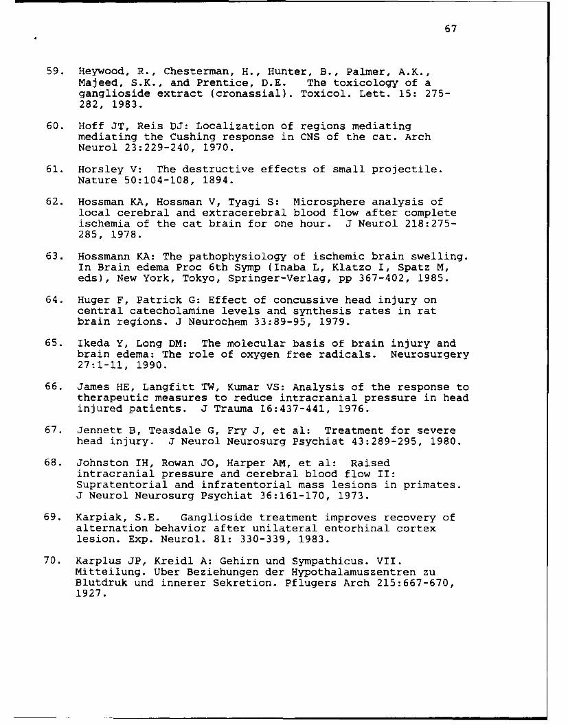
67
59. Heywood, R., Chesterman, H., Hunter, B., Palmer, A.K.,Majeed, S.K., and Prentice, D.E. The toxicology of aganglioside extract (cronassial). Toxicol. Lett. 15: 275-282, 1983.
60. Hoff JT, Reis DJ: Localization of regions mediatingmediating the Cushing response in CNS of the cat. ArchNeurol 23:229-240, 1970.
61. Horsley V: The destructive effects of small projectile.Nature 50:104-108, 1894.
62. Hossman KA, Hossman V, Tyagi S: Microsphere analysis oflocal cerebral and extracerebral blood flow after completeischemia of the cat brain for one hour. J Neurol 218:275-285, 1978.
63. Hossmann KA: The pathophysiology of ischemic brain swelling.In Brain edema Proc 6th Symp (Inaba L, Klatzo I, Spatz M,eds), New York, Tokyo, Springer-Verlag, pp 367-402, 1985.
64. Huger F, Patrick G: Effect of concussive head injury oncentral catecholamine levels and synthesis rates in ratbrain regions. J Neurochem 33:89-95, 1979.
65. Ikeda Y, Long DM: The molecular basis of brain injury andbrain edema: The role of oxygen free radicals. Neurosurgery27:1-11, 1990.
66. James HE, Langfitt TW, Kumar VS: Analysis of the response totherapeutic measures to reduce intracranial pressure in headinjured patients. J Trauma 16:437-441, 1976.
67. Jennett B, Teasdale G, Fry J, et al: Treatment for severehead injury. J Neurol Neurosurg Psychiat 43:289-295, 1980.
68. Johnston IH, Rowan JO, Harper AM, et al: Raisedintracranial pressure and cerebral blood flow II:Supratentorial and infratentorial mass lesions in primates.J Neurol Neurosurg Psychiat 36:161-170, 1973.
69. Karpiak, S.E. Ganglioside treatment improves recovery ofalternation behavior after unilateral entorhinal cortexlesion. Exp. Neurol. 81: 330-339, 1983.
70. Karplus JP, Kreidl A: Gehirn und Sympathicus. VII.Mitteilung. Uber Beziehungen der Hypothalamuszentren zuBlutdruk und innerer Sekretion. Pflugers Arch 215:667-670,1927.
68
71. Katahira K, Mikami H, Tsunetoshi T, et al: Increase in theblood pressure and decrease in the norepinephrine release inthe ventrolateral medulla during intraventricularadministration of hypertonic NaCl. Pflugers Arch Eur JPhysiol 414:719-725, 1989.
72. Kety SS, Schmidt CF: The effect of active and passivehyperventilation on cerebral blood flow, cerebral oxygenconsumption, cardiac output and blood pressure of normalyoung men. J Clin Invest 25:107-119, 1946
73. Kety SS, Schmidt CF: The effects of altered arterialtensions of CO2 and oxygen on CBF and cerebral oxygenconsumption of normal young men. J CLin Invest 27:484-492,1948
74. Kinuta Y, Kikuchi H, Ischikawa M, et al: Lipid peroxidationin focal cerebral ischemia. J Neurosurg 71:421-429, 1989
75. Kmiecak-Kolada K, Felinska W, Stachura Z, et al:Concentrations of biogenic amines and their metabolites indifferent parts of brain after experimental cerebralconcussion. Pol J Pharmacol Pharm 39:47-53, 1987.
76. Kobilansky C, Lanzinger I, Philippu A: Release of endogenouscatecholamines in the nucleus tractus solitarii duringexperimentally induced blood pressure changes. Naunyn-Schmiedeberg's Arch Pharmacol 337:125-130, 1988.
77. Kocsis B, Fedina L, Pasztor E: Two phase change ofsympathetic rhythms in brain ischemia, Cushing reaction, andasphyxia. Am J Physiol 256:R120-R132, 1989.
78. Kontos HA, Raper AJ, Patterson Jr JL: Analysis ofvasoactivity of local pH, PCO and bicarbonate on pialvessels. Stroke 8:358-360, 1977
79. Kontos HA, Wei EP, Povlishock JT, et al: Oxygen radicalsmediate the cerebral arteriolar dilation from arachidonateand bradykinin in cats. Circ Res 55:295-303, 1984
80. Kontos HA, Povilshock JT: Oxygen radicals in brain injury.CNS Trauma 3:257-263, 1986
81. Kontos HA, Wei EP: Superoxide production in experimentalbrain injury. J Neurosurg 64:803-807-, 1986
82. Kovach AGB: Cerebral circulation in hypoxia and ischemia.In Alan R Liss, Inc Prespectives in Shock Research, pp 147-158, 1988
83. Kuehl FA, Ekgan RA: Prostaglandins, arachidonic acid, andinflammation. Science 210:978-984, 1980
- - . ." - i "I I

69
84. Kuschinsky W, Wahl M: Local and chemical neurogenicregulation of cerebral vascular resistance. Physiol Rev58:656-685, 1978
85. Kvetnansky R, Kopin IJ, Saavedra JM: Changes in epinephrinein individual hypothalamic nuclei after immobilizationstress. Brain Res 155:387-390, 1978.
86. Lai EK, Crossley S, Ridhar R, et al: In vivo spin trappingof free radicals generated in brain, spleen, liver andduring radiation of mice. Arch Biochem Biophys 244:156-160,1986
87. Lambertsen CJ, Kough RE, Cooper DT, et al: Oxygen toxicity.Effcts in man of oxygen inhalation at 1 and 3.5 atmospheresupon blood gas transport, cerebral circulation and cerebralmetabolism. J Appl Physiol 5:471-486, 1953
88. Langfitt TW, Gennarelli TA: Can outcome from head injury beimproved? J Neurosurg 56:19-25, 1982
89. Lassen NA: Brain extracellular pH: the main factorcontrolling cerebral blood flow. Scand J Clin Lab Invest22:247-251, 1968
90. Lassen NA: Control of cerebral circulation in health anddisease. Circ Res 34:749-760, 1974
91. Lassen NA, Christensen MS: Physiology of cerebral bloodflow. Br J Anesthesiol 48:719-734, 1976.
92. Levett JM, Johns LM, Replogle RL, et al: Cardiovasculareffects of experimental cerebral missile injury in primates.Surg Neurol 13:59-65, 1980.
93. Lewelt W, Jenkins LW, Miller JD: Autoregulation of CBFafter experimental percussion injury of the brain. JNeurosurg 53:500-511, 1980.
94. Lewelt W, Jenkins LW, Miller JD: Effects of experimentalfluid percussion injury of the brain on cerebrovascularreactivity to hypoxia and hypercapnia. J Neurosurg 56:332-338, 1982.
95. Liebler DC, Kling DS, Reed DJ: Antioxidant protection ofhospholipid bilayers by alpha-tocopherol. J Biol Chem261:12114-12119, 1986.
96. Lundberg N, Kjallquist A, Bien C: Reduction of increasedintracranial pressure by hyperventilation. Acta PsychiatScand (Suppl) 139:1-64, 1959.

'70
97. Machlin LJ, Bendich A: Free radical tissue damage:Protective role of antioxidant nutrients. FASEB J 1:441-445, 1987.
98. MacKenzie ET, McCulloch J, Harper AM: Influence ofendogenous norepinephrine on cerebral blood flow andmetabolism. Am J Physiol 231:489-494, 1976.
99. Mahadik, S.P., Vilim, F., Korenovsky, A., and Karpiak, S.E.GM1 ganglioside protects nucleus basalis from excitotoxindamage: Reduced cortical cholinergic losses and animalmortality. J. Neurosci. Res. 20: 479-483, 1988.
100. Matsuura S, Sakamoto H, Hayashida Y, et al: Efferentdischarges of sympathetic and parasympathetic nerve fibersduring increased intracranial pressure in anesthetized catsin absence and presence of pressor response. Brain Res305:291-301, 1984.
101. McCay PB, Lai EK, Poyer JL, et al: Oxygen- and carbon-centered free radical formation during carbon tetrachloridemetabolism. J Biol Chem 259:2135-2143, 1984
102. McCord JM: Oxygen derived free radicals in post ischemictissue injury. N Engl J Med 312:159-163, 1985
103. McDowall DG: Interrelationships between blood oxygentensions and CBF, in Payne JP and Hill DW eds: Oxygenmeasurements in blood and tissues. London: J and AChurchill, pp 205-219, 1966
104. McHenry LC, Slocum HC, Bivens ME, et al: Hyperventilationin awake and anesthetized man: Effcts on cerebral bloodflow and cerebral metabolism. Arch Neurol 12:270-277, 1965
105. Meyer JS, Kondo A, Nomura F, et al: Cerebral hemodynmicsand metabolism following experimental head injury. JNeurosurg 32:304-319, 1970.
106. Millen JE, Glauser FL, Zimmerman M: Physiological effectsof controlled concussive brain trauma. J Appl Physiol:Respirat Environ Exercise Physiol 49:856-862, 1980.
107. Miller JD, Stanek AE, Langfitt TW: Cerebral blood flowregulation during experimental brain compression. JNeurosurg 39:186-196. 1973.
108. Miller JD, Sweet RC, Narayan R, et al: Early insult to theinjured brain. JAMA 240:439-442, 1978.
109. Mitamura JA, Seligman ML, Solomon JJ, et al: Loss ofessential membrane lipids and ascorbic acid from rat brainfollowing cryogenic injury and protection bymethylprednisolone. Neurol Res 3:329-344, 1981.

71
110. Nilsson B, Ponten U, Voigt G: Experimental head injury inthe rat. Part 2: Mechanics, pathophysiology and morphologyin an impact acceleration trauma model. J Neurosurg47:241-251, 1977.
111. Nilsson B, Nordstrom CH: Experimental head injury in therat. Part 3: CBF and oxygen consumption after concussiveimpact acceleration. J Neurosurg 47:262-273, 1977.
112. Norberg K, Siesjo BK: Cerebral metabolism in hypoxichypoxia. I Pattern of activation of glycolysis: a re-evaluation. Brain Res 86:31-44, 1975.
113. Pannier JL, Leusen I: Circulation to the brain of the ratduring acute and prolonged respiratory changes in the acid-base balance. Pflugers Arch 338:347-359, 1973.
114. Papo I, Caruselli G: Effects on ICP of stopping controlledventilation. Neurochirurgica 21:157-163, 1978.
115. Pappius HM, Wolfe LS: Functional disturbances in brainfollowing injury: Search for underlying mechanisms.Neurochem Res 8:63-72, 1983.
116. Pasztor E, Fedina L, Kocsis B, et al: Activity ofperipheral sympathetic efferent nerves in experimentalSubarachnoideal hemorrhage. Part I: Observations at thetime of intracranial hypertension. Acta Neurochir 79:125-131, 1986.
117. Philippu A: Regulation of blood pressure by centralneurotransmitters and neuropeptides. Rev Physiol BiochemPharmacol 111:1-115, 1988.
118. Philippu A, Dietl H, Sinha JN: Rise in blood pressureincreases the release of endogenous catecholamines in theanterior hypothalamus of the cat. Naunyn-Schmiedeberg'sArch Pharmacol 310:237-240, 1980.
119. Philippu A, Schartner P: Inhibition by locally appliedalpha-adrenoreceptor agents of the depressor response tostimulation of the anterior hypothalamus. Naunyn-Schmiedeberg's Arch Pharmacol 295:1-7, 1976.
120. Pietronigro DD, Hovsepuan M, Demopoulos HB, et al: Loss ofascorbic acid from injured feline spinal cord. J Neurochem41:1072-1076, 1983
121. Polis A: Recherches experimentales sur la commutionCerebrale Rev de Chir, 273:645-655, 1884

72
122. Poole GV Jr, Prough DS, Johnson JC, et al: Effects ofresuscitation from hemorrhagic shock on cerebralhemodynamics in the presence of an intracranial mass. TheJ of Trauma 27:18-23, 1987
123. Price DJE, Murray A: The influence of hypoxia andhypotension on recovery from head injury. Injury 3:218-224, 1972
124. Pryor WA: The role of free radical reactions in biologicalsystems, in Pryor WA (ed): Free Radicals in Biology. NewYork, Academic Press, pp 1-49, 1976
125. Pryor WA: Oxy-radicals and related species: Theirformation, life times and reactions. Ann Rev Physiol48:657-667, 1986
126. Przuntek H, Guimaraes S, Philippu A: Importance ofadrenergic neurons of the brain for the rise of bloodpressure evoked by hypothalamic stimulation. Naunyn-Schmiedeberg's Arch Pharmacol 271:311-319, 1971.
127. Quintin L, Gillon JY, Ghignone M, et al: Baroreflex-linkedvariations of catecholamine metabolism in the caudalventrolateral medulla: An in vivo electrochemical study.Brain Res 425:319-336, 1987.
128. Raichle ME, Posner JB, Plum F: Cerebral blood flow duringand after hyperventilation. Arch Neurol 23:394-403, 1970
129. Ramana Reddy SV, Yaksh TL, Anderson RE, et al: Effect oflocus coeruleus lesions on the response of cerebral bloodflow and cardiac output to altered PaCO2 Brain Research365:278-288, 1986.
130. Rapella CE, and Green HD: Autoregulaton of canine cerebralblood flow. Cir Res XIV:I-205-I-212, 1964.
131. Reivich M: Arterial PCO 2 and cerebral hemodynamics. Am JPhysiol 206:25-35, 1964
132. Rubio R, Berne RN, Bockman EL, et al: Relationship betweenadenosine concentration and oxygen supply in rat brain. AmJ Physiol 228:1896-1902, 1975
133. Roozekrans NTP, Porsius AJ, van Zwieten PA: Comparisonbetween the pressor response and the rise in plasmacatecholamines by acutely elevated intracranial pressure.Arch Int Pharmacodyn 240:143-157, 1979.

73
134. Rosner MJ, Newsome HH, Becker DP: Mechanical braininjury:the sympathoadrenal response. J Neurosurgery 61:76-86, 1984.
135. Roth KA, Mefford IM, Barchas JD: Epinephrine,norepinephrine, dopamine and serotonin: differentialeffects of acute and chronic stress on regional brainamines. Brain Res 239:417-424, 1982.
136. Saavedra JM, Kvetnansky R, Kopin IJ: Adrenaline,noradrenaline and dopamine levels in specific brain stemareas of acutely immobilized rats. Brain Res 160:271-280,1979.
137. Saavedra JM, Torda T: Increased brain stem and decreasedhypothalamic adrenaline-forming enzyme after acute andrepeated immobilization stress in the rat.Neuroendocrinology 31:142-146, 1980.
138. Sabel, B.A., Dunbar, G.L., and Stein, D.G. Gangliosidesminimize behavioral deficits and enhance structural repairafter brain injury. J. Neurosci. Res. 12: 429-443, 1984.
139. Sabel, B.A., Slavin, M.D., and Stein, D.G. GM1ganglioside treatment facilitates behavioral recovery frombilateral brain damage. Science 225: 340-342, 1984.
140. Sadoshima S, Thames M, Heistad D: Cerebral blood flowduring elevation of intracranial pressure: role ofsympathetic nerves. Am J Physiol 241:H78-H84, 1981.
141. Saunders ML, Miller JD, Stablein D, et al: The effects ofgraded experimental trauma on cerebral blood flow andresponsiveness to carbon dioxide. J Neurosurg 51:18-26,1979
142. Sauter AM, Baba Y, Stone EA, et al: Effect of stress and ofphenylethanolamine-N-methyltransferase inhibition oncentral norepinephrine and epinephrine levels. Brain Res144:415-419, 1978.
143. Siesjo BK, Bendek G, Koide T, et al: Influence of acidosison lipid peroxidation in brain tissues in vitro. J CerebBlood Flow Metab 5:253-258, 1985
144. Sinha JN, Dietl H, Philippu A: Effect of a fall of bloodpressure on the release of catecholamines in thehypothalamus. Life Sci 26:1751-1760, 1980.
145. Slater G, Vladeck BC, Bassin R, et al: Sequential changesin cerebral blood flow and distribution of flow within thebrain during hemorrhagic shock. Ann of Surg 181:1-4, 1975

74
146. Soblosky JS, Rogers NL, Adams AJ, et al: Central andperipheral biogenic amine effects of brain missile woundingand increased intracranial pressure. Submitted to JNeurosurg
147. Spector R: Vitamin homeostasis in the central nervoussystem. N Engl J Med 296:1393-1398, 1977
148. Struyker-Boudier H, Smeets G, Brouwer G, et al:Hypothalamic alpha-adrenergic receptors in cardiovascularregulation. Neuropharmacology 13:837-841, 1974.
149. Tauber I, Babior BM: Neutrophil oxygen reduction: Theenzymes and the productions. Adv Free Radical Biol Med1:265-307, 1985
150. Thompson RK, Malina S: Dynamic axial brain-stem distortionas a nechanism explaining the cardio-respiratory changes inincreased intracranial pressure. J Neurosurg 16:664-675,1959.
151. Toffano, G., Agnati, L.F., Fuxe, K., Aldinio, C.,Consolazione, A., Valenti, G., and Savoini, G. Effect ofGM1 canglioside treatment on the recovery of dopaminergicnigro-striatal neurons after different types of lesion.Acta Physiol. Scand. 122: 313-321, 1984.
152. Toffano, G., Savoini, G., Aporti, F., Calzolari, S.,Consclazione, A., Maura, G., Marchi, M., Raiteri, M., andAgna.i, L.F. The functional recovery of damaged brain:The effect of GM1 monoganglioside. J. Neurosci. Res. 12:397-t08, 1984.
153. Toffano, G., Savoini, G., Moroni, F., Lombardi, G., Calza,L., ,.gnati, and L.F. GMI ganglioside stimulates theregei eration of dopaminergic neurons in the central nervoussystEm. Brain Res. 261: 163-166, 1983.
154. Torbrti D, Church D, Keller JM, et al: Free radicalgeneiation during development of CNS toxicity in awakerats. (Abstract) Undersea Biomed Res Suppl 17:164, 1990
155. Torbati D, Jacks AF, Carey ME, et al: Cerebralcardiovascular and respiratory variables after anexperimental brain missile wound. J Neurotrauma. In Press
156. Wahl M, Deetjen P, Thurau K, et al: Micropunctureevaluation of the importance of perivascular pH for thearteriolar diameter on the brain surface. Pfluegers Arch316:152-163, 1970
75
157. Watkins FP, Pearce BP, Stainer MC: Physical effects of thepenetration of head simulants by steel spheres. NATODefence study group II, Chemical Defence Establishment,Porton Down, Salisbury Witts, 1983.
158. Webster JE, Gurdjian ES: Acute physiological effects ofgunshot and other penetrating wounds of the brain. JNeurophysiol 6:255-262, 1943
159. Wei EP, Kontos HA, Deitrich WD, et al: Inhibition by freeradical scavengers and by cyclooxygenase inhibitors in pialarteriolar abnormalities from concussive brain injury incats. Circ Res 48:95-103, 1981
160. Wei EP, Navari RM, Kontos JH. et al: Oxygen consumptionand responsiveness of pial arteries following acuteexperimental brain injury. Circ 56 (Suppl 43): 111-121,1977
161. Wei EP, Dietrich WD, Pavlishock JT, et al: Functionalmorphological and metabolic abnormalities of the cerebvralmicrocirculation after concussive brian injury in cats.Circ Res 46:37-47, 1980
162. Willmore LJ, Rubin JJ: Formation of malondialdehyde andfocal brain edema induced by subpial injection of FeC12into rat isocortex. Brain Res 24:113-119, 1982
163. Yavorsky M, Almaden P, King CG: The vitamin C content ofhuman tissues. J Biol Chem 106:525-529, 1934
164. Zierski J: Blood flwo in brain structures during increasedICP. Acta Neurochirurgica (Suppl) 40:95-116, 1987

76 Appendix 1
We define reactivity (R) to hypercapnia as:
dCBFdPaCO 2
where: dCBF = Normocapnic CBF - Hypercapnia CBFand, dPaCO 2 = Normocapnic PaCO 2, - Hypercapnic PaCO2
In this formula vascular reactivity which increases CBF has a positivevalue (vasodilation) and that which decreases CBF a negative one(vasoconstriction).
To differentiate between vasodilatory a ; vasoconstrictive valuesof reactivity during hypocapnia, we de-fined activity to hypocapniaas:
-dCBFdPaCO 2
This will allow us to present R values in response to hypocapnia as
negative numbers indicating vasoconstriction.
Reactivity to hypoxia was defined as:
-dCBFdPaO2
where dPaO 2 = normoxic PaO 2 - Hypoxic PaC 2
In this formula too, positive values indicate vasodilation andnegative values indicate vasoconstriction.

77
PUBLISHED ARTICLES
1. McKowen H.C., Farrell J.B., Carey M.E. and Soblosky J.S.: rCBFfollowing an experimental missile wound to the brain. IntracranialPressure VII (J.T. Hoff and A.L. Betz, eds.), Springer-Verlag,Berlin, pp.620-622, 1988.
2. Torbati D., Farrell J.B., Davidson J.F. and Carey M.E.: Effect ofhemorrhagic hypotension on CBF and ICP after brain wounding inanesthetized paralyzed cats. Intracranial Pressure VII (J.T. Hoffand A.L. Betz, eds.), Springer-Verlag, Berlin, pp.657-659, 1988.
3. Carey M.E., Sarna G.S., Farrell J.B. and Happel L.T.: Experimentalmissile wound to the brain. J. Neurosurgery 71:754-764 1989.
4. Carey M.E., Sarna G.S. and Farrell J.B.: Brain edema following anexperimental missile wound to the brain. J. Neurotrauma 7:13-19,1990.
5. Carey M.E., Sarna G.S., Farrell J.B.: Brain edema after aiexperimental missile wound. Advances in Neurology (D.M. Long,ed.), 52:301-305, 1990
6. Torbati D, Jacks AF, Carey ME, Davidson JF, Farrell JB: Cerebral,cardiovascular and respiratory variables after an experimentalbrain missile wound. J. Neurotrauma (In Press).
7. Soblosky JS, Rogers NL, Adams JA, Farrell JB, Davidson JF, CareyME: Central and peripheral biogenic amine effects of brain missilewounding (BMW) and increased intracranial pressure. J.Neurosurgery (In Press).

78
ARTICLES UNDER PREPARATION
1. McKowen HC, Soblosky JS, Farrell JB, et al: Regional cerebralblood flow following an experimental missile wound of the brain.
2. Awasthi D, Church DF, Torbati D, et al: Free radical reactionsafter traumatic brain injury in rats.
3. Torbati D, Carey ME, Davidson, JF: Regional cerebral blood flowautoregulation following brain missile wounding in anesthetizedcats.
4. Torbati D, Carey ME: Cerebrovascular responsiveness to hypoxia,hyperoxia and hyperoxic-hypercapnia following brain missilewounding in the cat.
5. Torbati D, Carey ME: Cerebrovascular responsiveness to arterialCO2 after brain missile wounding in the cat.
6. Church D, Torbati D, Awashti D, et al: Ascorbyl radical andoxidative stress following experimental traumatic brain injury.

79
ABSTRACTS PRESENTED
1. Carey ME, Sarna GS, Farrell JB: Pathophysiological effects ofmissile wounding in the cat. NATO Conference, November, 1984
2. Sarna GS, Farrell JB, Carey ME: Brain water and electrolytechanges following penetrating head injury. AANS Denver, CO., Abstrp.206, 1986
3. Sarna GS, Shohami E, Carey ME, et al: Activation of arachidonicacid metabolism following penetrating brain injury. Soc NeurosciAbstr, 12(l):85, 1986
4. Farrell JB, Sarna GS, Carey ME: Blood brain barrier breakdown andrestitution following brain injury. Soc Neurosci Abstr, 13(2):774,1987
5. McKowen C, Farrell JB, Carey ME, Soblosky JS: Focal and globalcerebral blood flow changes in experimental penetrating missilewounds to the brain. AANS Toronto, Canada, p.222, 1988
6. McKowen C, Farrell JB, Carey ME: rCBF following an experimentalmissile wound to the brain. ICP and Brain Injury VII Sci Symp,p.201, 1988
7. Torbati D, Carey ME, Davidson JF, Farrell JB: Cerebral blood flowautoregulation after penetrating brain injury in anesthetizedcats. The FASEB J, 2:1291, 1988
8. Torbati D, Farrell JB, Davidson JF, Carey ME: Effect ofhemorrhagic hypotension on CBF and ICP after brain missile woundin anesthetized paralyzed cats. ICP and Brain Injury VII Sci Symp,p.201, 1988
9. Torbati D, Carey ME, Davidson JF, Farrell JB: Regional cerebralblood flow responsiveness to hypoxia and hypercapnia before andafter brain missile wounding in anesthetized paralyzed cats. SocNeurosci, pt.1, p.621, 1988
10. Torbati D, Davidson JF, Carey ME: Peripheral and circulatorycorrelates of brain missile wounding in anesthetized paralyzedcats. Physiologist, 31:149, 1988

80
11. Awasthi D, Church D, Torbati D, et al: The generation of freeradicals in experimental traumatic brain injury. AANS Washington,DC., p.163, 1989
12. Jacks AF, Torbati D, Davidson JF, Carey ME: Circulatory andrespiratory functions following brain missile wound inanesthetized cats. The FASEB J, 3:1167, 1989
13. Torbati D, Carey ME: Effect of normobaric hyperoxia on regionalcerebral blood flow before and after brain missile wounding inanesthetized paralyzed cats. Undersea Biomed Res Suppl 16:78, 1989
14. Torbati D, Jacks AF, Davidson JF, Carey ME: Regional cerebralblood flow following brain missile wounding in surviving and non-surviving anesthetized cats. Soc Neurosci, pt.l, p.28, 1989
15. Torbati D, Carey ME: Effect of hyperventilation on regional CBFfollowing brain missile wounding in anesthetized cats. JNeurotrauma, 6(3):205, 1989
16. Soblosky JS, Rogers NL, Adams JA, Davidson JF, Farrell JB: Effectsof brain missile wounding (BMW) and increased intracranialpressure (ICP) on brain biogenic amines and plasma catecholaminesin anesthetized cats. The FASEB J, 4(4):A900, 1990
17. Torbati D, Davidson JF, Carey ME: Autoregulation of regionalcerebral blood flow (rCBF) during intracranial hypertensionassociated with hemorrhagic hypotension (HH) in anesthetizedparalyzed cats. The FASEB J, 4(4):AI097, 1990
18. Torbati D, Davidson JF, Carey ME: Cerebrovascular responsivenessto hyperoxic-hypercapnia after brain missile wounding inanesthetized cats. Neurosci Abstr Book, pt.l, p.741, 1990
19. Torbati D, Church DF, Pryor WA, Carey ME: Ascorbyl radical andoxidative stress in the CNS trauma. Free Rad Biol Med 9 (Suppl1):43, 1990
20. Torbati D, Church DF, Carey ME, Pryor, WA: Early detection of freeradicals after closed-head injury in rats. The FASEB J, 5(4):A891,1991