Embed Size (px)
Citation preview

An Immunohistochemical Study of Ovarian Follicle
Histogenesis in the Early Post-hatch Japanese Quail
(Coturnix coturnix japonica)
M.-C. MADEKUROZWA
Department of Anatomy and Physiology, University of Pretoria, Private bag X04,
Onderstepoort 0110, South Africa. Tel: +27 12 529 8417. Fax: +27 12 529 8320.
e-mail: [email protected]
With 6 figures
Short version of title: Early post-hatch ovarian development
Summary
The early post-hatch development of immunoreactivity to vimentin, desmin,
smooth muscle actin (SMA) and laminin, in relation to follicle histogenesis,
was described in the present study. Ovigerous cords in day old quails
contained pre-granulosa and oocytes. Pre-granulosa cells at the cortico-
medullary junction were vimentin immunopositive. A laminin immunopositive
basement membrane and desmin immunopositive mesenchymal cells lined
the ovigerous cords. Ovigerous cords in 3 day old quails contained developing
primordial follicles, the vimentin immunopositive pre-granulosa cells of which
were partially encircled by a basement membrane and desmin
immunopositive mesenchymal cells. In 5 to 7 day old quails, ovigerous cords
formed an outer cortical region, while primordial follicles formed the inner
cortical region. Early pre-vitellogenic follicles were present in 9 to 13 day old
quails. Underlying the granulosa cells of these follicles was a laminin

2
immunopositive basement membrane and a layer of desmin immunopositive
thecal cells. Early and late pre-vitellogenic follicles dominated the ovary in 15
to 17 day old quails. The thecal layer in these follicles was desmin
immunopositive, but SMA immunonegative.
The results of the study have shown that the process of primordial
follicle development in the Japanese quail is similar to that reported in
mammals. The study suggests that in the quail pre-granulosa cells originate
predominantly from the medulla. The study has shown that, in the Japanese
quail, thecal cells are derived from desmin immunopositive mesenchymal
cells lining the ovigerous cords.
Key words: quail, ovary, folliculogenesis, immunohistochemistry, cytoskeletal
proteins, laminin.
Introduction
Several studies have demonstrated the presence of the cytoskeletal proteins,
vimentin, desmin and smooth muscle actin (SMA) in the ovaries of birds and
mammals (Van Nassauw et al., 1992a,b; Marettova and Maretta, 2002;
Madekurozwa and Kimaro, 2006; Madekurozwa, 2007). Studies have shown
that the sequences of the intermediate filaments, vimentin and desmin, as
well as the microfilament actin have been conserved during evolution (Nelson
and Traub, 1982; Kabsch and Vandekerckhove, 1992; Galkin et al., 2010). It
is known that vimentin, desmin and SMA are involved in cellular differentiation
and support (Amsterdam and Aharoni, 1994; Galou et al., 1997).
Vimentin has been localized in the granulosa cells of healthy and
atretic follicles, while desmin and SMA have been demonstrated in cortical

3
and thecal smooth muscle cells (Van den Hurk et al., 1995; Khan-Dawood et
al., 1996; Madekurozwa and Kimaro, 2006). In addition to providing a
structural framework for the ovary, thecal and cortical smooth muscle cells are
involved in the ovulatory process (Martin and Talbot, 1981; Van Nassauw et
al., 1992b; 1993).
In addition to desmin and SMA, smooth muscle cells are characterized
by the presence of an enclosing basement membrane, which is
immunopositive for the glycoprotein laminin (Abd-Elmaksoud, 2009). It is
known that the basement membrane plays an important role in follicular
formation, maturation and atresia (Bagavandoss et al., 1983; Mazaud et al.,
2005). The sequence of laminin has been conserved through evolution (Mota
et al., 1988; Hynes and Zhao, 2000). The evolutionary conservation of
sequences in laminin has made it possible to use rabbit anti-laminin
antibodies to localize laminin in the quail ovary (Rodler and Sinowatz, 2011).
Despite the importance of cytoskeletal proteins and laminin in cellular
differentiation, no information is available on post-hatch developmental
changes of these constituents in birds. In addition, studies on ovarian
development in birds have focussed mainly on embryonic stages, with limited
information being available on the post-hatch period (Yoshimura and
Nishikori, 2004). In contrast, several studies have been conducted on the
developing ovary in the mammalian neonate (Hirshfield and DeSanti, 1995;
Ullmann, 1996; Mazaud et al., 2005; Guittot et al., 2006; Kerr et al., 2006).
The present study describes follicle histogenesis, in the post-hatch
Japanese quail, in relation to changes in the distribution of vimentin, desmin,
SMA and laminin immunoreactivity.

4
Materials and Methods
A total of 45 newly hatched Japanese quail chicks were maintained in a
temperature-controlled room, with a light regime of 14 h light: 10 h dark. Feed
and water were provided ad libitum. Five birds were administered with an
overdose of sodium pentobarbital anaesthetic (Sagatal, May and Baker,
South Africa) at 1, 3, 5, 7, 9, 11, 13, 15 and 17 days post-hatch. All the
procedures used in this study were approved by the Animal Use and Care
Committee of the University of Pretoria.
The thoraco-abdominal cavity was cut open, and the ovary was
removed. Ovarian tissue was immersion-fixed in 4% phosphate buffered
formaldehyde for 48 h. After fixation, tissues were processed routinely for
histology and embedded in paraffin wax. The immunostaining technique was
performed on 5 mm thick sections using a Universal LSAB-plus kit, Peroxidase
(DakoCytomation, Glostrup, Denmark).
Sections were deparaffinized and endogenous peroxidase activity was
blocked, using a 3% (v/v) hydrogen peroxide solution in water for 5 min. The
slides were then rinsed in a 0.01 M phosphate buffered saline solution (PBS,
pH 7.4) for 5 min. Thereafter, the sections for vimentin, desmin and SMA
immunohistochemistry were microwaved at 750 W for three cycles of 7 min
each. After being allowed to cool for 20 min the sections were rinsed with
PBS. Antigen retrieval on sections for laminin immunohistochemistry was
performed by incubating the sections with Proteinase K (Dakocytomation,
Glostrup, Denmark) in a 0.05 mol/L Tris-HCl (pH 7.6) solution for 6 min.
The sections were incubated for 30 min at room temperature with
monoclonal antibodies against desmin, vimentin and smooth muscle actin at

5
dilutions of 1:50, 1:100 and 1:50 respectively. In addition, a polyclonal laminin
antibody at a dilution of 1:100 was used. The primary antisera were
purchased from Dakocytomation, Glostrup, Denmark. The mouse anti-human
desmin, as well as the mouse anti-human smooth muscle actin antibodies
used in this study have previously been used to immunolocalize actin in
smooth muscle cells of the quail testis and excurrent ducts (Aire and Ozegbe,
2007, 2008). The mouse anti-bovine vimentin antibody used in the present
study has been used to immunolocalize vimentin in fibroblasts of the quail
testis and excurrent ducts (Aire and Ozegbe, 2007, 2008). In addition, the
antibody has been used to immunolocalize vimentin in the surface epithelium
of the quail ovary (Rodler and Sinowatz, 2011).
After the incubation with primary antibodies the slides were rinsed with
PBS and then incubated for 15 min with a biotinylated secondary antibody
(LSAB-plus kit). Thereafter, the slides were rinsed in PBS and subsequently
incubated for 15 min with the streptavidin peroxidase component of the LSAB-
plus staining kit. Slides were then rinsed in PBS and bound antibody was
visualized after the addition of a 3,3’-diaminobenzidine tetrachloride solution
(LSAB-plus kit). The sections were counter-stained with Mayer’s
haematoxylin.
In the negative controls the monoclonal desmin, vimentin and SMA
antibodies were replaced with mouse IgG1 (Dakocytomation, Glostrup,
Denmark), while the polyclonal laminin antibody was substituted with rabbit
immunoglobulin fraction (Dakocytomation, Glostrup, Denmark). The negative
control reagents were diluted to the same concentration as the primary
antibodies. Smooth muscle was used as a positive control for desmin, SMA

6
and laminin, while tonsillar tissue was used as a positive control for vimentin.
Variations in the immunostaining of sections used in this study were minor. No
background staining was detected in the negative control sections, while
positive immunostaining for vimentin, desmin, SMA and laminin was observed
in the control sections.
Results
1 day old quails
The cortex was composed of ovigerous cords, which extended from the
surface epithelium to the medulla (Fig. 1). The ovigerous cords were
composed of oocytes and pre-granulosa cells. Pre-granulosa cells at the
cortico-medullary junction and occasional surface epithelium cells displayed
vimentin immunostaining (Fig. 2a). The surface epithelium and ovigerous
cords were lined by a continuous laminin immunopositive basement
membrane, which resulted in contact between surface epithelial cells and the
ovigerous cords (Fig. 2b). Underlying the basement membrane of the
ovigerous cords were desmin immunopositive mesenchymal cells (Fig. 1 &
2c). The mesenchymal cells were SMA immunonegative. The tunica media of
blood vessels between the ovigerous cords and within the medulla displayed
desmin, laminin and SMA immunostaining (Fig. 2d).
3 day old quails
The ovigerous cords contained oocytes and pre-granulosa cells, except at the
cortico-medullary junction where developing primordial follicles (approximately
20 to 30 mm) were observed. The vimentin immunopositive pre-granulosa

7
cells of developing primordial follicles were partially encircled by an
invaginating laminin immunopositive basement membrane (Fig. 3b).
Peripheral to the basement membrane were desmin immunopositive
mesenchymal cells (differentiating thecal cells), which extended into the
ovigerous cords from the interstitium (Fig. 3a).
5 to 7 day old quails
A laminin immunopositive basement membrane and desmin immunopositive
mesenchymal cells subdivided the cortex into two regions. The outer cortical
region contained ovigerous cords, while the inner region contained primordial
follicles. The vimentin immunopositive granulosa cells of the primordial
follicles (Fig. 4a) were enclosed by a continuous laminin immunopositive
basement membrane, which was surrounded by a desmin immunopositive
thecal layer (Fig. 4b). The thecal layer was immunonegative for SMA, but
displayed weak laminin immunostaining.
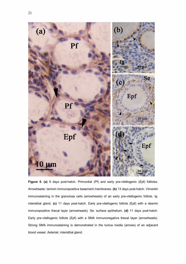
9 to 13 day old quails
Due to the absence of ovigerous cords, the surface epithelium was lined by a
continuous laminin immunopositive basement membrane. The cortex was
composed of primordial follicles and early pre-vitellogenic follicles, which were
approximately 100 to 150 mm in diameter (Fig. 5a). The oocyte in early pre-
vitellogenic follicles was surrounded by a simple cuboidal to columnar
granulosa layer, which was vimentin immunopositive (Fig. 5b). Underlying the
granulosa layer was a laminin immunopositive basement membrane (Fig. 5a),
and a single layer of desmin immunopositive thecal cells (Fig. 5c). The thecal
8
layer was smooth muscle actin immunonegative (Fig. 5d), but demonstrated
weak laminin immunostaining.
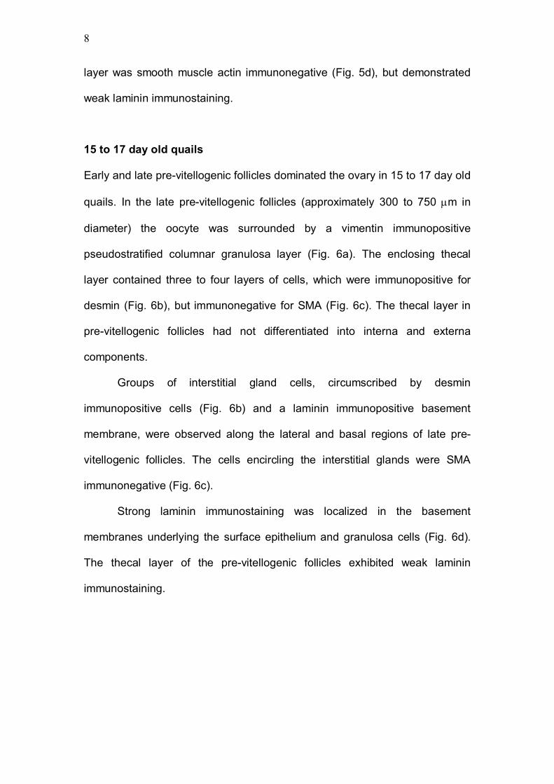
15 to 17 day old quails
Early and late pre-vitellogenic follicles dominated the ovary in 15 to 17 day old
quails. In the late pre-vitellogenic follicles (approximately 300 to 750 mm in
diameter) the oocyte was surrounded by a vimentin immunopositive
pseudostratified columnar granulosa layer (Fig. 6a). The enclosing thecal
layer contained three to four layers of cells, which were immunopositive for
desmin (Fig. 6b), but immunonegative for SMA (Fig. 6c). The thecal layer in
pre-vitellogenic follicles had not differentiated into interna and externa
components.
Groups of interstitial gland cells, circumscribed by desmin
immunopositive cells (Fig. 6b) and a laminin immunopositive basement
membrane, were observed along the lateral and basal regions of late pre-
vitellogenic follicles. The cells encircling the interstitial glands were SMA
immunonegative (Fig. 6c).
Strong laminin immunostaining was localized in the basement
membranes underlying the surface epithelium and granulosa cells (Fig. 6d).
The thecal layer of the pre-vitellogenic follicles exhibited weak laminin
immunostaining.

9
DISCUSSION
The study describes the development of immunoreactivity to vimentin,
laminin, desmin and SMA, in relation to follicle histogenesis, in the post-hatch
quail ovary. The morphology of primordial, early and late pre-vitellogenic
follicles, observed in the present study, was similar to that described in the
adult Japanese quail (Van Nassauw et al., 1992a,b), ostrich (Madekurozwa
and Kimaro, 2006) and emu (Madekurozwa, 2007).
The process of primordial follicle development, in the post-hatch
Japanese quail, is similar to that of the mouse (Frojdman et al., 1995), rat
(Hirshfield and DeSanti, 1995; Guittot et al., 2006), brushtail possum
(Ullmann, 1996) and sheep (Juengel et al., 2002; Sawyer et al., 2002).
In the present study, the formation of primordial follicles was evident
from day one post-hatch, with pre-granulosa cells at the cortico-medullary
interface displaying vimentin immunostaining. The localization of vimentin
immunopositive pre-granulosa cells at the cortico-medullary interface
suggests that in the Japanese quail pre-granulosa cells originate
predominantly from the medulla. This is supported by research on ovarian
development in the brushtail possum (Ullmann, 1996), which has shown that
pre-granulosa cells located in the cortico-medullary region are derived from
disintegrating cellular cords located in the medulla (Ullmann, 1996). Further
research by Juengel et al. (2002) and Sawyer et al. (2002) has shown that
upon establishment of a basement membrane around the ovigerous cords
and below the surface epithelium, pre-granulosa cells of developing primordial
follicles can only be recruited from the surface epithelium. In the present study
few cells in the surface epithelium displayed vimentin immunostaining. This

10
finding suggests that the surface epithelium contributes a limited number of
pre-granulosa cells in the post-hatch quail ovary.
In the Japanese quail primordial follicles were isolated from the
ovigerous cords once the basement membrane had completely surrounded
the pre-granulosa cells of developing follicles. Ultrastructural and
immunofluorescence studies on follicle histogenesis in the rat (Mazaud et al.
2005) have shown that the process of primordial follicle detachment involves
the formation of clefts within the ovigerous cords, into which basement
membrane components are deposited. These findings correlate well with the
results of the present study, in which the extension of the developing
basement membrane between cells of the ovigerous cords was clearly
demonstrated by laminin immunostaining.
Studies on the formation of the thecal layer in mammals have shown
that thecal cells are derived from mesenchymal cells, which migrate into the
ovigerous cords from the adjacent interstitium (Hirshfield, 1991; Mazaud et al.
2005). Although several studies have described the development of oocytes
and granulosa cells in birds, little mention is made of the formation of the
thecal cell layer. The current study has shown that desmin immunopositive
mesenchymal cells from the cortico-medullary junction and areas between the
ovigerous cords are incorporated into developing primordial follicles to form
the thecal layer.
Although both desmin and SMA are markers for smooth muscle cells,
the thecal cells in primordial and pre-vitellogenic follicles of the Japanese
quail were immunopositive for desmin, but consistently immunonegative for
SMA. Studies on the Japanese quail (Van Nassauw et al., 1992a,b), ostrich

11
(Madekurozwa and Kimaro, 2006) and emu (Madekurozwa, 2007) have
shown that the distribution of desmin and SMA in the avian ovary is species
dependent. Furthermore, these studies have shown that the distribution of
desmin and SMA changes with follicular maturation. In the ostrich, thecal cells
of primordial follicles were desmin and SMA immunonegative, while the thecal
cells of pre-vitellogenic follicles were immunopositive for both desmin and
SMA (Madekurozwa and Kimaro, 2006). In contrast, in the emu desmin
immunostaining was localized in pericytes in the theca interna, while SMA
immunostaining was demonstrated in the theca externa of vitellogenic follicles
(Madekurozwa, 2007).
In the present study weak laminin immunostaining was observed in the
thecal cells of primordial and pre-vitellogenic follicles. Research conducted by
Bagavandoss et al. (1983), on the developing ovary in the rat, has shown that
the thecal layer synthesizes the laminin component of the basement
membranes associated with ovarian follicles. Using immunohistochemical and
immunoelectron microscopic techniques, Leardkamolkarn and Abrahamson
(1992) localized laminin within both thecal and granulosa cells, indicating that
these cells are involved in the synthesis of the basement membrane in
ovarian follicles.
In conclusion, the results of the present study have shown that the
processes of primordial follicle development from ovigerous cords are similar
to that observed in mammals. Based on the concentration of vimentin
immunopositive pre-granulosa cells at the cortico-medullary interface, it would
appear that in the Japanese quail granulosa cells originate predominantly
from the medulla. Furthermore, the results indicate that thecal cells are

12
derived from desmin immunopositive mesenchymal cells located at the
cortico-medullary junction and in the areas between adjacent ovigerous cords.
Acknowledgements
The author thanks staff in the Department of Pathology, University of Pretoria,
for their assistance. The National Research Foundation funded this study.
References
Abd-Elmaksoud, A, 2009: Comparative expression of laminin and smooth
muscle actin in the testis and epididymis of poultry and rabbit. J. Mol.
Histol. 40, 407-416.
Aire, T.A., and P.C. Ozegbe, 2007: The testicular capsule and peritubular
tissue of birds: morphometry, histology, ultrastructure and
immunohistochemistry. J. Anat. 210, 731-740.
Aire, T.A., and P.C. Ozegbe, 2008: Immunohistochemistry of the cytoskeleton
in the excurrent ducts of the testis in birds of the Galloanserae
monophyly. Cell Tiss. Res. 333, 311-321.
Amsterdam, A., and D. Aharoni, 1994: Plasticity of cell organization during
differentiation of normal and oncogene transformed granulosa cells.
Microsc. Res. Tech. 27, 108-124.
Bagavandoss, P., A.R. Midgley, and M. Wicha, 1983: Developmental
changes in the ovarian follicular basement membrane detected by
immunofluorescence and electron microscopy. J. Histochem.
Cytochem. 31, 633-640.
Frojdman, K., P. Ekblom, L. Sorokin, A. Yagi, and L.J. Pelliniemi, 1995:

13
Differential distribution of laminin chains in the development and sex
differentiation of mouse internal genitalia. Int. J. Dev. Biol. 39, 335-344.
Galkin, V.E., A. Orlova, G. Schroder, and E.H. Egelman, 2010: Structural
polymorphism in F actin. Nat. Struct. Mol. Biol. 17, 1318-1323.
Galou, M., J. Gao, J. Humbert, M. Mericskay, Z. Li, D. Paulin, and P. Vicart,
1997: The importance of intermediate filaments in the adaptation of
tissues to mechanical stress: evidence from gene knockout studies.
Biol. Cell. 89, 85-97.
Guittot, S.M., C.J. Guigon, N. Coudouel, and S. Magre, 2006: Consequences
of fetal irradiation on follicle histogenesis and early follicle development
in rat ovaries. Biol. Reprod. 75, 749-759.
Hirshfield, A.N., 1991: Theca cells may be present at the outset of follicular
growth. Biol. Reprod. 44, 1157-1162.
Hirshfield, A.N., and A.M. DeSanti, 1995: Patterns of ovarian cell proliferation
in rats during the embryonic period and the first three weeks
postpartum. Biol. Reprod. 53, 1208-1221.
Hynes, R.O., and Q. Zhao, 2000: The evolution of cell adhesion. J. Cell. Biol.
150, F89-F95.
Juengel, J.L., H.R. Sawyer, P.R. Smith, L.D. Quirke, D.A. Heath, S. Lun, S.J.
Wakefield, and K.P. McNatty, 2002: Origins of follicular cells and
ontogeny of steroidogenesis in ovine fetal ovaries. Mol. Cell. Endocr.
191, 1-10.
Kabsch, W., and J. Vandekerckhove, 1992: Structure and function of actin.
Annu. Rev. Biophys. Biolmol. Struct. 21, 49-76.
Kerr, J.B., R. Duckett, M. Myers, K.L. Britt, T. Mladenovska, and J.K. Findlay,

14
2006: Quantification of healthy follicles in the neonatal and adult mouse
ovary: evidence for maintenance of primordial follicle supply. 132, 95-
109.
Khan-Dawood, F.S., M.Y. Dawood, and S. Tabibzadeh, 1996:
Immunohistochemical analysis of the microanatomy of primate ovary.
Biol. Reprod. 54, 734-742.
Leardkamolkarn, V., and D.R. Abrahamson, 1992: Immunoelectron
microscopic localization of laminin in rat ovarian follicles. Anat. Rec.
233, 41-52.
Madekurozwa, M.-C., 2007: An immunohistochemical study of the
distribution of intermediate filaments in the ovary of the emu (Dromaius
novaehollandiae). Anat. Histol. Embryol. 36, 336-342.
Madekurozwa, M.-C., and W.H. Kimaro, 2006: A morphological and
immunohistochemical study of healthy and atretic follicles in the ovary
of the sexually immature ostrich (Struthio camelus). Anat. Histol.
Embryol. 35, 253-258.
Marettova, E., and M. Maretta, 2002: Demonstration of intermediate
filaments in sheep ovary. Acta Histochem. 104, 431-434.
Martin, G.G., and P. Talbot, 1981: The role of follicular smooth muscle
cells in hamster ovulation. J. Exp. Zool. 216, 469-482.
Mazaud, S., R. Guyot, C.J. Guigon, N. Coudouel, B. Le Magueresse-
Battistoni, and S. Magre, 2005: Basal membrane remodeling during
follicle histogenesis in the rat ovary: contribution of proteinases of the
MMP and PA families. Dev. Biol. 277, 403-416.
Mota, G.F.A., C.R.W. Carneiro, L. Gomes, and J.D. Lopes, 1988: Monoclonal

15
antibodies to Staphylococcus aureus laminin-binding proteins cross
react with mammalian cells. Infection and Immunity. 56, 1580-1584.
Nelson, J.W., and P. Traub, 1982: Intermediate (10 nm) filament proteins and
the Ca 2+ - activated proteinase specific for vimentin and desmin in the
cells from fish to man: an example of evolutionary conservation. J. Cell.
Sci. 57, 25-49.
Rodler, D., and F. Sinowatz, 2011: Immunohistochemical and ultrastructural
characterization of the ovarian surface epithelium of Japanese quail
(Coturnix japonica). Ani. Sci. J. 82, 307-313.
Sawyer, H.R., P. Smith, D.A. Heath, J.L. Juengel, St. J. Wakefield, and K.P.
McNatty, 2002: Formation of ovarian follicles during fetal development
in sheep. Biol. Reprod. 66, 1134-1150.
Ullmann, S.L., 1996: Development of the ovary in the brushtail possum
Trichosurus vulpecula (Marsupialia). J. Anat. 189, 651-665.
Van den Hurk, R., G. Dijkstra, F.N. Van Mil, S.C.J. Hulshof, and S.G.A.M. Van
den Ingh, 1995: Distribution of the intermediate filament proteins
vimentin, keratin and desmin in the bovine ovary. Mol. Reprod. Dev.
41, 459-467.
Van Nassauw, L., F. Harrisson, and M. Callebaut, 1992a: Immunolocalization
of smooth muscle-like cells in the quail ovary. Eur. J. Morph. 30, 275-
288.
Van Nassauw, L., M. Callebaut, F. Harrisson, and D.W. Scheuermann, 1992b:
Smooth muscle cells in the walls of ovarian follicles in the Japanese
quail. Cell Tiss. Res. 269, 49-56.
Van Nassauw, L., S.U. Sys, F. Harrisson, and M. Callebaut, 1993: In vitro

16
study of the contractility of the wall of the preovulatory follicle in the
Japanese quail. Biol. Reprod. 49, 359-364.
Yoshimura, Y., and M. Nishikori, 2004: Identification of apoptotic oocytes
in the developing ovary of embryonic and post-hatched chicks in
Japanese quail (Coturnix japonica). J. Poult. Sci. 41, 64-68.

17
Figure 1. 1 day post-hatch. Survey photomicrograph of the ovary. Se: surface epithelium.
Asterisks: ovigerous cords. Arrows: desmin immunopositive cells. Med: medulla.

18
Figure 2. 1 day post-hatch. (a) Vimentin immunopositive pre-granulosa cells (arrows) line
oocytes (asterisks) at the cortico-medullary junction. Se: surface epithelium. Med: medulla.
(b) A laminin immunopositive basement membrane (arrows) lines ovigerous cords (Ovc).
Arrowheads: pre-granulosa cells.(c) Desmin immunopositive mesenchymal cells (arrows)
extend between ovigerous cords (Ovc). Se: surface epithelium. Med: medulla. (d) SMA
immunostaining (arrow) in the tunica media of a blood vessel adjacent to ovigerous cords
(asterisks). Isolated cells (arrowheads) at the cortico-medullary junction display SMA
immunostaining. Se: surface epithelium. Med: medulla.

19
Figure 3. 3 days post-hatch. (a) Developing primordial follicles (asterisks) partially enclosed
by desmin immunopositive mesenchymal cells (arrows). Ovc: ovigerous cord. Med: medulla.
(b) A laminin immunopositive basement membrane (arrowheads) lines ovigerous cords (Ovc)
and a developing primordial follicle (asterisk). Note the absence of a basement membrane
below the surface epithelium (Se). Med: medulla.

20
Figure 4. (a) 5 days post-hatch. Vimentin immunopositive pre-granulosa (arrows) and
granulosa cells (arrowheads). Asterisks: oocytes. Pf: primordial follicle. Med: medulla. (b) 7
days post-hatch. Primordial follicles (Pf) with desmin immunopositive thecal cells (arrows).
Ovc: ovigerous cord. Med: medulla.
21
Figure 5. (a) 9 days post-hatch. Primordial (Pf) and early pre-vitellogenic (Epf) follicles.
Arrowheads: laminin immunopositive basement membranes. (b) 13 days post-hatch. Vimentin
immunostaining in the granulosa cells (arrowheads) of an early pre-vitellogenic follicle. Ig:
interstitial gland. (c) 11 days post-hatch. Early pre-vitellogenic follicle (Epf) with a desmin
immunopositive thecal layer (arrowheads). Se: surface epithelium. (d) 11 days post-hatch.
Early pre-vitellogenic follicle (Epf) with a SMA immunonegative thecal layer (arrowheads).
Strong SMA immunostaining is demonstrated in the tunica media (arrows) of an adjacent
blood vessel. Asterisk: interstitial gland.

22
Figure 6. (a) 15 days post-hatch. Vimentin immunopositive granulosa cells (arrows) in a late
pre-vitellogenic follicle. T: thecal layer. Se: surface epithelium. (b) 15 days post-hatch.
Desmin immunopositive thecal cells (arrows) in a late pre-vitellogenic follicle (Lpf). Desmin
immunopositive cells (arrowheads) encircle interstitial glands (Ig). (c) 17 days post-hatch.
Late pre-vitellogenic follicle (Lpf) with a SMA immunonegative thecal layer (arrows). Strong
SMA immunoreactivity is demonstrated in the tunica media (arrowheads) of adjacent blood
vessels. Ig: Interstitial gland. (d) 17 days post-hatch. Weak laminin immunostaining in the
thecal layers (T) of adjacent late pre-vitellogenic follicles. Strong laminin immunoreactivity is
demonstrated in the basement membranes (arrowheads) of granulosa cell layers (Gr) and
blood vessels (asterisks).