Embed Size (px)
Citation preview
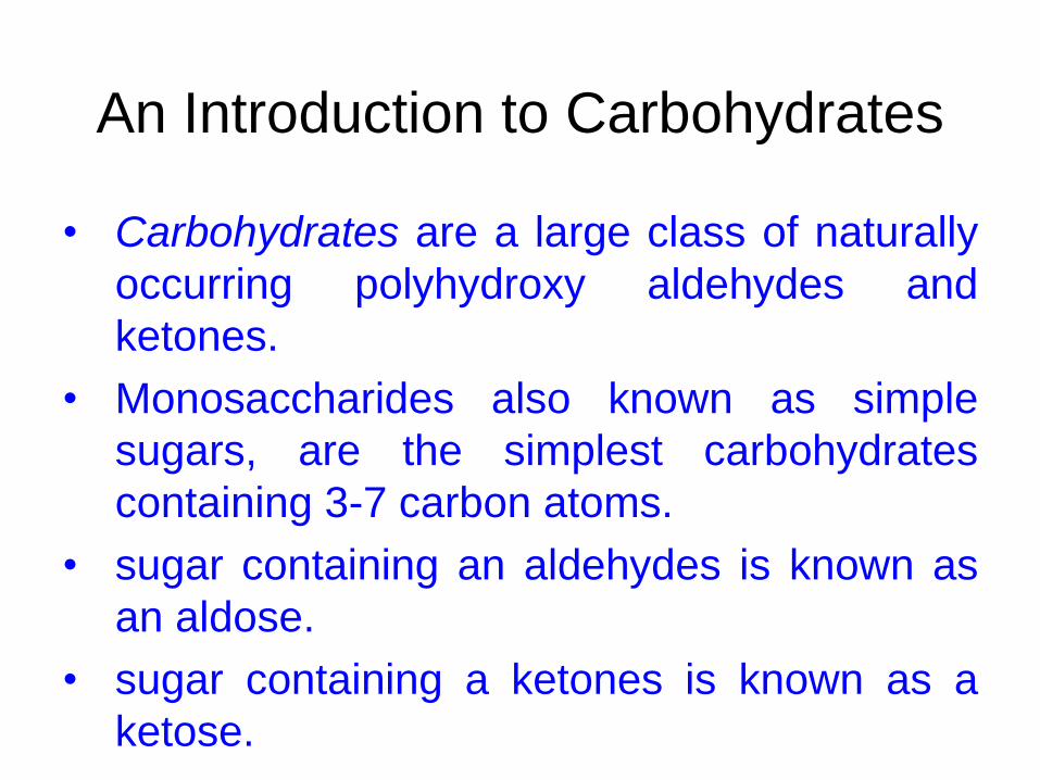
An Introduction to Carbohydrates
• Carbohydrates are a large class of naturally
occurring polyhydroxy aldehydes and
ketones.
• Monosaccharides also known as simple
sugars, are the simplest carbohydrates
containing 3-7 carbon atoms.
• sugar containing an aldehydes is known as
an aldose.
• sugar containing a ketones is known as a
ketose.
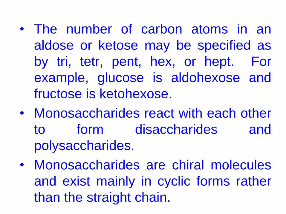
• The number of carbon atoms in an
aldose or ketose may be specified as
by tri, tetr, pent, hex, or hept. For
example, glucose is aldohexose and
fructose is ketohexose.
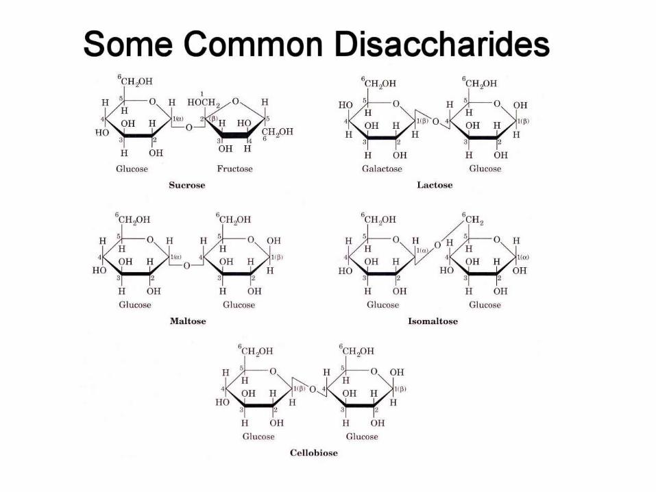
• Monosaccharides react with each other
to form disaccharides and
polysaccharides.
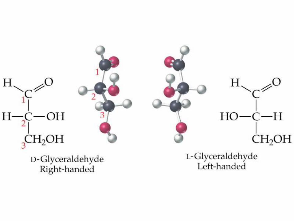
• Monosaccharides are chiral molecules
and exist mainly in cyclic forms rather
than the straight chain.
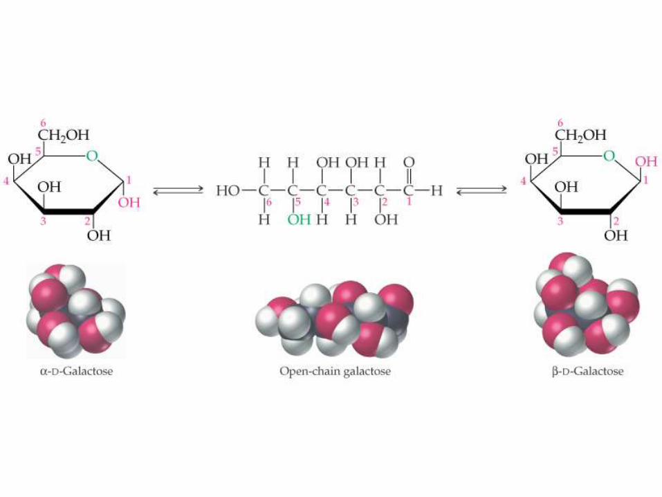
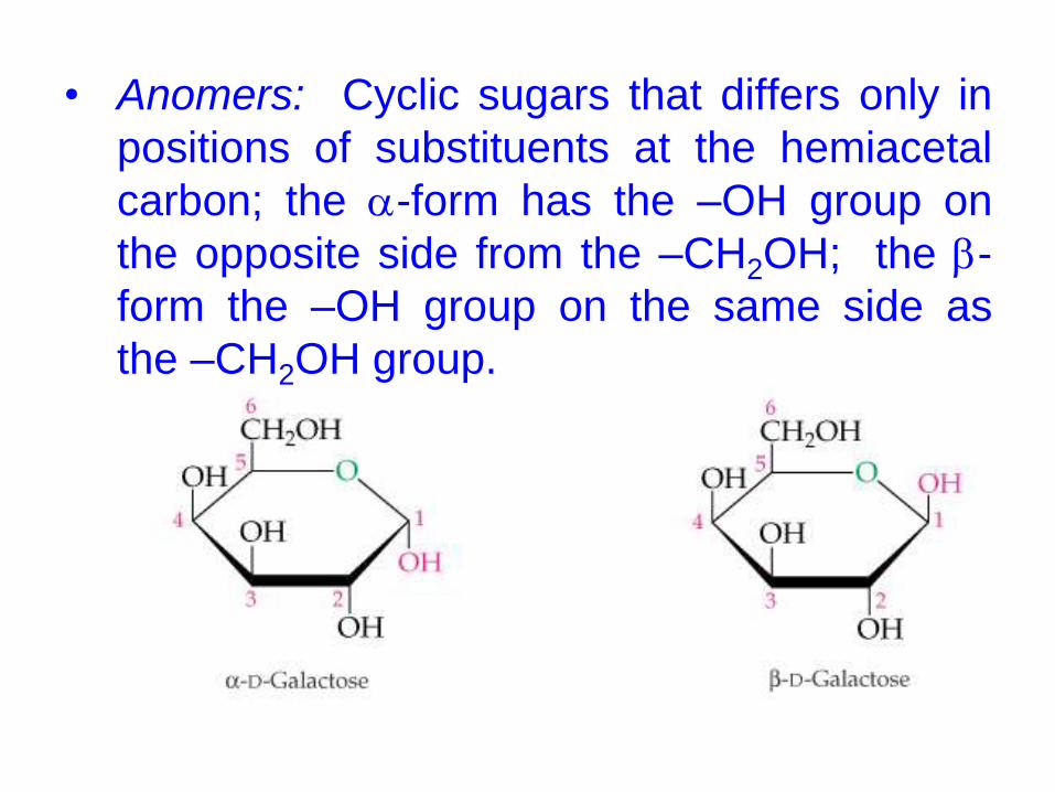
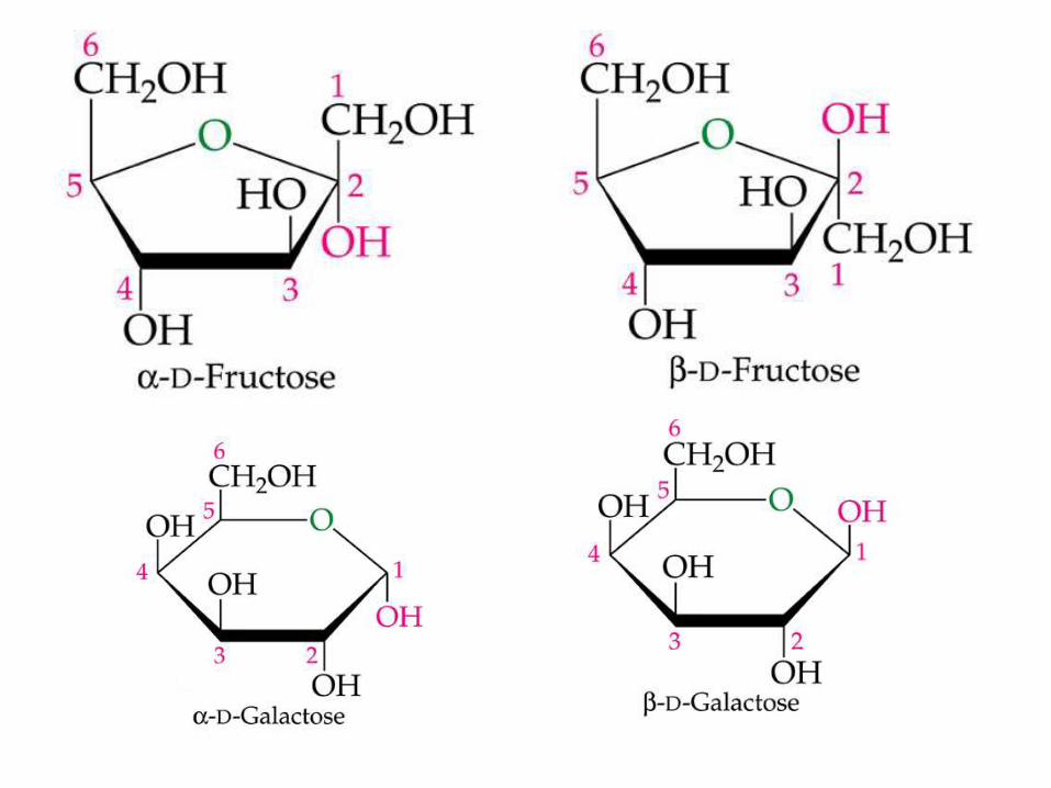
• Anomers: Cyclic sugars that differs only in
positions of substituents at the hemiacetal
carbon; the a-form has the –OH group on
the opposite side from the –CH2OH; the b-
form the –OH group on the same side as
the –CH2OH group.
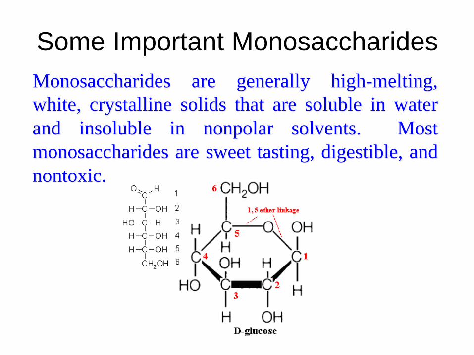
Some Important Monosaccharides
Monosaccharides are generally high-melting,
white, crystalline solids that are soluble in water
and insoluble in nonpolar solvents. Most
monosaccharides are sweet tasting, digestible, and
nontoxic.
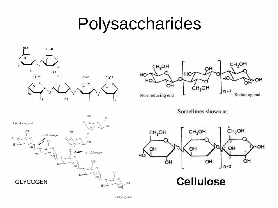
Polysaccharides
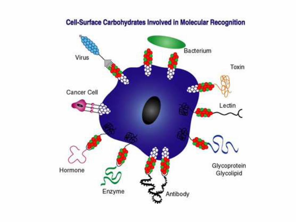
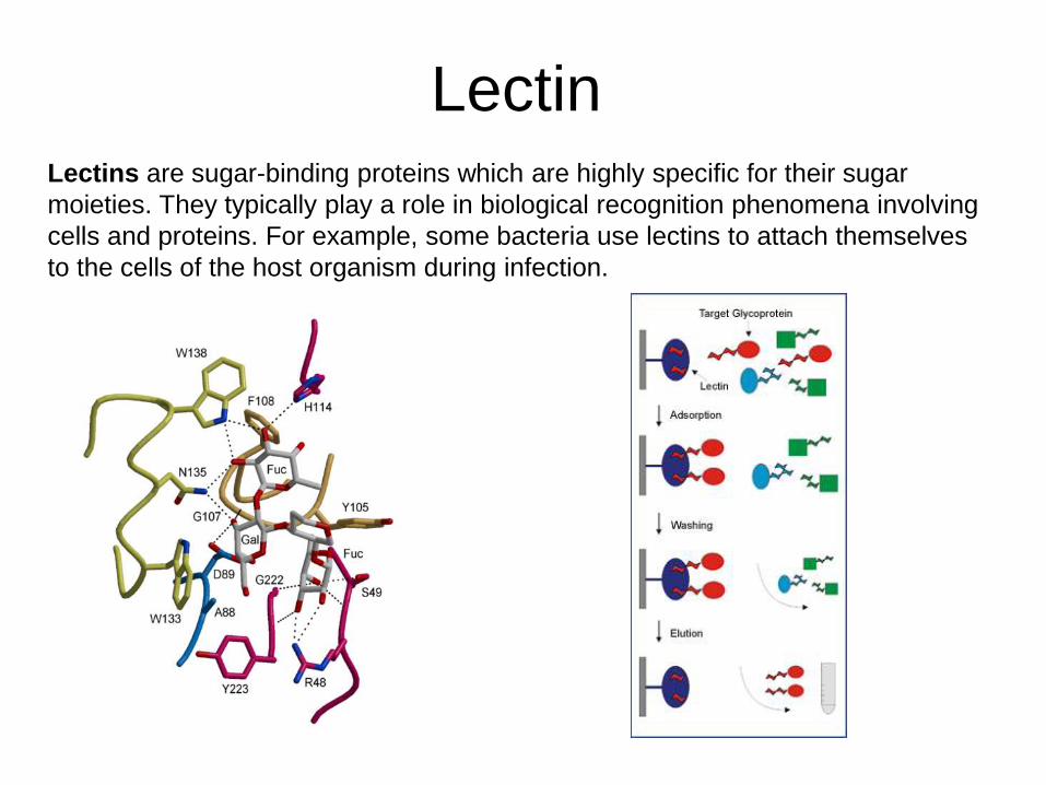
LectinLectins are sugar-binding proteins which are highly specific for their sugar
moieties. They typically play a role in biological recognition phenomena involving
cells and proteins. For example, some bacteria use lectins to attach themselves
to the cells of the host organism during infection.
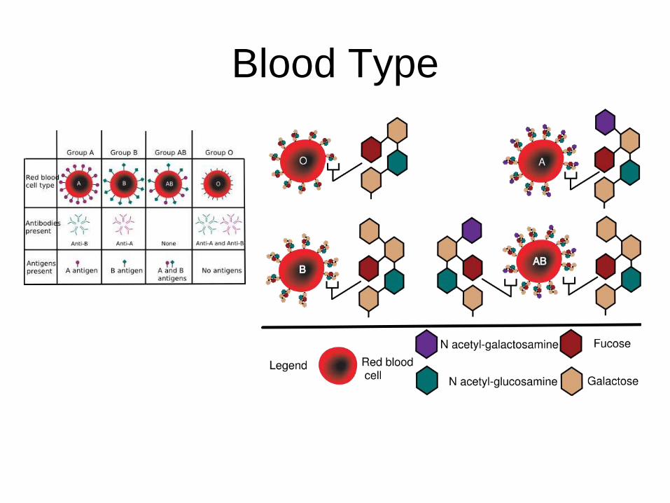
Blood Type
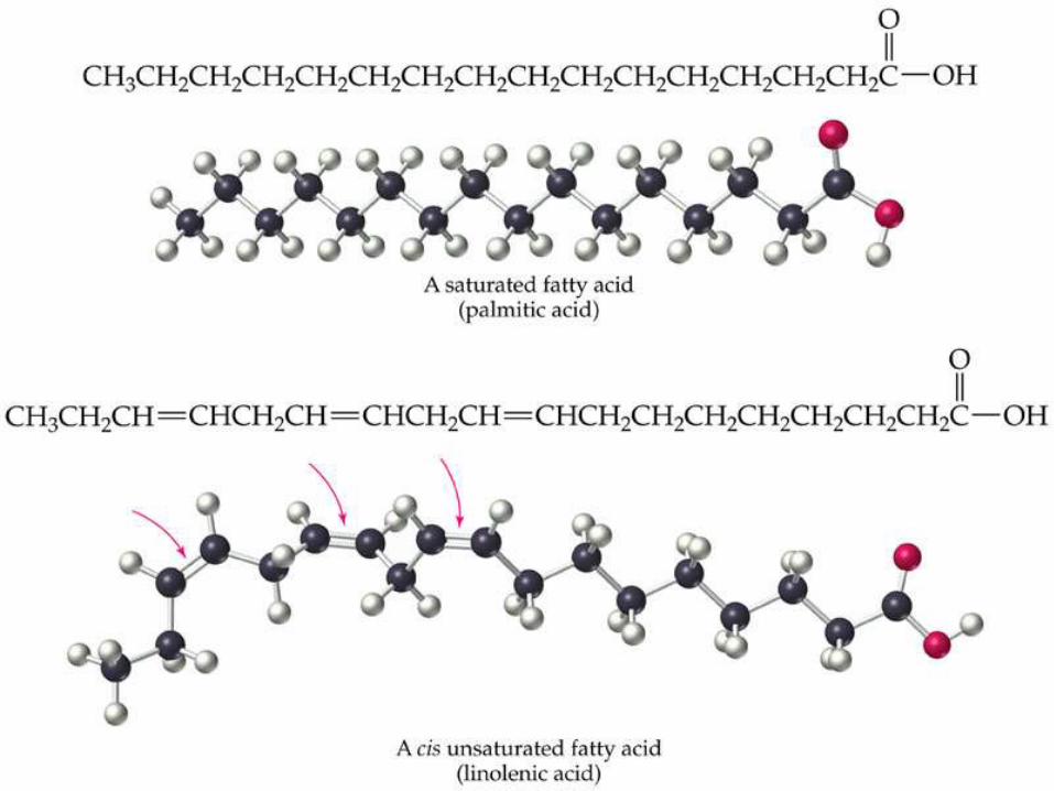
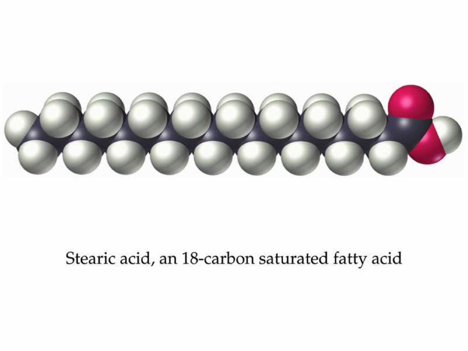
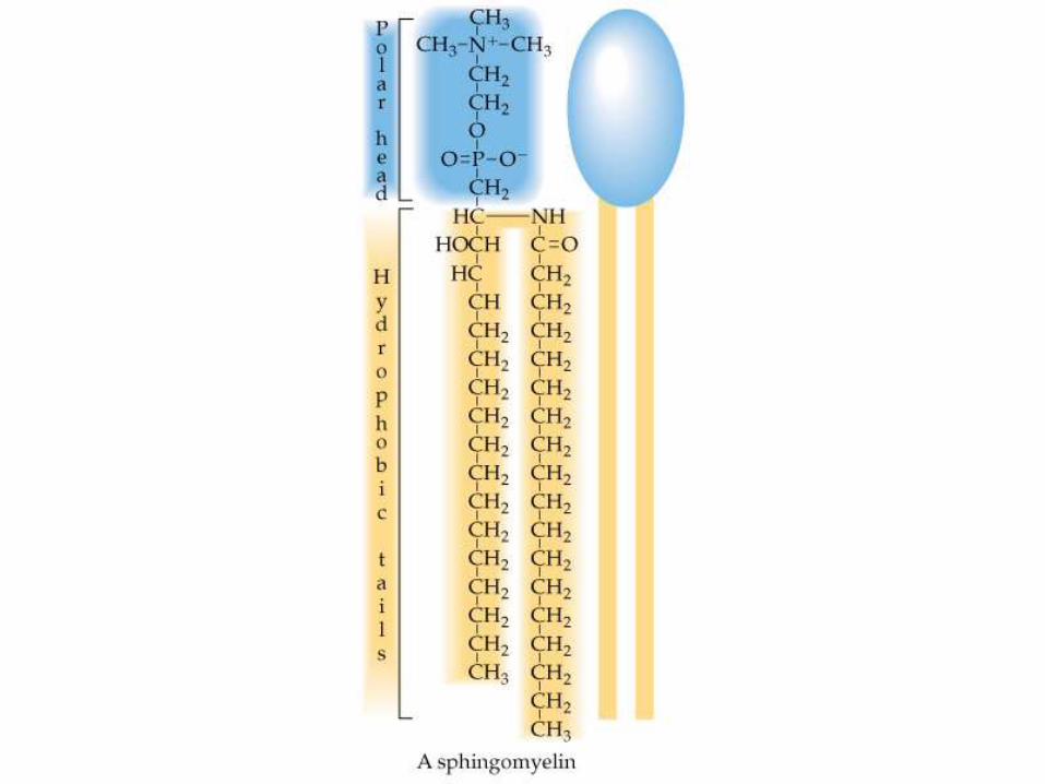
Lipid
• Lipids are naturally occurring molecules
from plants or animals that are soluble in
nonpolar organic solvents.
• Lipid molecules contain large
hydrocarbon portion and not many polar
functional group, which accounts for their
solubility behavior.
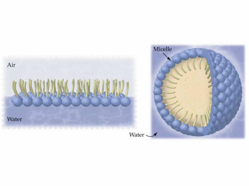
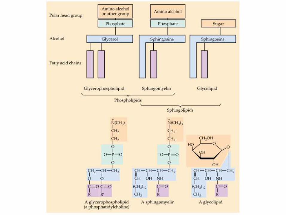
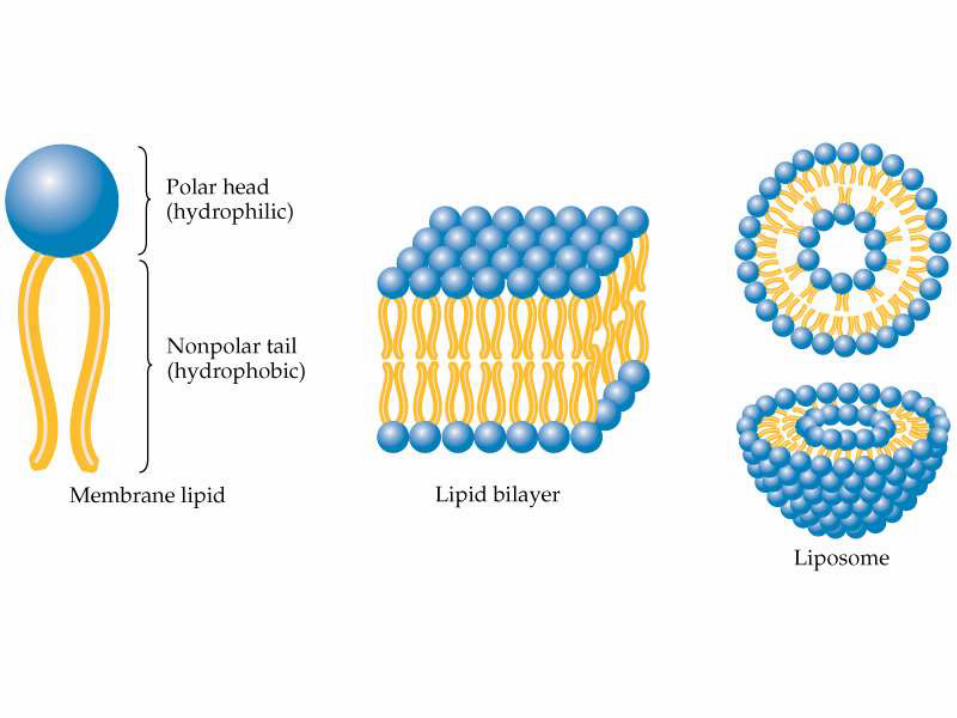
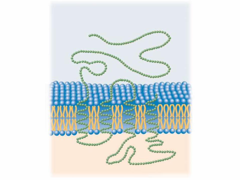
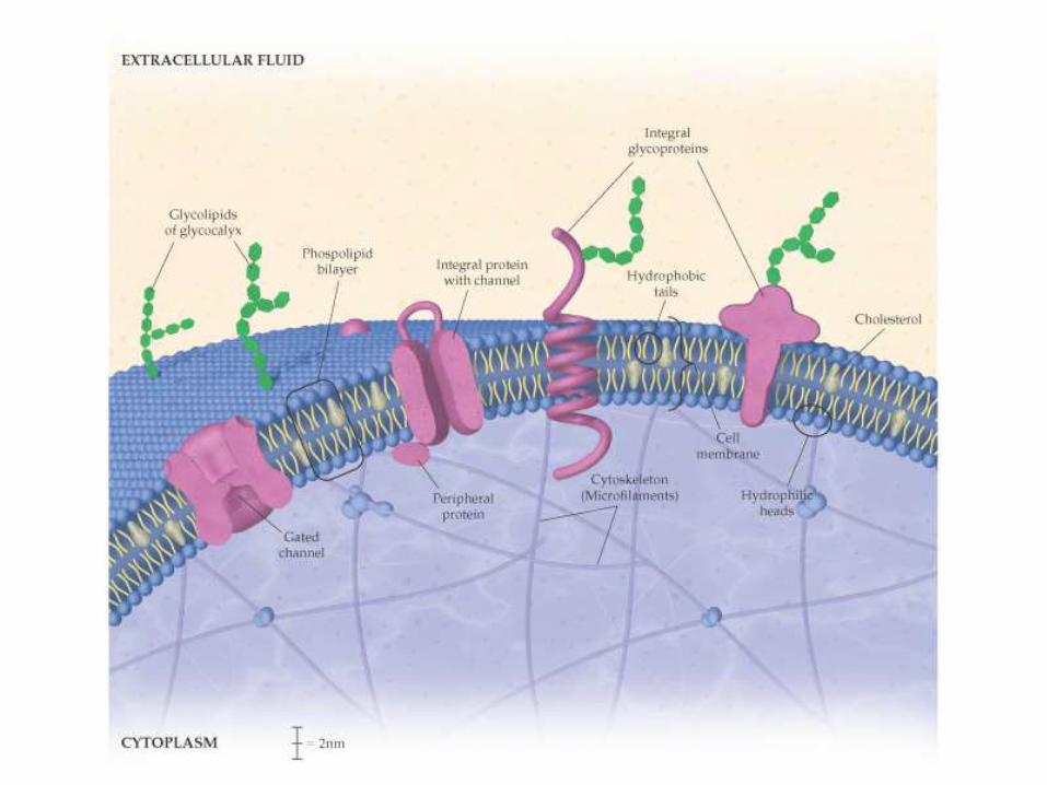
• Cell membranes are composed of a fluid likephospholipid bilayer.
• The bilayer incorporates cholesterol, proteins,and glycolipids.
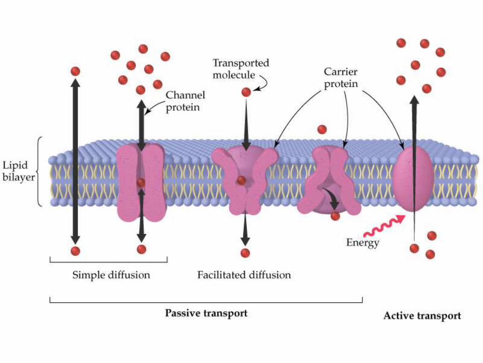
• Small nonpolar molecules cross by diffusionthrough the lipid bilayer.
• Small ions and polar molecules diffuse throughthe aqueous media in protein pores.
• Glucose and certain other substances crosswith the aid of proteins without energy input.
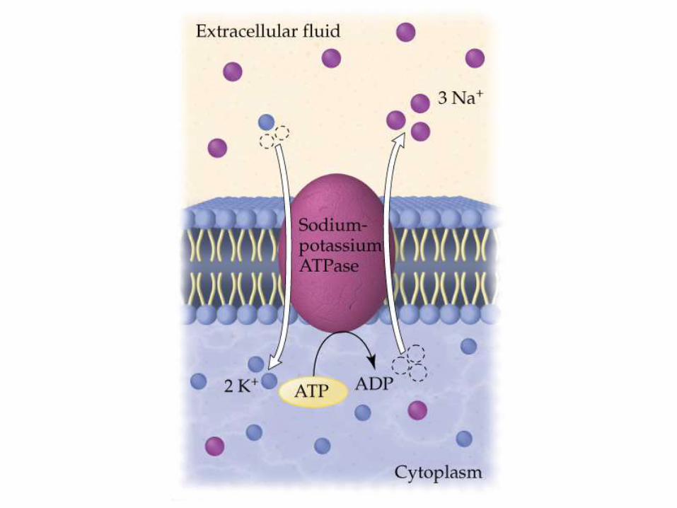
• Na+, K+, and other substances that maintainconcentration gradients inside and outside thecell cross with expenditure of energy and theaid of proteins.
Properties of cell membranes:
• Small ions and polar molecules diffuse
through the aqueous media in protein
pores.
• Glucose and certain other substances
cross with the aid of proteins without
energy input.
• Na+, K+, and other substances that
maintain concentration gradients inside
and outside the cell cross with
expenditure of energy and the aid of
proteins.
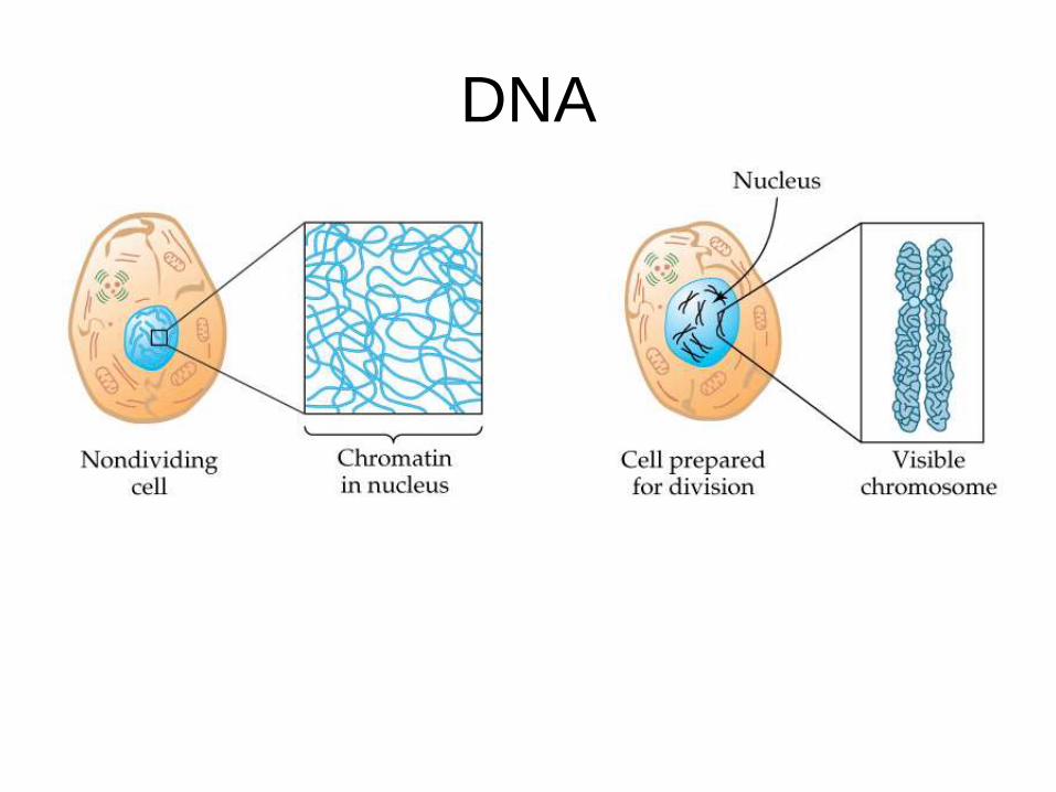
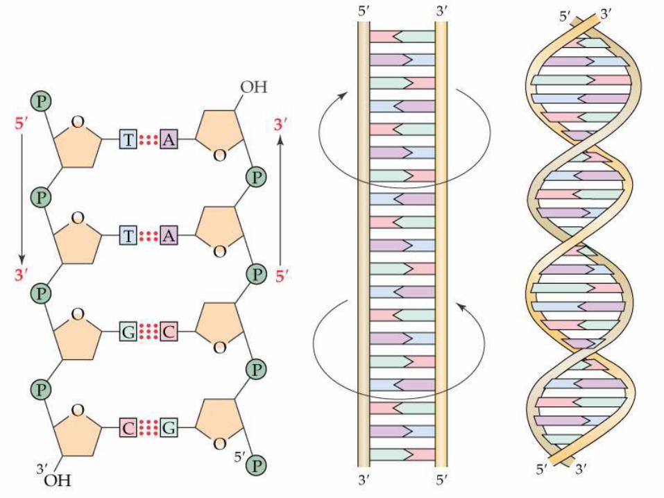
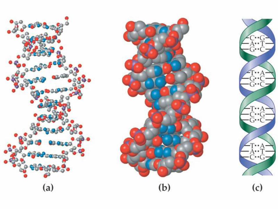
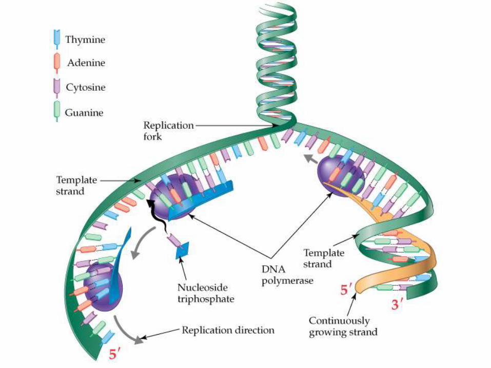
DNA
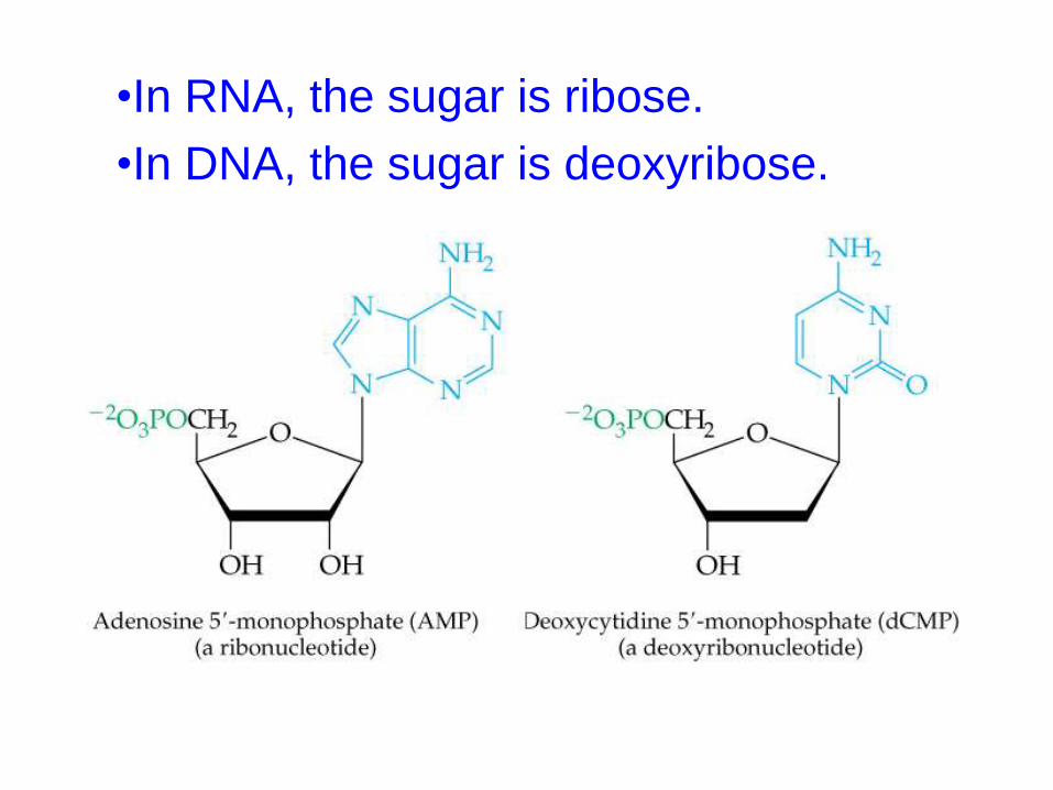
•In RNA, the sugar is ribose.
•In DNA, the sugar is deoxyribose.
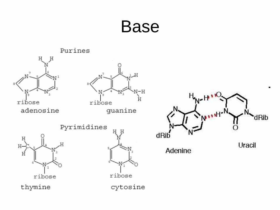
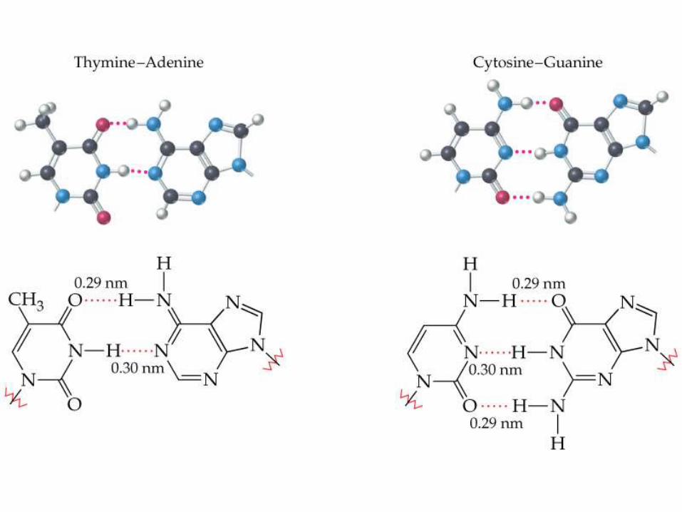
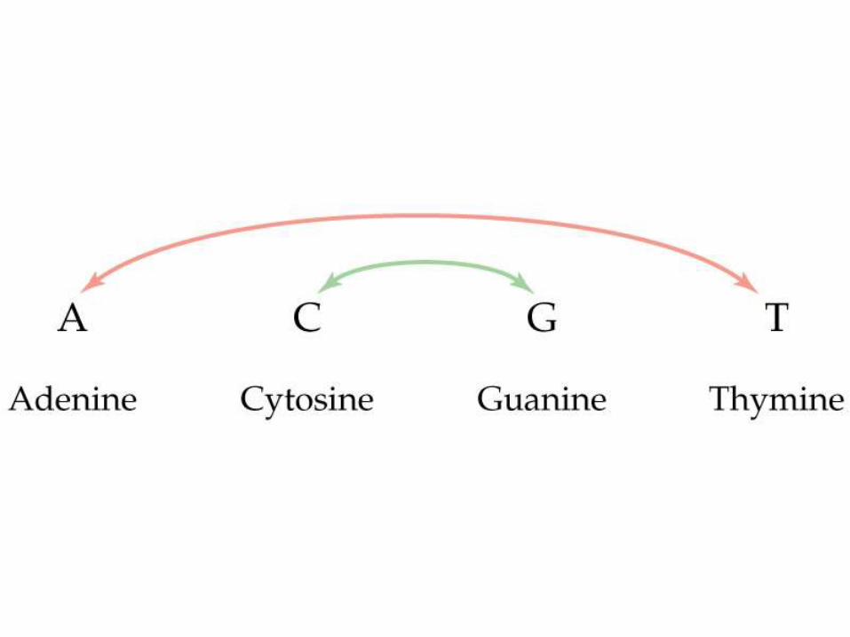
Base
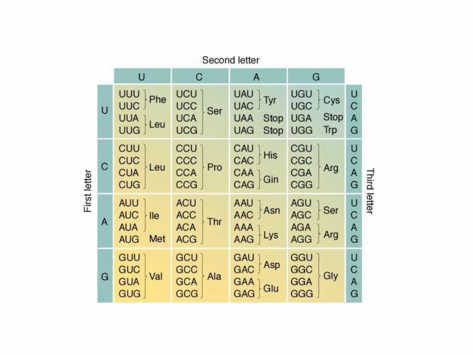
•The following three RNA make it possible for theencoded information carried by the DNA to be putto use in the synthesis of proteins.
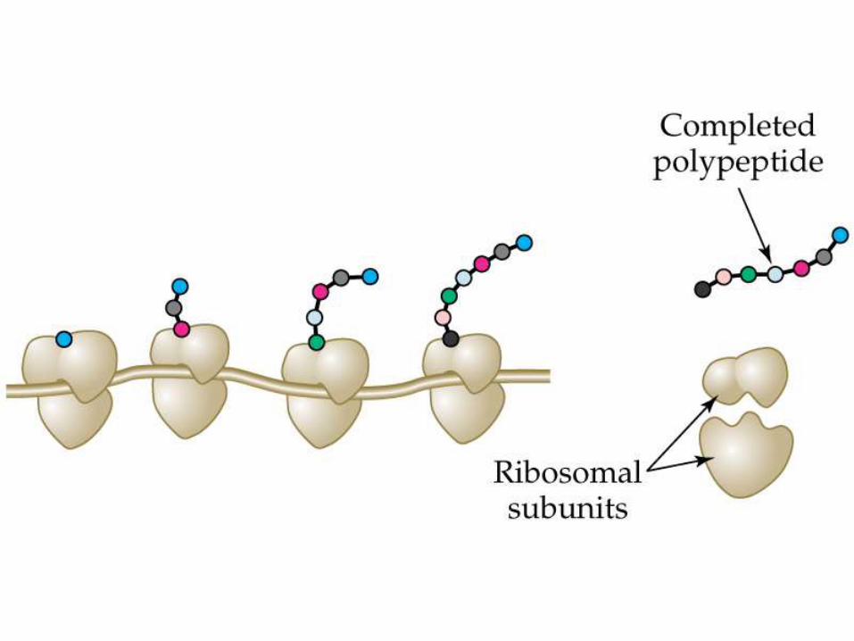
•Ribosome RNA: The granular organelles in thecell where protein synthesis takes place. Theseorganelles are composed of protein and ribosomalRNA (rRNA).
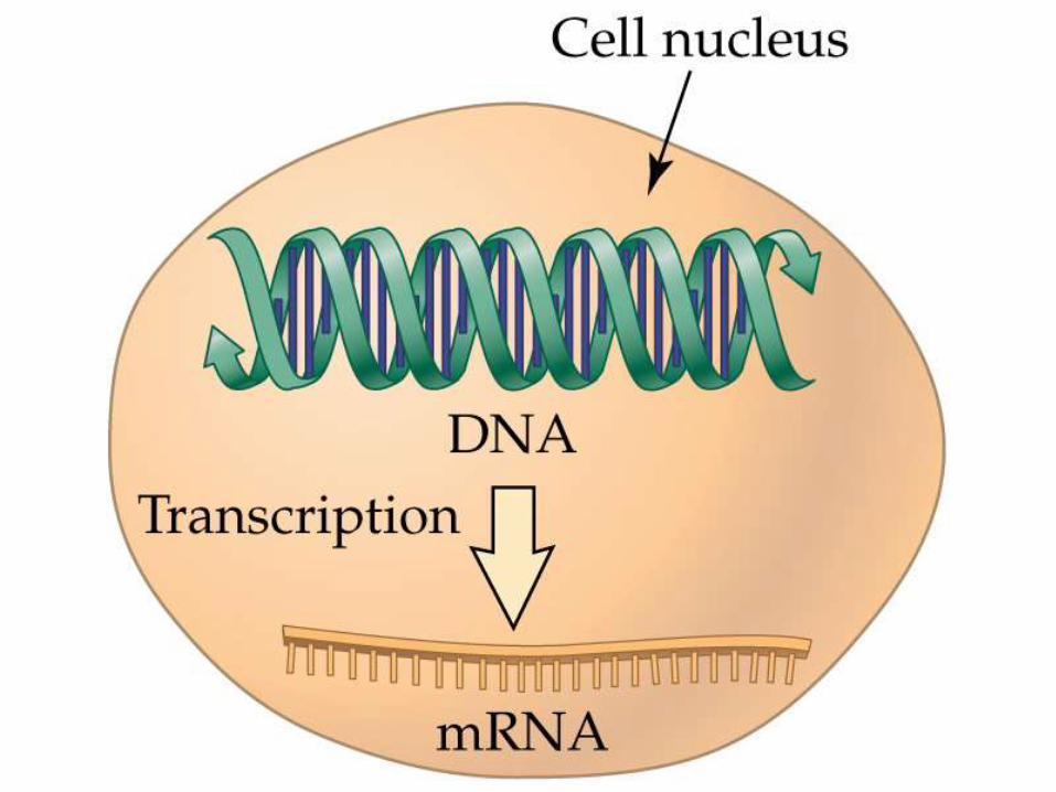
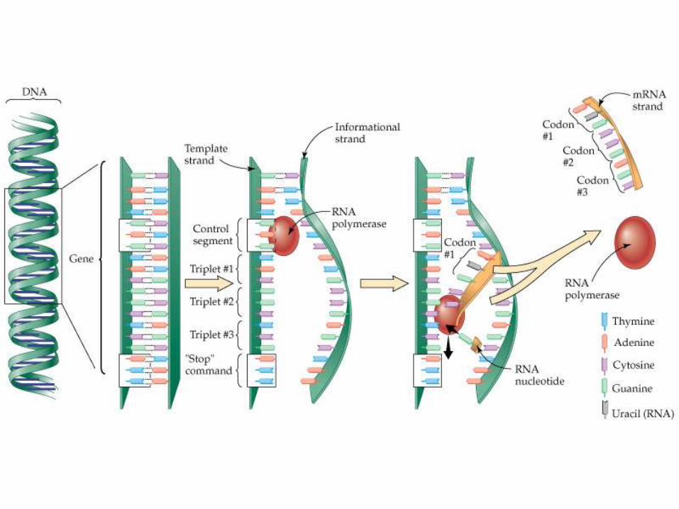
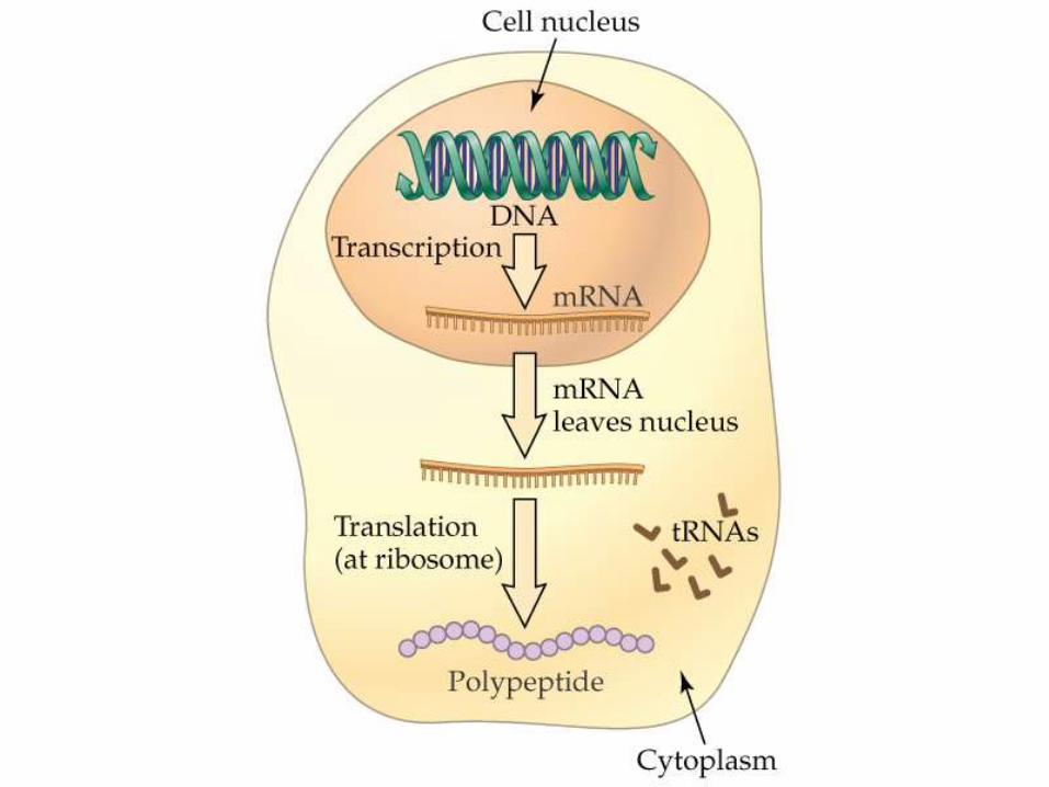
•Messenger RNA (mRNA): The RNA that carriesthe code transcribed from DNA and directs proteinsynthesis.
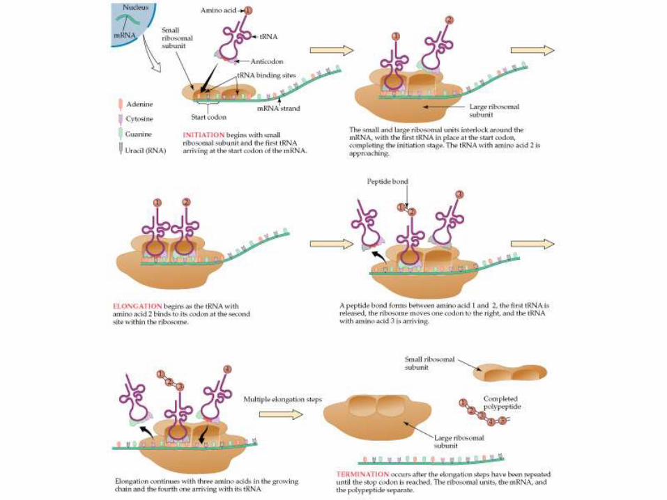
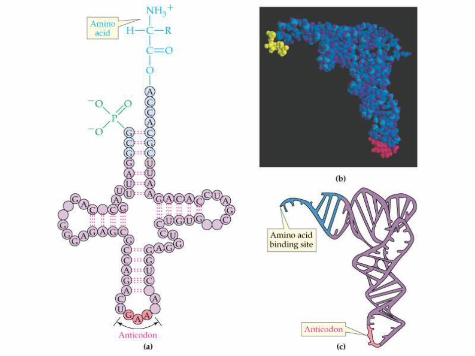
•Transfer RNA (tRNA): The smaller RNA thatdelivers amino acids one by one to protein chainsgrowing at ribosomes. Each tRNA recognizes andcarries only one amino acid.

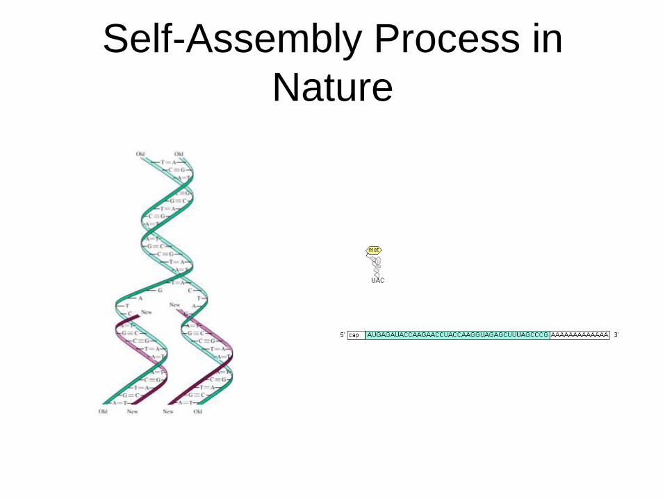
Self-Assembly Process in
Nature

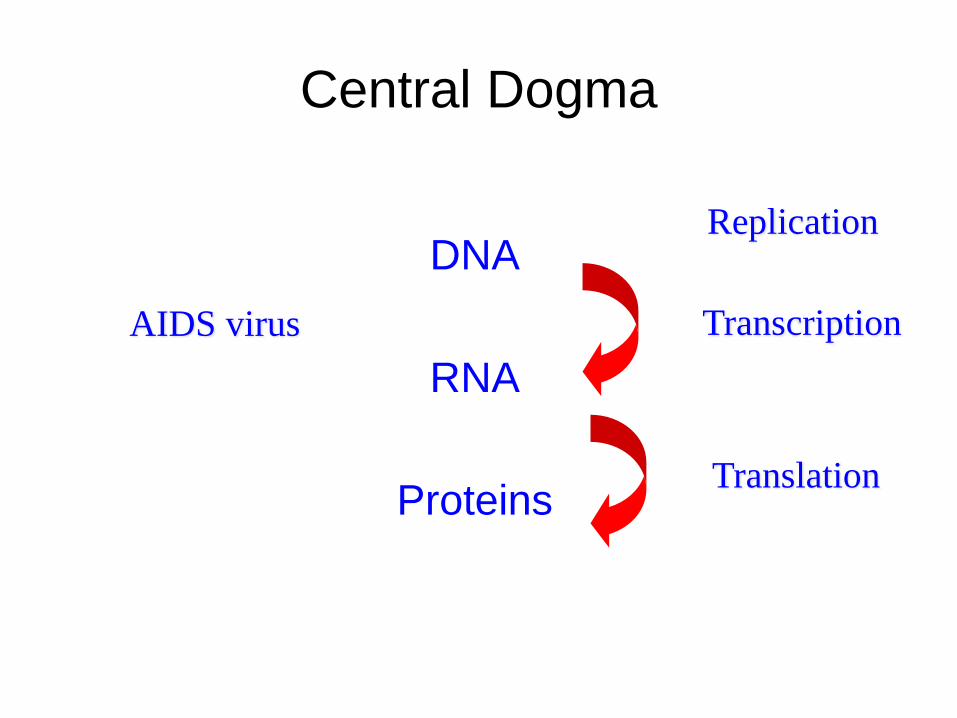
Central Dogma
DNA
RNA
Proteins
Transcription
Translation
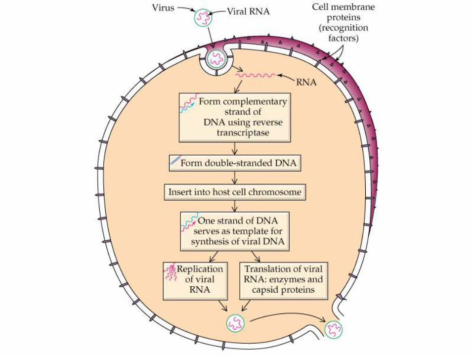
AIDS virus
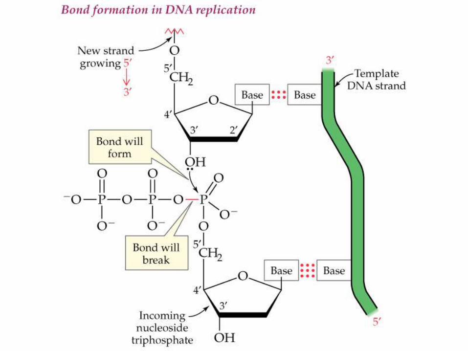
Replication
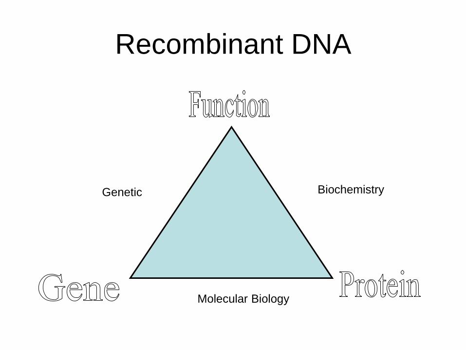
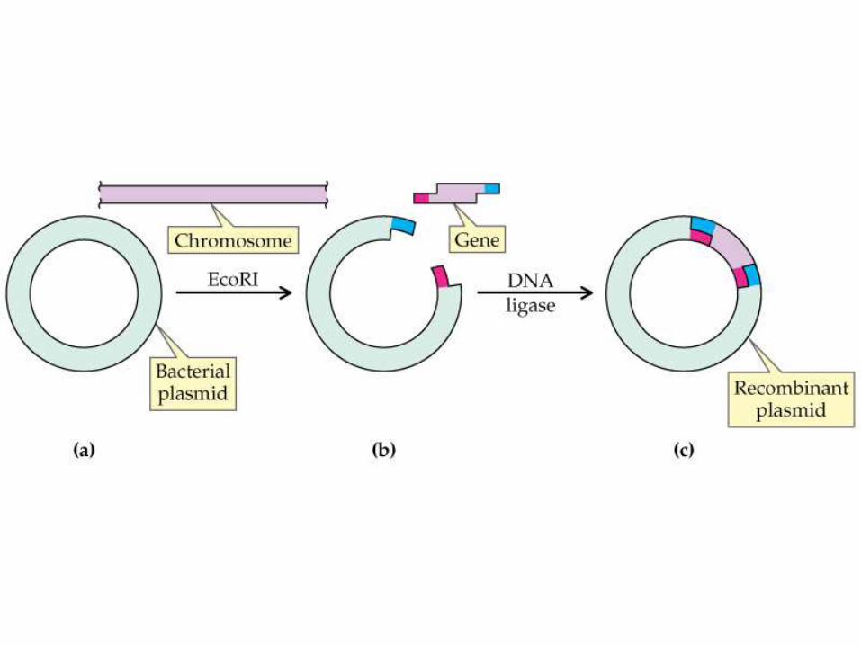
Recombinant DNA
BiochemistryGenetic
Molecular Biology
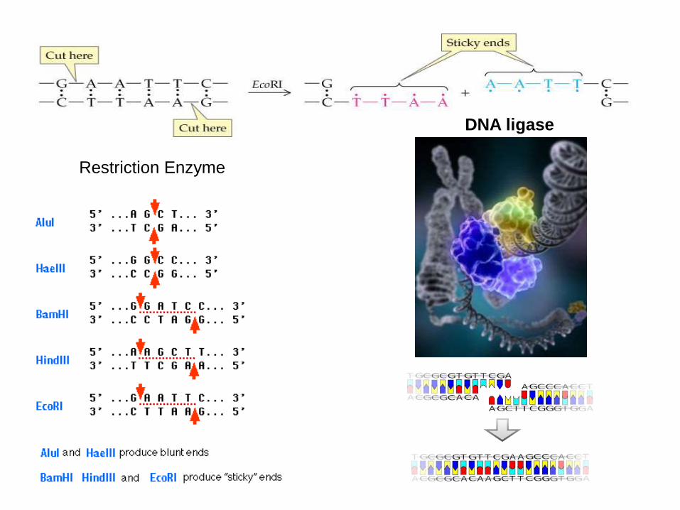
DNA ligase
Restriction Enzyme
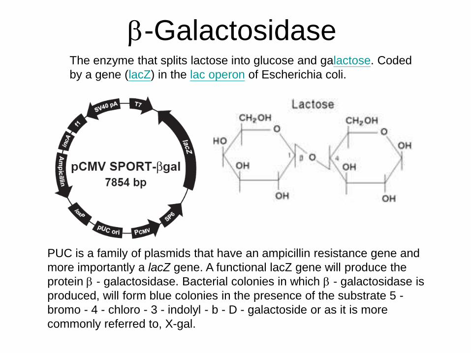
b-Galactosidase
PUC is a family of plasmids that have an ampicillin resistance gene and
more importantly a lacZ gene. A functional lacZ gene will produce the
protein b - galactosidase. Bacterial colonies in which b - galactosidase is
produced, will form blue colonies in the presence of the substrate 5 -
bromo - 4 - chloro - 3 - indolyl - b - D - galactoside or as it is more
commonly referred to, X-gal.
The enzyme that splits lactose into glucose and galactose. Coded
by a gene (lacZ) in the lac operon of Escherichia coli.
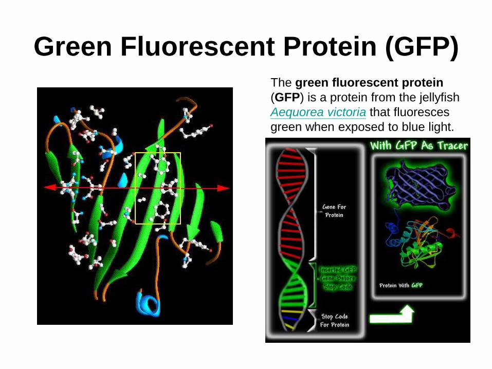
Green Fluorescent Protein (GFP)The green fluorescent protein
(GFP) is a protein from the jellyfish
Aequorea victoria that fluoresces
green when exposed to blue light.
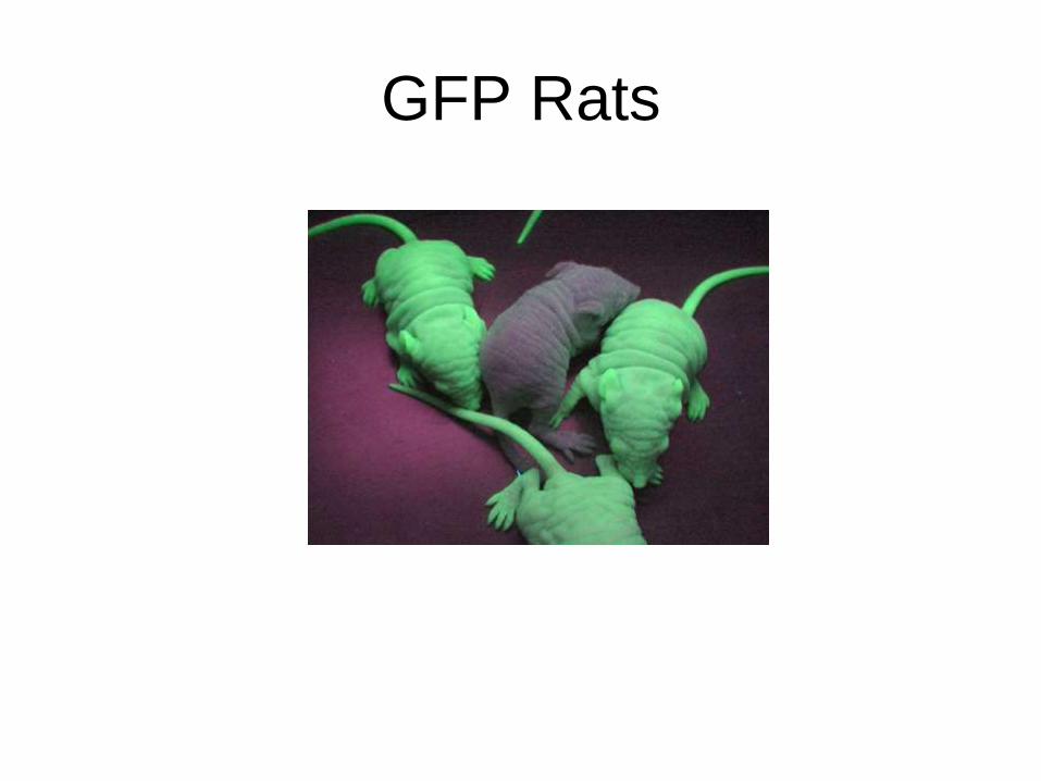
GFP Rats
Central Dogma
DNA
RNA
Proteins
Transcription
Translation
AIDS virus
Replication
Life
• Replication: reproduction
• Function: catalytic functions
• RNA world:
• Virus is not alive
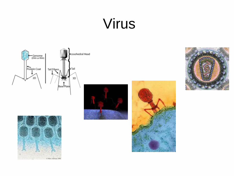
Virus
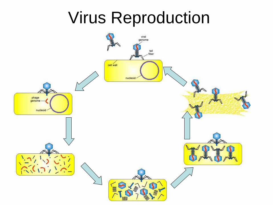
Virus Reproduction
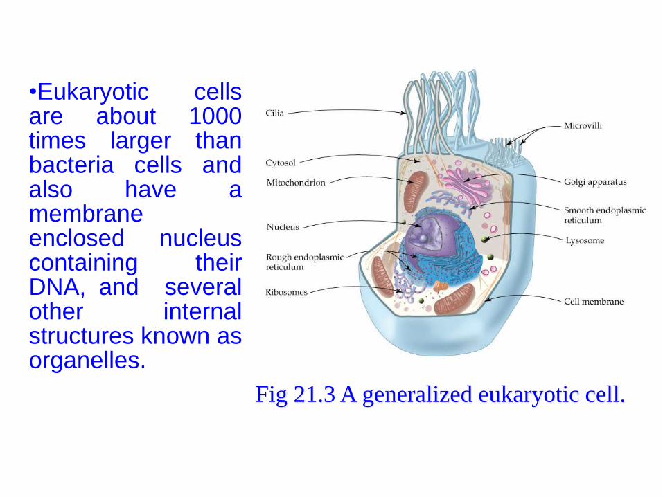
•Eukaryotic cellsare about 1000times larger thanbacteria cells andalso have amembraneenclosed nucleuscontaining theirDNA, and severalother internalstructures known asorganelles.
Fig 21.3 A generalized eukaryotic cell.
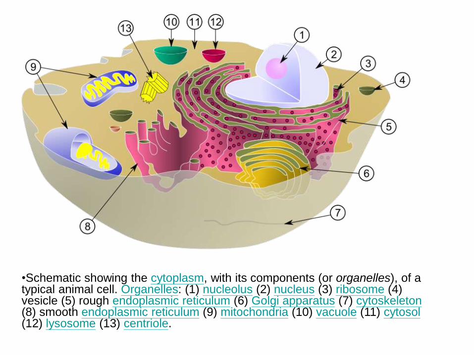
•Schematic showing the cytoplasm, with its components (or organelles), of a typical animal cell. Organelles: (1) nucleolus (2) nucleus (3) ribosome (4) vesicle (5) rough endoplasmic reticulum (6) Golgi apparatus (7) cytoskeleton(8) smooth endoplasmic reticulum (9) mitochondria (10) vacuole (11) cytosol(12) lysosome (13) centriole.
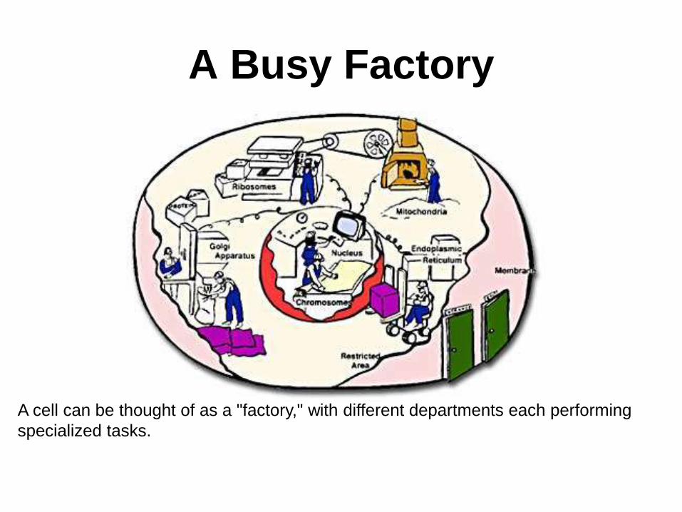
A Busy Factory
A cell can be thought of as a "factory," with different departments each performing
specialized tasks.
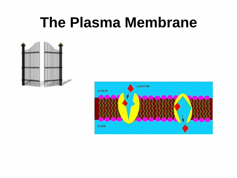
The Plasma Membrane
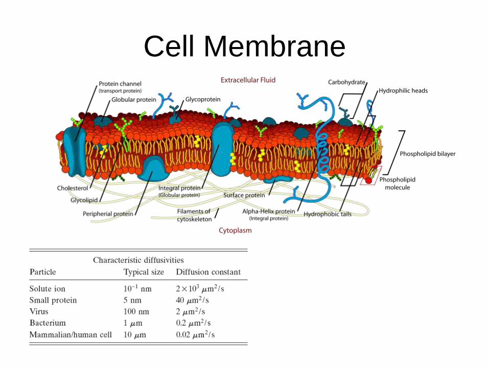
Cell Membrane
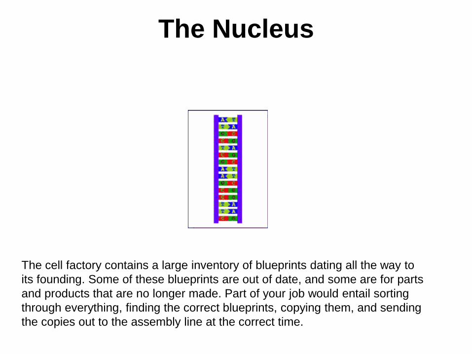
The Nucleus
The cell factory contains a large inventory of blueprints dating all the way to
its founding. Some of these blueprints are out of date, and some are for parts
and products that are no longer made. Part of your job would entail sorting
through everything, finding the correct blueprints, copying them, and sending
the copies out to the assembly line at the correct time.
Nucleus
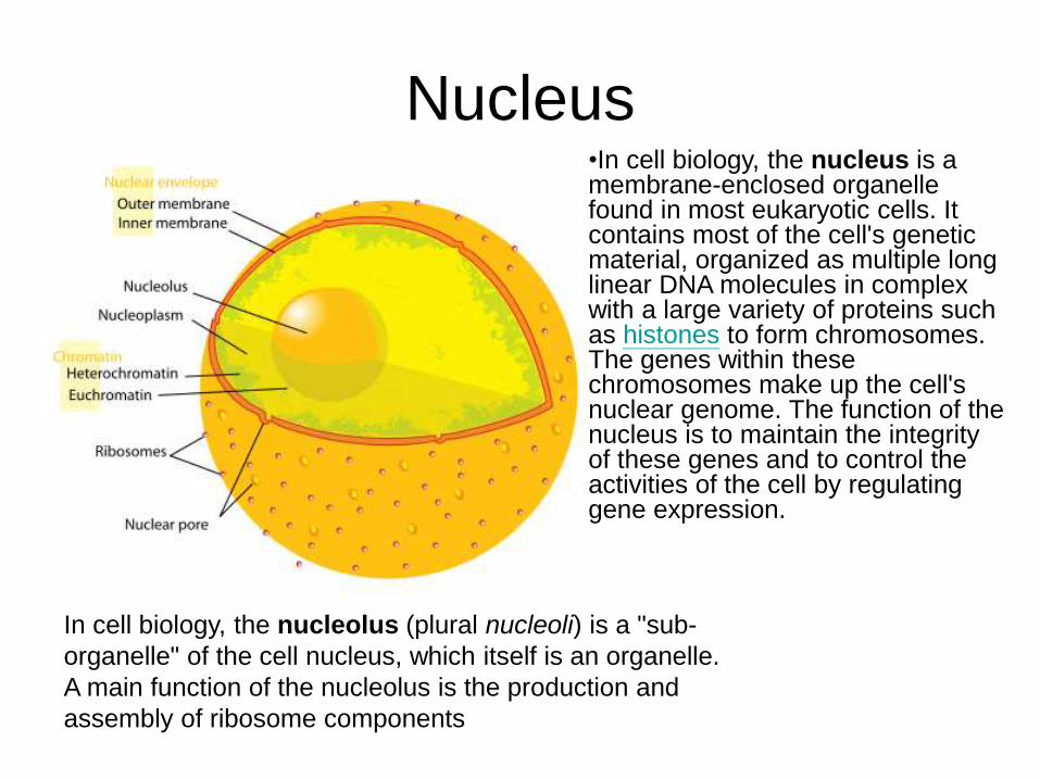
In cell biology, the nucleolus (plural nucleoli) is a "sub-
organelle" of the cell nucleus, which itself is an organelle.
A main function of the nucleolus is the production and
assembly of ribosome components
•In cell biology, the nucleus is a membrane-enclosed organelle found in most eukaryotic cells. It contains most of the cell's genetic material, organized as multiple long linear DNA molecules in complex with a large variety of proteins such as histones to form chromosomes. The genes within these chromosomes make up the cell's nuclear genome. The function of the nucleus is to maintain the integrity of these genes and to control the activities of the cell by regulating gene expression.
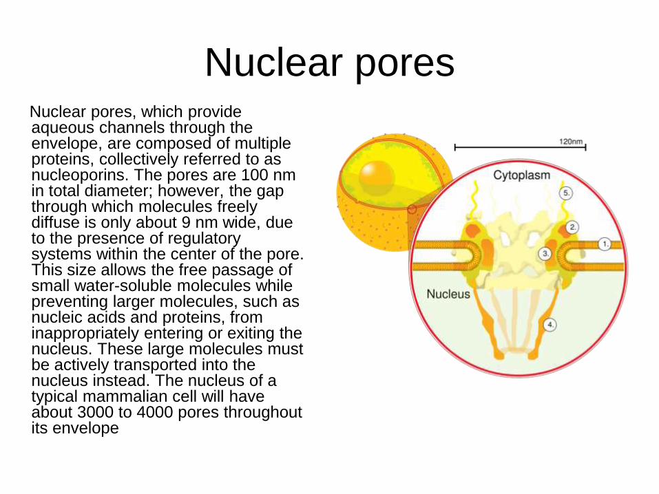
Nuclear poresNuclear pores, which provide aqueous channels through the envelope, are composed of multiple proteins, collectively referred to as nucleoporins. The pores are 100 nm in total diameter; however, the gap through which molecules freely diffuse is only about 9 nm wide, due to the presence of regulatory systems within the center of the pore. This size allows the free passage of small water-soluble molecules while preventing larger molecules, such as nucleic acids and proteins, from inappropriately entering or exiting the nucleus. These large molecules must be actively transported into the nucleus instead. The nucleus of a typical mammalian cell will have about 3000 to 4000 pores throughout its envelope
Nuclear localizing sequence
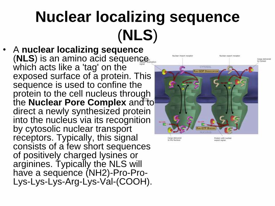
(NLS)• A nuclear localizing sequence
(NLS) is an amino acid sequence which acts like a 'tag' on the exposed surface of a protein. This sequence is used to confine the protein to the cell nucleus through the Nuclear Pore Complex and to direct a newly synthesized protein into the nucleus via its recognition by cytosolic nuclear transport receptors. Typically, this signal consists of a few short sequences of positively charged lysines or arginines. Typically the NLS will have a sequence (NH2)-Pro-Pro-Lys-Lys-Lys-Arg-Lys-Val-(COOH).
The Ribosomes and the ER
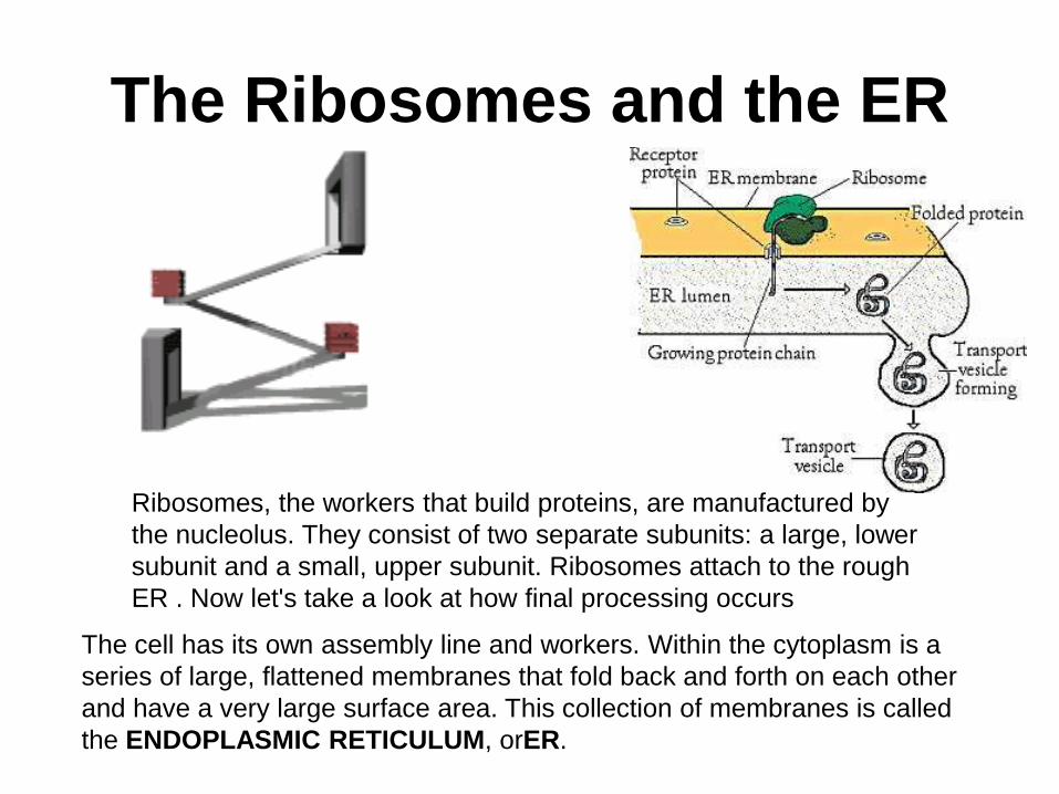
The cell has its own assembly line and workers. Within the cytoplasm is a
series of large, flattened membranes that fold back and forth on each other
and have a very large surface area. This collection of membranes is called
the ENDOPLASMIC RETICULUM, orER.
Ribosomes, the workers that build proteins, are manufactured by
the nucleolus. They consist of two separate subunits: a large, lower
subunit and a small, upper subunit. Ribosomes attach to the rough
ER . Now let's take a look at how final processing occurs
Ribosome
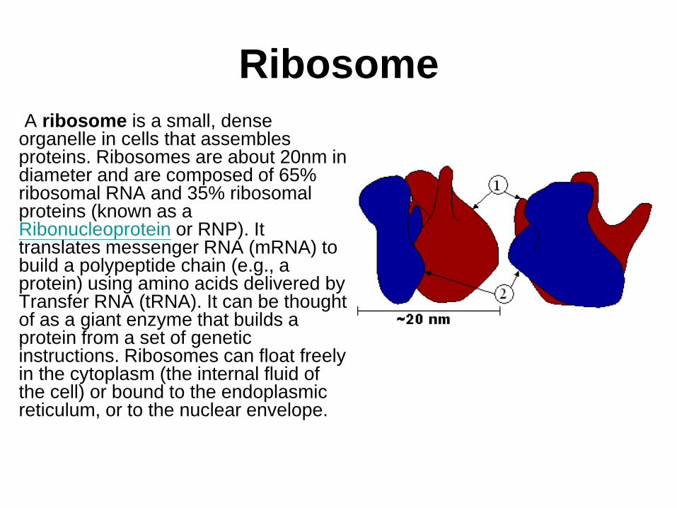
A ribosome is a small, dense organelle in cells that assembles proteins. Ribosomes are about 20nm in diameter and are composed of 65% ribosomal RNA and 35% ribosomal proteins (known as a Ribonucleoprotein or RNP). It translates messenger RNA (mRNA) to build a polypeptide chain (e.g., a protein) using amino acids delivered by Transfer RNA (tRNA). It can be thought of as a giant enzyme that builds a protein from a set of genetic instructions. Ribosomes can float freely in the cytoplasm (the internal fluid of the cell) or bound to the endoplasmic reticulum, or to the nuclear envelope.
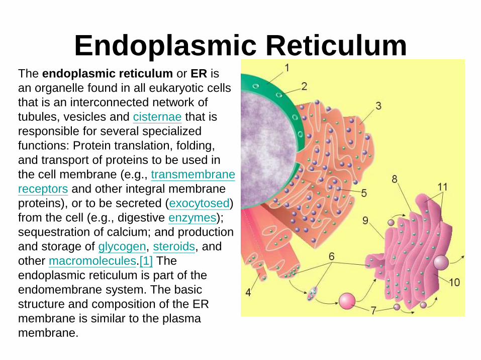
Endoplasmic ReticulumThe endoplasmic reticulum or ER is
an organelle found in all eukaryotic cells
that is an interconnected network of
tubules, vesicles and cisternae that is
responsible for several specialized
functions: Protein translation, folding,
and transport of proteins to be used in
the cell membrane (e.g., transmembrane
receptors and other integral membrane
proteins), or to be secreted (exocytosed)
from the cell (e.g., digestive enzymes);
sequestration of calcium; and production
and storage of glycogen, steroids, and
other macromolecules.[1] The
endoplasmic reticulum is part of the
endomembrane system. The basic
structure and composition of the ER
membrane is similar to the plasma
membrane.
Rough endoplasmic reticulum
• The surface of the rough endoplasmic reticulum is studded with protein-manufacturing ribosomes giving it a "rough" appearance. But it should be noted that these ribosomes are not resident of the endoplasmic reticulum incessantly. The ribosomes only bind to the ER once it begins to synthesize a protein destined for sorting. The membrane of the rough endoplasmic reticulum is continuous with the outer layer of the nuclear envelope. Although there is no continuous membrane between the rough ER and the Golgi apparatus, membrane bound vesicles shuttle proteins between these two compartments. The rough endoplasmic reticulum works in concert with the Golgi complex to target new proteins to their proper destinations
Smooth endoplasmic reticulum
• The smooth endoplasmic reticulum has functions in several metabolic processes, including synthesis of lipids, metabolism of carbohydrates and calcium concentration, and attachment of receptors on cell membrane proteins. It is connected to the nuclear envelope. Smooth endoplasmic reticulum is found in a variety of cell types (both animal and plant) and it serves different functions in each. It consists of tubules and vesicles that branch forming a network. In some cells there are dilated areas like the sacs of rough endoplasmic reticulum. The network of smooth endoplasmic reticulum allows increased surface area for the action or storage of key enzymes and the products of these enzymes. The smooth endoplasmic reticulum is known for its storage of calcium ions in muscle cells.
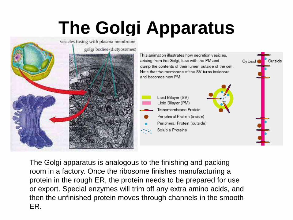
The Golgi Apparatus
The Golgi apparatus is analogous to the finishing and packing
room in a factory. Once the ribosome finishes manufacturing a
protein in the rough ER, the protein needs to be prepared for use
or export. Special enzymes will trim off any extra amino acids, and
then the unfinished protein moves through channels in the smooth
ER.
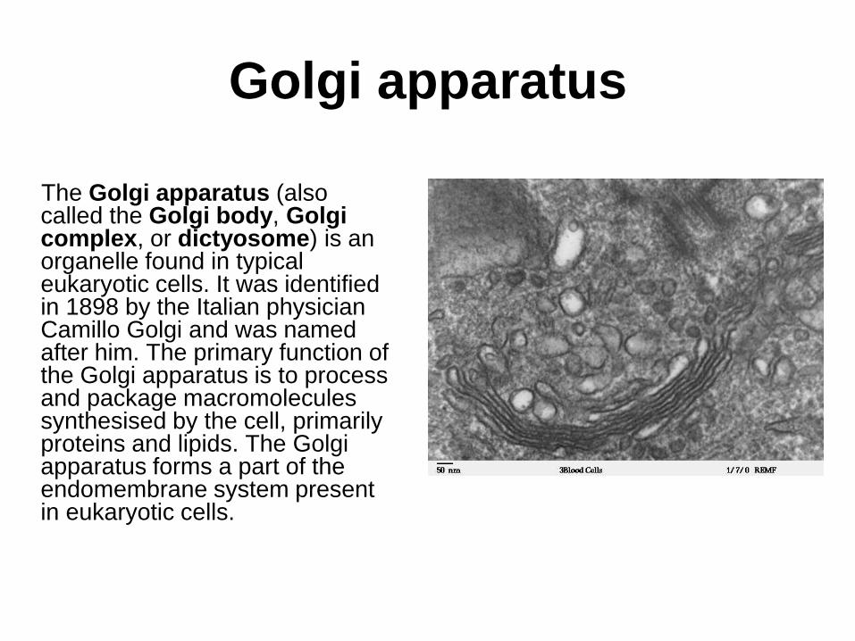
Golgi apparatus
The Golgi apparatus (also called the Golgi body, Golgi complex, or dictyosome) is an organelle found in typical eukaryotic cells. It was identified in 1898 by the Italian physician Camillo Golgi and was named after him. The primary function of the Golgi apparatus is to process and package macromolecules synthesised by the cell, primarily proteins and lipids. The Golgi apparatus forms a part of the endomembrane system present in eukaryotic cells.
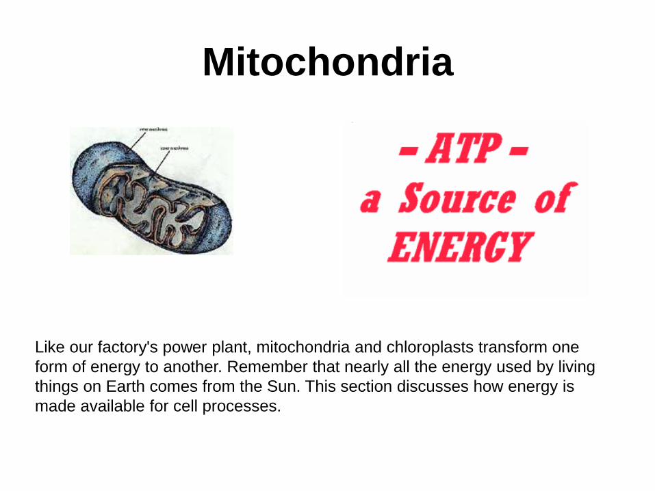
Mitochondria
Like our factory's power plant, mitochondria and chloroplasts transform one
form of energy to another. Remember that nearly all the energy used by living
things on Earth comes from the Sun. This section discusses how energy is
made available for cell processes.
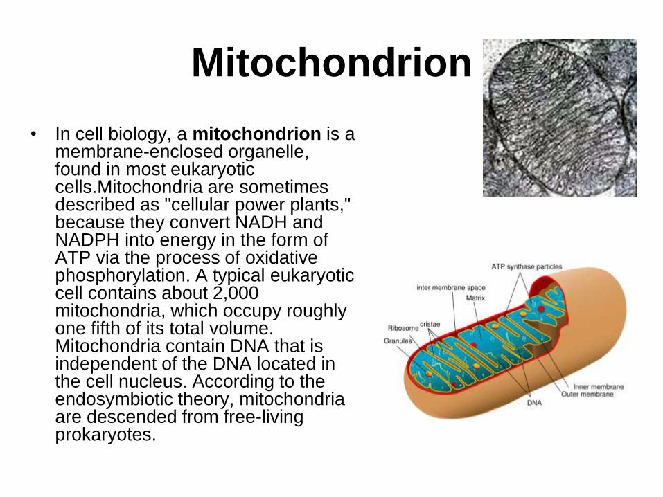
Mitochondrion
• In cell biology, a mitochondrion is a membrane-enclosed organelle, found in most eukaryotic cells.Mitochondria are sometimes described as "cellular power plants," because they convert NADH and NADPH into energy in the form of ATP via the process of oxidative phosphorylation. A typical eukaryotic cell contains about 2,000 mitochondria, which occupy roughly one fifth of its total volume. Mitochondria contain DNA that is independent of the DNA located in the cell nucleus. According to the endosymbiotic theory, mitochondria are descended from free-living prokaryotes.
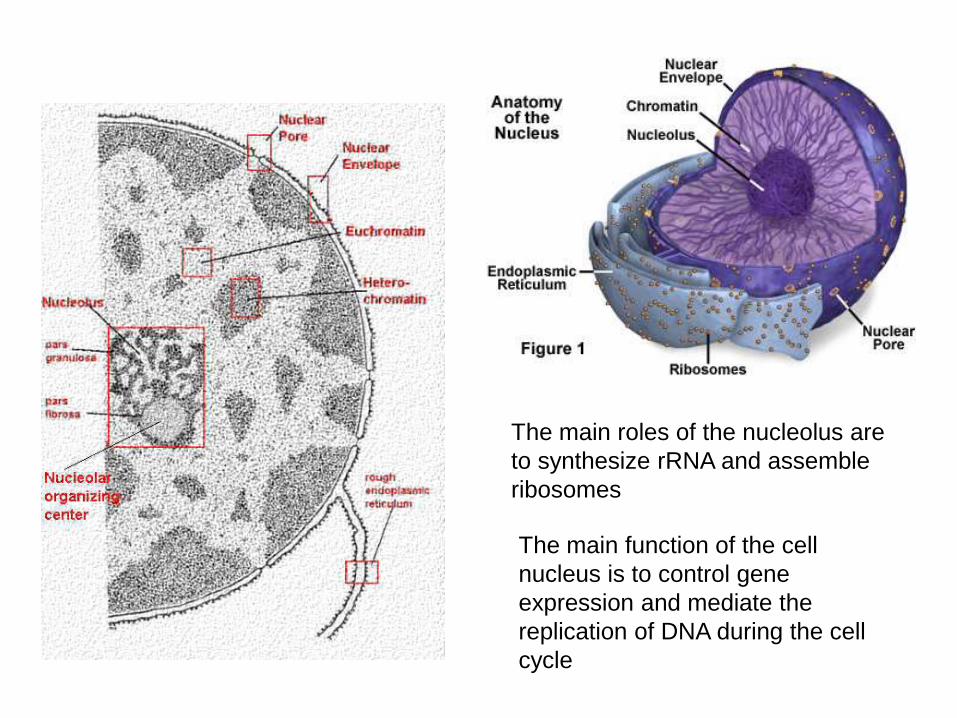
The main roles of the nucleolus are
to synthesize rRNA and assemble
ribosomes
The main function of the cell
nucleus is to control gene
expression and mediate the
replication of DNA during the cell
cycle
Lysosomes
• Lysosomes are organelles that contain digestive enzymes (acid hydrolases). They digest excess or worn out organelles, food particles, and engulfed viruses or bacteria. The membrane surrounding a lysosome prevents the digestive enzymes inside from destroying the cell. Lysosomes fuse with vacuoles and dispense their enzymes into the vacuoles, digesting their contents. They are built in the Golgi apparatus. The name lysosome derives from the Greek words lysis, which means dissolution or destruction, and soma, which means body. They are frequently nicknamed "suicide-bags" or "suicide-sacs" by cell biologists due to their role in autolysis.
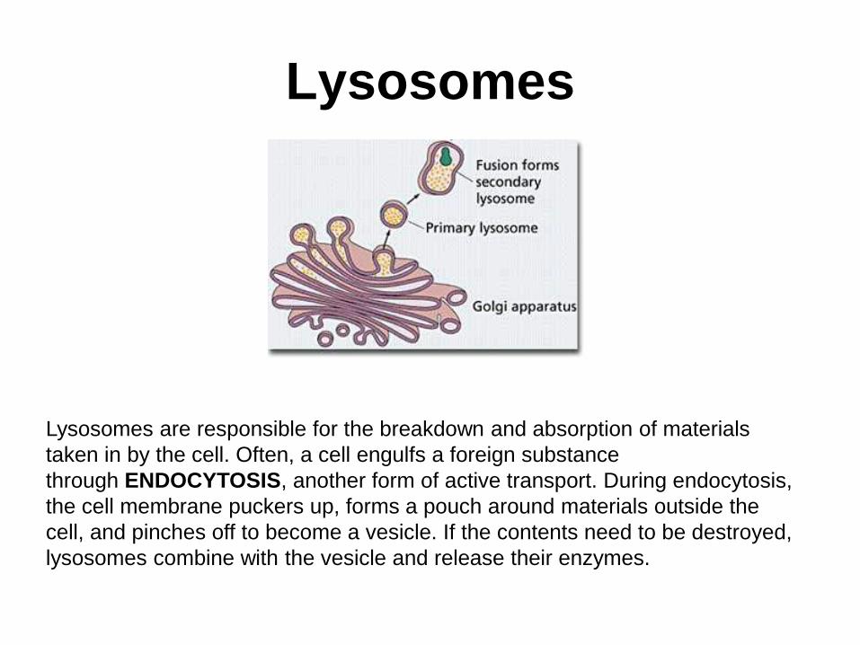
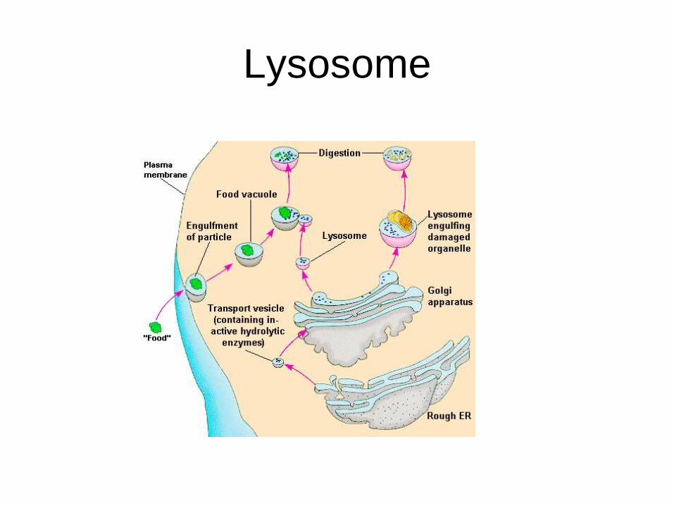
Lysosomes
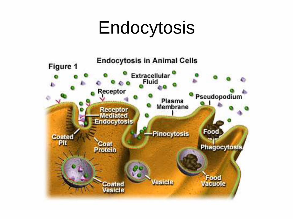
Lysosomes are responsible for the breakdown and absorption of materials
taken in by the cell. Often, a cell engulfs a foreign substance
through ENDOCYTOSIS, another form of active transport. During endocytosis,
the cell membrane puckers up, forms a pouch around materials outside the
cell, and pinches off to become a vesicle. If the contents need to be destroyed,
lysosomes combine with the vesicle and release their enzymes.
Lysosome
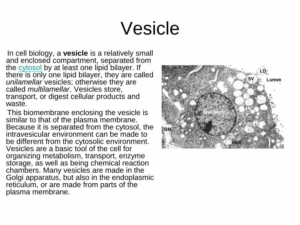
VesicleIn cell biology, a vesicle is a relatively small and enclosed compartment, separated from the cytosol by at least one lipid bilayer. If there is only one lipid bilayer, they are called unilamellar vesicles; otherwise they are called multilamellar. Vesicles store, transport, or digest cellular products and waste.
This biomembrane enclosing the vesicle is similar to that of the plasma membrane. Because it is separated from the cytosol, the intravesicular environment can be made to be different from the cytosolic environment. Vesicles are a basic tool of the cell for organizing metabolism, transport, enzyme storage, as well as being chemical reaction chambers. Many vesicles are made in the Golgi apparatus, but also in the endoplasmic reticulum, or are made from parts of the plasma membrane.
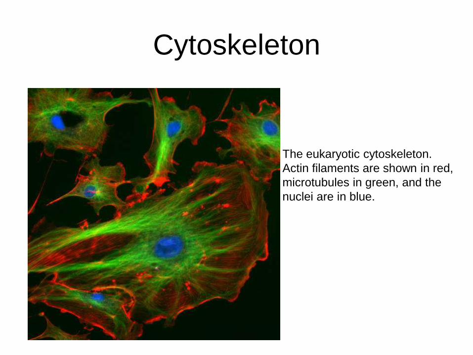
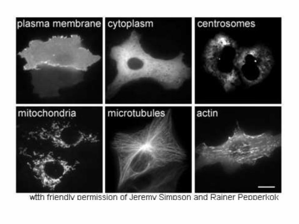
Cytoskeleton
The eukaryotic cytoskeleton.
Actin filaments are shown in red,
microtubules in green, and the
nuclei are in blue.
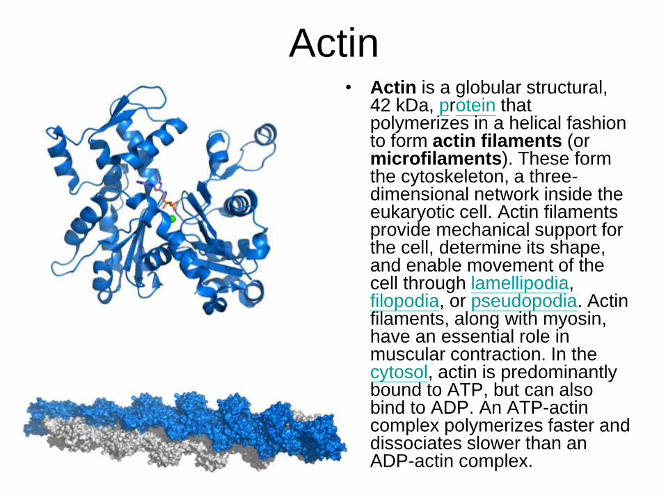
Actin• Actin is a globular structural,
42 kDa, protein that polymerizes in a helical fashion to form actin filaments (or microfilaments). These form the cytoskeleton, a three-dimensional network inside the eukaryotic cell. Actin filaments provide mechanical support for the cell, determine its shape, and enable movement of the cell through lamellipodia, filopodia, or pseudopodia. Actin filaments, along with myosin, have an essential role in muscular contraction. In the cytosol, actin is predominantly bound to ATP, but can also bind to ADP. An ATP-actin complex polymerizes faster and dissociates slower than an ADP-actin complex.
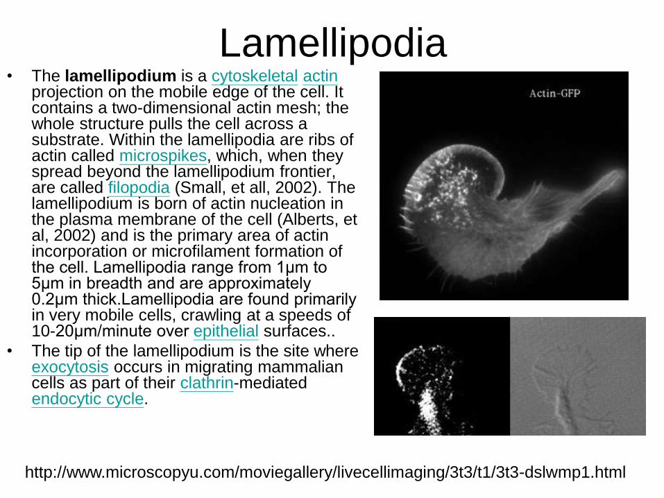
Lamellipodia • The lamellipodium is a cytoskeletal actin
projection on the mobile edge of the cell. It contains a two-dimensional actin mesh; the whole structure pulls the cell across a substrate. Within the lamellipodia are ribs of actin called microspikes, which, when they spread beyond the lamellipodium frontier, are called filopodia (Small, et all, 2002). The lamellipodium is born of actin nucleation in the plasma membrane of the cell (Alberts, et al, 2002) and is the primary area of actin incorporation or microfilament formation of the cell. Lamellipodia range from 1μm to 5μm in breadth and are approximately 0.2μm thick.Lamellipodia are found primarily in very mobile cells, crawling at a speeds of 10-20μm/minute over epithelial surfaces..
• The tip of the lamellipodium is the site where exocytosis occurs in migrating mammalian cells as part of their clathrin-mediated endocytic cycle.
http://www.microscopyu.com/moviegallery/livecellimaging/3t3/t1/3t3-dslwmp1.html
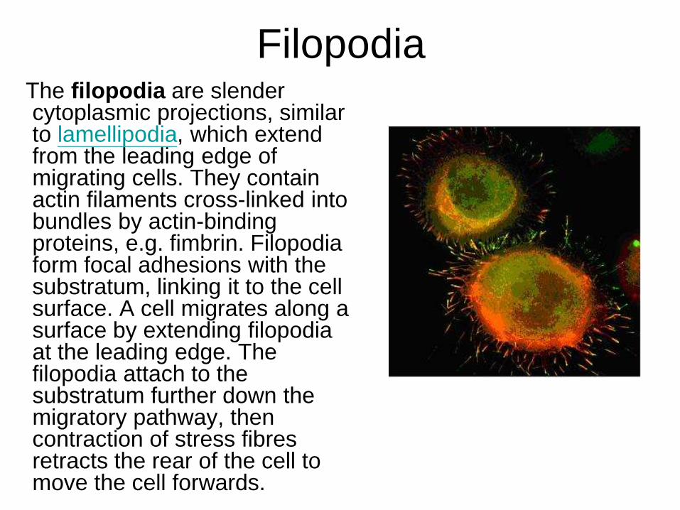
Filopodia The filopodia are slender cytoplasmic projections, similar to lamellipodia, which extend from the leading edge of migrating cells. They contain actin filaments cross-linked into bundles by actin-binding proteins, e.g. fimbrin. Filopodia form focal adhesions with the substratum, linking it to the cell surface. A cell migrates along a surface by extending filopodia at the leading edge. The filopodia attach to the substratum further down the migratory pathway, then contraction of stress fibres retracts the rear of the cell to move the cell forwards.
Focal adhesion
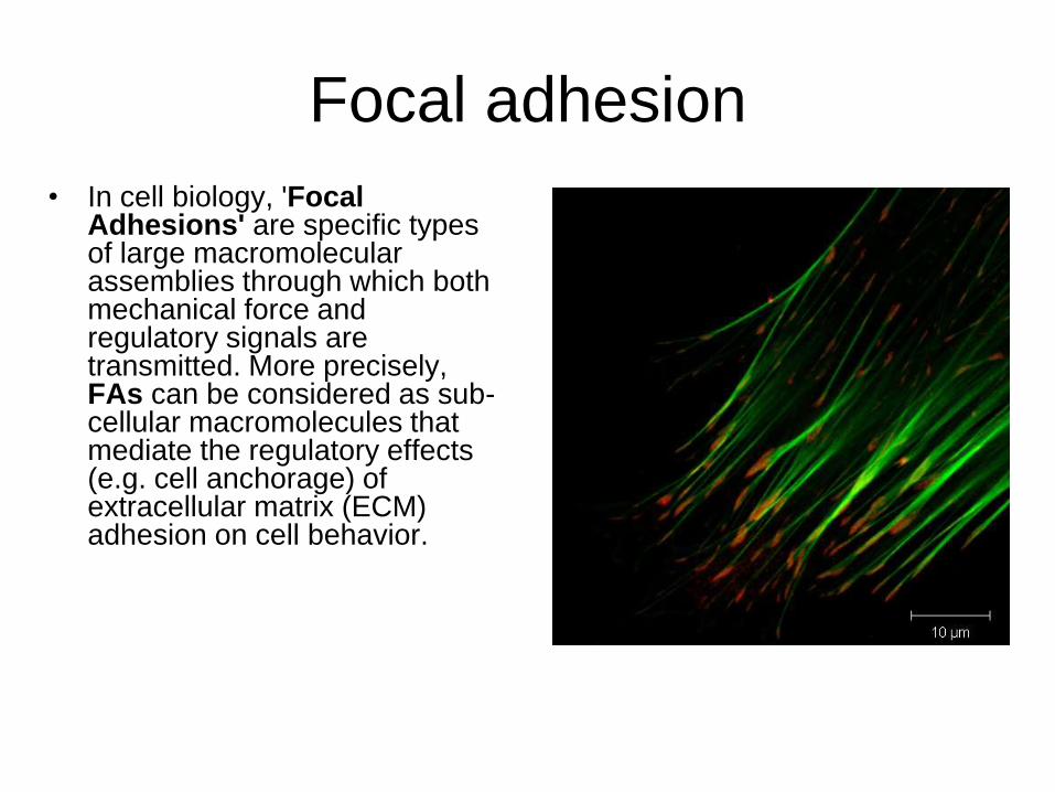
• In cell biology, 'Focal Adhesions' are specific types of large macromolecular assemblies through which both mechanical force and regulatory signals are transmitted. More precisely, FAs can be considered as sub-cellular macromolecules that mediate the regulatory effects (e.g. cell anchorage) of extracellular matrix (ECM) adhesion on cell behavior.
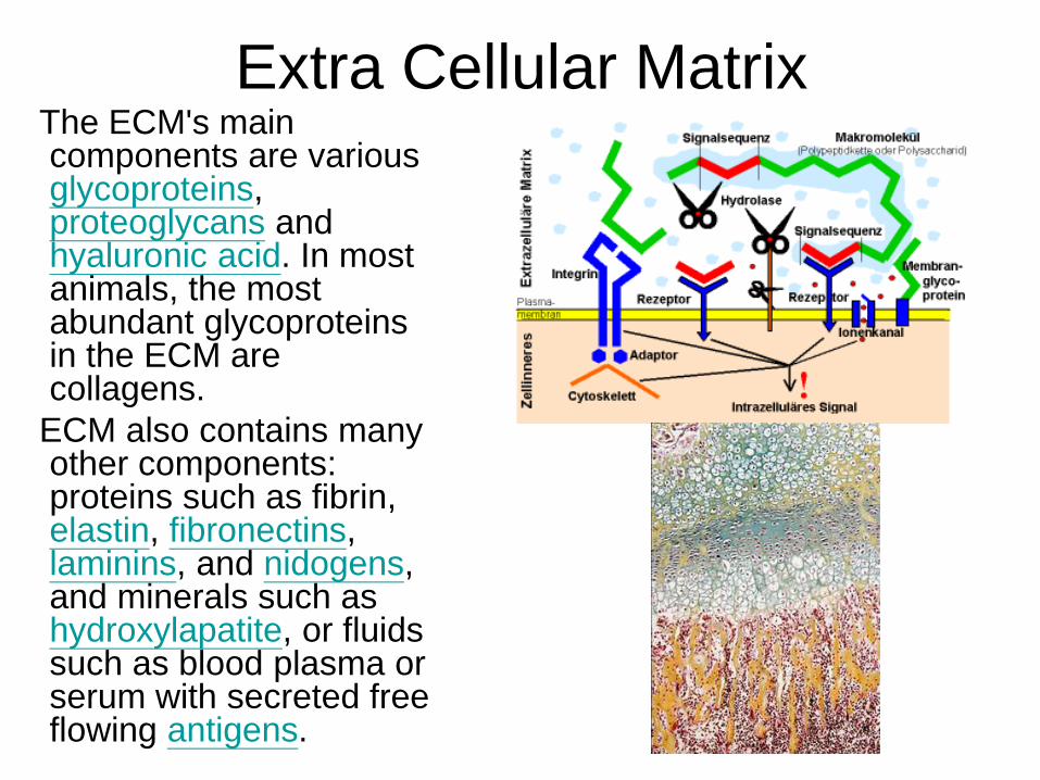
Extra Cellular MatrixThe ECM's main components are various glycoproteins, proteoglycans and hyaluronic acid. In most animals, the most abundant glycoproteins in the ECM are collagens.
ECM also contains many other components: proteins such as fibrin, elastin, fibronectins, laminins, and nidogens, and minerals such as hydroxylapatite, or fluids such as blood plasma or serum with secreted free flowing antigens.
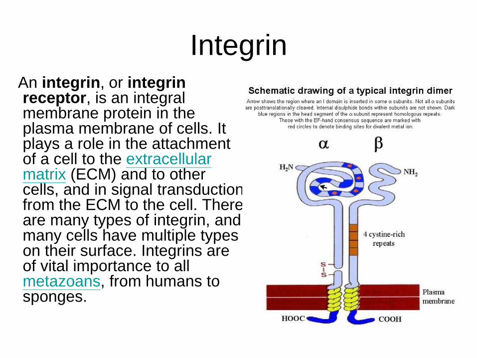
IntegrinAn integrin, or integrin receptor, is an integral membrane protein in the plasma membrane of cells. It plays a role in the attachment of a cell to the extracellular matrix (ECM) and to other cells, and in signal transduction from the ECM to the cell. There are many types of integrin, and many cells have multiple types on their surface. Integrins are of vital importance to all metazoans, from humans to sponges.
Endocytosis


![Chapter 22 Carbohydrates - Hashemite University [Compatibility Mode].pdf · Chapter 22 Carbohydrates. Introduction Classification of Carbohydrates ... Polyhydroxy Compounds Compounds](https://img.pdfslide.net/doc/110x75/5ad7d1ab7f8b9a3e578cb827/chapter-22-carbohydrates-hashemite-university-compatibility-modepdfchapter.jpg)