Embed Size (px)
Citation preview

M I N E R A L O G I C A L MAGAZINE, MARCH 1985, VOL. 49, PP. 31-36
An investigation of nephrite jade by electron microscopy
M. DORLING AND J. ZUSSMAN
Department of Geology, University of Manchester, Manchester M13 9PL
ABSTRACT. TWO specimens of tremolite and one of richterite, all with nephrite jade texture, have been examined by transmission electron microscopy using ion-thinning for specimen preparation. The specimens contain clusters of very small lath-like crystallites with z- axes approximately parallel but in a range of azimuthal orientations. It is suggested that these clusters which are themselves in varied orientations are the result of post-tectonic recrystallization of strained amphibole crystals, the new crystals inheriting the z-axis orientations of the old. The extreme toughness of nephrite jade is attributed to a number of the sub-microscopic features observed, including the sizes, habits, and orientations of its crystallites, and the nature of its grain boundaries.
KEYWORDS: electron microscopy, nephrite jade, tre- molite, richterite.
T HE extreme toughness of nephrite jade, a form of tremolite-actinolite, has been the subject of several investigations by scanning electron microscopy (SEM) (e.g. Bradt et al., 1973; Rowcliffe and Fr/ihauf, 1977) and by transmission electron micro- scopy (TEM) (e.g. Hutchison et al., 1976; Mallinson et al., 1977; Jefferson et al., 1978; Mallinson, 1980; Mallinson et al., 1980). In general terms the toughness has been attributed to a texture of sub-microscopic interlocking crystals. The SEM method does not lend itself readily to the deter- mination of crystal orientation or to the study of microstructural details such as grain boundaries and crystal defects, and the TEM work was carried out on dispersions of crystal grains which yielded predominantly crystals viewed perpendicular to their axis of elongation (z-axis). The study reported here attempts a more detailed analysis of the texture by employing TEM on randomly sectioned and ion-thinned specimens, yielding information from electron images and diffraction patterns of crystals in a wide variety of orientations, including views down the z-axis.
Specimens. Two specimens of nephrite were examined; their formulae, determined from electron microprobe analyses on the basis of twenty- three oxygen equivalents [i.e. assuming O22(OH)2],
�9 Copyright the Mineralogical Society
were as follows: (1) Cal.98(Mg4.6sFeo.25Cro.04 Alo.06)x5.03 (Si7.s8Alo.12)x8.00022(OH)2 from Verla Irene, Sweetwater, Wyoming, USA; (2) Cal.97 (Mg4.85Fe0.15~s.00Sis.02022(OH)2 from Lander, Wyoming, USA.
Preliminary observations on crushed grains and thin sections by polarizing microscopy confirmed that the grain size for these specimens was sub- microscopic or at the best at the margin of re- solution. Sweeping extinction was observed from aggregates of crystallites. A small fragment (< 1/ 10 mm in width), when mounted on a single crystal X-ray goniometer yielded powder (not single- crystal) diffraction patterns characteristic of poly- crystalline aggregates of more or less randomly oriented crystallites.
Electron microscopy. Specimens for transmission electron microscopy were prepared by conven- tional methods (e.g. Champness and Lorimer, 1971) using ion-beam thinning. Electron micrographs revealed the lath-like morphology of the nephrite crystallites and showed that their grain size, though small, covered quite a wide range. It was clear also that although the orientations of crystals are over- all random, there is a good degree of order, in that crystals occur in bundles or clusters with z-axes approximately parallel. The clusters themselves (each containing something like 30 to over 100 crystallites) are not in any orientational relation- ship with respect to one another (fig. 1). Electron diffraction patterns from areas containing longi- tudinal sections of crystal bundles show that the deviation from parallelism among its crystallites is up to about 5 deg. The crystallites are seen to be far from parallel-sided.
Cross-sections (perpendicular to z) of crystals are particularly interesting, since they reveal better than overlapping longitudinal crystals the nature of grain boundaries and certain other micro- structural features. Crystals within a quasi-parallel bundle exhibit a wide range of azimuthal orienta- tions (around z); evidence for this is provided by high resolution lattice images and diffraction

32 M. D O R L I N G AND J. Z U S S M A N
F1GS. 1 and 2. FIG. l (left). Transmission eleciron micrograph of random section through a nephrite specimen (no. 1), showing many different orientations of the crystallites. In this micrograph longitudinal sections of crystallite bundles appear to form a 'herring-bone' texture. The area outlined by a broken line contains a cluster of crystallites viewed approximately perpendicular to z. Note the curvature of the crystal terminations (e.g. at P). FIG. 2 (right). Electron micrograph (of nephrite, no. 2) showing how electron beam damage occurs preferentially along multiple chain defects and grain boundaries, revealing the shapes of crystallites and their orientations even at low magnifications. Some
examples of (010) defects and y axis directions are marked.
patterns, but also by means of the readily visible multiple-chain defects on (010) even at low magnifi- cation which have been described by others for longitudinal sections (e.g. Jefferson et al., 1978). The visibility of these defects is enhanced consider- ably because electron beam damage occurs pre- ferentially along them (and at grain boundaries) (fig. 3). While helpful in this respect, beam damage makes it necessary, when examining detail of grain boundaries and defects, to use short beam exposures.
Changes in lattice orientation at grain boun- daries are usually quite marked and abrupt, so that atoms at these boundaries are probably displaced from their normal lowest energy positions, and this may be a factor that facilitates beam damage. Many of the grain boundaries are irregular and apparently curved (fig. 3) rather than parallel to a prominent lattice plane, a feature that might in some circumstances lead to the elimination of boundaries (i.e. crystal growth), but the misorientations prob- ably hinder this. The large boundary area associated with small grain size and irregular boundaries can serve to accommodate stress which would other- wise lead to deformation and fracture; this helps to explain the toughness of nephrite. In addition, if a crack does develop in a crystal, the misorientation of neighbouring grains will hinder or prevent its further propagation. Mis-orientations which are effective in this way occur in the two senses; the azimuthal variation of crystallites (ranging up to 90 deg.) within the clusters, and the more or less
random orientations of the bundles themselves many of which meet one another at high angle.
Cross-sections show that the individual crystal- lites tend to be lath-shaped and show consi- derable variation in dimensions (fig. 4). The laths are flattened approximately on (100); the shorter dimension (thickness) ranges from < 0.03 to 0.5/~m and the larger dimension (width) from 0.05 to > 1/~m. Few, if any, crystallites have widths greater than 3 #m. The lengths of crystallites, esti- mated from longitudinal sections of crystal dusters, range approximately from 2/~m to over 5 #m.
Whereas it is well known that the crystals of all amphiboles tend to show elongation in the z direction (increasingly so from prismatic to acicular to asbestiform varieties), it is not generally recognized that a further distinction, though not as marked, occurs with respect to the x and y direc- tions, the latter being the larger of the two. We have observed this in our TEM studies of cross-sections of finely acicular and asbestiform amphiboles, as well as for nephrites.
Examinat ion of apparently curved grain boun- daries at higher magnifications reveals that some of them are partially defined by low-index faces ( 110}, (100), and sometimes (010) (fig. 2). Longitudinal sections are less suitable for showing the nature of these {hk0} boundaries but they do show apparently curved boundaries near the tips of the lath-like crystallites (e.g. fig. 1).
Multiple-chain defects on (010) have been ob- served in both longitudinal and cross-sections of

NEPHRITE JADE 33
FIG. 3. Electron micrograph of cross-sections of quasi-parallel crystallites of nephrite (specimen l). Azimuthal mis-orientation is indicated by multiple chain defects on (010) visible as linear features, e.g. those marked L. (020) lattice
fringes are resolved in some crystals (e.g. crystal A).
nephrite crystals and appear to be more abundant than those occurring in other kinds of amphibole. In terms of pyroxene and amphibole-like chain units (1- and 2- respectively), the multiples observed are: 3- and 4-. They are seen to occur in isolation or in repeating fashion, e.g. in fig. 5, where in some regions as many as 10 regular repeats of triple chains can be seen. In some crystals coherent interfaces have been observed (as by Jefferson et al., 1978) between double- and triple-chain regions. The percentage of non-amphibole-chain structure appears to range from about 1 ~ in some crystals to about 5 ~o in others.
In the smallest crystals multiple-chain defects run right across whereas in others some terminate within the crystal, sometimes at a sub-grain boundary (fig. 6). In the larger crystals, the strains caused by planar defects can reside in dislocations or fault planes although some pairs of defects compensate one another in the manner described
by Mallinson (1980). In small crystals the defects can run right across, building up only little strain which is relieved at the grain boundary. It might be argued that a high density of(010) defects and their associated strains do in fact contribute towards the formation of sub-grain boundaries.
Discussion. In publications to date on the textures of nephrites revealed by electron microscopy there has been little or no discussion of the likely conditions of formation of nephrite jade suggested by such textures. Some suggestions may be made based upon the above observations and the analogy (though approximate) of metallographic textures. In the latter the finest grain sizes are attained in materials which are most heavily cold- worked and annealed at the lowest possible re- crystallization temperature for the shortest possible time for full recrystallization (Chadwick, 1980). Rapid recrystallization, albeit at relatively low temperature, may also explain the abundance of

34 M. D O R L I N G AND J. ZUSSMAN
Fla. 4. Electron micrograph of cross-sections (perpendicular to z) of nephrite (no. 1) crystallites. Note that individual crystals tend to be flattened with y as the larger dimension. The large crystal 'B' shows the formation of small crystallites
at different stages of development (arrowed).
planar defects. Veblen (1981), in considering the growth mechanism for multiple chain defects and the normal amphibole region between them, refers to 'massive transformations' (analogous to those in metals) which may occur rapidly by recrystalliza- tion, The observed combination of planar and curved interfaces is also typical of recrystallization and grain growth during an annealing process as shown in studies of metals (e.g, Cahn, 1970) and rocks (e.g. Voll, 1960; Kretz, 1966; Lister and Price, 1978).
These suggestions are compatible also with what is known about the natural occurrences of nephrites. The two main types of occurrences are: (a) by contact metamorphism of dolomitic rocks where a reaction of the type dolomite + quartz + H/O --) tremolite + calcite + CO2 can be invoked, and (b) involving metasomatism in the region of contact between serpentinites and other basic rocks.
In the context of (a) Nichol (1974) refers to 'the later tectonic recrystallization oftremolite' and also to local zones of 'more intense deformation'. We suggest that, in the specimens studied, each bundle of crystallites in nephrite relates to an antecedent single crystal of tremolite the orientation of which controls the z-orientation of the quasi-parallel aggregate of nephrite crystallites even though re- crystallization has occurred. Evidence that this process has occurred is apparent in the specimens studied. In fig. 4, for example, the rather coarse grain 'B' appears to be undergoing recrystalliza- tion. The areas arrowed indicate crystallites at different stages of development. They are of similar size to those in the adjacent cluster of fully re- crystallized grains, and the large grain itself is of a similar size to a complete cluster. Some of the sub-grains within crystal 'B' show by means of the (010) defects that they are slightly rotated with respect to the parent crystal.
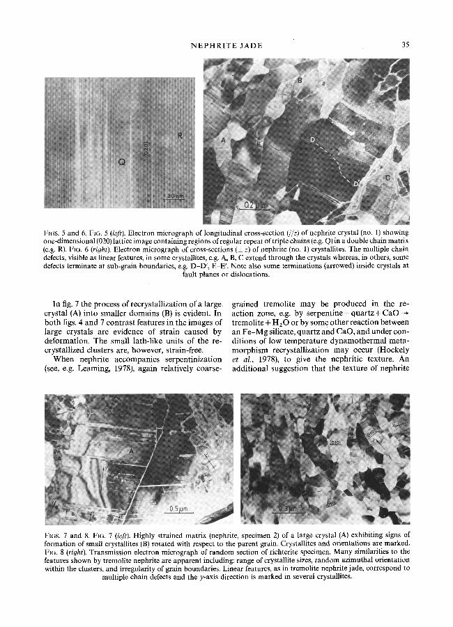
N E P H R I T E J A D E 35
FIGS. 5 and 6. FIG. 5 (left). Electron micrograph of longitudinal cross-section (//z) of nephrite crystal (no. 1) showing one-dimensional (020) lattice image containing regions of regular repeat of triple chains (e.g. Q) in a double chain matrix (e.g. R). FIG. 6 (right). Electron micrograph of cross-sections ( l z) of nephrite (no. 1) crystallites. The multiple chain defects, visible as linear features, in some crystallites, e.g. A, B, C extend through the crystals whereas, in others, some defects terminate at sub-grain boundaries, e.g. D-D', E-E'. Note also some terminations (arrowed) inside crystals at
fault planes or dislocations.
In fig. 7 the process of recrystallization of a large. crystal (A) into smaller domains (B) is evident. In both figs. 4 and 7 contrast features in the images of large crystals are evidence of strain caused by deformation. The small lath-like units of the re- crystallized clusters are, however, strain-free.
When nephrite accompanies serpentinization (see, e.g. Leaming, 1978), again relatively coarse-
grained tremolite may be produced in the re- action zone, e.g. by serpentine + quartz + CaO tremolite + H 2 0 or by some other reaction between an F e - M g silicate, quartz and CaO, and under con- ditions of low temperature dynamothermal meta- morphism recrystallization may occur (Hockely et al., 1978), to give the nephritic texture. An additional suggestion that the texture of nephrite
FIGS. 7 and 8. FIG. 7 (left). Highly strained matrix (nephrite, specimen 2) of a large crystal (A) exhibiting signs of formation of small crystallites (B) rotated with respect to the parent grain. Crystallites and orientations are marked. FIG. 8 (right). Transmission electron micrograph of random section of richterite specimen. Many similarities to the features shown by tremolite nephrite are apparent including: range of crystallite sizes, random azimuthal orientation within the clusters, and irregularity of grain boundaries. Linear features, as in tremolite nephrite jade, correspond to
multiple chain defects and the y-axis direction is marked in several crystallites.

36 M. D O R L I N G AND J. ZUS S M AN
may be related directly to the 'mesh texture' of serpentine itself (Learning, 1978) seems less likely.
Relating the texture of nephrite to pre-cursor coarse-grained tremolite is perhaps given further support by the description by Nichol (1974) of what are referred to by Learning as 'near nephrites' or 'semi-nephrites'. These occur in association with true nephrites and are relatively coarse-grained relict tremolite. The possibility of the reverse process, i.e. coarsening of nephrite, cannot however be ruled out (e.g. Turner, 1935).
In addition to the two specimens of nephrite described above we have investigated a white specimen (Warm Springs, California) which we found to have very similar features (fig. 8) and the toughness, and appearance of jade. This proved to have the composition not of tremolite but of richterite: Ko. 14Nao.50(Ca 1.24Nao.76)~2.0(Mg4.9 s Feo.02)~5.0Sis.03022(OH)z.
Conclusions. From the observations we have made it would appear that the tremolite in our specimens was formed by a metamorphic process and suffered some deformation, at the same time or later, which resulted in polygonization and formation of strained sub-grains. Subsequently the temperature rose sufficiently for the annealing out of strains and the lowering of energy by recrystal- lization into an aggregate of small strain-free lath-like crystallites. The fact that these new crystals and the old tend to have their z-axes aligned but are rotated about this direction is reminiscent of the textures reported for some other minerals (e.g. calcite, dolomite, quartz, and olivine). Accounts of these are given for example by Hobbs et al. (1976) and Vernon (1976). The rotations of recrystaUized grains have in some cases been attributed to a process of slip under the influence of non- hydrostatic stress. Alternatively they may result from multiple and independent nucleation in a range of azimuthal orientations about a preferred direction, this, the direction of rapid growth (z-axis) being inherited from the host crystal. The latter explanation seems more likely here in view of wide spread of orientations of both the recrystallized and original tremolite crystals. A more conclusive study with regard to tectonic history would, however, require the examination of many more specimens and knowledge of the field relationships of the rocks from which they are collected.
With regard to the toughness of nephrite jade, it may be suggested, for reasons given in the text, that contributions to this property are made by the following features: (1) very small average size of lath-like crystallites; (2) non-parallel sides of lath-like crystallites; (3) azimuthal misorienta- tions between laths within bundles; (4) irregular grain boundaries between laths within bundles; (5)
bundles of crystallites randomly oriented with irregular boundaries where they meet.
In addition two more general features are its monomineralic nature and the lack of alteration products at crystal margins.
Acknowledgements. We wish to thank Professor W. S. MacKenzie, Professor T. Zoltai and Johns-Manville Co. Ltd. for providing specimens, and Dr J. P. Platt for helpful discussion.
R E F E R E N C E S
Bradt, R. C., Newnham, R. E., Biggers, J. V. (1973) Am. Mineral. 58, 727-32.
Cahn, R. W. (1970) Recovery and recrystallization. In Physical metallurgy. 2nd edn. (R. W. Cahn, ed.). North- Holland Publ. Co. Amsterdam.
Chadwick, G. A. (1980) Metallography of phase trans- formations. Central Printing Unit, Univ. Southampton.
Champness, P. E., and Lorimer, G. W. (1971) Contrib. Mineral. Petrol. 33, 171-83.
Hobbs, B. E., Means, W. D., and Williams, P. F. (1976) An outline of structural geology. John Wiley and Sons, Inc. New York.
Hockely, J. J., Birch, W. D., and Worner, H. K. (1978) J. Geol. Soc. Austral. 25~ 249-54.
Hutchison, J. L., Jefferson, D. A., and Mallinson, L. G. (1976) Mater. Res. Bull. l l , 1557 62.
Jefferson, D. A., Mallinson, L. G., Hutchison, J. L., and Thomas, J. L. (1978) Contrib. Mineral. Petrol. 66, 1-4.
Kretz, R. (1966) J. Petrol. 7, 68 94. Learning, S. F. (1978) Jade in Canada. Paper 78-19,
Energy, Mines and Resources, Canada. Lister, G. S., and Price, G. P. (1978) Tectonophys. 49,
37 78. Mallinson, L. G. (1980) Acta Crystallogr. 36A, 378-81. - -Hutch i son , J. L., and Jefferson, D. A. (1977) J. Chem.
Soc., Chem. Comm. 910. Jefferson, D. A., Thomas, J. M., and Hutchison,
J. L. (1980) Phil. Trans. R. Soc. London A, 295, 537- 52.
Nichol, D. (1974) Nephrite jade deposits near Cowell, Eyre Peninsula. Dept. Mines, South Australia.
Rowcliffe, D. J., and Friihauf, V. (1977) J. Mater. Sci. 12, 35-42.
Turner, F. J. (1935) Trans. Proc. R. Soc. New Zealand, 65, part 2, October 1935.
Veblen, D. R. (1981) Non-classical pyriboles and poly- somatic reactions in biopyriboles. In Amphiboles and other hydrous pyribotes--mineralo#y. (D. R. Vehlen, ed.). Reviews in Mineralogy 9A, Mineral. Soc. Am. 223.
Vernon, R. H. (1976) Metamorphic Processes. Reactions and microstructure development. Alien and Unwin, Ltd. London.
Voll, G. (1960) Liverpool Manchester Geol. J. 2, 503 67.
[Manuscript received 20 September 1984; revised 3 October 1984]