Embed Size (px)
Citation preview
1130-0108/2017/109/12/870-874Revista española de enfeRmedades digestivas© Copyright 2017. sepd y © ARÁN EDICIONES, S.L.
Rev esp enfeRm dig2017, Vol. 109, N.º 12, pp. 870-874
Ortuño-Moreno MI, Ferri-Ñíguez B, Martínez-Barba E, Fernández-Hernández JA. An ulcerated gastric ulcer and pseudotumour with pancreatic affectation associated with immunoglobulin G4-related disease: a case report and litera-ture review. Rev Esp Enferm Dig 2017;109(12):870-874.
DOI: 10.17235/reed.2017.4996/2017
Received: 12-04-2017Accepted: 12-09-2017
Correspondence: María Isabel Ortuño-Moreno. Department of Pathology. Hospital Clínico Universitario Virgen de la Arrixaca. Ctra. Madrid-Cartage-na, s/n. 30120 El Palmar, Murcia. Spaine-mail: [email protected]
CASE REPORT
An ulcerated gastric ulcer and pseudotumour with pancreatic affectation associated with immunoglobulin G4-related disease: a case report and literature reviewMaría Isabel Ortuño-Moreno1, Belén Ferri-Ñíguez1, Enrique Martínez-Barba1 and Juan Ángel Fernández-Hernández2
1Department of Pathology, and 2Department of General Surgery and Digestive Diseases. Hospital Clínico Universitario Virgen de la Arrixaca. El Palmar, Murcia. Spain
ABSTRACT
We report the case of a 67 year old male who presented with a nine year history of a gastric ulcer with symptoms of hematemesis and melena. Histological analysis identified fibrotic lesions and the accumulation of immunoglobulin G4-positive plasma cells with no evidence of malignancy. The lesion extended into the pancreas, where histological lesions and gastric lesions were also observed. This is a case of an ulcerated gastric ulcer and pseudo-tumor with pancreatic affection that is associated with immunoglobulin G4-related disease.
Key words: Immunoglobulin G4. Gastric pseudotumor. Gastric ulcer.
INTRODUCTION
Immunoglobulin G4-related disease is a multisystem inflammatory disorder that can present with dysfunction of various organs, the pancreas is the most frequently involved and presents as autoimmune pancreatitis (1,2). There are few cases described in literature that describe an association with the stomach. In most cases a mass or pseudotumour (3,4) is involved and ulcers are rare (5-7). This is the first case described in Spain of the disease pre-senting as a pseudo-tumor with a gastric ulcer and pancre-atic involvement.
CASE REPORT
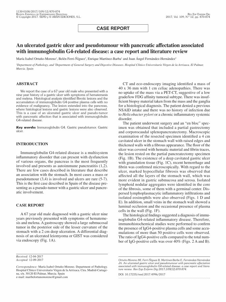
A 67 year old male diagnosed with a gastric ulcer nine years previously presented with symptoms of hemateme-sis and melena. A gastroscopy showed a large submucosal tumor in the posterior side of the lesser curvature of the stomach with a 2 cm deep ulceration. A differential diag-nosis of an ulcerated leiomyoma or GIST was considered via endoscopy (Fig. 1A).
CT and eco-endoscopy imaging identified a mass of 40 x 36 mm with 1 cm celiac adenopathies. There was no uptake of the mass via a PET-CT, suggestive of a low grade/low FDG affinity tumoral subtype. There was insuf-ficient biopsy material taken from the mass and the ganglia for a histological diagnosis. The patient denied a previous NSAID intake and there was no history of infection due to Helicobacter pylori or a chronic inflammatory systemic disorder.
The patient underwent surgery and an “en bloc” spec-imen was obtained that included a partial gastrectomy and corporocaudal splenopancreatectomy. Macroscopic examination of the resected specimen identified a 4 cm cavitated ulcer in the stomach wall with raised edges and thickened walls with a fibrous appearance. The floor of the ulcer was covered with hematic material and fibrin traces, the lesion rested on the partial pancreatectomy specimen (Fig. 1B). The existence of a deep cavitated gastric ulcer with granulation tissue (Fig. 1C), recent hemorrhage and fibrin was confirmed microscopically. With regard to the ulcer, marked hypocellular fibrosis was observed that affected all the layers of the stomach wall, which was more evident in gastric submucosa and serosa. Isolated lymphoid nodular aggregates were identified in the core of the fibrosis, some of them with a germinal center. Dis-persed lymphoplasmacytic inflammatory infiltrations and isolated eosinophils were also observed (Figs. 1 D and E). In addition, small veins in the stomach wall showed a luminal occlusion and the occasional presence of plasma cells in the wall (Fig. 1F).
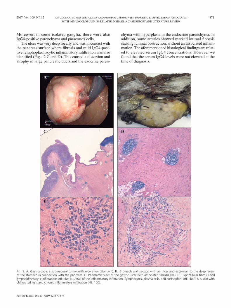
The histological findings suggested a diagnosis of immu-noglobulin G4-related inflammatory disease. Therefore, immunohistochemical studies were performed to confirm the presence of IgG4-positive plasma cells and some accu-mulations of more than 50 positive cells were observed. The ratio of IgG4-positive cells compared to the total num-ber of IgG-positive cells was over 40% (Figs. 2 A and B).
2017, Vol. 109, N.º 12 AN ULCERATED GASTRIC ULCER AND PSEUDOTUMOUR WITH PANCREATIC AFFECTATION ASSOCIATED 871 WITH IMMUNOGLOBULIN G4-RELATED DISEASE: A CASE REPORT AND LITERATURE REVIEW
Rev esp enfeRm Dig 2017;109(12):870-874
Fig. 1. A. Gastroscopy: a submucosal tumor with ulceration (stomach). B. Stomach wall section with an ulcer and extension to the deep layers of the stomach in connection with the pancreas. C. Panoramic view of the gastric ulcer with associated fibrosis (HE). D. Hypocellular fibrosis and lymphoplasmacytic infiltrations (HE. 40). E. Detail of the inflammatory infiltration, (lymphocytes, plasma cells, and eosinophils) (HE. 400). F. A vein with obliterated light and chronic inflammatory infiltration (HE. 100).
C
E
A
D
F
B
Moreover, in some isolated ganglia, there were also IgG4-positive parenchyma and paracortex cells.
The ulcer was very deep focally and was in contact with the pancreas surface where fibrosis and mild IgG4-posi-tive lymphoplasmacytic inflammatory infiltration was also identified (Figs. 2 C and D). This caused a distortion and atrophy in large pancreatic ducts and the exocrine paren-
chyma with hyperplasia in the endocrine parenchyma. In addition, some arteries showed marked intimal fibrosis causing luminal obstruction, without an associated inflam-mation. The aforementioned histological findings are relat-ed to elevated serum IgG4 concentrations. However we found that the serum IgG4 levels were not elevated at the time of diagnosis.
872 M. I. ORTUÑO-MORENO ET AL. Rev esp enfeRm Dig
Rev esp enfeRm Dig 2017;109(12):870-874
DISCUSSION
IgG4-related disease is a chronic multisystem inflam-matory disorder that can manifest in different ways. This includes different diseases such as autoimmune pancre-atitis, sclerosing cholangitis, retroperitoneal fibrosis or inflammatory pseudo-tumors. The most frequent symp-tom is pancreaticobiliary involvement and gastrointestinal involvement is rare (1,2).
Although this disease can affect different organs, there are several aspects that suggest that it is the same condition. These include mass-like lesions that can be mistaken for malignant or benign tumors, synchronous or metachronous involvement of a relatively constant group of organs, elevat-ed serum IgG4 concentrations, a characteristic histological appearance, high concentrations of IgG4-positive plasma cells and a high ratio of IgG4/IgG (1,2,8). The presentation of high serum IgG4 levels varies among different series, ranging from 44% to 100% (9). Therefore normal levels of serum IgG4 do not exclude a diagnosis, as in this case.
Histologically, the disease is characterized by a dense lymphoplasmacytic infiltration, dense fibrosis that is usu-ally arranged in a storiform pattern and obliterative phle-bitis (1,2,8). A definitive diagnosis of IgG4-related disease requires at least two of these three features. Even though it is the least common feature, obliterative phlebitis is a unique characteristic of this disease. Medium-sized veins are affected and light is obliterated or partially obliterated due to fibrosis and lymphoplasmacytic infiltration. Some-times, arteries are also affected (10). A high concentration of IgG4-positive plasma cells or a total IgG4/IgG ratio of over 40% is required for a definitive diagnosis (1,5,7,8,10).
There appear to be two phases of this disease. It is thought that there is an early stage of the disease in which lymphoplasmacytic infiltration is predominant and a late stage with an evolution to extensive fibrosis and decreased inflammation. This disease usually responds well to immu-nosuppressant treatment with steroids, mainly during an early stage. During late stages, fibrosis spread can be mis-taken for a pseudotumor, which depending on the loca-
Fig. 2. A. Immunohistochemistry of total IgG in one of the lymphoid aggregates of the stomach (HE. 100). B) Immunohistochemistry of IgG4 in the same lymphoid aggregate (HE. 100). C. Lymphoplasmacytic infiltration and fibrosis in the pancreas (HE. 100). D. Immunohistochemistry of IgG4 in the same lymphoid aggregate (HE. 100).
C D
A B
2017, Vol. 109, N.º 12 AN ULCERATED GASTRIC ULCER AND PSEUDOTUMOUR WITH PANCREATIC AFFECTATION ASSOCIATED 873 WITH IMMUNOGLOBULIN G4-RELATED DISEASE: A CASE REPORT AND LITERATURE REVIEW
Rev esp enfeRm Dig 2017;109(12):870-874
tion, can be considered as a differential diagnosis of other tumours (5).
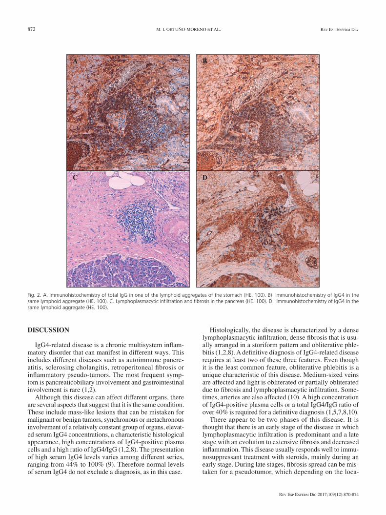
This disorder is better described in association with organs such as the pancreas and manifestation in the stomach is less frequent. Up to now, thirteen cases of gas-tric affection (including that described here) have been described worldwide (Table 1). The disease can present in different forms such as a mass or pseudotumor, an ulcer, or as both lesions in combination. The latter is less frequent and applies to the case reported here. When the disease presents as a pseudotumour, it usually grows as a transmu-ral mass or with a polypoid morphology, appearing more frequently in the stomach (3,4), colon or duodenal papilla. When presenting as a gastric ulcer, a marked thickening of the esophagus or stomach wall is usually evident (5,6). As far as we know, our case is the first one described in Spain in which this disease presents with an ulcerated mass with a secondary involvement of the adjacent pancreatic tissue.
In conclusion, this is an atypical case of an inflammato-ry pseudotumor associated with IgG4-related disease with mixed involvement, gastric and pancreatic. The histolo-gy of the lesions consists of marked fibrosis, lymphoid aggregates with IgG4-positive plasma cells and oblitera-tive phlebitis. These are typical features of the IgG4-relat-ed inflammatory disease. In order to provide an accurate diagnosis of this disease, a total IgG4/IgG ratio of over 40% is required.
Due to its manifestation and the morphological features described, this condition should be considered as a differential diagnosis of a rare case of a mass and gastric ulcer. When
histological characteristics of the biopsy include an ulcer with sclerotic fibrosis and high concentrations of plasma cells, an immunohistochemical staining of IgG4 should be performed and total IgG and IgG4 serum levels should be measured.
REFERENCES
1. Stone JH, Zen Y, Deshpande V. IgG4-related disease. N Engl J Med 2012;366:539-51. DOI: 10.1056/NEJMra1104650
2. Stone JH, Khosroshahi A, Deshpande V, et al. Recommendations for the nomenclature of IgG4-related disease and its individual organ system manifestations. Arthritis Rheum 2012;64:3061-7. DOI: 10.1002/art.34593
3. Rollins KE, Mehta SP, O’Donovan M, et al. Gastric IgG4-related autoimmune fibrosclerosing pseudotumour: a novel location. ISRN Gastroenterology 2011;2011:873087. DOI: 10.5402/2011/873087
4. Kim DH, Kim J, Park DH, et al. Immunoglobulin G4-related inflamma-tory pseudotumor of the stomach. Gastrointest Endosc 2012;76:451-2. DOI: 10.1016/j.gie.2011.07.061
5. Bateman AC, Sommerlad M, Underwood TJ. Chronic gastric ulcera-tion: a novel manifestation of IgG4-related disease? J Clin Pathol 2012;65:569-70.
6. Fujita T, Ando T, Sakakibara M, et al. Refractory gastric ulcer with abundant IgG4-positive plasma cell infiltration: a case report. World J Gastroenterol 2010;16:2183-6. DOI: 10.3748/wjg.v16.i17.2183
7. Moyer A, Schwartz M, Lim S, et al. IgG4-related disease in a non-healing gastric ulcer: case report. Int J Clin Exp Pathol 2016;9:6588-91
8. Deshpande V, Zen Y, Chan JK, et al. Consensus statement on the pathology of IgG4-related disease. Mod Pathol 2012;25:1181-92. DOI: 10.1038/modpathol.2012.72
9. Sah RP, Chari ST. Serologic issues in IgG4-related systemic disease and autoimmune pancreatitis. Curr Opin Rheumatol 2011;23:108-13. DOI: 10.1097/BOR.0b013e3283413469
10. Deshpande V, Gupta R, Sainani N, et al. Subclassification of autoimmune pancreatitis: a histologic classification with clinical significance. Am J Surg Pathol 2011;35:26-35. DOI: 10.1097/PAS.0b013e3182027717
Table 1. Cases registered to the present date of gastric involvement due to an immunoglobulin-G4-related disease
Case Study/Year Sex/Age Endoscopic findings Location
1 Fujita T, et al. (2010) F/73 3 cm ulcerLower part of the gastric body, lesser curvature
2 Rollins KE, et al. (2011) F/75 5 cm polypoid massMiddle part of the gastric body, greater curvature
3 Chetty R, et al. (2011)F/45M/60
1.5 cm noduleMultiple nodules of over 2.2 cm
FundusAntrum and pylorus
4 Bateman AC, et al. (2012) M/77 Diffuse ulcer Body
5 Kim DH, et al. (2012)F/54F/59
2.1 cm fixed mass3.3 cm sub-epithelial mass
Not specified
6 Na KY, et al. (2012) M/56 8 mm noduleLower part of the body, lesser curvature
7 Urban S, et al. (2014) M/73 5 cm ulcer Lesser curvature
8 Li X, et al. (2015) M/63 Two gastric ulcers of 4 and 3 cmFundus (4 cm)Antrum (3 cm)
9 Cheong HR, et al. (2016) F/27 4 cm sub-epithelial mass with superficial ulceration Fundus
10 Woo CG, et al. (2016) F/48 3.6 cm sub-epithelial tumor Posterior wall of the gastric body
11 Moyer A, et al. (2016) F/28 1 cm ulcer Pre-pylorus
12 Inoue D, et al. (2016) M/67 5 cm gastric mass and other systemic alterations Antrum
13 Present case (2016) M/67 Ulcer and 4 cm pseudotumor Posterior side of the lesser curvature


![Physical exercise for the treatment of non-ulcerated ...arquivos.info.ufrn.br/arquivos/20141612267d... · [Intervention Protocol] Physical exercise for the treatment of non-ulcerated](https://img.pdfslide.net/doc/110x75/5f0a5ba37e708231d42b3fe1/physical-exercise-for-the-treatment-of-non-ulcerated-intervention-protocol.jpg)