Embed Size (px)
Citation preview

1Shihui Zhang PhD, 1Shan Jiang PhD,
1Zhiyong Yang PhD, 2Ranlu Liu PhD,
1Yunpeng Yang MS, 1Honghua Liang MS
1. Tianjin University, Center for
Advanced Mechanisms and
Robotics, School of Mechanical
Engineering, Tianjin, China, 300072
2. Tianjin Institute of Urology&
Department of Urology, Second
Hospital of Tianjin Medical
University, 23 Pingjiang Road,
Hexi District, Tianjin, China, 300211
Keywords: -Prostate brachytherapy
-Ultrasound probe registration
-Deformable registration method
-MCC -Ultrasound
-Magnetic resonance imaging
Corresponding author: Shan Jiang PhD, Tianjin University,
Center of Advanced Mechanisms
and Robotics, School of Mechanical
Engineering, 92 Weijin Road,
Nankai District, Tianjin, China,
300072,
Tel: +86 13212154030;
Fax: +86-022-27401042;
Rece�ved:
9 September 2016
Accepted revised:
29 October 2016
An ultrasound image navigation robotic prostate
brachytherapy system based on US to MRI deformable
image registration method
AbstractObjective: This paper describes an ultrasound image navigation robotic prostate brachytherapy system. It uses a 2D ultrasound (US) probe rigidly �xed to a robotic needle insertion mechanism. Combined with the US probe registration and image registration, this system will help to navigate the prostate brachytherapy to increase the inserting accuracy. Subjects and Methods: The novelty of the system is that after the US probe registration using an improved iterative closest point (ICP) registration method, the initial regis-tration for the magnetic resonance imaging (MRI) and US image can be completely automatically. More-over, a deformable registration method based on statistical measurement was proposed to register US to MRI images intra-operatively. Results: The 6-degree of freedom (6-DOF) of robot and ultrasound probe are calibrated together with an accuracy of 0.9mm, allowing the needles to be precisely inserted to the seed targets after the image registration. Experiments were conducted by using US/MRI images ,capturing from patients. Results showed that the accuracies of probe registration and US-MRI registration were: 0.44± 0.12mm and 2.30±0.41mm, respectively. Conclusion: With the help of this robotic system, the accuracy and the costing of time for prostate brachytherapy will greatly improve.
Hell J Nucl Med 2016; 19(3): 223-230 Epub ahead of print: 8 November 2016 Published online: 10 December 2016
Introduction
Prostate cancer is now the most commonly diagnosed non-skin malignancy and is the main cause of cancer-related death in men. For example, in the USA, there will be 233,000 new cases of prostate cancer in 2014, accounting for more than a
quarter of all new (non-skin) cancer diagnoses of men, and 29,480 deaths from prostate cancer [1].
A number of treatment options are available, depending on a patient's age, medical history, and anatomy, as well as on the stage of the cancer. The most widely used treat-ment method among the available options is the permanent low-dose rate (LDR) bra-chytherapy which is a technique that involves the localized irradiation of the prostate by the permanent insertion of about 100 tiny radioactive seeds. Traditionally, the operators take the brachytherapy operation by using ultrasound (US) image to navigate it on the manual device. In order to increase the accuracy, stability, repeatability and dexterity of the surgery, there are many robotic prostate brachytherapy systems designed to help surgeons. Yu et al. (2010) designed and fabricated a device including Transrectal Ultraso-nography (TRUS) driver, gantry robot, needle inserter and a modular robotic platform for positioning the needle [2, 3], �tted to a cart with electronic housing, to perform mini-mally invasive interventions in prostate brachytherapy [4-6]. Other researchers [7] deve-loped a 6-DOF robot accomplished by 3 translating stages, 2 rotating joints and a needle rotation device, with the electromagnetic tracking system aimed to monitor trans-perineal needle placement in prostate brachytherapy. Other researchers [8-10] have also designed a robotic assistant system for US guided prostate brachytherapy, in which the puncture needles operated manually by a needle template.
Most of these robotic prostate brachytherapy systems use US images to navigate the operation process. However, the poor quality of the US image makes it not su�cient as a navigation method. In recent years, there are many scholars doing research on how to improve the quality of the US image and reach a consensus on the best way to use medi-cal image registration.
Medical image registration is the process of spatially aligning images in a common
Original Article
93 Hellenic Journal of Nuclear Medicine September-December 2016• www.nuclmed.gr223

coordinate space and aligning related features which exist in all images. This issue has been a widely investigated area in the past few decades, however remains challenging in particular for multi-modal registration. Often, di�erent mo-dalities complement each other well, which is relevant to a vast range of clinical applications for improving diagnosis, treatment planning, interventions, procedure follow-up, and screening. Many prostate minimally invasive surgeries motivate this work, magnetic resonance imaging (MRI) pro-vides a good visualization of the anatomy and tumors, while US is inexpensive and allows for intra-operative use to de-tect and correct for prostate deformation. Registering US and MRI images is a complex and di�cult process, largely because represents information from very di�erent physical properties. Besides, when using the US probe to take an image, the organs deform under the pressure of the probe. In order to register US and MRI images more accurately, deformable image registration (DIR) methods are proposed.
In modern image-guided online and o�-line adaptive radiation therapy, deformable image registration is a vital component which can establish a precise correspondence of two images which were collected with di�erent image modalities or at di�erent time voxel-to-voxel [11, 12]. The deformation vector �eld (DVF) which is the established cor-respondence by DIR has many applications in radiotherapy. For example, it can be used to promote auto-segmentation for tissue or organ changes in anatomy [13-18], assist to reconstruct high quality CBCT or 4D-CT images [19, 20], cal-culate accumulated dose in brachytherapy surgery [21-23], estimate organ motion [24-27] etc.
In the past few years, DIR has been investigated in detail and many new improved algorithms have been presented. Kim et al. (2010) proposed a B-spline algorithm whose ob-jective function combined organ contours and depicted manually particular points [28, 29]. Besides, a contour rigidity constraint was implemented into an adaptable De-mons algorithm to instruct its objective function [30]. Sch-reibmann et al (2006)proposed a method that using map-ped control volumes to improve the B-spline deformable calculation [31]. However, since the DIR calculation needed a lot of time, the real-time registration of the US image usu-ally used rigid transformation [32] and a mutual information (MI) based registration [33].
In this paper, we propose a robotic prostate brachythe-rapy system which includes robot parts and image guided brachytherapy system (IGBS). In our system, we use the US image as the navigation method through registered US and MRI images, which applies automatic pre-registration, replaces manual pre-registration after the probe working space calibration and applies a more e�cient deformation and registration method than the MI registration.
Subjects, Materials and Methods
System's descriptionGeneral layout
There are two main parts in our robotic prostate brachythe-rapy system, which are shown in Figure 1. The �rst part is the surgical robot which contains two sections (Figure 1), the lower layer is a positioning module and the upper one is an actuating robot. The positioning platform is at the top of the base support stands demanded to have the vertical and horizontal motions automatically actuated. The actuating robot is mounted on the positioning platform, consisting of an US probe holder and a guided module in a similar con�-guration to the conventional brachytherapy. The robot can manipulate the guided module to be manually translated to appropriate positions and rotated to proper angles in order to guide the puncture needle to remove the biopsy sample, or implant radioactive seeds through the needle. Besides, an electromagnetic position sensor attached to the US probe which will help to register the probe's working space to the MRI 3D reconstruction space that will be introduced in detail in the next section. The surgical robot will help surgeons to take the prostate brachytherapy surgery more stably.
F The general layout of the robotic prostate brachytherapy system.igure 1.
The second part is the IGBS system that contains three main parts: 3D organs reconstruction, 3D conformal dose planning and US navigation. In order to make full use of open-source packages and build a surgical navigation system, the visualization toolkit (VTK) and the insight toolkit (ITK) are employed for image visualization and image regis-tration respectively. The custom VTK classes are also used to acquire 3D position tracking information and 2D US images. In the navigation module, every component including ima-ge registration, US image acquisition and registered images visualization is integrated using multithreading techniques. After the US and MRI image registration, the quality of the US images will be improved by MRI images which can pro-vide appropriate anatomical structure context. Then the sur-geons can complete the prostate brachytherapy surgery more accurately.
Clinical work�owThe clinical work�ow that we have designed for our system is illustrated in the block diagram in Figure 2. At the begin-ning of the procedure, an innovative registration method with frame is used to register the ultrasound probe to the MRI 3D reconstruction space that could capture the US pros-
Original Article
93Hellenic Journal of Nuclear Medicine September-December 2016• www.nuclmed.gr 224

tate image which is very close to the corresponding MRI at the same position of the prostate. At the same time, the 3D conformal dose planning is made according to the pre-ope-ration MRI images that will instruct the operators to make needle trajectory planning.
F The clinical work�ow.igure 2.
After the probe registration, the US images captured in real-time are registered to pre-operation MRI images which together with the 3D conformal dose planning help the sur-geons to make needle trajectory planning. In this process a deformable non-rigid image registration method was used to register the US images to MRI images which can reach a reasonable accuracy and speed. Then, with the help of the registered image, the locating needle according to the dose planning and trajectory planning was �rst inserted into the target and after the locating needle other needles were in-serted.
Once the clinician is satis�ed with the �nal needle posi-tion, the seeds are inserted, while progressively removing the needle. A 3D US volume may be acquired to check the position of each seed separately or globally for all seeds inserted by a needle. This procedure is repeated until all se-eds have been distributed in the prostate
US probe registrationRegistration of US images to MRI images is a complex pro-cess in medical image processing. Traditionally, in order to register US images to MRI images fast and accurately, an ini-tial registration is usually made by the operators that puts the US images space position to the corresponding space position of the MRI images. This process usually costs a lot of time and is repeated many times by an experienced physi-
cian. We proposed an innovative registration method to im-prove this process by registering US probe to MRI 3D space in order to realize the automatic initial registration about US and MRI. This process describes a coordinates transforma-tion, from a coordinate system de�ned in the pre-operative-ly acquired image data (image or virtual model space), into a coordinate system de�ned by the localizer which is located in the left of the surgical robot (actual or patient space) [34].
At present, popular registration can be described as following: both and are referential Cartesian coordina- P Qtes, which can be de�ned as either image space or physical space of instrument and navigation localizer. The goal of registration between both and is to �nd out a transfor- P Q mation matrix , which enables the elements of both Q
P M P
and ( and ) satisfy an expression: Q p pP Q
(1)
Currently, several methods of image-to-patient registra-tion have been developed. Common methods include pai-red-point-based techniques with �ducial markers and surface-based registration using iterative closest point (ICP) [35].
In practical application, the number of the registration points is the main in�uence factor for the accuracy of ICP algorithm. If the number is large enough, the registration ac-curacy can satisfy the need of the clinical application. Too many points not only complicate the registration process but also expend the surgeons' more time and energy. To deal with these problems, an innovative concept called pre-operative registration based on ICP is proposed, which ma-kes use of a registration frame to obtain the positions of real and virtual registration points preoperatively. As a result of preoperative registration, a standard registration transfor-mation matrix is obtained [36]. This transformation mat-innMrix M can be used as the registration matrix in the progress inn
of navigation, surgery and surgeons don't need to repeat the complicated registration process. They just need to get more than three additional paired-points, which can ensure that the relative position between the real and virtual pati-ent are corresponding.
The calculations of the method are as follows: Firstly, pairs innof corresponding points and which act as registra-act mod
inn p p tion points in the innovative registration method are obtai-ned with the indentations of registration frame in actual and model space. Then as a result of innovative registration, a transformation matrix M is obtained [see calculation (2)]. inn
During the surgery (as mentioned above), surgeons just need to get more than three additional paired-points, which can ensure that the relative position in image between pati-ent and frame are corresponding. Then a series of corres-ponding points and , which attached to the phan-act mod
add addp ptom's skin, are selected as additional registration points. The
add addother points were gotten using multiplied by Mact inn p΄ pmod
add[see calculation (3)]. Points and were used as cor-mod modaddp΄ p
rection points in the innovative registration, then an additi-onal transformation matrixM between them was obtainedadd
Original Article
93 Hellenic Journal of Nuclear Medicine September-December 2016• www.nuclmed.gr225

using the leastsquare method[see calculation (4)]. The rotation and translation parameters (�,�,�) and (x,y,z) of M add
were solved through homogeneous equations. The position of virtual patient model will be adjusted automatically ac-cording to (�,�,�) and (x,y,z) respectively when the calcu-lations were performed. After that, the standard matrix can be used directly as registration result directly and the regis-tration process was immensely simpli�ed.
(2)
(3)
(4)
Figure 3. Experiments using registration frame, (a) The regular registration met-hod experiment frame, (b) The innovative registration frame with a number of mil-led indentations at prede�ned locations is a polymethyl methacrylate (PMMA) cu-bic frame (c) The relative position of the frame and the patient.
In order to verify the e�ectiveness of innovative regis-tration method, a registration frame is constructed, which serves as an arti�cial phantom for the experiments. The registration frame is a PMMA cubic frame, with a number of milled indentations at prede�ned locations across the fra-me's surface, which act as arti�cial landmarks points in the application [Figure 3(b)]. The diameter of the indentations matches exactly the diameter of the instrument's tip, allow-ing to precisely touch each of the points, which permitted us to perform an innovative preoperative registration combi-ning with datum points on the abdominal cavity phantom's skin. The virtual model of the registration frame was used to get the virtual position of the registration points and was imported to our own navigation software through the STL format.
We also did experiments to compare our method com-paring with the regular ICP method by using the frame sho-wn in Figure 3. Results will be shown in the Results section.
Deformable registration methodAfter completing the probe registration, the initial regis-tration of the US to MRI was performed at the same time. Then a deformable registration method is used to register US images to MRI in the intraprocedural registration process.
In this paper an alternative dependence measure propo-
sed by Rényi(1959) [37] was used to replace MI as the measu-rement of the deformable registration method, which can deal with multi-modal image registration, but does not need the estimation of continuous joint probability density func-tion. According to Rényi's theory, the maximum correlation coe�cient MCC is chosen as the measurement which can sa-tisfy the conditions proposed in the theory. Maximum corre-lation coe�cient de�ned as follows.
(5)
Where f and g are Borel measurable functions and V is the space of the functions. MCC (f(X), g(Y)) is the correlation co-e�cient of f(X) and g(Y).
(6)
where Var and Cov are on behalf of variance and covari-ance, respectively.
Let S(x) and T(x) be the source image and target image on the image domain �, respectively, and u(x) be the defor-mation �eld that deforms S(x) to T(x). So we used the theory of reproducing kernel Hilbert space (RKHS) to estimate the MCC between the deformed image s (x) s(x+u(x))and tar-u =get image T(x). We choose to use the following Gaussian ker-nel:
(7)
f gLet S (x) and T (x) be de�ned as:u
, , (8)
Then,
(9)
Now we can register the image S(x) to T(x) by solving the following minimization problem with respect to the defor-mation �eld u(x):
(10)
In this paper an iterative algorithm was used to �nd the minimizer u(x), �, � to complete the registration between US and MRI images. The registered US and MRI images together with 3D reconstructed organs and the robotic system will help the surgeons to make the brachytherapy surgery more commodiously and accurately.
In order to assess the accuracy of the registration method,
93Hellenic Journal of Nuclear Medicine September-December 2016• www.nuclmed.gr 226
Original Article
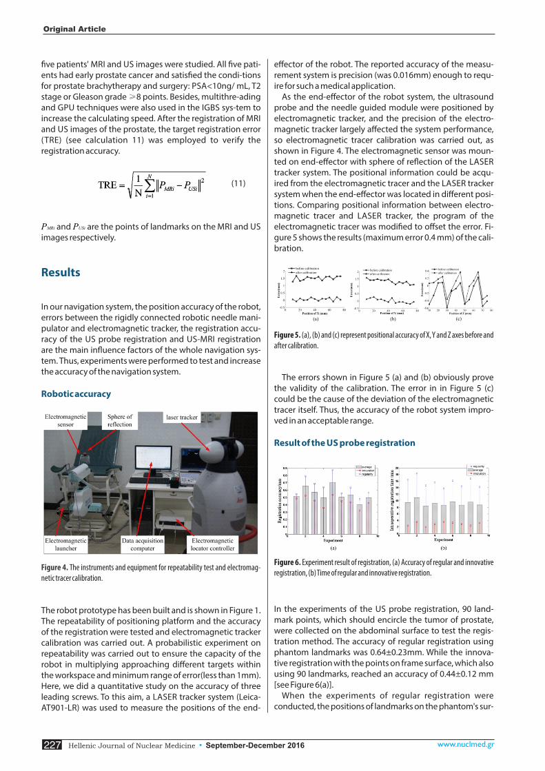
�ve patients' MRI and US images were studied. All �ve pati-ents had early prostate cancer and satis�ed the condi-tions for prostate brachytherapy and surgery: PSA<10ng/ mL, T2 stage or Gleason grade �8 points. Besides, multithre-ading and GPU techniques were also used in the IGBS sys-tem to increase the calculating speed. After the registration of MRI and US images of the prostate, the target registration error (TRE) (see calculation 11) was employed to verify the registration accuracy.
(11)
and are the points of landmarks on the MRI and US PMRi PUSi
images respectively.
Results
In our navigation system, the position accuracy of the robot, errors between the rigidly connected robotic needle mani-pulator and electromagnetic tracker, the registration accu-racy of the US probe registration and US-MRI registration are the main in�uence factors of the whole navigation sys-tem. Thus, experiments were performed to test and increase the accuracy of the navigation system.
Robotic accuracy
Figure 4. - The instruments and equipment for repeatability test and electromagnetic tracer calibration.
The robot prototype has been built and is shown in Figure 1. The repeatability of positioning platform and the accuracy of the registration were tested and electromagnetic tracker calibration was carried out. A probabilistic experiment on repeatability was carried out to ensure the capacity of the robot in multiplying approaching di�erent targets within the workspace and minimum range of error(less than 1mm). Here, we did a quantitative study on the accuracy of three leading screws. To this aim, a LASER tracker system (Leica-AT901-LR) was used to measure the positions of the end-
e�ector of the robot. The reported accuracy of the measu-rement system is precision (was 0.016mm) enough to requ-ire for such a medical application.
As the end-e�ector of the robot system, the ultrasound probe and the needle guided module were positioned by electromagnetic tracker, and the precision of the electro-magnetic tracker largely a�ected the system performance, so electromagnetic tracer calibration was carried out, as shown in Figure 4. The electromagnetic sensor was moun-ted on end-e�ector with sphere of re�ection of the LASER tracker system. The positional information could be acqu-ired from the electromagnetic tracer and the LASER tracker system when the end-e�ector was located in di�erent posi-tions. Comparing positional information between electro-magnetic tracer and LASER tracker, the program of the electromagnetic tracer was modi�ed to o�set the error. Fi-gure 5 shows the results (maximum error 0.4 mm) of the cali-bration.
Figure 5 . (a), (b) and (c) represent positional accuracy of X, Y and Z axes before and after calibration.
The errors shown in Figure 5 (a) and (b) obviously prove the validity of the calibration. The error in in Figure 5 (c) could be the cause of the deviation of the electromagnetic tracer itself. Thus, the accuracy of the robot system impro-ved in an acceptable range.
Result of the US probe registration
Figure 6 . Experiment result of registration, (a) Accuracy of regular and innovative registration, (b) Time of regular and innovative registration.
In the experiments of the US probe registration, 90 land-mark points, which should encircle the tumor of prostate, were collected on the abdominal surface to test the regis-tration method. The accuracy of regular registration using phantom landmarks was 0.64±0.23mm. While the innova-tive registration with the points on frame surface, which also using 90 landmarks, reached an accuracy of 0.44±0.12 mm [see Figure 6(a)].
When the experiments of regular registration were conducted, the positions of landmarks on the phantom's sur-
Original Article
93 Hellenic Journal of Nuclear Medicine September-December 2016• www.nuclmed.gr227
face were obtained intraopetatively. However, during the experiments of innovative registration, the positions of landmarks on the frame's surface were obtained by deve-lopers of surgery navigation system preoperatively. So the surgeon just needed to collect a few of additional paired-points and click the button. A higher registration precision intraopetatively will be thus gained. The intraoperative ti-mes of regular registration and innovative registration were 16±2min and 3±1min [Figure 6 (b)].
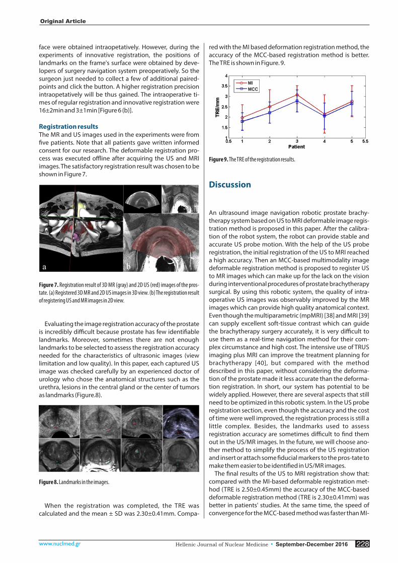
Registration resultsThe MR and US images used in the experiments were from �ve patients. Note that all patients gave written informed consent for our research. The deformable registration pro-cess was executed o�ine after acquiring the US and MRI images. The satisfactory registration result was chosen to be shown in Figure 7.
Figure 7. Registration result of 3D MR (gray) and 2D US (red) images of the pros-tate. (a) Registered 3D MR and 2D US images in 3D view. (b) The registration result of registering US and MR images in 2D view.
Evaluating the image registration accuracy of the prostate is incredibly di�cult because prostate has few identi�able landmarks. Moreover, sometimes there are not enough landmarks to be selected to assess the registration accuracy needed for the characteristics of ultrasonic images (view limitation and low quality). In this paper, each captured US image was checked carefully by an experienced doctor of urology who chose the anatomical structures such as the urethra, lesions in the central gland or the center of tumors as landmarks (Figure.8).
F Landmarks in the images.igure 8.
When the registration was completed, the TRE was calculated and the mean ± SD was 2.30±0.41mm. Compa-
red with the MI based deformation registration method, the accuracy of the MCC-based registration method is better. The TRE is shown in Figure. 9.
F The TRE of the registration results.igure 9.
Discussion
An ultrasound image navigation robotic prostate brachy-therapy system based on US to MRI deformable image regis-tration method is proposed in this paper. After the calibra-tion of the robot system, the robot can provide stable and accurate US probe motion. With the help of the US probe registration, the initial registration of the US to MRI reached a high accuracy. Then an MCC-based multimodality image deformable registration method is proposed to register US to MR images which can make up for the lack on the vision during interventional procedures of prostate brachytherapy surgical. By using this robotic system, the quality of intra-operative US images was observably improved by the MR images which can provide high quality anatomical context. Even though the multiparametric (mpMRI) [38] and MRI [39] can supply excellent soft-tissue contrast which can guide the brachytherapy surgery accurately, it is very di�cult to use them as a real-time navigation method for their com-plex circumstance and high cost. The intensive use of TRUS imaging plus MRI can improve the treatment planning for brachytherapy [40], but compared with the method described in this paper, without considering the deforma-tion of the prostate made it less accurate than the deforma-tion registration. In short, our system has potential to be widely applied. However, there are several aspects that still need to be optimized in this robotic system. In the US probe registration section, even though the accuracy and the cost of time were well improved, the registration process is still a little complex. Besides, the landmarks used to assess registration accuracy are sometimes di�cult to �nd them out in the US/MR images. In the future, we will choose ano-ther method to simplify the process of the US registration and insert or attach some �ducial markers to the pros-tate to make them easier to be identi�ed in US/MR images.
The �nal results of the US to MRI registration show that: compared with the MI-based deformable registration met-hod (TRE is 2.50±0.45mm) the accuracy of the MCC-based deformable registration method (TRE is 2.30±0.41mm) was better in patients' studies. At the same time, the speed of convergence for the MCC-based method was faster than MI-
Original Article
93Hellenic Journal of Nuclear Medicine September-December 2016• www.nuclmed.gr 228

based method (shown in Figure 10). So if the MCC-based registration method is used in real-time US image registra-tion, the duration of procedure may decrease a lot.
Figure 10. The results of iteration convergence for the MI and MCC
In conclusion, we have demonstrated an US image naviga-tion robotic system which combined the US probe regis-tration and MCC-based deformable registration method and proved its technical feasibility in prostate brachythe-rapy surgical. The technique proposed in this paper also has the latent energy to virtually improve the image guided navigation for other minimally invasive surgery just like radi-ofrequency ablation. The diagnostic capabilities for prostate cancer can also be improved through MR and US image registration and fusion. In the future, we will design another robotic system which will control needles directly replace the template to insert radioactive seeds and improve real-time registration.
Acknowledgment We gratefully acknowledge our research team at the Center for Advanced Mechanisms and Robotics, Tianjin University and the help of the Tianjin Institute of Urology& Department of Urology, Second Hospital of Tianjin Medical University. This research was partly supported by the Education Program for New Century Excellent Talents (NCET-10-0625), Key Technology and Development Program of the Tianjin Munic ipal S c ience and Technology Commiss ion (14ZCDZGX00490). No potential con�icts of interest were disclosed.
Bibliography1. Siegel R, Ma JM, Zou ZH et al. Cancer Statistics, 2014. Ca-a Cancer J.
for Clinicians 2014; 64: 9-29.2. Buzurovic I, Podder TK, Yu Y. Prediction control for brachytherapy
robotic system. Journal of Robotics 2010; 2010: ID 581840, 1-10 3. Buzurovic I, Podder T K, Yu Y. Robotic Systems for Radiation The-
rapy[M]. INTECH Open Access Publisher, 2012.4. Yan KG, Podder T, Yu Y et al. Flexible needle�tissue interaction
modeling with depth-varying mean parameter: preliminary study. Biomedical Engineering, IEEE Transactions on 2009; 56: 255-62.
5. Fu L, Liu H, Brasacchio R et al. Clinical Observation and Modeling of Postimplant Seed Displacement for Prostate Brachytherapy. Int J Rad Onco* Biol* Phys 2005; 63: S504-S5.
6. Ng W, Chung V, Vasan S et al., editors. Robotic radiation seed implantation for prostatic cancer. Engineering in Medicine and Biology Society, 1996 Bridging Disciplines for Biomedicine Proceedings of the 18th Annual International Conference of the IEEE; 1996: IEEE.
7. Meltsner M, Ferrier NJ, Thomadsen B. Observations on rotating needle insertions using a brachytherapy robot. Phys Med Biol 2007; 52: 6027-6037
8. Fichtinger G, Fiene J, Kennedy CW et al. Robotic assistance for ultrasound guided prostate brachytherapy. Medical Image Com-puting and Computer-Assisted Intervention�MICCAI 2007: Springer; 2007. p.119-27.
9. Fichtinger G, Fiene JP, Kennedy CW et al. Robotic assistance for ul-trasound-guided prostate brachytherapy. Med Image Analysis 2008 ;12: 535-45.
10. Krieger A, Iordachita II, Guion P et al. An MRI-compatible robotic system with hybrid tracking for MRI-guided prostate interven-tion. Biomed Engineer, IEEE Transactions on 2011; 58: 3049-60.
11. Hill DLG, Batchelor PG, Holden M et al. Medical image registration. Phys Med Biol 2001; 46: R1-R45.
12. Crum WR, Hartkens T, Hill DLG. Non-rigid image registration: the-ory and practice. Brit J Radiol 2004; 77: S140-S53.
13. Brock KK, Sharpe MB, Dawson LA et al. Accuracy of �nite element model-based multi-organ deformable image registration. Med Phys 2005; 32: 1647-59.
14. Lu WG, Olivera GH, Chen Q et al. Automatic re-contouring in 4D radiotherapy. Phys Med Biol 2006; 51: 1077-99.
15. Rietzel E, Chen GTY. Deformable registration of 4D computed to-mography data. Med Phys 2006; 33: 4423-30.
16. Chao M, Xie YQ, Xing L. Auto-propagation of contours for adaptive prostate radiation therapy. Phys Med Biol 2008; 53: 4533-42.
17. Wang H, Adam SG, Zhang LF et al. Performance evaluation of automatic anatomy segmentation algorithm on repeat or four-dimensional computed tomography images using deformable image registration method. Int J Radiat Oncol 2008; 72: 210-9.
18. Xie YQ, Chao M, Lee P et al. Feature-based rectal contour propa-gation from planning CT to cone beam CT. Med Phys 2008; 35: 4450-9.
19. Wu GR, Wang Q, Lian J et al. Reconstruction of 4D-CT from a Single Free-Breathing 3D-CT by Spatial-Temporal Image Registration. Lect Notes Comput Sc 2011;6801:686-98.
20. Ren L, Chetty IJ, Zhang J et al. Development and clinical evalua-tion of a three-dimensional cone-beam computed tomography estimation method using a deformation �eld map. Int J Radiat On-col* Biol* Phys 2012; 82: 1584-93.
21. Yan D, Ja�ray DA, Wong JW. A model to accumulate fractionated dose in a deforming organ. Int J Radiat Oncol 1999; 44: 665-75.
22. Rietzel E, Chen GT, Choi NC et al. Four-dimensional image-based treatment planning: Target volume segmentation and dose calculation in the presence of respiratory motion. Int J Radiat On-col* Biol* Phys 2005; 61: 1535-50.
23. Keall PJ, Joshi S, Vedam SS et al. Four-dimensional radiotherapy planning for DMLC-based respiratory motion tracking. Med Phys 2005; 32: 942-51.
24. Boldea V, Sharp GC, Jiang SB et al. 4D-CT lung motion estimation with deformable registration: Quanti�cation of motion nonline-arity and hysteresis. Med Phys 2008; 35: 1008-18.
25. Ehrhardt J, Werner R, Saring D et al. An optical �ow based method for improved reconstruction of 4D CT data sets acquired during free breathing. Med Phys 2007; 34: 711-21.
26. Yang DS, Lu W, Low DA et al. 4D-CT motion estimation using deformable image registration and 5D respiratory motion mode-ling. Med Phys 2008; 35: 4577-90.
27. Zeng RP, Fessler JA, Balter JM. Estimating 3-D respiratory motion from orbiting views by tomographic image registration. IEEE T Med Imaging 2007; 26: 153-63.
93 Hellenic Journal of Nuclear Medicine September-December 2016• www.nuclmed.gr229
Original Article

28. Kim J, Hammoud R, Pradhan D et al. Prostate Localization on Daily Cone-Beam Computed Tomography Images: Accuracy Assess- sment of Similarity Metrics. Int J Radiat Oncol 2010; 77: 1257-65.
29. Kim J, Kumar S, Liu C et al. A novel approach for establishing benchmark CBCT/CT deformable image registrations in prostate cancer radiotherapy. Phys Med Biol 2013; 8: 8077-97.
30. Gu XJ, Dong B, Wang J et al. A contour-guided deformable image registration algorithm for adaptive radiotherapy. Phys Med Biol 2013; 58: 1889-901.
31. Schreibmann E, Xing L. Image registration with auto-mapped con-trol volumes. Med Phys 2006; 33: 1165-79.
32. Xu S, Kruecker J, Turkbey B et al. Real-time MRI-TRUS fusion for guidance of targeted prostate biopsies. Comput Aided Surg 2008; 13: 255-64.
33. Zhang SH, Jiang S, Yang ZY et al. 2D Ultrasound and 3D MR Image Registration of the Prostate for Brachytherapy Surgical Navigation. Medicine 2015; 94: e1643(1-10).
34. Schmitz AC, Gianfelice D, Daniel BL et al. Image-guided focused
ultrasound ablation of breast cancer: current status, challenges, and future directions. Eur Radiol 2008; 18: 1431-41.
35. Wiles AD, Peters TM. Real-Time Estimation of FLE Statistics for 3-D Tracking With Point-Based Registration. IEEE T Med Imaging 2009; 28: 1384-98.
36. Spinczyk D, Karwan A, Copik M. Methods for abdominal respira-tory motion tracking. Comput Aided Surg 2014; 19: 34-47.
37. Rényi A. On measures of dependence. Acta Mathematica Acade-miae Scientiarum Hungaricae 1959; 10: 441-51.
38. Nicolae A M, Venugopal N, Ravi A. Trends in targeted prostate bra-chytherapy: from multiparametric MRI to nanomolecular radio-sensitizers. Cancer Nanotechnology 2016; 7: 1-17.
39. Kuo N, Lee J, Tempany C, et al. Mri-based prostate brachytherapy seed localizati. 2010 IEEE International Symposium on Biomedical Imaging: From Nano to Macro. IEEE 2010: 1397-400.
40. Reynier C, Troccaz J, Fourneret P, et al. MRI/TRUS data fusion for prostate brachytherapy. Preliminary results. Medical Physics 2004, 31: 1568-75.
Apollo and Artemis are killing the children of Niovi. Niovi mocked Lito, the mother of Artemis and Apollo, publicly and her children decided to take revenge for this assault.
93Hellenic Journal of Nuclear Medicine September-December 2016• www.nuclmed.gr 230
Original Article