Embed Size (px)
Citation preview
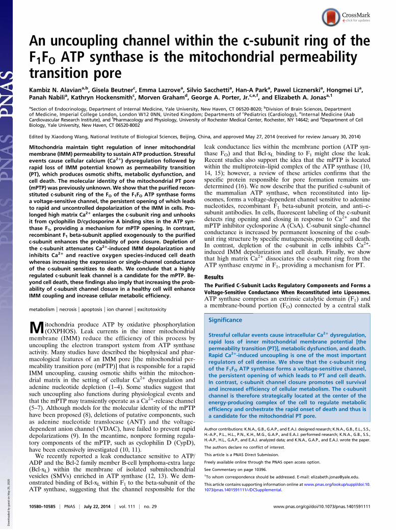
An uncoupling channel within the c-subunit ring of theF1FO ATP synthase is the mitochondrial permeabilitytransition poreKambiz N. Alaviana,b, Gisela Beutnerc, Emma Lazrovea, Silvio Sacchettia, Han-A Parka, Pawel Licznerskia, Hongmei Lia,Panah Nabilia, Kathryn Hockensmithc, Morven Grahamd, George A. Porter, Jr.c,e,f, and Elizabeth A. Jonasa,1
aSection of Endocrinology, Department of Internal Medicine, Yale University, New Haven, CT 06520-8020; bDivision of Brain Sciences, Departmentof Medicine, Imperial College London, London W12 0NN, United Kingdom; Departments of cPediatrics (Cardiology), eInternal Medicine (AabCardiovascular Research Institute), and fPharmacology and Physiology, University of Rochester Medical Center, Rochester, NY 14642; and dDepartment of CellBiology, Yale University, New Haven, CT 06520-8002
Edited by Xiaodong Wang, National Institute of Biological Sciences, Beijing, China, and approved May 27, 2014 (received for review January 30, 2014)
Mitochondria maintain tight regulation of inner mitochondrialmembrane (IMM) permeability to sustain ATP production. Stressfulevents cause cellular calcium (Ca2+) dysregulation followed byrapid loss of IMM potential known as permeability transition(PT), which produces osmotic shifts, metabolic dysfunction, andcell death. The molecular identity of the mitochondrial PT pore(mPTP) was previously unknown.We show that the purified recon-stituted c-subunit ring of the FO of the F1FO ATP synthase formsa voltage-sensitive channel, the persistent opening of which leadsto rapid and uncontrolled depolarization of the IMM in cells. Pro-longed high matrix Ca2+ enlarges the c-subunit ring and unhooksit from cyclophilin D/cyclosporine A binding sites in the ATP syn-thase F1, providing a mechanism for mPTP opening. In contrast,recombinant F1 beta-subunit applied exogenously to the purifiedc-subunit enhances the probability of pore closure. Depletion ofthe c-subunit attenuates Ca2+-induced IMM depolarization andinhibits Ca2+ and reactive oxygen species-induced cell deathwhereas increasing the expression or single-channel conductanceof the c-subunit sensitizes to death. We conclude that a highlyregulated c-subunit leak channel is a candidate for the mPTP. Be-yond cell death, these findings also imply that increasing the prob-ability of c-subunit channel closure in a healthy cell will enhanceIMM coupling and increase cellular metabolic efficiency.
metabolism | necrosis | apoptosis | ion channel | excitotoxicity
Mitochondria produce ATP by oxidative phosphorylation(OXPHOS). Leak currents in the inner mitochondrial
membrane (IMM) reduce the efficiency of this process byuncoupling the electron transport system from ATP synthaseactivity. Many studies have described the biophysical and phar-macological features of an IMM pore [the mitochondrial per-meability transition pore (mPTP)] that is responsible for a rapidIMM uncoupling, causing osmotic shifts within the mitochon-drial matrix in the setting of cellular Ca2+ dysregulation andadenine nucleotide depletion (1–4). Some studies suggest thatsuch uncoupling also functions during physiological events andthat the mPTP may transiently operate as a Ca2+-release channel(5–7). Although models for the molecular identity of the mPTPhave been proposed (8), deletions of putative components, suchas adenine nucleotide translocase (ANT) and the voltage-dependent anion channel (VDAC), have failed to prevent rapiddepolarizations (9). In the meantime, nonpore forming regula-tory components of the mPTP, such as cyclophilin D (CypD),have been extensively investigated (10, 11).We recently reported a leak conductance sensitive to ATP/
ADP and the Bcl-2 family member B-cell lymphoma-extra large(Bcl-xL) within the membrane of isolated submitochondrialvesicles (SMVs) enriched in ATP synthase (12, 13). We dem-onstrated binding of Bcl-xL within F1 to the beta-subunit of theATP synthase, suggesting that the channel responsible for the
leak conductance lies within the membrane portion (ATP syn-thase FO) and that Bcl-xL binding to F1 might close the leak.Recent studies also support the idea that the mPTP is locatedwithin the multiprotein–lipid complex of the ATP synthase (10,14, 15); however, a review of these articles confirms that thespecific protein responsible for pore formation remains un-determined (16). We now describe that the purified c-subunit ofthe mammalian ATP synthase, when reconstituted into lip-osomes, forms a voltage-dependent channel sensitive to adeninenucleotides, recombinant F1 beta-subunit protein, and anti–c-subunit antibodies. In cells, fluorescent labeling of the c-subunitdetects ring opening and closing in response to Ca2+ and themPTP inhibitor cyclosporine A (CsA). C-subunit single-channelconductance is increased by permanent loosening of the c-sub-unit ring structure by specific mutagenesis, promoting cell death.In contrast, depletion of the c-subunit in cells inhibits Ca2+-induced IMM depolarization and cell death. Finally, we showthat high matrix Ca2+ dissociates the c-subunit ring from theATP synthase enzyme in F1, providing a mechanism for PT.
ResultsThe Purified C-Subunit Lacks Regulatory Components and Forms aVoltage-Sensitive Conductance When Reconstituted into Liposomes.ATP synthase comprises an extrinsic catalytic domain (F1) anda membrane-bound portion (FO) connected by a central stalk
Significance
Stressful cellular events cause intracellular Ca2+ dysregulation,rapid loss of inner mitochondrial membrane potential [thepermeability transition (PT)], metabolic dysfunction, and death.Rapid Ca2+-induced uncoupling is one of the most importantregulators of cell demise. We show that the c-subunit ringof the F1FO ATP synthase forms a voltage-sensitive channel,the persistent opening of which leads to PT and cell death.In contrast, c-subunit channel closure promotes cell survivaland increased efficiency of cellular metabolism. The c-subunitchannel is therefore strategically located at the center of theenergy-producing complex of the cell to regulate metabolicefficiency and orchestrate the rapid onset of death and thus isa candidate for the mitochondrial PT pore.
Author contributions: K.N.A., G.B., G.A.P., and E.A.J. designed research; K.N.A., G.B., E.L., S.S.,H.-A.P., P.L., H.L., P.N., K.H., M.G., G.A.P., and E.A.J. performed research; K.N.A., G.B., S.S.,H.-A.P., H.L., G.A.P., and E.A.J. analyzed data; and K.N.A., G.A.P., and E.A.J. wrote the paper.
The authors declare no conflict of interest.
This article is a PNAS Direct Submission.
Freely available online through the PNAS open access option.
See Commentary on page 10396.1To whom correspondence should be addressed. E-mail: [email protected].
This article contains supporting information online at www.pnas.org/lookup/suppl/doi:10.1073/pnas.1401591111/-/DCSupplemental.
10580–10585 | PNAS | July 22, 2014 | vol. 111 | no. 29 www.pnas.org/cgi/doi/10.1073/pnas.1401591111
Dow
nloa
ded
by g
uest
on
May
25,
202
0
and a peripheral stator. Movement of protons down theirelectrochemical gradient through a translocator at the junc-tion between subunit-c and -a of FO provides the energy used by thealpha- and beta-subunits of F1 to synthesize ATP (17). The octa-meric c-subunit forms a central pore-like structure that could con-ceivably allow for uncoupling when exposed. To test this hypothesis,we analyzed the role of the c-subunit in IMM uncoupling. Mam-malian c-subunit is encoded on three separate nuclear genes(ATP5G1 to -3). Antibodies raised against the common sequencediscerned 8-kDa and 15-kDa bands (Fig. S1A). ATP5G1-encodedmyc/FLAG-tagged c-subunits were purified from HEK 293T cells(Fig. S1B). In the absence of denaturation, purified c-subunit wasdetected at ∼250 kDa, suggesting the presence of oligomers (Fig.S1C) (for endogenous oligomers, see Fig. 5G); similar c-subunit–containing complexes have been observed previously (18). Either anantibody raised against ATP5G or an ATP synthase (F1 binding)immunocapture antibody immunoprecipitated multiple subunits ofATP synthase (Fig. S2A), suggesting that the antibodies find epit-opes within F1 and FO in the assembled ATP synthase complex.The purified c-subunit was reconstituted into liposomes in its
native form. Immunoblotting confirmed the absence of regula-tory moieties in the purified c-subunit elution including ANT,VDAC1, CypD, ATP synthase b-subunit, and oligomycin sensi-tivity conferring protein (OSCP) (Fig. S1 C and E). Thec-subunit was localized to the liposomes by immunolabeling (Fig.S1D). Recordings of excised patches from the proteoliposomesdemonstrated a multiconductance channel with prominent sub-conductance states (Fig. 1A). Recordings of empty liposomesshowed no activity (n = 3). Patches contained an ∼100-pS con-ductance as a subconductance state of multiconductance activity,with peak conductances of up to ∼1.5–2 nS (Fig. 1A), similar toactivity described previously for the mitochondrial multipleconductance channel (MCC) (11, 19–21). Also consistent withMCC, channel activity showed negative rectification (Fig. 1B andFig. S3C). At very positive patch pipette potentials of over 100mV, channel conductances of ∼1.5 nS and ∼2 nS were alsoconsistently observed (Fig. 1 A–D).Such channel currents are not likely to represent movement of
protons through the ATP synthase proton translocator becauseof their very high conductance, lack of ion selectivity, and theabsence of the a-subunit in the liposome recordings. For exam-ple, the channel was only 1.5 times more selective for Na+/K+
(Fig. S3). In addition, although we could not specifically measureproton conductance electrophysiologically, it is unlikely thatprotons are excluded from such a large, nonselective channel. Insummary, the nonselective, high-conductance c-subunit channelcould conduct leak current that uncouples the IMM.In a previous study, we measured a large leak conductance in the
IMM directly by patch clamping SMVs isolated from native ratbrain (12). Here, we show that ATP blocks this activity with an EC50of ∼50 μM (Fig. S4 A and B). However, parallel experiments withpurified reconstituted c-subunit demonstrate decreased sensitivity toATP, with an EC50 of 660 μM (Fig. 1B and Fig. S4 C–F). Thesedata suggest that some ATP sensitivity is localized directly withinthe c-subunit itself and that other ATP binding sites are removedduring partial (urea-treated SMVs) or complete c-subunit purifi-cation. The purified c-subunit channel activity was equally attenu-ated by ATP, ADP, or AMP (Fig. S4F) so ATP hydrolysis by ATPsynthase is not likely to be required for its inhibition. Because thereappear to be multiple sites of inhibition by ATP within F1FO ATPsynthase, we used a more specific inhibitor of the purified c-subunit channel, the anti–pan-c-subunit antibody, which rap-idly and effectively attenuated purified c-subunit conductance(Fig. 1 C and D). We found similar antibody-mediated at-tenuation in SMVs after exposure to Ca2+, a known activator ofmPTP (Fig. 2A). These data strongly support the notion that the c-subunit forms the pore of the Ca2+-sensitive mPTP.A number of lines of evidence, including our ATP concen-
tration-dependence data, suggested that the purified c-subunitchannel lacked regulatory components that might control itsactivity in vivo. The mPTP is sensitive not only to high levels of
intramatrix Ca2+, but also to the protein CypD (22, 23), possiblyby its binding to OSCP on the F1 stator, and to CsA, which mayinterfere with the CypD–OSCP interaction (10, 15, 21). Weperformed electrophysiology on three preparations to study thefollowing: (i) purified recombinant c-subunit preparations lack-ing CypD and OSCP (Fig. S1 C and E) that were reconstitutedinto proteoliposomes, (ii) purified ATP synthase monomerslacking CypD (Fig. S2 B and C) reconstituted into proteolipo-somes, and (iii) mitochondria and SMVs containing endogenousCypD and OSCP (Fig. 2B). In keeping with the lack of Ca2+
and CsA sensitive sites present in the purified c-subunit ring, neitherCa2+ nor CsA had an effect on channel activity of the purifiedc-subunit (Fig. 1 C and D). Purified monomeric ATP synthasehad F1-related enzymatic activity and contained OSCP but
A
50pA0.1s
+220 mV
+140 mV
25pA1s
C-S
ub a
b.C
TL IgG
0
1000
2000
Pea
k co
nduc
tanc
e (p
S)
**
+140 mV
50pA1s
+180 mV
25pA1s
CTL
CTL
CTL
C-Sub ab.
IgG
CsA
C D
0 100 200 300 400 5000
50
100
Nor
mal
ized
N
Amplitude (pA)
CTL
1mM ATP
0mV
-100mV
100mV
Before ATP
-20
2040
1004030
20
-60(pA)-40
-20(pA)
20100
403020
50(s)
50(s)
B
CTL
Cyp
DC
a2+
CsA
0
500
1000
Peak
con
duct
ance
(pS)
CsACTL CypD+70 mV
20pA
5s
E F
100μM Ca2+
*
0 100 200 300 400 5000
50
100
Nor
mal
ized
N
Amplitude (PA)Normalized to Vh
PurifiedATP synthase
**
PurifiedC-Subunit
PurifiedC-Subunit
PurifiedC-Subunit
C
O2 O3
O1 C
O2 O3
O1
CTL
Ca2+
CsA
CTL
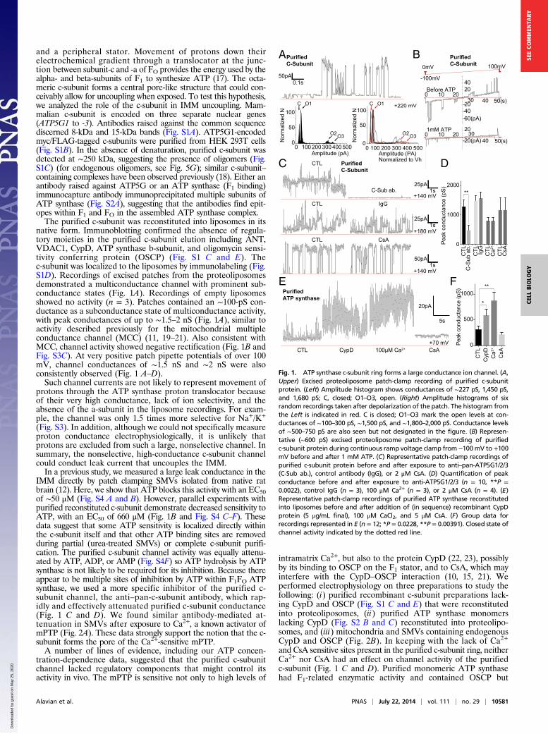
Fig. 1. ATP synthase c-subunit ring forms a large conductance ion channel. (A,Upper) Excised proteoliposome patch-clamp recording of purified c-subunitprotein. (Left) Amplitude histogram shows conductances of ∼227 pS, 1,450 pS,and 1,680 pS; C, closed; O1–O3, open. (Right) Amplitude histograms of sixrandom recordings taken after depolarization of the patch. The histogram fromthe Left is indicated in red. C is closed; O1–O3 mark the open levels at con-ductances of ∼100–300 pS, ∼1,500 pS, and ∼1,800–2,000 pS. Conductance levelsof ∼500–750 pS are also seen but not designated in the figure. (B) Represen-tative (∼600 pS) excised proteoliposome patch-clamp recording of purifiedc-subunit protein during continuous ramp voltage clamp from −100mV to +100mV before and after 1 mM ATP. (C) Representative patch-clamp recordings ofpurified c-subunit protein before and after exposure to anti–pan-ATP5G1/2/3(C-Sub ab.), control antibody (IgG), or 2 μM CsA. (D) Quantification of peakconductance before and after exposure to anti-ATP5G1/2/3 (n = 10, **P =0.0022), control IgG (n = 3), 100 μM Ca2+ (n = 3), or 2 μM CsA (n = 4). (E)Representative patch-clamp recordings of purified ATP synthase reconstitutedinto liposomes before and after addition of (in sequence) recombinant CypDprotein (5 μg/mL final), 100 μM CaCl2, and 5 μM CsA. (F) Group data forrecordings represented in E (n = 12; *P = 0.0228, **P = 0.00391). Closed state ofchannel activity indicated by the dotted red line.
Alavian et al. PNAS | July 22, 2014 | vol. 111 | no. 29 | 10581
CELL
BIOLO
GY
SEECO
MMEN
TARY
Dow
nloa
ded
by g
uest
on
May
25,
202
0
lacked CypD (Fig. S2 B and C) (15). When reconstituted intoliposomes, monomeric ATP synthase showed infrequent channelactivity significantly enhanced by the addition of recombinantCypD protein whether or not Ca2+ was present; channel ac-tivity was readily inhibited by CsA (Fig. 1 E and F). Finally, we re-corded channel activity of whole mitochondria, SMVs, and SMVsthat were exposed to urea to denature and remove extramem-brane proteins, including F1 components such as OSCP, beta-subunit, and CypD (Fig. 2B). Channel activity of mitochondriaand SMVs revealed robust Ca2+ and CsA responsiveness thatwas completely absent from the urea-exposed SMVs although1 mM ATP still rapidly inhibited the channel activity (Fig. 2 Cand D). These recordings confirm that Ca2+, CypD, and CsAregulate an IMM conductance by binding to an extramembraneprotein site, possibly within the OSCP-subunit of ATP synthase,and explain why Ca2+ and CsA have little effect on the con-ductance of the purified c-subunit channel.
The C-Subunit Leak Channel Undergoes Measurable ConformationalChanges upon Entering the Open and Closed States. To determinewhether the c-subunit ring structure within the ATP synthaseresponds to CsA and Ca2+ in cells, we adapted a fluorescentmethod, bipartite tetracysteine display (FlAsh), which detectsprotein–protein interactions in living cells and localizes proteinsto subcellular compartments (24–26). When two pairs of cysteinesplaced on proteins come in close proximity, they bind tightly to
the FlAsh dye, increasing its fluorescence intensity (Fig. 3A). Weexpressed in separate experiments two constructs, each contain-ing a cysteine pair inserted into ATP5G1 (C sub 1 or C sub 2)(Fig. 3 A–D). At 48 h after transfection, FlAsh fluorescence wasapparent at high levels at puncta colocalized with mitochondriawithin the cells (Fig. 3 B–D), suggesting that the individualc-subunit proteins were in relatively close proximity, as expected.However, only background FlAsh fluorescence was observed inthe absence of cysteine binding partners or after transfection ofa construct expressing a single pair of cysteines on OSCP (Fig. 3 Cand D). Specimens were then exposed to vehicle or CsA, followedby ionomycin to increase intracellular and matrix Ca2+ (Fig. 3 Cand D). In the C sub 1- and C sub 2-expressing cells, FlAshfluorescence was significantly higher in cells preexposed to CsA,suggesting that channel inhibition brings individual FlAsh-labeledc-subunits closer together, thus increasing the packing of thec-subunit ring. Ionomycin decreased FlAsh fluorescence, whichwas prevented by CsA, suggesting that pore opening loosens the
Closed+100 mV
CTL Ca2+ C-Sub ab.
400ms100p
A
+40 mV
+50 mV
CTL
2μM CsA
+50 mV
SMV+urea
Mito
SMV
10pA2s
CTL
CTL
Beta
cSub
50
15
C
B
D
OSCP37
Mito
SM
VS
MV
+ure
a
1mM ATP
Closed+60 mV
CTL IgG
400ms
20pA
***
**
CTL
Ca2+
IgG
0
500
0500
100015002000
A
2μM CsA
2μM CsA
Ca2+
Ca2+
Peak
cond
ucta
nce
(pS)
Peak
cond
ucta
nce
(pS)
1000
CTL
Ca2+
C-S
ub a
b.
CypD37
Ca2+
Ca2+
CTL
CsA
CTL
CsA
CTL
CsA
ATP
P eak
con
duct
anc e
(pS)
0
500
1000
1500
2000 * ***
IB:
SMV
Mito SMV SMV+urea
Closed
Closed
Closed
Ca2+
Ca2+
Ca2+
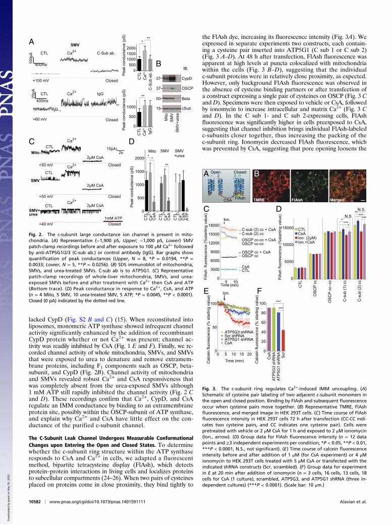
Fig. 2. The c-subunit large conductance ion channel is present in mito-chondria. (A) Representative (∼1,900 pS, Upper; ∼1,000 pS, Lower) SMVpatch-clamp recordings before and after exposure to 100 μM Ca2+ followedby anti-ATP5G1/2/3 (C-sub ab.) or control antibody (IgG). Bar graphs showquantification of peak conductances (Upper, N = 8, *P = 0.0194, **P =0.0033; Lower, N = 5, **P = 0.0256). (B) SDS immunoblot of mitochondria,SMVs, and urea-treated SMVs. C-sub ab is to ATP5G1. (C) Representativepatch-clamp recordings of whole-liver mitochondria, SMVs, and urea-exposed SMVs before and after treatment with Ca2+ then CsA and ATP(Bottom trace). (D) Peak conductance in response to Ca2+, CsA, and ATP(n = 4 Mito, 5 SMV, 10 urea-treated SMV, 5 ATP; *P = 0.0045, **P < 0.0001).Closed (0 pA) indicated by the dotted red line.
FlAshCSA
cc cc cccc
A
*****
***
****
*
E
D
B
C N.S.N.S.
0
3000
6000
9000
12000
15000
18000
Time (min)
FlA
sh f
luor
esce
nce
(%st
artin
g va
lue)
CTLCsA
Ion.
OSCP ccOSCP cc + CsA
OSCP cc-cc + CsA
***
***C
TL
OSC
P c c
OSC
P cc
--cc
C-s
ub(1
)cc
C-s
ub(2
)cc0
5000
10000
15000
F
FlA
sh fl
uore
scen
ce (%
star
ting
valu
e)
C-sub (2) cc + CsAC-sub (2) cc
5 10
OSCP cc-cc
0 5 10 15 200
50
100
Time (min)Cal
cein
fluo
resc
ence
(% s
tart
ing
valu
e)
Scr shRNAATP5G3 shRNA
Ion.
ATP5G1 shRNACsA
CsA
ATP
5G3
shR
NA
A TP
5G1
shR
NA
Scr
0
20
40
60
80
100
Cal
cein
fluo
resc
ence
(%st
artin
gva
lue)
Open Closed
TMRE FlAsh Merged
CTL
Ion. (2μM)Ion.+CsA
CsA
Fig. 3. The c-subunit ring regulates Ca2+-induced IMM uncoupling. (A)Schematic of cysteine pair labeling of two adjacent c-subunit monomers inthe open and closed position. Binding by FlAsh and subsequent fluorescenceoccur when cysteine pairs move together. (B) Representative TMRE, FlAshfluorescence, and merged image in HEK 293T cells. (C) Time course of FlAshfluorescence intensity in HEK 293T cells 72 h after transfection (CC-CC indi-cates two cysteine pairs, and CC indicates one cysteine pair). Cells werepretreated with vehicle or 2 μM CsA for 1 h and exposed to 2 μM ionomycin(Ion., arrow). (D) Group data for FlAsh fluorescence intensity (n = 12 datapoints and ≥3 independent experiments per condition; *P < 0.05, **P < 0.01,***P < 0.0001, N.S., not significant). (E) Time course of calcein fluorescenceintensity before and after addition of 1 μM (for CsA experiment) or 4 μMionomycin to HEK 293T cells treated with 5 μM CsA or transfected with theindicated shRNA constructs (Scr, scrambled). (F) Group data for experimentin E at 20 min after addition of ionomycin (n = 3 cells, 16 cells, 13 cells, 18cells for CsA (1 culture), scrambled, ATP5G3, and ATP5G1 shRNA (three in-dependent cultures) (***P < 0.0001). (Scale bar: 10 μm.)
10582 | www.pnas.org/cgi/doi/10.1073/pnas.1401591111 Alavian et al.
Dow
nloa
ded
by g
uest
on
May
25,
202
0
packing of the c-subunit ring. In cells in which two cysteine pairswere inserted on OSCP, FlAsh fluorescence was high, asexpected, but failed to respond to ionomycin or CsA (Fig. 3 Cand D), indicating no relative movement of the two cysteine pairsin response to the addition of Ca2+ when both were located onthe monomeric OSCP. Overall, these studies suggest that acti-vation or inhibition of the c-subunit channel results in changesin the structure of the c-subunit ring that are correlated withchanges in c-subunit channel conductance.
C-Subunit Depletion Prevents PT. Ca2+-induced PT can be quantifiedin live cells by examining IMM permeability and electrical potential(Δψm) (10, 11, 27). To determine whether the c-subunit ring isnecessary for such rapid uncoupling in intact cells, we depletedHEK 293T cells of c-subunit proteins by shRNA (Fig. 4A) andthen tested cells for IMM permeabilization using calcein/cobalt
quenching (7). After the addition of ionomycin to the control cells,a rapid drop in mitochondrial calcein fluorescence was measured,with a time course correlated with changes seen in FlAsh fluores-cence (compare Fig. 3 C and E); the drop in calcein fluorescencewas prevented by CsA or depletion of c-subunit isomers ATP5G1or ATP5G3 (Fig. 3 E and F). Next, we measured Δψm with thevoltage-dependent indicators JC-1 and TMRE, which decreasethe intensity of mitochondrial fluorescence upon IMM de-polarization (27). Compared with scrambled shRNA-transfectedcontrols, cells (on glycolytic media) transfected with c-subunitshRNA had similar starting Δψm (Fig. S5A), cytosolic ATP levels(Fig. S5B), and cell survival (MTT assay, n = 24 wells per con-dition). Ionomycin decreased Δψm in both untransfectedcontrol cells and those transfected with scrambled shRNA butnot after treatment with 2 μM CsA or depletion of ATP5G1 (Fig.S5 C and D). Together, these data suggest that the c-subunit is
M4100mV
WT
F
WTM1 M2 M3 M40
200
400
600
800
1000
WTM1 M2 M3 M40.0
0.5
1.0
1.5
G
Aver
age
cond
ucta
nce(
pS)
15kDA
37kDA
CTL
shR
NA
sh+W
Tsh
+M4
Scr
.
0.0
0.5
1.0
CTL
shR
NA
sh+W
T
sh+
M40
10
20
30
40
50
C-Sub.
Gapdh
H JI
E
Scr
.
10pA0.2s
**
**
******
***
******
10pA0.2s
******
***
CTL Scr
G1
shR
NA
G3
shR
NA
G1
O/E
C Sub15kDA
GAPDH37kDA
BAX20kDA
ANT37kDA
A
CTL M4
Veh
.+G
lut.
CsA
+Glu
t.
Veh
.+H
2O2
CsA
+H2O
2
scr shRNA G1 shRNA G3 shRNA
CTL M4
Veh
.+H
2O2
CsA
+H2O
2
scr shRNA G1 shRNA G3 shRNA
Veh
.+G
lut.
CsA
+Glu
t.
Scr G1 G30
20
40
60
80PI
-pos
itive
cells
(% G
FPex
pres
sing
cel
ls)
GlutGlut+CsA
Scr G1 G30
20
40
60
80
PI-p
ositi
vece
ll s(%
GFP
expr
essi
ng c
ells
)
H2O2H2O2+CsA
CTL M40
20
40
60
80
PI-p
ositi
vece
ll s(%
GFP
expr
essi
ng c
ells
)
H2O2H2O2+CsA
CTL M40
20
40
60
80
100
PI-p
ositi
vece
lls(%
GFP
expr
essi
ng c
ells
)
Glut.Glut.+CsA
CTL
shR
NA
shW
T
sh+M
4
Scr
.
Rel
ativ
e N
Po
(pos
t/pre
ATP
)
Pyk
notic
nuc
lei (
% to
tal)
Sur
viva
l rat
e (n
orm
aliz
ed to
CTL
)
B C
K L
2
3
D
M4WT
vg1
2
3
456
7
8
1
456
7
8
*
*****
*
*****
*******
*******
********
Purified C-Subunit
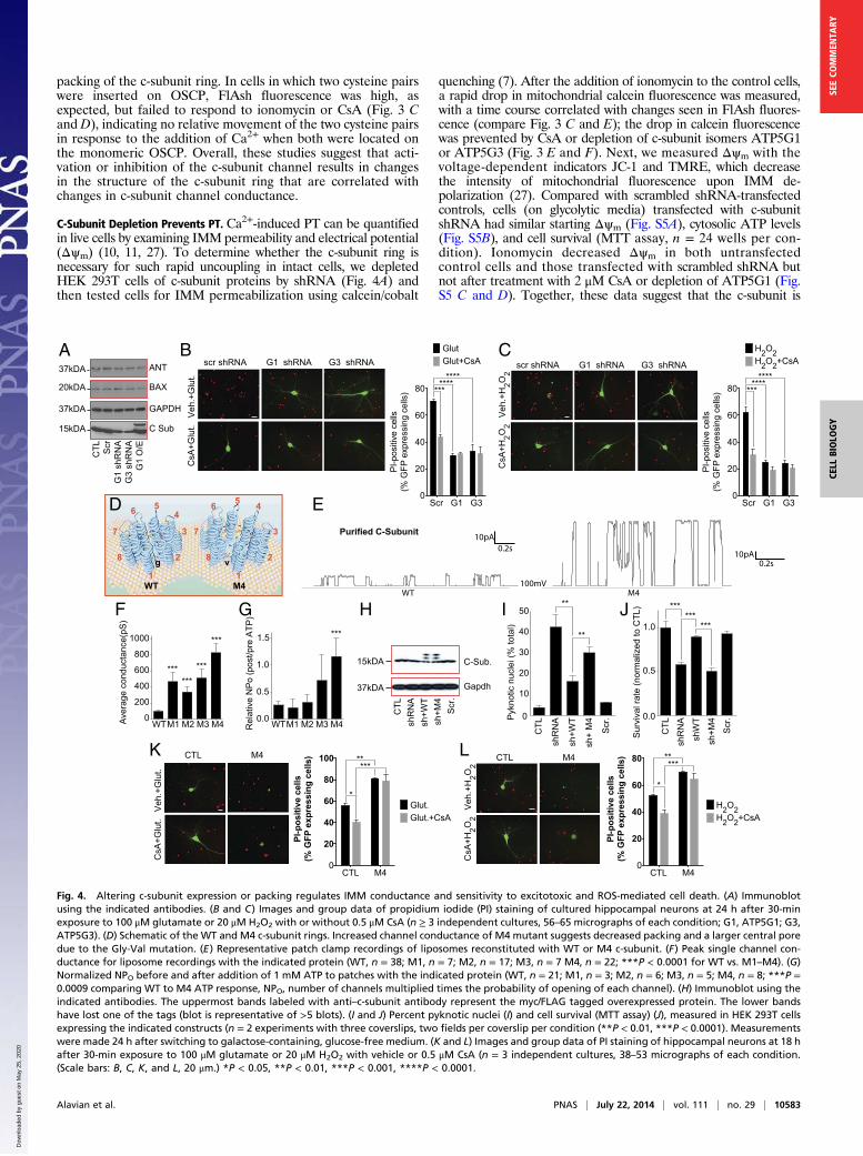
Fig. 4. Altering c-subunit expression or packing regulates IMM conductance and sensitivity to excitotoxic and ROS-mediated cell death. (A) Immunoblotusing the indicated antibodies. (B and C) Images and group data of propidium iodide (PI) staining of cultured hippocampal neurons at 24 h after 30-minexposure to 100 μM glutamate or 20 μM H2O2 with or without 0.5 μM CsA (n ≥ 3 independent cultures, 56–65 micrographs of each condition; G1, ATP5G1; G3,ATP5G3). (D) Schematic of the WT and M4 c-subunit rings. Increased channel conductance of M4 mutant suggests decreased packing and a larger central poredue to the Gly-Val mutation. (E) Representative patch clamp recordings of liposomes reconstituted with WT or M4 c-subunit. (F) Peak single channel con-ductance for liposome recordings with the indicated protein (WT, n = 38; M1, n = 7; M2, n = 17; M3, n = 7 M4, n = 22; ***P < 0.0001 for WT vs. M1–M4). (G)Normalized NPO before and after addition of 1 mM ATP to patches with the indicated protein (WT, n = 21; M1, n = 3; M2, n = 6; M3, n = 5; M4, n = 8; ***P =0.0009 comparing WT to M4 ATP response, NPO, number of channels multiplied times the probability of opening of each channel). (H) Immunoblot using theindicated antibodies. The uppermost bands labeled with anti–c-subunit antibody represent the myc/FLAG tagged overexpressed protein. The lower bandshave lost one of the tags (blot is representative of >5 blots). (I and J) Percent pyknotic nuclei (I) and cell survival (MTT assay) (J), measured in HEK 293T cellsexpressing the indicated constructs (n = 2 experiments with three coverslips, two fields per coverslip per condition (**P < 0.01, ***P < 0.0001). Measurementswere made 24 h after switching to galactose-containing, glucose-free medium. (K and L) Images and group data of PI staining of hippocampal neurons at 18 hafter 30-min exposure to 100 μM glutamate or 20 μM H2O2 with vehicle or 0.5 μM CsA (n = 3 independent cultures, 38–53 micrographs of each condition.(Scale bars: B, C, K, and L, 20 μm.) *P < 0.05, **P < 0.01, ***P < 0.001, ****P < 0.0001.
Alavian et al. PNAS | July 22, 2014 | vol. 111 | no. 29 | 10583
CELL
BIOLO
GY
SEECO
MMEN
TARY
Dow
nloa
ded
by g
uest
on
May
25,
202
0
necessary for rapid IMM permeabilization and membrane de-polarization induced by PT.
Depletion of the C-Subunit Attenuates, Whereas Overexpression ofMutant Large Conductance C-Subunit Aggravates, Cell Death. Be-cause PT is the hallmark of certain forms of cell death, partic-ularly after ischemia in brain and heart (28, 29), we testedwhether c-subunits were required for Ca2+- and H2O2-induceddeath. We found that, as previously reported, CsA attenuatedH2O2- or glutamate-induced death in cultured hippocampalneurons (Fig. 4 B and C). In addition, although depletion ofATP5G1 or ATP5G3 by shRNA did not affect protein levels ofBax or ANT (Fig. 4A), it attenuated H2O2- or glutamate-induceddeath to a similar extent to that of CsA (Fig. 4 B and C). Thesestudies suggest that c-subunit expression is required for excito-toxicity and reactive oxygen species (ROS)-associated cell death.To further demonstrate that the c-subunit ring creates an
uncoupling pore, we mutated four highly conserved glycineswithin the first (N terminus) alpha-helical region of the c-subunit(Fig. S6). These glycines may be responsible for the tight packingof the c-subunit molecules within the ring structure and controlmembrane coupling and OXPHOS (Fig. 4D) (30–32). To de-crease this tight packing, we made four individual constructscontaining glycine-to-valine mutations (M1–M4), and all fourmutant proteins when reconstituted into liposomes demon-strated increased single-channel conductance averaging overmultiple traces (including those with subconductance states only)compared with WT c-subunit; the conductance of M4 was thelargest of the four (Fig. 4 E and F). We also found that M4abolished attenuation of channel activity by ATP (Fig. 4G).Because these changes in the c-subunit ring could decreasemetabolic efficiency and thus increase cell death, we determinedwhether the mutations altered cell survival by combining intrinsicc-subunit depletion with overexpression of shRNA-resistant WTand M4 constructs (Fig. 4H). After depletion of c-subunits,forcing OXPHOS using glucose-free, galactose-containing me-dium increased cell death whereas overexpression of the WT, butnot M4, c-subunit rescued these effects (Fig. 4 I and J). There-fore, we overexpressed M4 c-subunit in neurons and determinedeffects on H2O2- or glutamate-induced cell death in normalmedium. Neuronal death was markedly increased in cells over-expressing M4, and this death was not inhibited by CsA (Fig. 4 Kand L). These data demonstrate that mutations that loosen thepacking of c-subunits of ATP synthase permanently increase theconductance of the c-subunit ring, decrease sensitivity to CsAand ATP, and increase cell death under oxidative conditions orin response to PT-inducing stimuli.
F1 Is Required for Inhibition of C-Subunit Channel Activation, and FOReleases F1 During PT. Because our previous studies suggested thatthe F1 beta-subunit may control leak current (12, 13) and otherstudies suggested that F1 removal uncouples bacterial mem-branes (33), we hypothesized that positioning of F1 over thec-subunit ring may be required for adequate coupling andinhibition of the c-subunit leak conductance. To detect whethercomponents of F1 bind to and inhibit the c-subunit channel, weapplied purified individual F1 proteins to reconstituted c-subunitchannels (Fig. 5A). Although gamma-, delta-, and epsilon-sub-units had no effect, purified beta-subunit protein attenuated thec-subunit conductance in both the small and large conductancemodes (Fig. 5 B–D), suggesting that the beta-subunit can bind tothe c-subunit directly to inhibit pore activity in vitro.Our findings that removal of the F1 proteins with urea un-
masked, and adding back the beta-subunit inhibited, the con-ductance of the c-subunit pore raised the intriguing possibilitythat physical uncoupling of F1 from FO could increase c-subunitpore conductance and initiate PT. Therefore, we determinedwhether release of F1 from c-subunit rings was associated withPT. Indeed, using immunocapture of the whole ATP synthase(experimental design in Fig. S7), Ca2+-induced swelling releasedc-subunit oligomers (15 kDa, 120 kDa, and 250 kDa) seen in
denaturing or native immunoblots whereas PT inhibitors (CsA,ADP) prevented the release fromATP synthase (Fig. 5 E,G, andH).Furthermore, c-subunit release was not detectable in mitochondriafrom CypD KOmice, even when swelling was induced with very high
B
5pA1s
0mV-100mV
100pA
-100
-200 (pA)
100
200
1050 (s)
04030
20
C
ß0.0
0.5
1.0
1.5
2.0
A
DBeta
Beta
CTL
0.5s2pA
5pA
10pAEpsilon
Delta
Gamma
0.5s
0.5sα dß b ε γ
75
10
37
50
2515
c δ
100mV
100mV
100mV
120mV
Rat
io (N
Po p
re/p
ost p
rote
in)
**
E
ca
H+OSCP
dF6
γ
δ
α ß
b ε
H+H+
Ca2+CypDI
caH+
OSCP
dF6
γδ
α ß
bε
H+H+H+
ADPATP
Pi
CsA Bcl-xLADP/ATP
ε γδ
Abs
orba
nce
(A/A
0)+/
- SE
MTime [sec]
200 400
1.0
0.8
0.9
1.1WT
Control
60 μM Ca2+
60 μM Ca2++ CsA60 μM Ca2++ ADP
200 400600
CypD null
1 mM Ca2++ CsA1 mM Ca2+
1.0
0.8
0.9
1.1
Time [sec]
Control60 μM Ca2+
% D
ensi
ty+/
- SE
M
75
100
150
* 15 kD120/250 kD
CsA ADP60 μM0
125
175WT CypD null
Sup
erna
tant
15Eluate
250
120
15
CsA ADP60 μM0Ca2+
Inhibitor1 mM60 μM
CsA
F
G H
Open Closed
Ca2+ Ca2+
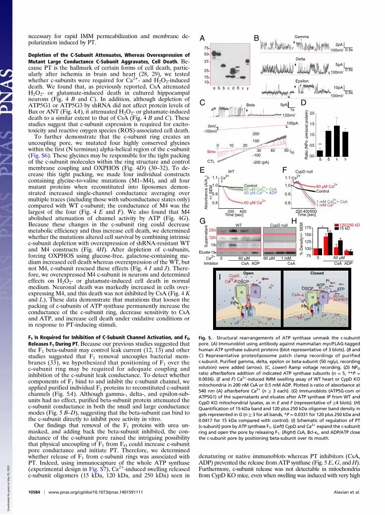
Fig. 5. Structural rearrangements of ATP synthase unmask the c-subunitpore. (A) Immunoblot using antibody against mammalian myc/FLAG-taggedhuman ATP synthase subunit proteins (blot representative of 3 blots). (B andC ) Representative proteoliposome patch clamp recordings of purifiedc-subunit. Purified gamma, delta, epsilon or beta-subunit (50 ng/μL recordingsolution) were added (arrow). (C, Lower) Ramp voltage recording. (D) NPOratio after/before addition of indicated ATP synthase subunits (n = 5, **P =0.0036). (E and F) Ca2+-induced IMM swelling assay of WT heart or CypD KOmitochondria in 200 nM CsA or 0.5 mM ADP. Plotted is ratio of absorbance at540 nm (A) after/before Ca2+ (n ≥ 3 each). (G) Immunoblots (ATP5G-com orATP5G1) of the supernatants and eluates after ATP synthase IP from WT andCypD KO mitochondrial lysates, as in E and F (representative of ≥4 blots). (H)Quantification of 15-kDa band and 120 plus 250 kDa oligomer band density ingels represented in G (n ≥ 3 for all bands, *P = 0.0331 for 120 plus 250 kDa and0.0417 for 15 kDa compared with control). (I) Schematic of regulation of PT(c-subunit) pore by ATP synthase F1. (Left) CypD and Ca2+ expand the c-subunitring and open the pore by releasing F1. (Right) CsA, Bcl-xL, and ADP/ATP closethe c-subunit pore by positioning beta-subunit over its mouth.
10584 | www.pnas.org/cgi/doi/10.1073/pnas.1401591111 Alavian et al.
Dow
nloa
ded
by g
uest
on
May
25,
202
0
concentrations of Ca2+ (Fig. 5 F and G) (22). This release of c-sub-units was not due to mitochondrial rupture because insignificantamounts of c-subunit were found in the postmitochondrial superna-tant after Ca2+-induced swelling, which, in contrast, did release cy-tochrome c (Fig. S8). These data suggest that CypD-mediated Ca2+binding to F1 destabilizes ATP synthase, causing unmasking of the c-subunit ring, which initiates PT (Figs. 5I and Fig. S7B).
DiscussionMitochondrial PT was first described in the 1950s (10) whereas theelectrophysiologic properties of the mPTP (or MCC) were de-scribed in the 1980s and 1990s as a voltage- and Ca2+-sensitivemulticonductance channel (conductances from 100 pS to 2 nS (1,19–21)). mPTP is inhibited by CsA and ADP. We find similarbiophysical features in the purified c-subunit, yet it is not sensitiveto Ca2+ and it is resistant to CsA. However, the original recordingsof MCC/mPTP used preparations of IMM or mitoplasts thatcontain now appreciated regulatory components such as CypD,and we recapitulate the data of these reports using SMVs andpurified ATP synthase. Thus, we conclude that the major regula-tory sites for the mPTP, which provide sensitivity to Ca2+, CypD,CsA, and ADP, reside in F1. These sites are removed during pu-rification of the c-subunit or by stripping F1 from FO using urea,exposing a lower-affinity adenine nucleotide binding site (Figs. 1and 2 and Figs. S1 and S3). Furthermore, the purified c-subunitring largely lacks cation selectivity, similar to the MCC. For thesereasons, we suggest that the c-subunit ring is the pore of the mPTP.If the c-subunit comprises the mPTP, then there exists a re-
lationship between the opening of the mPTP and ATP synthaseactivity. If the mPTP is open, ATP hydrolysis, rather than synthesis,occurs, leading to a rundown of energy and further opening of themPTP. In addition, eliminating the c-subunit may disrupt assemblyand function of ATP synthase, compromising metabolism in cellsthat depend on OXPHOS (Fig. 4 H–J). However, glycolytic con-ditions prevent this toxicity unless stress occurs (excitotoxicity,H2O2) (Fig. 4 and Fig. S5). Overall, too much pore activity willprevent ATP production and too little mPTP activity, although
enhancing the efficiency of OXPHOS, could be detrimental to cellsurvival by preventing an escape valve for Ca2+ and ROS.In summary, we find that the long-sought molecular pore of
the mPTP is a heretofore-undetected ion channel located withinthe c-subunit ring of the mammalian ATP synthase that may beexposed during physical uncoupling of the F1 and FO complexes.Depletion of the c-subunit prevents mitochondrial PT, attenu-ating excitotoxicity- and ROS-induced death in primary neurons.Mutations of the transmembrane domain that loosen c-subunitpacking increase the size of the c-subunit channel conductanceand predispose cells to death in a CsA-resistant manner. Thec-subunit channel is inhibited by the beta-subunit of ATP syn-thase, regulated also by Bcl-xL to improve metabolism (12). Froma pathophysiologic standpoint, induction of PT unmasks c-subunitrings that create the mPTP. We suggest that the c-subunit ring isso situated as to regulate both metabolism and death.
Materials and MethodsPurification of the F1FO ATP Synthase Subunit Proteins and C-Subunit Knockdown.Cells were transfected to express or knock down (shRNA) WT or mutant humanATP5G1 or -3.
Electrophysiology. SMV, mitochondrial, or proteoliposome recordings weremade by forming a giga-ohm seal in intracellular solution. Group data werequantified in terms of conductance.
Bipartite Tetracysteine Display. Two cysteine residues were placed on the Nterminus of the c-subunit sequence before the first alpha-helical region (onthe intermembraneous space side).
Statistical Analysis. Data in graphs are shown as mean ± SEM. Statisticalcomparisons included t tests or ANOVA and Kruskal–Wallis tests with posthoc testing (P < 0.05).
ACKNOWLEDGMENTS. We thank Dr. Leonard K. Kaczmarek for insightfulscientific discussion and constructive review of the manuscript. This workwas supported by National Institutes of Health Grant NS064967 (to E.A.J.)and American Heart Association Grant 12GRNT12060233 (to G.A.P.).
1. Petronilli V, Szabò I, Zoratti M (1989) The inner mitochondrial membrane containsion-conducting channels similar to those found in bacteria. FEBS Lett 259(1):137–143.
2. Antonenko YN, Smith D, Kinnally KW, Tedeschi H (1994) Single-channel activity in-duced in mitoplasts by alkaline pH. Biochim Biophys Acta 1194(2):247–254.
3. Bernardi P, et al. (1992) Modulation of the mitochondrial permeability transitionpore: Effect of protons and divalent cations. J Biol Chem 267(5):2934–2939.
4. Bernardi P (1992) Modulation of the mitochondrial cyclosporin A-sensitive perme-ability transition pore by the proton electrochemical gradient: Evidence that the porecan be opened by membrane depolarization. J Biol Chem 267(13):8834–8839.
5. Jonas EA, Buchanan J, Kaczmarek LK (1999) Prolonged activation of mitochondrialconductances during synaptic transmission. Science 286(5443):1347–1350.
6. Hüser J, Blatter LA (1999) Fluctuations in mitochondrial membrane potential causedby repetitive gating of the permeability transition pore. Biochem J 343(Pt 2):311–317.
7. Hom JR, et al. (2011) The permeability transition pore controls cardiac mitochondrialmaturation and myocyte differentiation. Dev Cell 21(3):469–478.
8. Haworth RA, Hunter DR (2000) Control of the mitochondrial permeability transitionpore by high-affinity ADP binding at the ADP/ATP translocase in permeabilized mi-tochondria. J Bioenerg Biomembr 32(1):91–96.
9. Kokoszka JE, et al. (2004) The ADP/ATP translocator is not essential for the mito-chondrial permeability transition pore. Nature 427(6973):461–465.
10. Bernardi P (2013) The mitochondrial permeability transition pore: A mystery solved?Front Physiol 4:95.
11. Elrod JW, Molkentin JD (2013) Physiologic functions of cyclophilin D and the mito-chondrial permeability transition pore. Circ J 77(5):1111–1122.
12. Alavian KN, et al. (2011) Bcl-xL regulates metabolic efficiency of neurons through in-teraction with the mitochondrial F1FO ATP synthase. Nat Cell Biol 13(10):1224–1233.
13. Chen YB, et al. (2011) Bcl-xL regulates mitochondrial energetics by stabilizing theinner membrane potential. J Cell Biol 195(2):263–276.
14. Bonora M, et al. (2013) Role of the c subunit of the FO ATP synthase in mitochondrialpermeability transition. Cell Cycle 12(4):674–683.
15. Giorgio V, et al. (2013) Dimers of mitochondrial ATP synthase form the permeabilitytransition pore. Proc Natl Acad Sci USA 110(15):5887–5892.
16. Chinopoulos C, Szabadkai G (2013) What makes you can also break you: Mitochon-drial permeability transition pore formation by the c subunit of the F(1)F(0) ATP-synthase? Front Oncol 3:25.
17. Watt IN, Montgomery MG, Runswick MJ, Leslie AG, Walker JE (2010) Bioenergetic costof making an adenosine triphosphate molecule in animal mitochondria. Proc NatlAcad Sci USA 107(39):16823–16827.
18. Havlícková V, Kaplanová V, Nusková H, Drahota Z, Houstek J (2010) Knockdown of F1
epsilon subunit decreases mitochondrial content of ATP synthase and leads to accu-
mulation of subunit c. Biochim Biophys Acta 1797(6-7):1124–1129.19. Zorov DB, Kinnally KW, Perini S, Tedeschi H (1992) Multiple conductance levels in rat
heart inner mitochondrial membranes studied by patch clamping. Biochim Biophys
Acta 1105(2):263–270.20. Szabó I, Zoratti M (1992) The mitochondrial megachannel is the permeability tran-
sition pore. J Bioenerg Biomembr 24(1):111–117.21. Szabó I, Bernardi P, Zoratti M (1992) Modulation of the mitochondrial megachannel
by divalent cations and protons. J Biol Chem 267(5):2940–2946.22. Baines CP, et al. (2005) Loss of cyclophilin D reveals a critical role for mitochondrial
permeability transition in cell death. Nature 434(7033):658–662.23. Nakagawa T, et al. (2005) Cyclophilin D-dependent mitochondrial permeability transi-
tion regulates some necrotic but not apoptotic cell death. Nature 434(7033):652–658.24. Luedtke NW, Dexter RJ, Fried DB, Schepartz A (2007) Surveying polypeptide and
protein domain conformation and association with FlAsH and ReAsH. Nat Chem Biol
3(12):779–784.25. Hoffmann C, et al. (2005) A FlAsH-based FRET approach to determine G protein-
coupled receptor activation in living cells. Nat Methods 2(3):171–176.26. Adams SR, et al. (2002) New biarsenical ligands and tetracysteine motifs for protein labeling
in vitro and in vivo: Synthesis and biological applications. J AmChem Soc 124(21):6063–6076.27. Nicholls DG (2012) Fluorescence measurement of mitochondrial membrane potential
changes in cultured cells. Methods Mol Biol 810:119–133.28. Brown DA, O’Rourke B (2010) Cardiac mitochondria and arrhythmias. Cardiovasc Res
88(2):241–249.29. Crompton M (1999) The mitochondrial permeability transition pore and its role in cell
death. Biochem J 341(Pt 2):233–249.30. Norris U, Karp PE, Fimmel AL (1992) Mutational analysis of the glycine-rich region of
the c subunit of the Escherichia coli F0F1 ATPase. J Bacteriol 174(13):4496–4499.31. Pogoryelov D, et al. (2007) The oligomeric state of c rings from cyanobacterial F-ATP
synthases varies from 13 to 15. J Bacteriol 189(16):5895–5902.32. Vonck J, et al. (2002) Molecular architecture of the undecameric rotor of a bacterial
Na+-ATP synthase. J Mol Biol 321(2):307–316.33. Mitome N, Suzuki T, Hayashi S, Yoshida M (2004) Thermophilic ATP synthase has
a decamer c-ring: Indication of noninteger 10:3 H+/ATP ratio and permissive elastic
coupling. Proc Natl Acad Sci USA 101(33):12159–12164.
Alavian et al. PNAS | July 22, 2014 | vol. 111 | no. 29 | 10585
CELL
BIOLO
GY
SEECO
MMEN
TARY
Dow
nloa
ded
by g
uest
on
May
25,
202
0