Embed Size (px)
Citation preview
CASE REPORT Open Access
An unusual presentation of acutemyocardial infarction in physiotherapydirect access: findings from a case reportLorenzo Storari1 , Valerio Barbari1 , Fabrizio Brindisino2 , Marco Testa1 and Maselli Filippo1*
Abstract
Background: Shoulder pain (SP) may originate from both musculoskeletal and visceral conditions. Physiotherapists(PT) may encounter patients with life-threatening pathologies that mimic musculoskeletal pain such as AcuteMyocardial Infarction (AMI). A trained PT should be able to distinguish between signs and symptoms ofmusculoskeletal or visceral origin aimed at performing proper medical referral.
Case presentation: A 46-y-old male with acute SP lasting from a week was diagnosed with right painfulmusculoskeletal shoulder syndrome, in two successive examinations by the emergency department physicians.However, after having experienced a shift of the pain on the left side, the patient presented to a PT. The PTrecognized the signs and symptoms of visceral pain and referred him to the general practitioner, which identified acardiac disease. The final diagnosis was acute myocardial infarction.
Conclusion: This case report highlights the importance of a thorough patient screening examination, especially forpatients treated in an outpatient setting, which allow distinguishing between signs and symptoms ofmusculoskeletal from visceral diseases.
Keywords: Anterior wall myocardial infarction, Differential diagnosis, Referral and consultation, Shoulder pain,Physiotherapy
BackgroundShoulder pain (SP) is one of the most common musculo-skeletal (MSK) disorders in the general population witha prevalence ranging from 7 to 27% among adults youn-ger than 70 years-old [1]. SP is the third most commonMSK disease after lower back and neck pain, further-more, it is also one of the most prevalent complaints inoutpatient clinic [1, 2], and in emergency department(ED) [3, 4]. Most of the patients consulting healthcareprofessionals for SP (in primary and secondary care)have been diagnosed with subacromial impingement
syndrome, rotator cuff tendinopathy and adhesive capsu-litis [1, 5, 6]. However, in addition to the musculoskel-etal or mechanic origin of pain, healthcare professionalsmust be alert that visceral [7], or serious potential life-threatening illnesses may refer pain to the shoulder, suchas cardiovascular disorders [7–9]. In fact, patients withcardiovascular pathologies may present crushing, sub-sternal chest pain, abdominal pain, jaw pain, neck pain,diaphoresis, dyspnea, fatigue and arm or shoulder re-ferred pain as main signs and symptoms [8, 9], withsome differences given by sex [8].Among cardiovascular pathologies, high rates of both
incidence and prevalence are represented by acute myo-cardial infarction (AMI )[10, 11, 11], which most com-mon sites of pain are chest, upper limb and abdomen[12, 13]. Unfortunately, these possible referred pain
© The Author(s). 2021 Open Access This article is licensed under a Creative Commons Attribution 4.0 International License,which permits use, sharing, adaptation, distribution and reproduction in any medium or format, as long as you giveappropriate credit to the original author(s) and the source, provide a link to the Creative Commons licence, and indicate ifchanges were made. The images or other third party material in this article are included in the article's Creative Commonslicence, unless indicated otherwise in a credit line to the material. If material is not included in the article's Creative Commonslicence and your intended use is not permitted by statutory regulation or exceeds the permitted use, you will need to obtainpermission directly from the copyright holder. To view a copy of this licence, visit http://creativecommons.org/licenses/by/4.0/.The Creative Commons Public Domain Dedication waiver (http://creativecommons.org/publicdomain/zero/1.0/) applies to thedata made available in this article, unless otherwise stated in a credit line to the data.
* Correspondence: [email protected] of Neurosciences, Rehabilitation, Ophthalmology, Genetic andMaternal Infantile Sciences (DINOGMI), University of Genova - Campus ofSavona, Savona, ItalyFull list of author information is available at the end of the article
Storari et al. Archives of Physiotherapy (2021) 11:5 https://doi.org/10.1186/s40945-021-00099-x
locations could lead to misleading diagnosis even in EDcontext [14]. Therefore, healthcare professionals, includ-ing physiotherapist (PT), should evaluate each possibleunderlying cause during the evaluation of patients refer-ring SP [9, 15, 16]. Currently, there is sparse literatureregarding cases of differential diagnosis made by PT inpatients with musculoskeletal diseases [17–19], espe-cially in case of cardiogenic referred pain [20]. Moreover,it is not available at present any reports related to anacute life-threatening condition such as AMI. We reportthe case of a patient presented to an outpatient physio-therapy clinic with SP that quickly shifted from the rightto the left shoulder, which has been properly recognizedas visceral related pain by the PT, and finally referred toan appropriate medical management.
Case presentationA 46-y-old tiler male presented to the ED complainingof right SP during the previous week due to increasedworkload (13–14 h per day laying bulky tiles, instead ofhis usual 8 h of daily work). His vitals parameters werepulse oxygen saturation level of 99%, blood pressure(BP) of 130/90 mmHg, respiratory rate (RR) of 16 rpm,
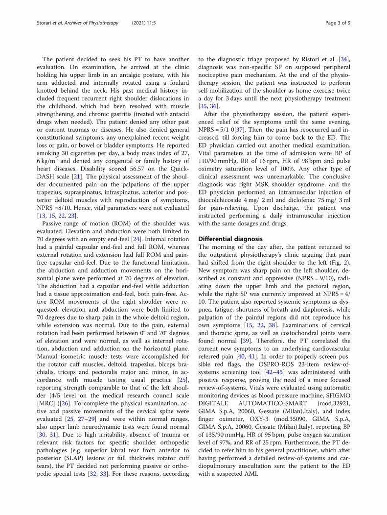
and heart rate (HR) of 72 bpm. Cardiopulmonary andneurological examinations were unremarkable. The pa-tient denied family history for cardiovascular disease,any medication intake in the previous 48 h, and declaredbeing smoker since 20 years. Physical examination docu-mented worsening of pain on palpation of the acromialand scapular region stated with numeric pain rating(NPRS) scale at 8/10, and there were no other abnormal-ities in other physical examinations (Fig. 1). The painwas described as dull and intense, lasting from a week,with a subtle onset. He was diagnosed by the ED phys-ician with right painful musculoskeletal (MSK) shouldersyndrome and treated with an intramuscular injection ofdiclofenac 75 mg/mL and ketorolac 30 mg/mL, with norelief. Accordingly, to reduce the pain and calm downthe patient, the physician prescribed an infusion of salinesolution (100 mL and 250 mL) and ranitidine hydro-chloride 50mg/5 mL and a diazepam dose 10 mg/ 2 mL,which led to a sensible improvement of the pain.Upon discharge, the patient was advised to take eperi-
sone hydrochloride 100 mg twice and ibuprofen 600 mgtwice a day for 5 days, rest from work for 1 week and aprescription of antalgic electrotherapies.
Fig. 1 Body chart. Symptoms at 1st ED visit. In red NPRS 8/10 at rest
Storari et al. Archives of Physiotherapy (2021) 11:5 Page 2 of 9
The patient decided to seek his PT to have anotherevaluation. On examination, he arrived at the clinicholding his upper limb in an antalgic posture, with hisarm adducted and internally rotated using a foulardknotted behind the neck. His past medical history in-cluded frequent recurrent right shoulder dislocations inthe childhood, which had been resolved with musclestrengthening, and chronic gastritis (treated with antaciddrugs when needed). The patient denied any other pastor current traumas or diseases. He also denied generalconstitutional symptoms, any unexplained recent weightloss or gain, or bowel or bladder symptoms. He reportedsmoking 30 cigarettes per day, a body mass index of 27,6 kg/m2 and denied any congenital or family history ofheart diseases. Disability scored 56.57 on the Quick-DASH scale [21]. The physical assessment of the shoul-der documented pain on the palpations of the uppertrapezius, supraspinatus, infraspinatus, anterior and pos-terior deltoid muscles with reproduction of symptoms,NPRS =8/10. Hence, vital parameters were not evaluated[13, 15, 22, 23].Passive range of motion (ROM) of the shoulder was
evaluated. Elevation and abduction were both limited to70 degrees with an empty end-feel [24]. Internal rotationhad a painful capsular end-feel and full ROM, whereasexternal rotation and extension had full ROM and pain-free capsular end-feel. Due to the functional limitation,the abduction and adduction movements on the hori-zontal plane were performed at 70 degrees of elevation.The abduction had a capsular end-feel while adductionhad a tissue approximation end-feel, both pain-free. Ac-tive ROM movements of the right shoulder were re-quested: elevation and abduction were both limited to70 degrees due to sharp pain in the whole deltoid region,while extension was normal. Due to the pain, externalrotation had been performed between 0° and 70° degreesof elevation and were normal, as well as internal rota-tion, abduction and adduction on the horizontal plane.Manual isometric muscle tests were accomplished forthe rotator cuff muscles, deltoid, trapezius, biceps bra-chialis, triceps and pectoralis major and minor, in ac-cordance with muscle testing usual practice [25],reporting strength comparable to that of the left shoul-der (4/5 level on the medical research council scale[MRC] )[26]. To complete the physical examination, ac-tive and passive movements of the cervical spine wereevaluated [25, 27–29] and were within normal ranges,also upper limb neurodynamic tests were found normal[30, 31]. Due to high irritability, absence of trauma orrelevant risk factors for specific shoulder orthopedicpathologies (e.g. superior labral tear from anterior toposterior (SLAP) lesions or full thickness rotator cufftears), the PT decided not performing passive or ortho-pedic special tests [32, 33]. For these reasons, according
to the diagnostic triage proposed by Ristori et al .[34],diagnosis was non-specific SP on supposed peripheralnociceptive pain mechanism. At the end of the physio-therapy session, the patient was instructed to performself-mobilization of the shoulder as home exercise twicea day for 3 days until the next physiotherapy treatment[35, 36].After the physiotherapy session, the patient experi-
enced relief of the symptoms until the same evening,NPRS = 5/1 0[37]. Then, the pain has reoccurred and in-creased, till forcing him to come back to the ED. TheED physician carried out another medical examination.Vital parameters at the time of admission were BP of110/90 mmHg, RR of 16 rpm, HR of 98 bpm and pulseoximetry saturation level of 100%. Any other type ofclinical assessment was unremarkable. The conclusivediagnosis was right MSK shoulder syndrome, and theED physician performed an intramuscular injection ofthiocolchicoside 4 mg/ 2 ml and diclofenac 75mg/ 3 mlfor pain-relieving. Upon discharge, the patient wasinstructed performing a daily intramuscular injectionwith the same dosages and drugs.
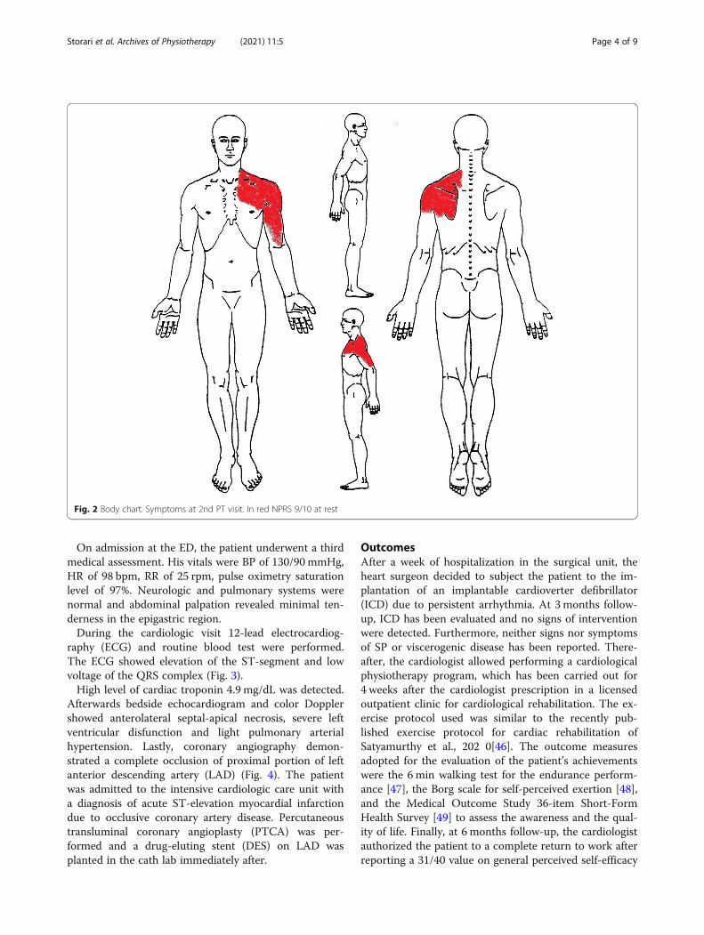
Differential diagnosisThe morning of the day after, the patient returned tothe outpatient physiotherapy’s clinic arguing that painhad shifted from the right shoulder to the left (Fig. 2).New symptom was sharp pain on the left shoulder, de-scribed as constant and oppressive (NPRS = 9/10), radi-ating down the upper limb and the pectoral region,while the right SP was currently improved at NPRS = 4/10. The patient also reported systemic symptoms as dys-pnea, fatigue, shortness of breath and diaphoresis, whilepalpation of the painful regions did not reproduce hisown symptoms [15, 22, 38]. Examinations of cervicaland thoracic spine, as well as costochondral joints werefound normal [39]. Therefore, the PT correlated thecurrent new symptoms to an underlying cardiovascularreferred pain [40, 41]. In order to properly screen pos-sible red flags, the OSPRO-ROS 23-item review-of-systems screening tool [42–45] was administered withpositive response, proving the need of a more focusedreview-of-systems. Vitals were evaluated using automaticmonitoring devices as blood pressure machine, SFIGMODIGITALE AUTOMATICO-SMART (mod.32921,GIMA S.p.A, 20060, Gessate (Milan),Italy), and indexfinger oximeter, OXY-3 (mod.35090, GIMA S.p.A,GIMA S.p.A, 20060, Gessate (Milan),Italy), reporting BPof 135/90 mmHg, HR of 95 bpm, pulse oxygen saturationlevel of 97%, and RR of 25 rpm. Furthermore, the PT de-cided to refer him to his general practitioner, which afterhaving performed a detailed review-of-systems and car-diopulmonary auscultation sent the patient to the EDwith a suspected AMI.
Storari et al. Archives of Physiotherapy (2021) 11:5 Page 3 of 9
On admission at the ED, the patient underwent a thirdmedical assessment. His vitals were BP of 130/90 mmHg,HR of 98 bpm, RR of 25 rpm, pulse oximetry saturationlevel of 97%. Neurologic and pulmonary systems werenormal and abdominal palpation revealed minimal ten-derness in the epigastric region.During the cardiologic visit 12-lead electrocardiog-
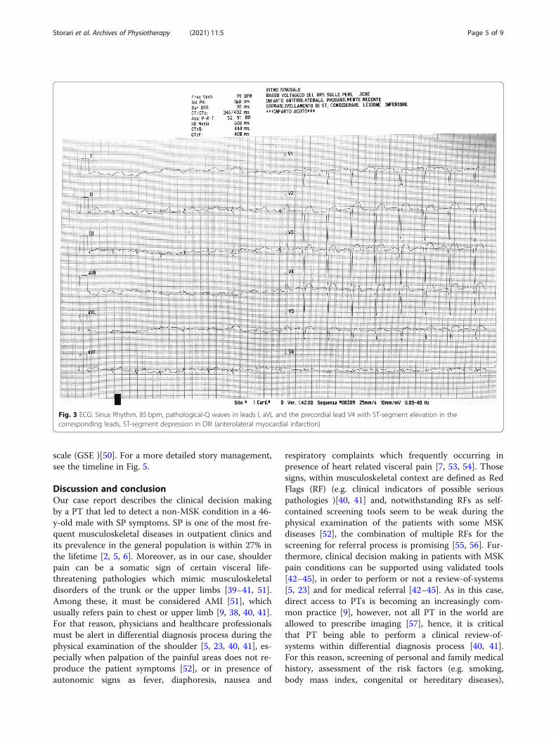
raphy (ECG) and routine blood test were performed.The ECG showed elevation of the ST-segment and lowvoltage of the QRS complex (Fig. 3).High level of cardiac troponin 4.9 mg/dL was detected.
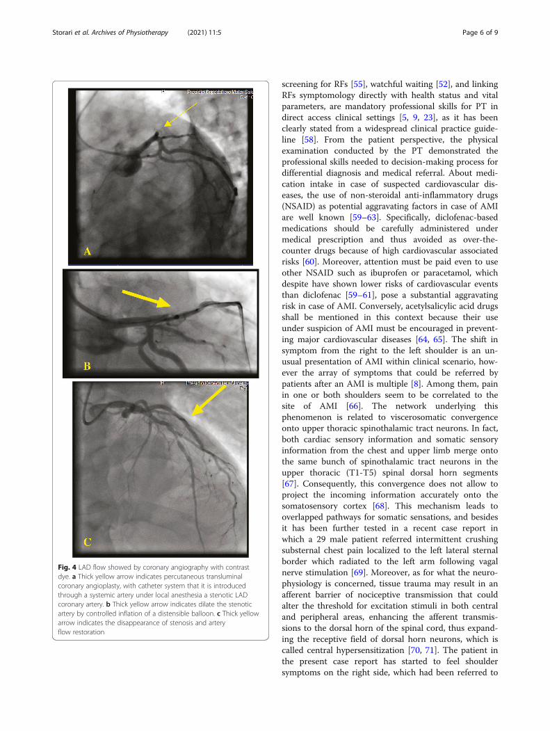
Afterwards bedside echocardiogram and color Dopplershowed anterolateral septal-apical necrosis, severe leftventricular disfunction and light pulmonary arterialhypertension. Lastly, coronary angiography demon-strated a complete occlusion of proximal portion of leftanterior descending artery (LAD) (Fig. 4). The patientwas admitted to the intensive cardiologic care unit witha diagnosis of acute ST-elevation myocardial infarctiondue to occlusive coronary artery disease. Percutaneoustransluminal coronary angioplasty (PTCA) was per-formed and a drug-eluting stent (DES) on LAD wasplanted in the cath lab immediately after.
OutcomesAfter a week of hospitalization in the surgical unit, theheart surgeon decided to subject the patient to the im-plantation of an implantable cardioverter defibrillator(ICD) due to persistent arrhythmia. At 3 months follow-up, ICD has been evaluated and no signs of interventionwere detected. Furthermore, neither signs nor symptomsof SP or viscerogenic disease has been reported. There-after, the cardiologist allowed performing a cardiologicalphysiotherapy program, which has been carried out for4 weeks after the cardiologist prescription in a licensedoutpatient clinic for cardiological rehabilitation. The ex-ercise protocol used was similar to the recently pub-lished exercise protocol for cardiac rehabilitation ofSatyamurthy et al., 202 0[46]. The outcome measuresadopted for the evaluation of the patient’s achievementswere the 6 min walking test for the endurance perform-ance [47], the Borg scale for self-perceived exertion [48],and the Medical Outcome Study 36-item Short-FormHealth Survey [49] to assess the awareness and the qual-ity of life. Finally, at 6 months follow-up, the cardiologistauthorized the patient to a complete return to work afterreporting a 31/40 value on general perceived self-efficacy
Fig. 2 Body chart. Symptoms at 2nd PT visit. In red NPRS 9/10 at rest
Storari et al. Archives of Physiotherapy (2021) 11:5 Page 4 of 9
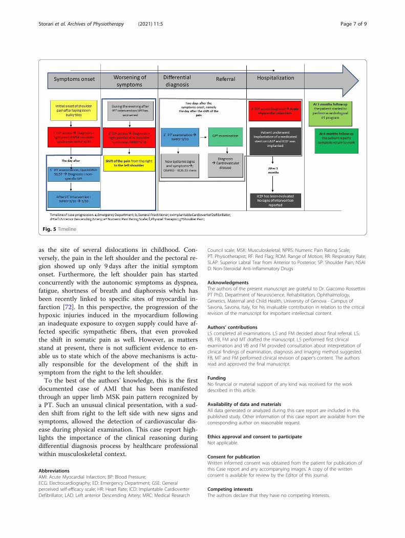
scale (GSE )[50]. For a more detailed story management,see the timeline in Fig. 5.
Discussion and conclusionOur case report describes the clinical decision makingby a PT that led to detect a non-MSK condition in a 46-y-old male with SP symptoms. SP is one of the most fre-quent musculoskeletal diseases in outpatient clinics andits prevalence in the general population is within 27% inthe lifetime [2, 5, 6]. Moreover, as in our case, shoulderpain can be a somatic sign of certain visceral life-threatening pathologies which mimic musculoskeletaldisorders of the trunk or the upper limbs [39–41, 51].Among these, it must be considered AMI [51], whichusually refers pain to chest or upper limb [9, 38, 40, 41].For that reason, physicians and healthcare professionalsmust be alert in differential diagnosis process during thephysical examination of the shoulder [5, 23, 40, 41], es-pecially when palpation of the painful areas does not re-produce the patient symptoms [52], or in presence ofautonomic signs as fever, diaphoresis, nausea and
respiratory complaints which frequently occurring inpresence of heart related visceral pain [7, 53, 54]. Thosesigns, within musculoskeletal context are defined as RedFlags (RF) (e.g. clinical indicators of possible seriouspathologies )[40, 41] and, notwithstanding RFs as self-contained screening tools seem to be weak during thephysical examination of the patients with some MSKdiseases [52], the combination of multiple RFs for thescreening for referral process is promising [55, 56]. Fur-thermore, clinical decision making in patients with MSKpain conditions can be supported using validated tools[42–45], in order to perform or not a review-of-systems[5, 23] and for medical referral [42–45]. As in this case,direct access to PTs is becoming an increasingly com-mon practice [9], however, not all PT in the world areallowed to prescribe imaging [57], hence, it is criticalthat PT being able to perform a clinical review-of-systems within differential diagnosis process [40, 41].For this reason, screening of personal and family medicalhistory, assessment of the risk factors (e.g. smoking,body mass index, congenital or hereditary diseases),
Fig. 3 ECG: Sinus Rhythm, 85 bpm, pathological-Q waves in leads I, aVL and the precordial lead V4 with ST-segment elevation in thecorresponding leads, ST-segment depression in DIII (anterolateral myocardial infarction)
Storari et al. Archives of Physiotherapy (2021) 11:5 Page 5 of 9
screening for RFs [55], watchful waiting [52], and linkingRFs symptomology directly with health status and vitalparameters, are mandatory professional skills for PT indirect access clinical settings [5, 9, 23], as it has beenclearly stated from a widespread clinical practice guide-line [58]. From the patient perspective, the physicalexamination conducted by the PT demonstrated theprofessional skills needed to decision-making process fordifferential diagnosis and medical referral. About medi-cation intake in case of suspected cardiovascular dis-eases, the use of non-steroidal anti-inflammatory drugs(NSAID) as potential aggravating factors in case of AMIare well known [59–63]. Specifically, diclofenac-basedmedications should be carefully administered undermedical prescription and thus avoided as over-the-counter drugs because of high cardiovascular associatedrisks [60]. Moreover, attention must be paid even to useother NSAID such as ibuprofen or paracetamol, whichdespite have shown lower risks of cardiovascular eventsthan diclofenac [59–61], pose a substantial aggravatingrisk in case of AMI. Conversely, acetylsalicylic acid drugsshall be mentioned in this context because their useunder suspicion of AMI must be encouraged in prevent-ing major cardiovascular diseases [64, 65]. The shift insymptom from the right to the left shoulder is an un-usual presentation of AMI within clinical scenario, how-ever the array of symptoms that could be referred bypatients after an AMI is multiple [8]. Among them, painin one or both shoulders seem to be correlated to thesite of AMI [66]. The network underlying thisphenomenon is related to viscerosomatic convergenceonto upper thoracic spinothalamic tract neurons. In fact,both cardiac sensory information and somatic sensoryinformation from the chest and upper limb merge ontothe same bunch of spinothalamic tract neurons in theupper thoracic (T1-T5) spinal dorsal horn segments[67]. Consequently, this convergence does not allow toproject the incoming information accurately onto thesomatosensory cortex [68]. This mechanism leads tooverlapped pathways for somatic sensations, and besidesit has been further tested in a recent case report inwhich a 29 male patient referred intermittent crushingsubsternal chest pain localized to the left lateral sternalborder which radiated to the left arm following vagalnerve stimulation [69]. Moreover, as for what the neuro-physiology is concerned, tissue trauma may result in anafferent barrier of nociceptive transmission that couldalter the threshold for excitation stimuli in both centraland peripheral areas, enhancing the afferent transmis-sions to the dorsal horn of the spinal cord, thus expand-ing the receptive field of dorsal horn neurons, which iscalled central hypersensitization [70, 71]. The patient inthe present case report has started to feel shouldersymptoms on the right side, which had been referred to
Fig. 4 LAD flow showed by coronary angiography with contrastdye. a Thick yellow arrow indicates percutaneous transluminalcoronary angioplasty, with catheter system that it is introducedthrough a systemic artery under local anesthesia a stenotic LADcoronary artery. b Thick yellow arrow indicates dilate the stenoticartery by controlled inflation of a distensible balloon. c Thick yellowarrow indicates the disappearance of stenosis and arteryflow restoration
Storari et al. Archives of Physiotherapy (2021) 11:5 Page 6 of 9
as the site of several dislocations in childhood. Con-versely, the pain in the left shoulder and the pectoral re-gion showed up only 9 days after the initial symptomonset. Furthermore, the left shoulder pain has startedconcurrently with the autonomic symptoms as dyspnea,fatigue, shortness of breath and diaphoresis which hasbeen recently linked to specific sites of myocardial in-farction [72]. In this perspective, the progression of thehypoxic injuries induced in the myocardium followingan inadequate exposure to oxygen supply could have af-fected specific sympathetic fibers, that even provokedthe shift in somatic pain as well. However, as mattersstand at present, there is not sufficient evidence to en-able us to state which of the above mechanisms is actu-ally responsible for the development of the shift insymptom from the right to the left shoulder.To the best of the authors’ knowledge, this is the first
documented case of AMI that has been manifestedthrough an upper limb MSK pain pattern recognized bya PT. Such an unusual clinical presentation, with a sud-den shift from right to the left side with new signs andsymptoms, allowed the detection of cardiovascular dis-ease during physical examination. This case report high-lights the importance of the clinical reasoning duringdifferential diagnosis process by healthcare professionalwithin musculoskeletal context.
AbbreviationsAMI: Acute Myocardial Infarction; BP: Blood Pressure;ECG: Electrocardiography; ED: Emergency Department; GSE: Generalperceived self-efficacy scale; HR: Heart Rate; ICD: Implantable CardioverterDefibrillator; LAD: Left anterior Descending Artery; MRC: Medical Research
Council scale; MSK: Musculoskeletal; NPRS: Numeric Pain Rating Scale;PT: Physiotherapist; RF: Red Flag; ROM: Range of Motion; RR: Respiratory Rate;SLAP: Superior Labral Tear from Anterior to Posterior; SP: Shoulder Pain; NSAID: Non-Steroidal Anti-Inflammatory Drugs
AcknowledgmentsThe authors of the present manuscript are grateful to Dr. Giacomo RossettiniPT PhD, Department of Neuroscience, Rehabilitation, Ophthalmology,Genetics, Maternal and Child Health, University of Genova - Campus ofSavona, Savona, Italy, for his invaluable contribution in relation to the criticalrevision of the manuscript for important intellectual content.
Authors’ contributionsLS completed all examinations. LS and FM decided about final referral. LS,VB, FB, FM and MT drafted the manuscript. LS performed first clinicalexamination and VB and FM provided consultation about interpretation ofclinical findings of examination, diagnosis and imaging method suggested.FB, MT and FM performed clinical revision of paper’s content. The authorsread and approved the final manuscript.
FundingNo financial or material support of any kind was received for the workdescribed in this article.
Availability of data and materialsAll data generated or analyzed during this care report are included in thispublished study. Other information of this case report are available from thecorresponding author on reasonable request.
Ethics approval and consent to participateNot applicable.
Consent for publicationWritten informed consent was obtained from the patient for publication ofthis Case report and any accompanying images. A copy of the writtenconsent is available for review by the Editor of this journal.
Competing interestsThe authors declare that they have no competing interests.
Fig. 5 Timeline
Storari et al. Archives of Physiotherapy (2021) 11:5 Page 7 of 9
Author details1Department of Neurosciences, Rehabilitation, Ophthalmology, Genetic andMaternal Infantile Sciences (DINOGMI), University of Genova - Campus ofSavona, Savona, Italy. 2Department of Medicine and Health Science“Vincenzo Tiberio”, University of Molise c/o Cardarelli Hospital, C/da Tappino,Campobasso, Italy.
Received: 26 November 2020 Accepted: 19 January 2021
References1. Luime JJ, Koes BW, Hendriksen IJM, Burdorf A, Verhagen AP, Miedema HS,
Verhaar JAN. Prevalence and incidence of shoulder pain in the generalpopulation; a systematic review. Scand J Rheumatol. 2004;33:73–81.
2. Greving K, Dorrestijn O, Winters JC, Groenhof F, van der Meer K, Stevens M,Diercks RL. Incidence, prevalence, and consultation rates of shouldercomplaints in general practice. Scand J Rheumatol. 2012;41:150–5.
3. Lawrence RL, Braman JP, Laprade RF, Ludewig PM. Comparison of 3-dimensional shoulder complex kinematics in individuals with and withoutshoulder pain, part 1: Sternoclavicular, acromioclavicular, andscapulothoracic joints. J Orthop Sports Phys Ther. 2014;44:636–45.
4. Rekola KE, Keinanen-Kiukaanniemi S, Takala J. Use of primary health servicesin sparsely populated country districts by patients with musculoskeletalsymptoms: consultations with a physician. J Epidemiol Community Health.1993;47:153–7.
5. Mitchell C, Adebajo A, Hay E, Carr A. Shoulder pain: diagnosis andmanagement in primary care. Br Med J. 2005;331:1124–8.
6. Malavolta EA, Gracitelli MEC, Assunção JH, de MR PG, AZF d S, AAF N.Shoulder disorders in an outpatient clinic: an epidemiological study. ActaOrtop Brasil. 2017;25:78–80.
7. Sikandar S, Dickenson AH. Visceral pain: the ins and outs, the ups anddowns. Curr Opin Support Palliat Care. 2012;6:17–26.
8. Berg J, Björck L, Dudas K, Lappas G, Rosengren A. Symptoms of a first acutemyocardial infarction in women and men. Gender Medicine. 2009;6:454–62.
9. Weber MD. Screening and evaluation of the cardiovascular and pulmonarysystems in patients presenting with upper extremity impairments. J HandTher. 2010;23:127–39.
10. Mohseni J, Kazemi T, Maleki M, Beydokhti H. A systematic review on theprevalence of acute myocardial infarction in Iran. Heart Views. 2017;18:125.
11. Bradley SM, Borgerding JA, Wood GB, Maynard C, Fihn SD. Incidence, riskfactors, and outcomes associated with in-hospital acute myocardialinfarction. JAMA Netw Open. 2019;2:e187348.
12. Čulić V, Eterović D, Mirić D, Silić N. Symptom presentation of acutemyocardial infarction: influence of sex, age, and risk factors. Am Heart J.2002;144:1012–7.
13. Bruyninckx R, Aertgeerts B, Bruyninckx P, Buntinx F. Signs and symptoms indiagnosing acute myocardial infarction and acute coronary syndrome: adiagnostic meta-analysis. Br J Gen Pract. 2008;58:105–11.
14. Krolak K. Claims case study: missed myocardial infarctions: Cire; 2018.https://curi.com/news/missed-mi/. Accessed 18 Nov 2020
15. Primary Care for the Physical Therapist - 3rd Edition. https://www.elsevier.com/books/primary-care-for-the-physical-therapist/boissonnault/978-0-323-63897-5. Accessed 18 Nov 2020.
16. Cook CE, Décary S. Higher order thinking about differential diagnosis. Braz JPhys Ther. 2020;24:1–7.
17. VanWye WR, Pinerola J, Ogle KC, Wallmann HW. Screening for referral by asports physical therapist reveals an effort thrombosis in a collegiate pitcher:a CASE report. Int J Sports Phys Ther. 2016;11:607–13.
18. Slaven EJ, Mathers J. Differential diagnosis of shoulder and cervical pain: acase report. J Man Manipulative Ther. 2010;18:191–6.
19. Mathers JJ. Differential diagnosis of a patient referred to physical therapywith neck pain: a case study of a patient with an atypical presentation ofangina. J Man Manipulative Ther. 2012;20:214–8.
20. Perry M. Differential diagnosis: musculoskeletal pain vs. cardiogenic pain.Research innovation scholarship entrepreneurship 18-11–2020; 2012.
21. Mintken PE, Glynn P, Cleland JA. Psychometric properties of the shorteneddisabilities of the arm, shoulder, and hand questionnaire (QuickDASH) andnumeric pain rating scale in patients with shoulder pain. J Shoulder ElbSurg. 2009;18:920–6.
22. Gräni C, Senn O, Bischof M, Cippà PE, Hauffe T, Zimmerli L, Battegay E,Franzen D. Diagnostic performance of reproducible chest wall tenderness
to rule out acute coronary syndrome in acute chest pain: a prospectivediagnostic study. BMJ Open. 2015;5:e007442.
23. House J, Mooradian A. Evaluation and management of shoulder pain inprimary care clinics. South Med J. 2010;103:1129–35.
24. Petersen CM, Hayes KW. Consruct validity of Cyriax’s selective tensionexamination: association of end-feels with pain at the knee and shoulder. JOrthop Sports Phys Ther. 2000;30:512–27.
25. Orthopedic Physical Assessment - 5th Edition. https://www.elsevier.com/books/orthopedic-physical-assessment/magee/978-0-7216-0571-5. Accessed30 Oct 2020.
26. Paternostro-Sluga T, Grim-Stieger M, Posch M, Schuhfried O, Vacariu G,Mittermaier C, Bittner C, Fialka-Moser V. Reliability and validity of theMedical Research Council (MRC) scale and a modified scale for testingmuscle strength in patients with radial palsy. J Rehabil Med. 2008;40:665–71.
27. Jull G, Bogduk N, Marsland A. The accuracy of manual diagnosis for cervialzygapophysial joint pain syndromes. Med J Aust. 1988;148:233–6.
28. Cooper G, Bailey B, Bogduk N. Cervical zygapophysial joint pain maps. PainMed. 2007;8:344–53.
29. Siegenthaler A, Eichenberger U, Schmidlin K, Arendt-Nielsen L, Curatolo M.What does local tenderness say about the origin of pain? An investigationof cervical zygapophysial joint pain. Anesth Analg. 2010;110:923–7.
30. Vanti C, Conteddu L, Guccione A, Morsillo F, Parazza S, Viti C, Pillastrini P. The upperlimb Neurodynamic test 1: intra- and Intertester reliability and the effect of severalrepetitions on pain and resistance. J Manip Physiol Ther. 2010;33:292–9.
31. Coppieters M, Stappaerts K, Janssens K, Jull G. Reliability of detecting “onsetof pain” and “submaximal pain” during neural provocation testing of theupper quadrant. Physiother Res Int. 2002;7:146–56.
32. Lewis JS. Rotator cuff tendinopathy/subacromial impingement syndrome: isit time for a new method of assessment? Br J Sports Med. 2009;43:259–64.
33. Hegedus EJ, Goode A, Campbell S, Morin A, Tamaddoni M, Moorman CT,Cook C. Physical examination tests of the shoulder: a systematic review withmeta-analysis of individual tests. Br J Sports Med. 2008;42:80–92.
34. Ristori D, Miele S, Rossettini G, Monaldi E, Arceri D, Testa M. Towards anintegrated clinical framework for patient with shoulder pain. ArchPhysiother. 2018. https://doi.org/10.1186/s40945-018-0050-3.
35. Ribeiro DC, Sole G, Venkat R, Shemmell J. Differences between clinician-and self-administered shoulder sustained mobilization on scapular andshoulder muscle activity during shoulder abduction: a repeated-measuresstudy on asymptomatic individuals. Musculoskelet Sci Pract. 2017;30:25–33.
36. Vicenzino B, Paungmali A, Teys P. Mulligan’s mobilization-with-movement,positional faults and pain relief: current concepts from a critical review ofliterature. Man Ther. 2007;12:98–108.
37. Michener LA, Snyder AR, Leggin BG. Responsiveness of the numeric painrating scale in patients with shoulder pain and the effect of surgical status. JSport Rehabil. 2011;20:115–28.
38. Albarran J, Durham B, Gowers J, Dwight J, Chappell G. Is the radiation ofchest pain a useful indicator of myocardial infarction? A prospective studyof 541 patients. Accid Emerg Nurs. 2002;10:2–9.
39. Amsterdam EA, Wenger NK, Brindis RG, et al. 2014 AHA/ACC guideline forthe management of patients with non-st-elevation acute coronarysyndromes: a report of the American college of cardiology/American heartassociation task force on practice guidelines. Circulation. 2014;130:e344–426.
40. Goodman CC. Screening for medical problems in patients with upperextremity signs and symptoms. J Hand Ther. 2010;23:105–26.
41. Differential Diagnosis for Physical Therapists - 6th Edition. https://www.elsevier.com/books/differential-diagnosis-for-physical-therapists/heick/978-0-323-47849-6. Accessed 30 Oct 2020.
42. George SZ, Beneciuk JM, Lentz TA, Wu SS. The optimal screening forprediction of referral and outcome (OSPRO) in patients withmusculoskeletal pain conditions: a longitudinal validation cohort from theUSA. BMJ Open. 2017. https://doi.org/10.1136/bmjopen-2016-015188.
43. Beneciuk JM, Lentz TA, He Y, Wu SS, George SZ. Prediction of persistentmusculoskeletal pain at 12 months: a secondary analysis of the optimalscreening for prediction of referral and outcome (OSPRO) validation cohortstudy. Phys Ther. 2018;98:290–301.
44. George SZ, Beneciuk JM, Lentz TA, Wu SS, Dai Y, Bialosky JE, Zeppieri G.Optimal screening for prediction of referral and outcome (OSPRO) formusculoskeletal pain conditions: results from the validation cohort. J OrthopSports Phys Ther. 2018;48:460–75.
45. George SZ, Beneciuk JM, Bialosky JE, Lentz TA, Zeppieri G, Pei Q, Wu SS.Development of a review-of-systems screening tool for orthopaedic
Storari et al. Archives of Physiotherapy (2021) 11:5 Page 8 of 9
physical therapists: results from the optimal screening for prediction ofreferral and outcome (OSPRO) cohort. J Orthop Sports Phys Ther. 2015;45:512–26.
46. Satyamurthy A, Prabhu N, Padmakumar R, Babu AS. Feasibility of anexercise-based cardiac rehabilitation algorithm in patients followingpercutaneous coronary intervention for acute coronary syndrome. IndianHeart J. 2020;72:289–92.
47. Bellet RN, Adams L, Morris NR. The 6-minute walk test in outpatient cardiacrehabilitation: validity, reliability and responsiveness-a systematic review.Physiotherapy (United Kingdom). 2012;98:277–86.
48. Aamot IL, Forbord SH, Karlsen T, Støylen A. Does rating of perceivedexertion result in target exercise intensity during interval training in cardiacrehabilitation? A study of the Borg scale versus a heart rate monitor. J SciMed Sport. 2014;17:541–5.
49. Kweon S, Sohn MK, Jeong JO, Kim S, Jeon H, Lee H, Ahn SC, Park SH, Jee S.Quality of life and awareness of cardiac rehabilitation program in peoplewith cardiovascular diseases. Ann Rehabil Med. 2017;41:248–56.
50. Zotti AM, Balestroni G, Cerutti P, Ferrario SR, Angelino E, Miglioretti M.Application of the general perceived self-efficacy scale in cardiovascularrehabilitation. Monaldi Arch Chest Dis. 2016. https://doi.org/10.4081/monaldi.2007.451.
51. Thygesen K, Alpert JS, Jaffe AS, et al. Third universal definition of myocardialinfarction. Eur Heart J. 2012;33:2551–67.
52. Cook CE, George SZ, Reiman MP. Red flag screening for low back pain:nothing to see here, move along: a narrative review. Br J Sports Med. 2018;52:493–6.
53. Body R, Carley S, Wibberley C, McDowell G, Ferguson J, Mackway-Jones K.The value of symptoms and signs in the emergent diagnosis of acutecoronary syndromes. Resuscitation. 2010;81:281–6.
54. Maselli F, Palladino M, Barbari V, Storari L, Rossettini G, Testa M. Thediagnostic value of red flags in thoracolumbar pain: a systematic review.Disabil Rehabil. 2020. https://doi.org/10.1080/09638288.2020.1804626.
55. Finucane LM, Downie A, Mercer C, Greenhalgh SM, Boissonnault WG, Pool-Goudzwaard AL, Beneciuk JM, Leech RL, Selfe J. International framework forred flags for potential serious spinal pathologies. J Orthop Sports Phys Ther.2020;50:350–72.
56. Roffi M, Patrono C, Collet JP, et al. 2015 ESC guidelines for the managementof acute coronary syndromes in patients presenting without persistent ST-segment elevation: task force for the management of acute coronarysyndromes in patients presenting without persistent ST-segment elevationof the european society of cardiology (ESC). Eur Heart J. 2016;37:267–315.
57. Boyles RE, Gorman I, Pinto D, Ross MD. Physical therapist practice and therole of diagnostic imaging. J Orthop Sports Phys Ther. 2011;41:829–37.
58. Clinical practice guidelines for the management of rotator cuff syndrome inthe workplace Clinical Practice Guidelines. https://www.guidelinecentral.com/summaries/clinical-practice-guidelines-for-the-management-of-rotator-cuff-syndrome-in-the-workplace/#section-society. Accessed 5 Nov 2020.
59. Baigent C, Bhala N, Emberson J, et al. Vascular and upper gastrointestinaleffects of non-steroidal anti-inflammatory drugs: meta-analyses of individualparticipant data from randomised trials. Lancet. 2013;382:769–79.
60. Schmidt M, Sørensen HT, Pedersen L. Diclofenac use and cardiovascularrisks: series of nationwide cohort studies. BMJ (Online). 2018. https://doi.org/10.1136/bmj.k3426.
61. Schmidt M, Lamberts M, Olsen AMS, et al. Cardiovascular safety of non-aspirin non-steroidal anti-inflammatory drugs: review and position paper bythe working group for cardiovascular pharmacotherapy of the EuropeanSociety of Cardiology. Eur Heart J. 2016;37:1015–23.
62. Bally M, Dendukuri N, Rich B, Nadeau L, Helin-Salmivaara A, Garbe E, BrophyJM. Risk of acute myocardial infarction with NSAIDs in real world use: Bayesianmeta-analysis of individual patient data. BMJ (Online). 2017;357:1909.
63. Masclee GMC, Straatman H, Arfè A, et al. Risk of acute myocardial infarctionduring use of individual NSAIDs: a nested case-control study from the SOSproject. PLoS One. 2018;13:e0204746.
64. Ittaman S v, JJ VW, Rezkalla SH. The role of aspirin in the prevention ofcardiovascular disease. Clin Med Res. 2014;12:147–54.
65. Gelbenegger G, Postula M, Pecen L, Halvorsen S, Lesiak M, SchoergenhoferC, Jilma B, Hengstenberg C, Siller-Matula JM. Aspirin for primary preventionof cardiovascular disease: a meta-analysis with a particular focus onsubgroups. BMC Med. 2019. https://doi.org/10.1186/s12916-019-1428-0.
66. Culic V, Miric D, Eterovic D. Correlation between symptomatology and siteof acute myocardial infarction. Int J Cardiol. 2001;77:163–8.
67. Foreman RD, Garrett KM, Blair RW. Mechanisms of cardiac pain. ComprPhysiol. 2015;5:929–60.
68. Leach A, Fisher M. Myocardial ischaemia and cardiac pain – a mysteriousrelationship. Br J Pain. 2013;7:23–30.
69. Nichols JB, McCallum AP, Khattar NK, Wei GZ, Gopinathannair R, Nauta HJW,Neimat JS. Pseudoanginal chest pain associated with vagal nervestimulation: a case report. BMC Neurol. 2020. https://doi.org/10.1186/s12883-020-01693-5.
70. McDowell TS. Peripheral mechanisms of pain transmission and modulation.Pain: Springer International Publishing; 2019. p. 37–40.
71. Yam MF, Loh YC, Tan CS, Adam SK, Manan NA, Basir R. General pathways ofpain sensation and the major neurotransmitters involved in pain regulation.Int J Mol Sci. 2018. https://doi.org/10.3390/ijms19082164.
72. Rahman MN, Artani A, Baloch F, Hussain B. Severity of chest pain amongacute myocardial infarction patients with diagonal branch vessel disease: apilot study. Cureus. 2019. https://doi.org/10.7759/cureus.5519.
Publisher’s NoteSpringer Nature remains neutral with regard to jurisdictional claims inpublished maps and institutional affiliations.
Storari et al. Archives of Physiotherapy (2021) 11:5 Page 9 of 9