Embed Size (px)
Citation preview
An update on ancillary
techniques in the diagnosis
of soft tissue tumors.
Andrew Horvai, MD, PhD
Clinical Professor,
Pathology

Disclosures
I have nothing to disclose.
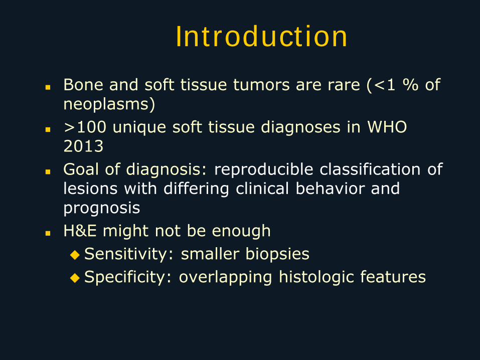
Introduction
Bone and soft tissue tumors are rare (<1 % of neoplasms)
>100 unique soft tissue diagnoses in WHO 2013
Goal of diagnosis: reproducible classification of lesions with differing clinical behavior and prognosis
H&E might not be enough
Sensitivity: smaller biopsies
Specificity: overlapping histologic features
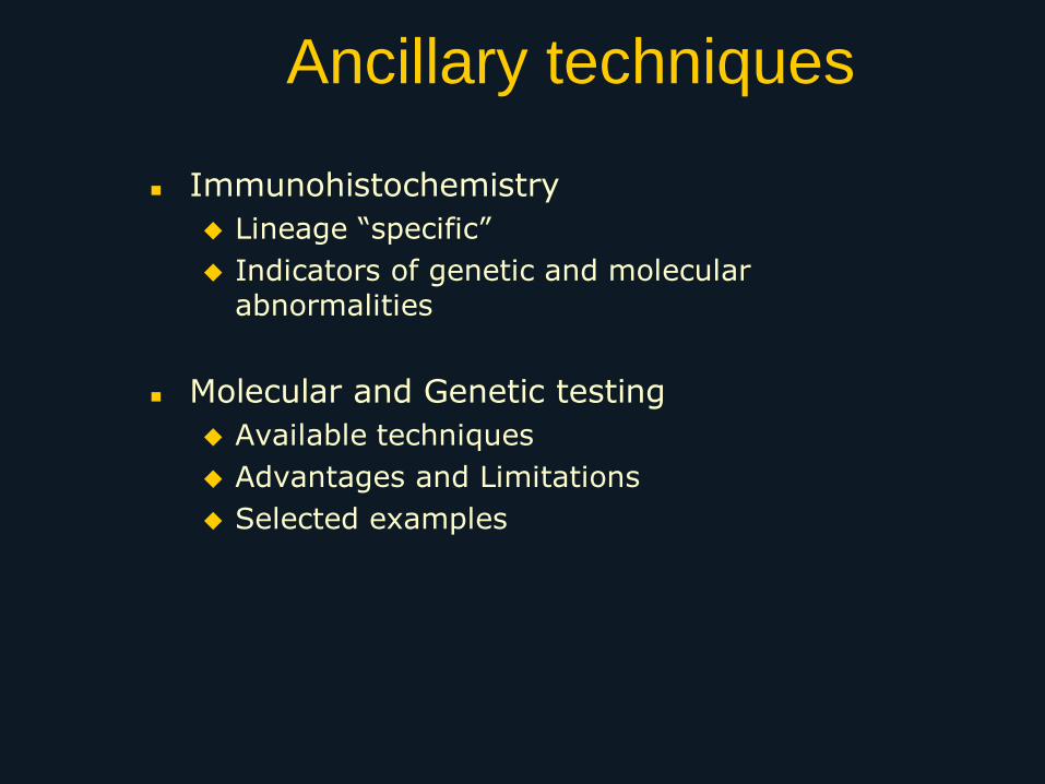
Ancillary techniques
Immunohistochemistry
Lineage “specific”
Indicators of genetic and molecular abnormalities
Molecular and Genetic testing
Available techniques
Advantages and Limitations
Selected examples
Immunohistochemistry (IHC)
1. Lineage specific proteins
2. Indicators of genetic and molecular abnormalities (amplifications, deletions, translocations, point mutations)
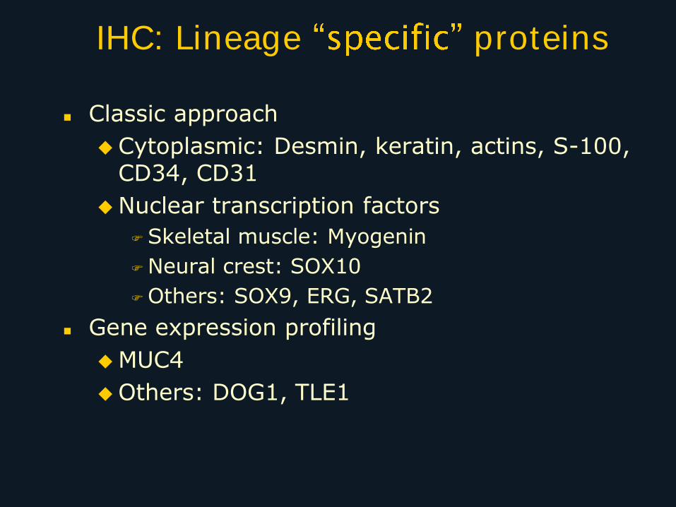
IHC: Lineage proteins
Classic approach
Cytoplasmic: Desmin, keratin, actins, S-100, CD34, CD31
Nuclear transcription factors
Skeletal muscle: Myogenin
Neural crest: SOX10
Others: SOX9, ERG, SATB2
Gene expression profiling
MUC4
Others: DOG1, TLE1
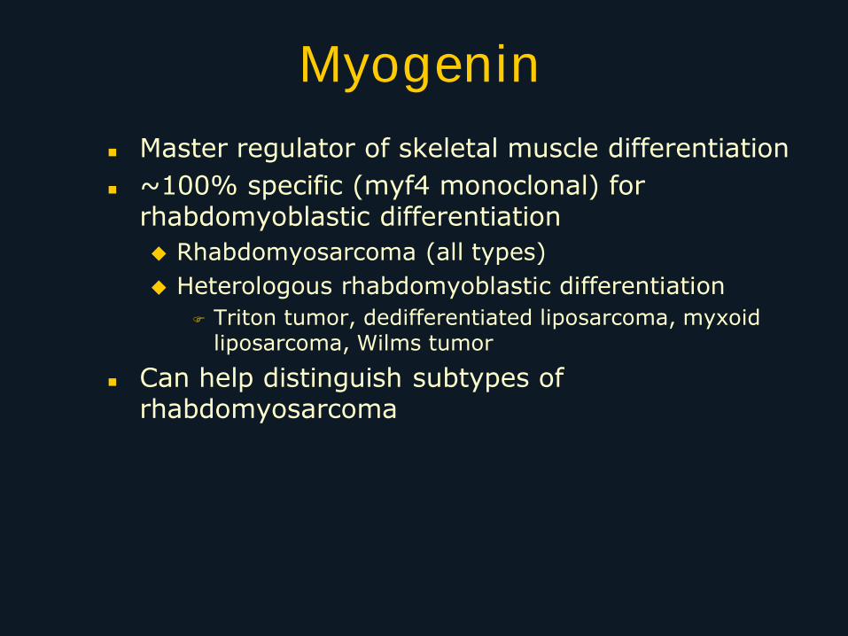
Myogenin
Master regulator of skeletal muscle differentiation
~100% specific (myf4 monoclonal) for rhabdomyoblastic differentiation
Rhabdomyosarcoma (all types)
Heterologous rhabdomyoblastic differentiation
Triton tumor, dedifferentiated liposarcoma, myxoid liposarcoma, Wilms tumor
Can help distinguish subtypes of rhabdomyosarcoma
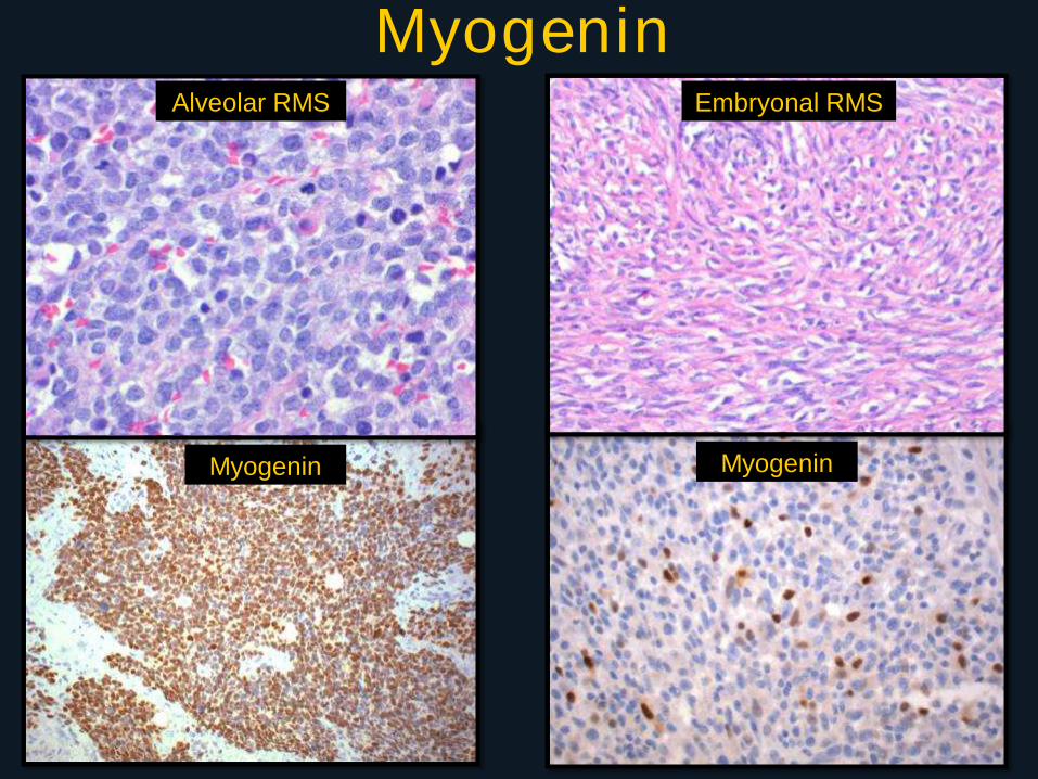
Alveolar RMS Embryonal RMS
Myogenin Myogenin
Myogenin
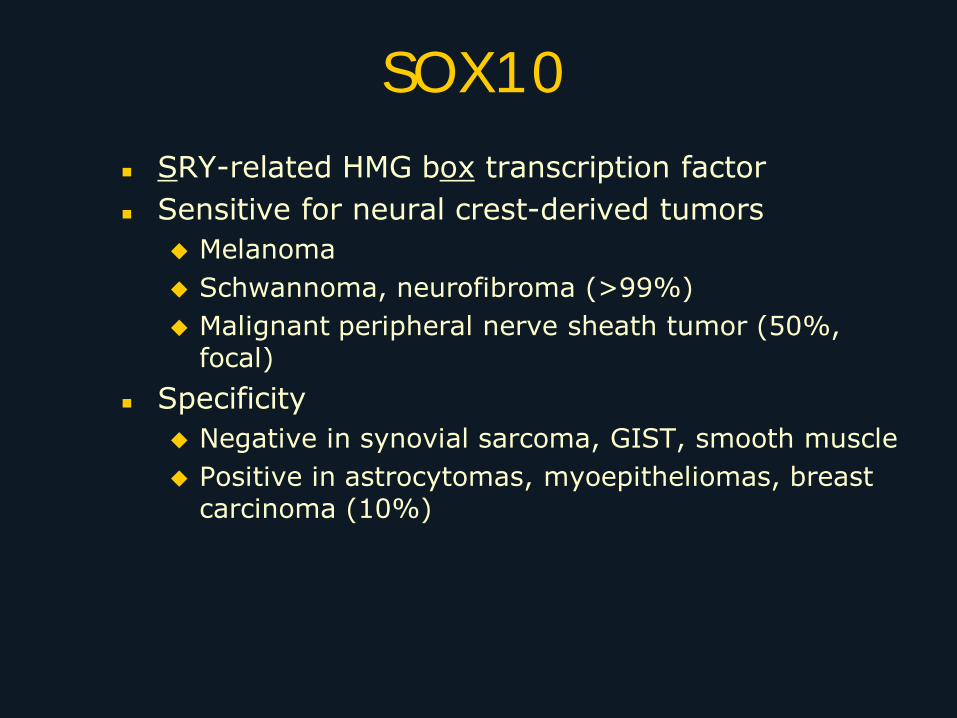
SOX10
SRY-related HMG box transcription factor
Sensitive for neural crest-derived tumors
Melanoma
Schwannoma, neurofibroma (>99%)
Malignant peripheral nerve sheath tumor (50%, focal)
Specificity
Negative in synovial sarcoma, GIST, smooth muscle
Positive in astrocytomas, myoepitheliomas, breast carcinoma (10%)
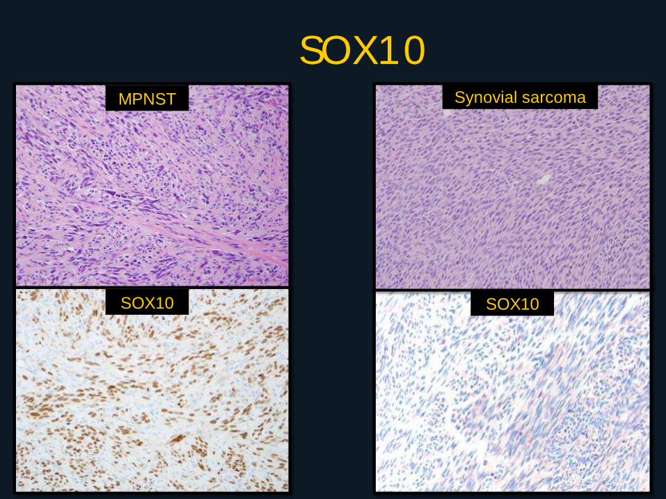
SOX10 MPNST
SOX10
Synovial sarcoma
SOX10
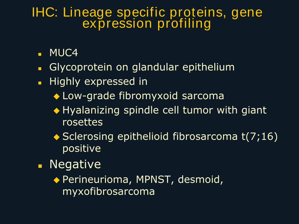
IHC: Lineage specific proteins, gene expression profiling
MUC4
Glycoprotein on glandular epithelium
Highly expressed in
Low-grade fibromyxoid sarcoma
Hyalanizing spindle cell tumor with giant rosettes
Sclerosing epithelioid fibrosarcoma t(7;16) positive
Negative
Perineurioma, MPNST, desmoid, myxofibrosarcoma
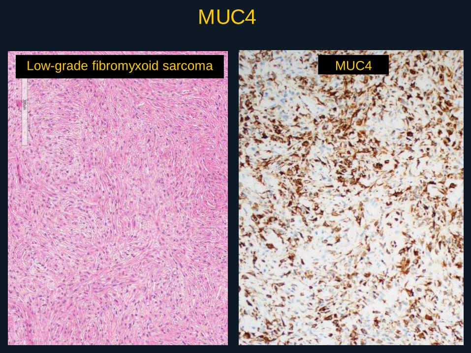
MUC4
Low-grade fibromyxoid sarcoma MUC4
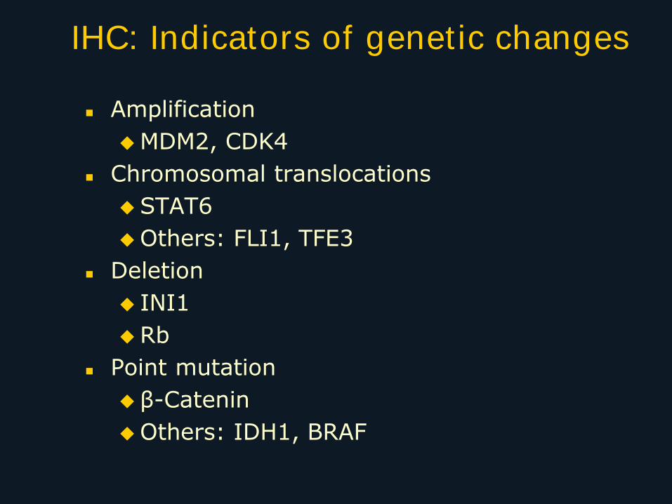
IHC: Indicators of genetic changes
Amplification
MDM2, CDK4
Chromosomal translocations
STAT6
Others: FLI1, TFE3
Deletion
INI1
Rb
Point mutation
β-Catenin
Others: IDH1, BRAF
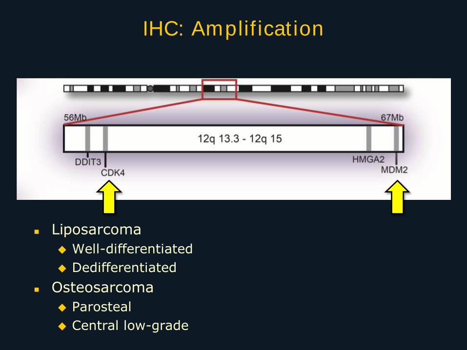
Liposarcoma
Well-differentiated
Dedifferentiated
Osteosarcoma
Parosteal
Central low-grade
IHC: Amplification
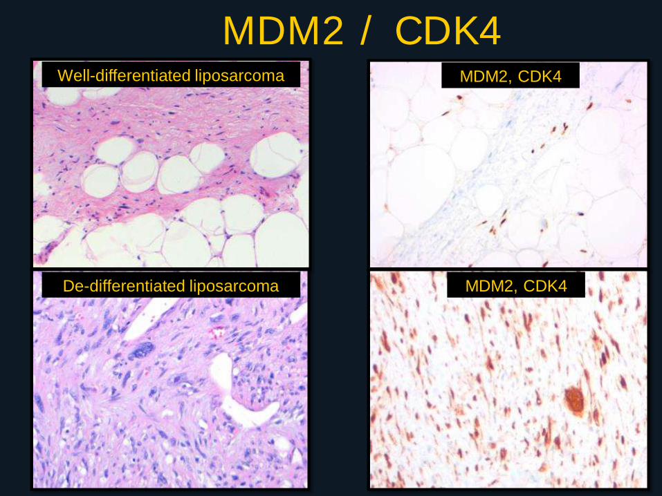
MDM2 / CDK4 Well-differentiated liposarcoma
De-differentiated liposarcoma
MDM2, CDK4
MDM2, CDK4
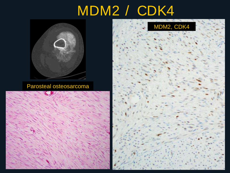
MDM2 / CDK4
Parosteal osteosarcoma
MDM2, CDK4
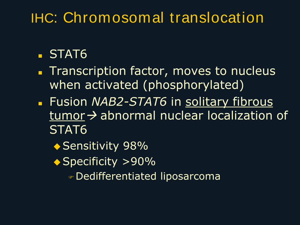
IHC: Chromosomal translocation
STAT6
Transcription factor, moves to nucleus when activated (phosphorylated)
Fusion NAB2-STAT6 in solitary fibrous tumor abnormal nuclear localization of STAT6
Sensitivity 98%
Specificity >90%
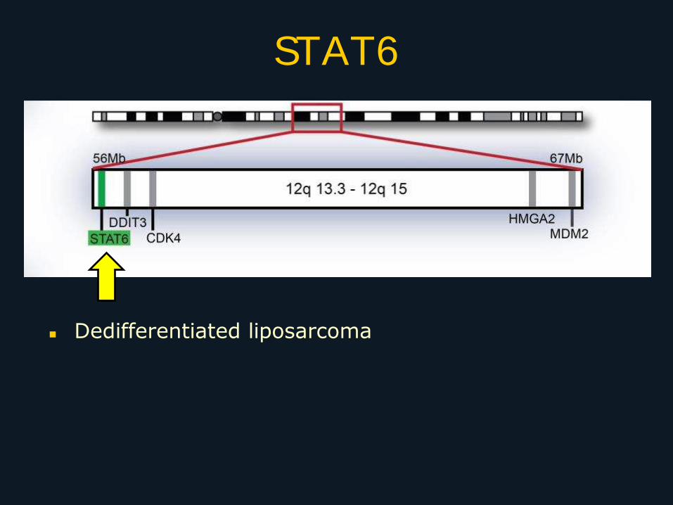
Dedifferentiated liposarcoma
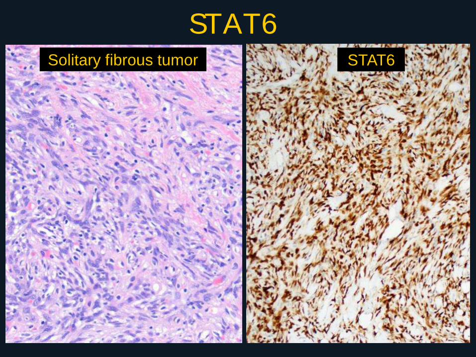
STAT6 Solitary fibrous tumor STAT6
STAT6
Dedifferentiated liposarcoma
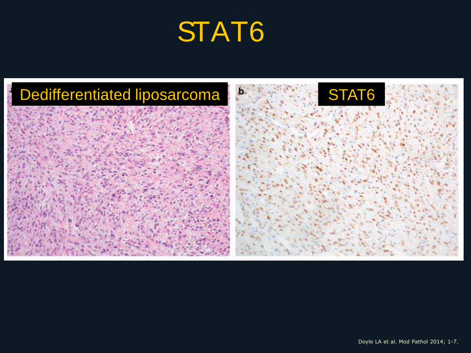
STAT6
Doyle LA et al. Mod Pathol 2014; 1-7.
Dedifferentiated liposarcoma STAT6
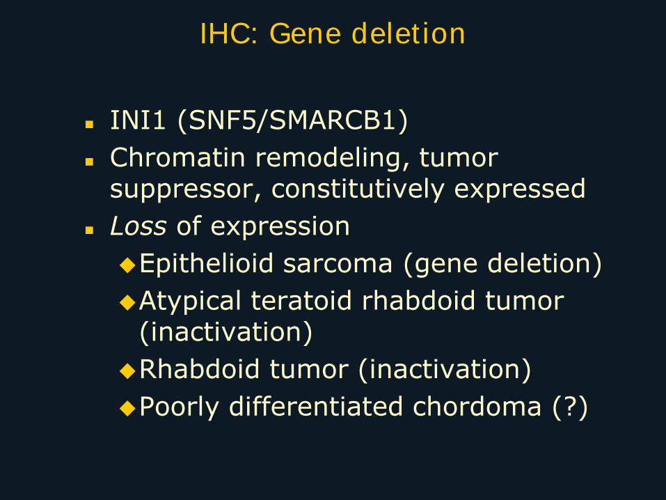
INI1 (SNF5/SMARCB1)
Chromatin remodeling, tumor suppressor, constitutively expressed
Loss of expression
Epithelioid sarcoma (gene deletion)
Atypical teratoid rhabdoid tumor (inactivation)
Rhabdoid tumor (inactivation)
Poorly differentiated chordoma (?)
IHC: Gene deletion
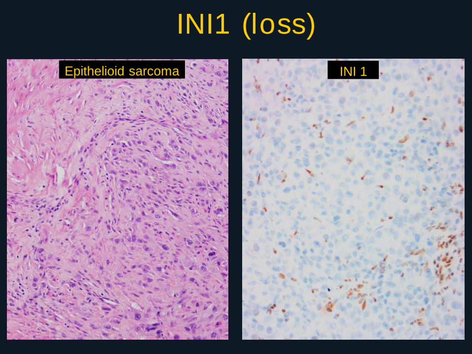
INI1 (loss)
Epithelioid sarcoma INI 1
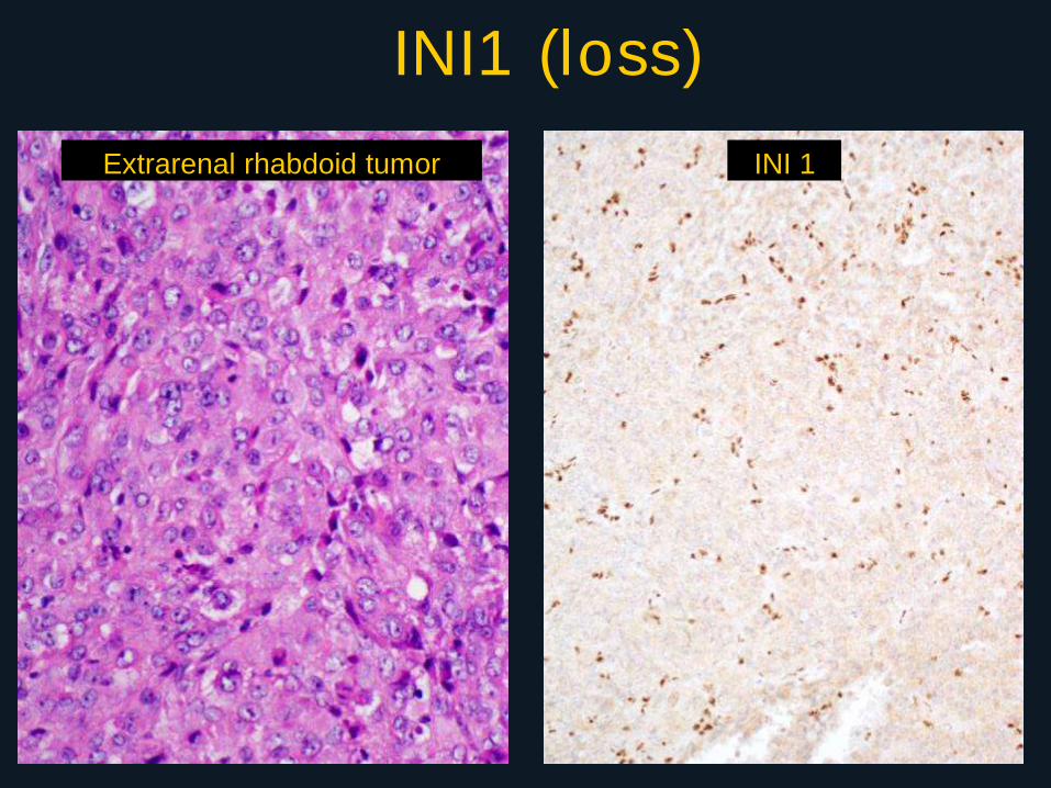
INI1 (loss)
Extrarenal rhabdoid tumor INI 1
Rb
Retinoblastoma gene 13q14
Tumor suppressor
Deleted or mutated
Spindle cell lipoma
Pleomorphic lipoma
Mammary type myofibroblastoma
Cellular angiofibroma
Retained in
Other benign and malignant lipomatous tumors
Solitary fibrous tumor
IHC: Gene deletion or mutation
24
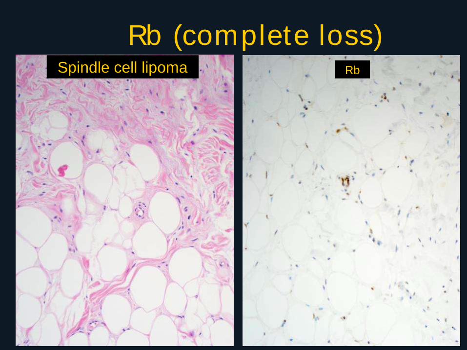
Rb (complete loss) Spindle cell lipoma Rb
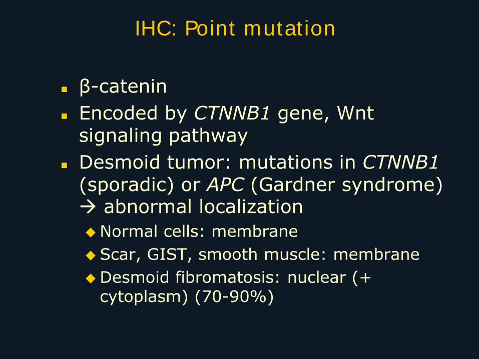
IHC: Point mutation
β-catenin
Encoded by CTNNB1 gene, Wnt signaling pathway
Desmoid tumor: mutations in CTNNB1 (sporadic) or APC (Gardner syndrome) abnormal localization
Normal cells: membrane
Scar, GIST, smooth muscle: membrane
Desmoid fibromatosis: nuclear (+ cytoplasm) (70-90%)
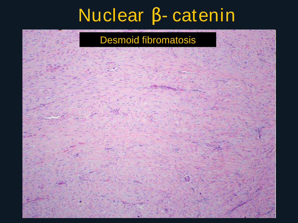
Nuclear -catenin Desmoid fibromatosis
Genetic and molecular testing
Purpose
Classification:
Separation of tumors into clinically meaningful categories based on reproducible changes
Prognostic:
Alveolar versus embryonal rhabdomyosarcoma
Myxoid versus well-differentiated liposarcoma
Predictive:
Therapeutic target from fusion gene product
Techniques
Cytogenetic: Karyotype, FISH
Molecular: RT-PCR, Sanger sequencing, MLPA, array
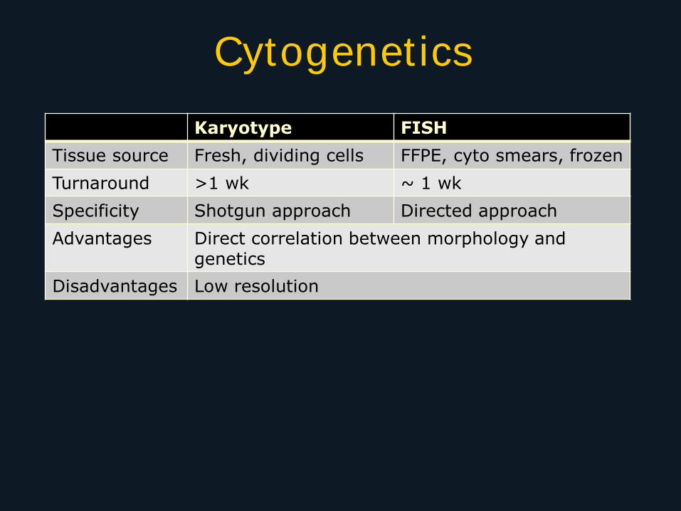
Cytogenetics
Karyotype FISH
Tissue source Fresh, dividing cells FFPE, cyto smears, frozen
Turnaround >1 wk ~ 1 wk
Specificity Shotgun approach Directed approach
Advantages Direct correlation between morphology and genetics
Disadvantages Low resolution
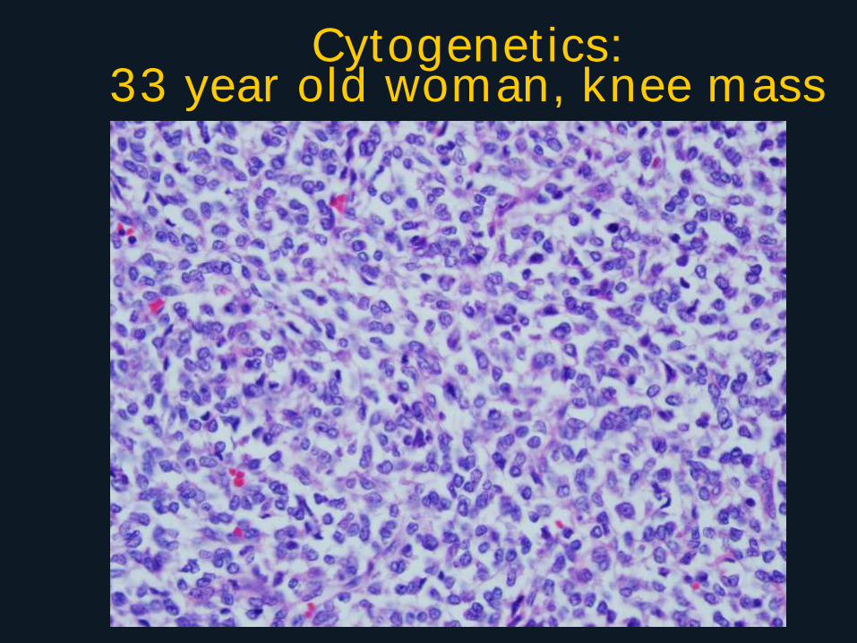
Cytogenetics: 33 year old woman, knee mass
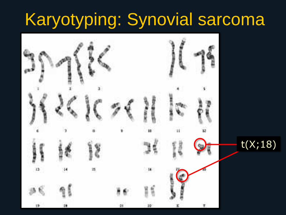
Karyotyping: Synovial sarcoma
t(X;18)
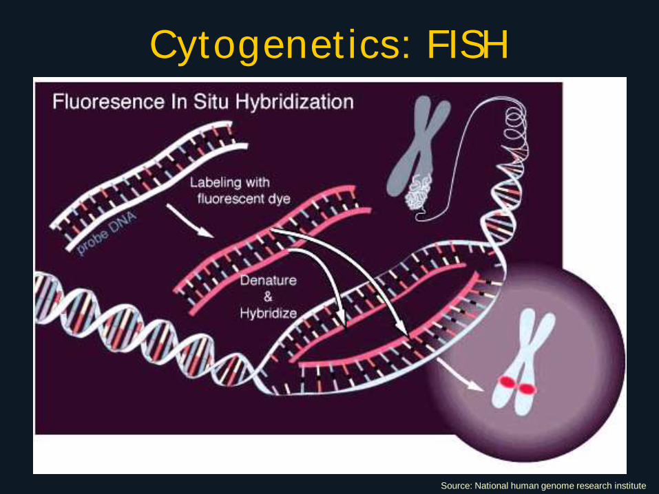
Cytogenetics: FISH
Source: National human genome research institute
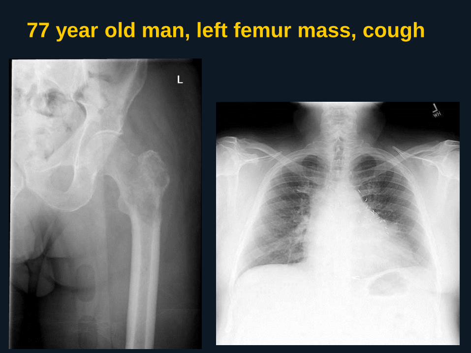
77 year old man, left femur mass, cough
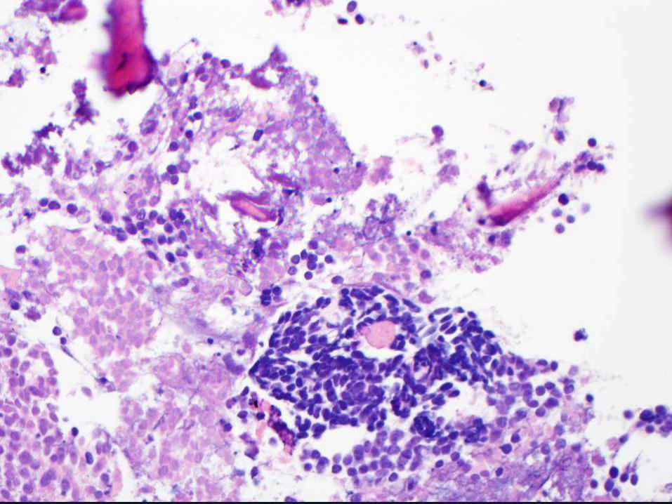
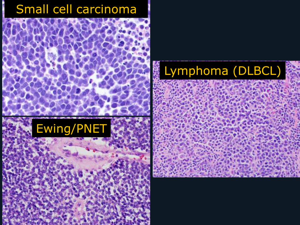
Small cell carcinoma
Ewing/PNET
Lymphoma (DLBCL)
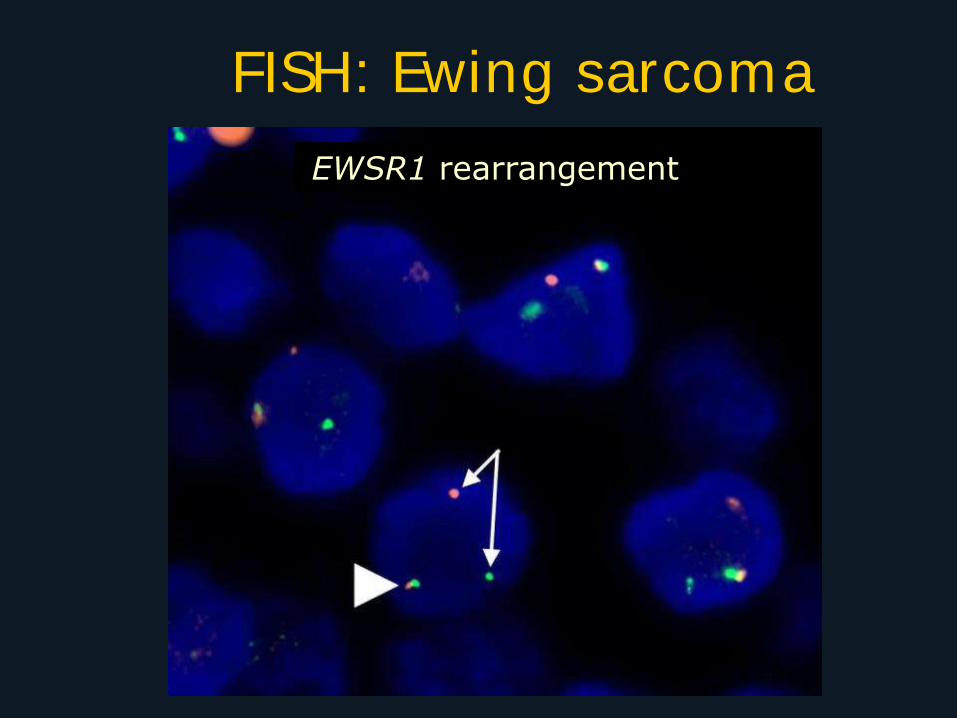
FISH: Ewing sarcoma
EWSR1 rearrangement
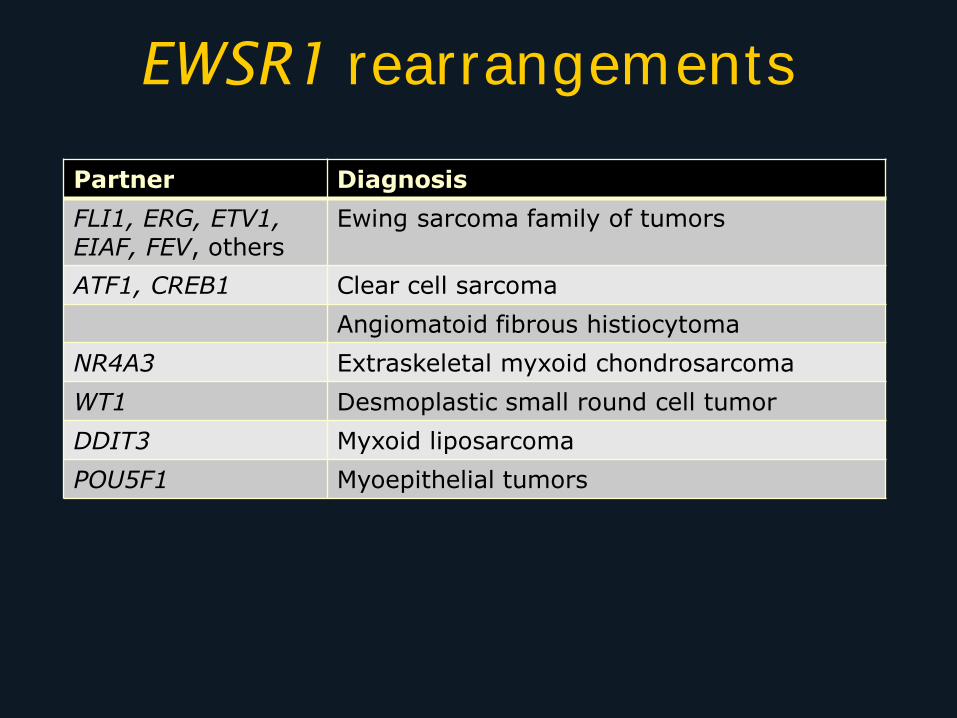
EWSR1 rearrangements
Partner Diagnosis
FLI1, ERG, ETV1, EIAF, FEV, others
Ewing sarcoma family of tumors
ATF1, CREB1 Clear cell sarcoma
Angiomatoid fibrous histiocytoma
NR4A3 Extraskeletal myxoid chondrosarcoma
WT1 Desmoplastic small round cell tumor
DDIT3 Myxoid liposarcoma
POU5F1 Myoepithelial tumors
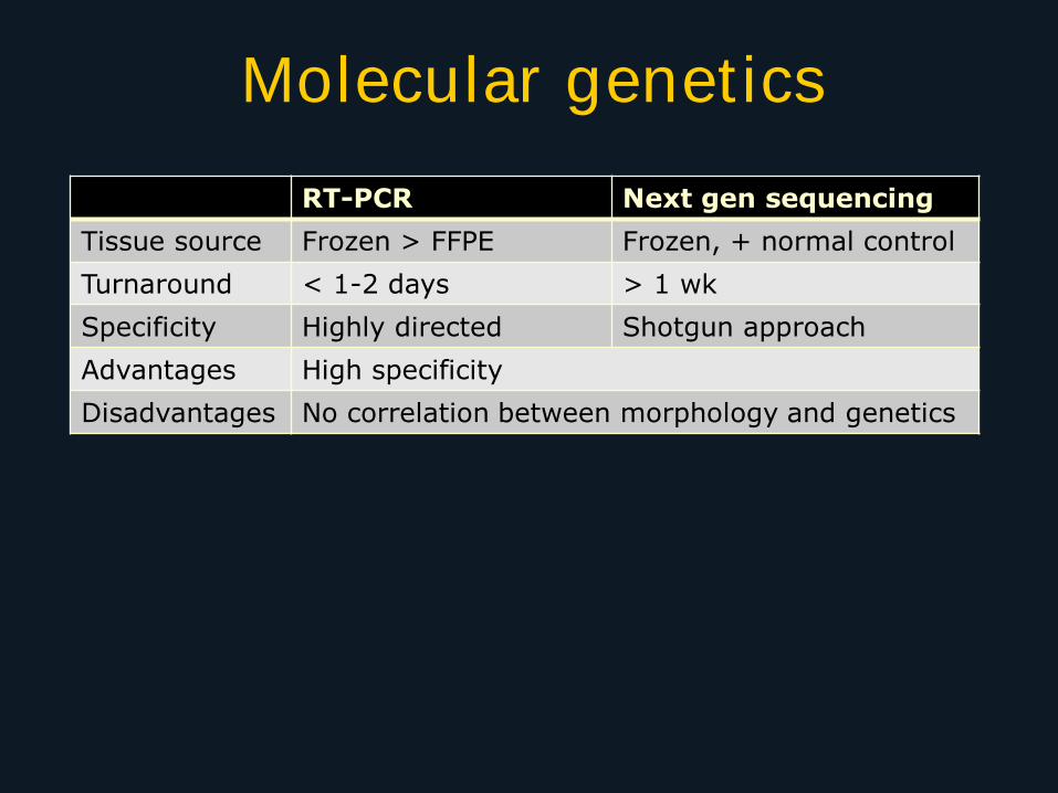
Molecular genetics
RT-PCR Next gen sequencing
Tissue source Frozen > FFPE Frozen, + normal control
Turnaround < 1-2 days > 1 wk
Specificity Highly directed Shotgun approach
Advantages High specificity
Disadvantages No correlation between morphology and genetics
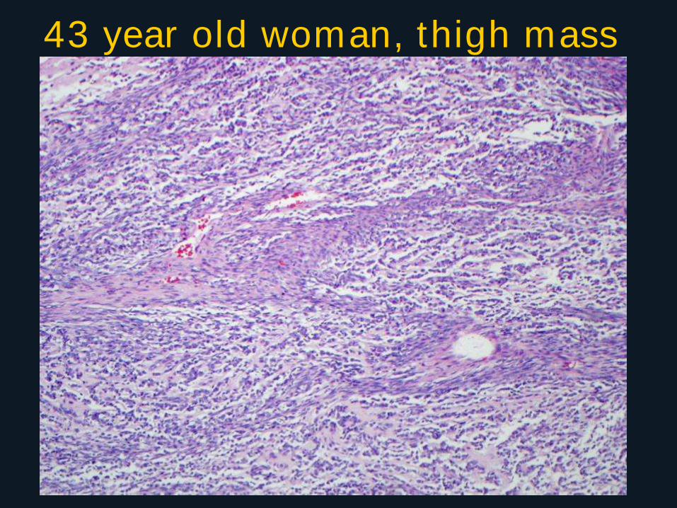
43 year old woman, thigh mass
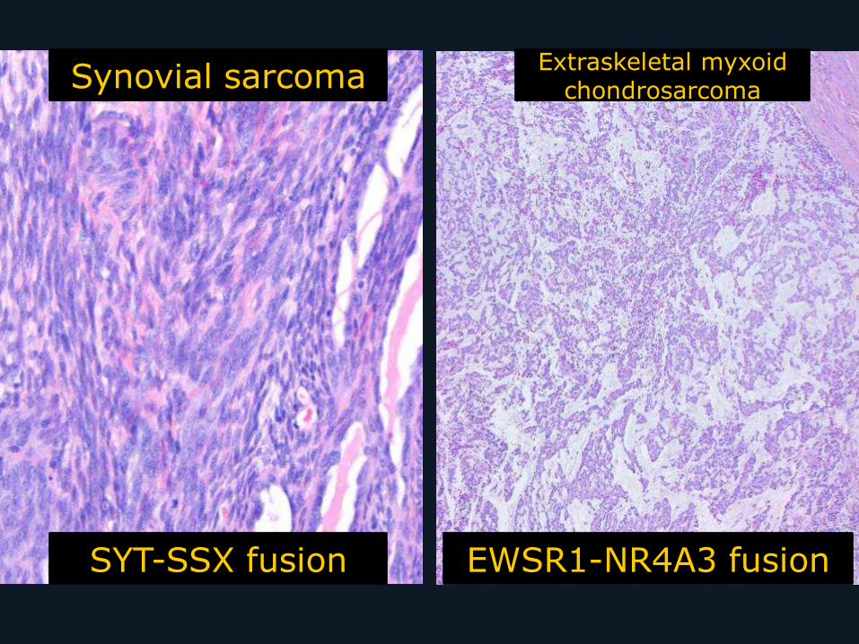
Synovial sarcoma Extraskeletal myxoid
chondrosarcoma
SYT-SSX fusion EWSR1-NR4A3 fusion
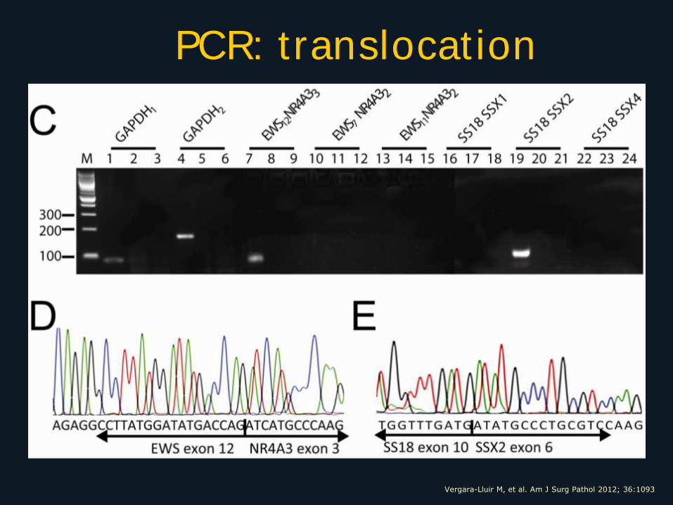
PCR: translocation
Vergara-Lluir M, et al. Am J Surg Pathol 2012; 36:1093
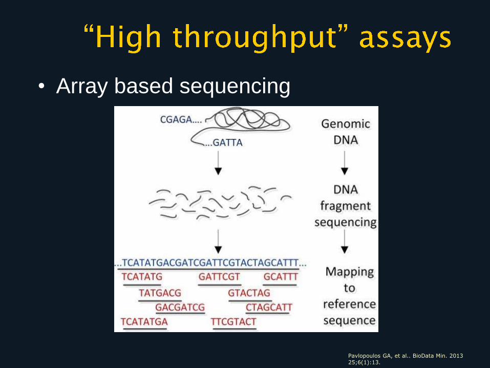
Pavlopoulos GA, et al.. BioData Min. 2013 25;6(1):13.
• Array based sequencing
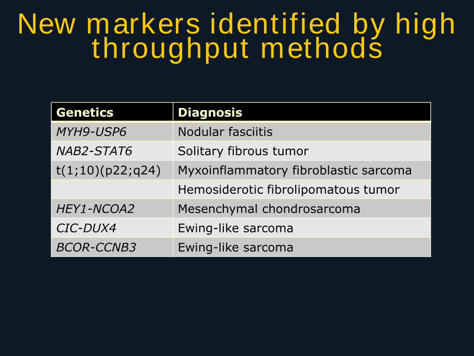
New markers identified by high throughput methods
Genetics Diagnosis
MYH9-USP6 Nodular fasciitis
NAB2-STAT6 Solitary fibrous tumor
t(1;10)(p22;q24) Myxoinflammatory fibroblastic sarcoma
Hemosiderotic fibrolipomatous tumor
HEY1-NCOA2 Mesenchymal chondrosarcoma
CIC-DUX4 Ewing-like sarcoma
BCOR-CCNB3 Ewing-like sarcoma
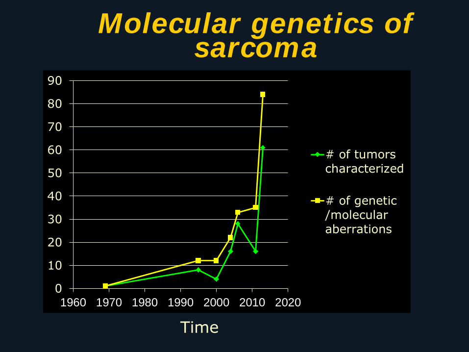
0
10
20
30
40
50
60
70
80
90
1960 1970 1980 1990 2000 2010 2020
# of tumorscharacterized
# of genetic/molecularaberrations
Molecular genetics of sarcoma
Time
Take-home messages
Lineage-specific is a relative term
IHC for nuclear transcription factors offer advantages over older cytoplasmic proteins
IHC can indirectly detect tumor-specific genetic and molecular abnormalities
Gene and molecular abnormalities can be detected directly by more specialized methods
High throughput methods can rapidly screen an entire tumor genome and may allow personalized medicine
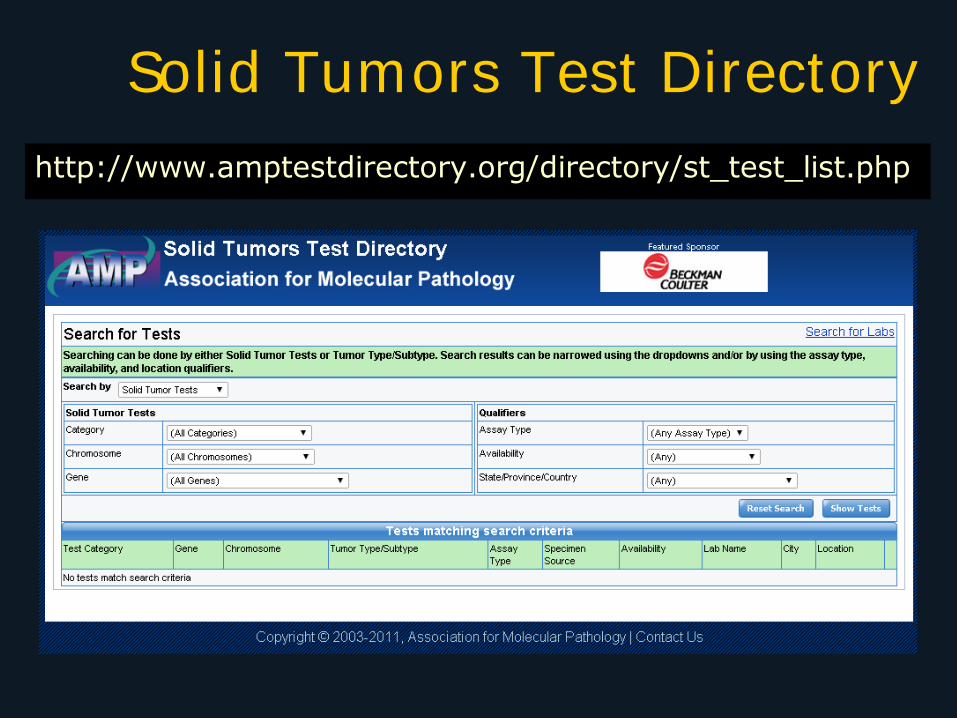
Solid Tumors Test Directory
http://www.amptestdirectory.org/directory/st_test_list.php