Embed Size (px)
Citation preview
© SJ GIRLING Page 1 15/11/2012
ANAESTHESIA OF RABBITS, RODENTS AND FERRETS
Simon J Girling BVMS (Hons) DZooMed CBiol MIBiol MRCVS
RCVS Recognised Specialist Zoo & Wildlife Medicine
Anaesthesia of the Domestic Rabbit
Clinical Examination
It is important that a full clinical examination of the rabbit is performed prior to any
attempts to anaesthetise. Common sense should prevail and harsh sounding chests on
auscultation or a history of >12 hours anorexia should immediately sound warning bells.
Gastro-intestinal stasis is a potential complication of general anaesthesia and can be
life-threatening in rabbits within 24 hours-often due to a Clostridia spp. overgrowth and
exotoxin release. Remember that rabbits are obligate nasal breathers and that if the
nares are pus filled, or there is evidence of dacrocystitis, this will result in respiratory
compromise and probably lung pathology.
If a long period of reduced food intake in an obese or over-weight rabbit has occurred-
pre-anaesthetic blood tests to check for hepatic lipidosis are advisable.
Pre-anaesthetic Care
Preanaesthetic Management
Rabbits do not need to be starved prior to anaesthetic as they have a tight pre-cardiac
sphincter and cannot vomit unless in extremis. Indeed it is deleterious to starve rabbits
as this will increase the risk of post-anaesthetic GI stasis, hepatic lipidosis and other
complications.
Preanaesthetic Medications
Glycopyrrolate – This should be used instead of atropine, as 60% of rabbits possess a
serum atropinesterase rendering atropine ineffective. Doses of 0.1mg/kg SC may be
given. In general, anticholinergics are not often used in routine anaesthetics in rabbits as
they tend to increase the viscosity of respiratory secretions and increase the likelihood of
airway blockage.
Acepromazine – This can be used safely as a premedicant in rabbits, and may provide
enough restraint to allow minor radiographic procedures. Doses of 1mg/kg SC are
© SJ GIRLING Page 2 15/11/2012
recommended. Disadvantages include a longer post-anaesthetic recovery phase and
therefore return to eating than premedicants which may be part/fully reversed.
Fentanyl/Fluanisone – This is the Hypnorm® combination and is often used at 0.5ml/kg
bodyweight to induce sedation. It may be used at 0.1ml/kg to provide a light plane of pre-
anaesthetic sedation which facilitates gaseous induction. An advantage of this drug
(apart from the fact that it is licensed for use in rabbits) is that it is partly reversible (the
fentanyl part) with partial opioid agonists such as buprenorphine or butorphanol.
Medetomidine – This may be used on its own or combined with ketamine to produce
mild sedation right through (in combination with ketamine) to full surgical anaesthesia.
Doses of 0.1mg/kg medetomidine SC or IV may be used to provide very light sedation,
through to 0.25mg/kg medetomidine with 15mg/kg ketamine to provide anaesthesia.
General Anaesthesia
Injectable Agents
Fentanyl/Fluanisone - This may be used as sedation only on its own at a dose of
0.5 mls/kg intramuscularly (see data sheets). This produces sedation and
immobilisation for 30-60 minutes, but its analgesic effect due to the opioid derivative
fentanyl will persist for some time after. To provide anesthetic depth, fentanyl/fluanisone
may be combined with diazepam (0.3 ml Hypnorm® to 2 mg/kg diazepam)
intraperitoneally, or intravenously (but in separate syringes as they do not mix), or with
midazolam (0.3 ml Hypnorm® to 2 mg/kg midazolam) intramuscularly or intraperitoneally
in the same syringe. Alternatively the Hypnorm® may be given IM first and then 15
minutes later the midazolam is given IV into the lateral ear vein. These two
combinations provide good analgesia and muscle relaxation with duration of anaesthesia
of 20-40 minutes. The fentanyl part may be reversed with buprenorphine or butorphanol
given intravenously, or in emergencies the drug naloxone at 0.1 mg/kg intramuscularly
or intravenously may be given, but this provides no substitute analgesia.
Fentanyl/fluanisone combinations are well tolerated in most rabbits, but they can
produce respiratory depression and hypoxia, which can lead to cardiac arrhythmias and
even arrest.
© SJ GIRLING Page 3 15/11/2012
Medetomidine+Ketamine – As mentioned above, medetomidine at 0.25mg/kg combined
with 15mg/kg ketamine can provide surgical anaesthesia. Individual rabbits will vary in
their response to this combination and the reader should be aware that these drugs are
not licensed for use in rabbits. The alpha 2 agonists such as medetomidine produce the
same side-effects in rabbits as are observed in cats and dogs with hypotension,
hyperglycaemia, cardiac arrhythmias etc are seen. The advantage is that medetomidine
may be reversed with atipamazole at 1mg/kg.
Medetomidine+Ketamine+Butorphanol – This combination allows the further reduction of
the dosages of medetomidine and ketamine and so minimise their unwanted side-
effects. This author has used dosages of 0.1mg/kg medetomidine with 5mg/kg ketamine
and 0.5mg/kg butorphanol given SC/IM or by slow IV. This can provide light surgical
anaesthesia for 15-20 minutes and may be deepened or prolonged by intubation and
volatile gaseous anaesthesia such as isoflurane or sevoflurane. Again, the dose of
medetomidine may be reversed with atipamazole at 0.5mg/kg (i.e. the same volume as
that of Domitor® given).
Inhalational Anaesthetics
Isoflurane – This is the main gaseous anaesthetic of choice at present, although
sevoflurane has some advantages (see below). It has the advantage that it is licensed in
rabbits, and respiratory arrest occurs prior to cardiac arrest (unlike the situation with
halothane). It is easier to induce and maintain rabbits that have been premedicated with
a low dose of fentanyl/fluanisone or an alpha 2 agonist/triple combination. The rabbit
may then be induced by face mask as follows:-
In sternal recumbancy a clear face mask is applied to the nose and mouth and 100%
oxygen only is supplied for 2 minutes. If regular respiration occurs, the Isotec is turned
up to 0.5% isoflurane in 100% oxygen. If regular respiration occurs this is maintained for
2 minutes and then turned up to 1%. The process is continued in 0.5% increments until a
surgical plane of anaesthesia is achieved (usually 1.5-2% isoflurane). At this point
intubation may be performed or the rabbit maintained on a face mask.
Sevoflurane – This has the advantage that it is less irritant to the mucus membranes and
therefore better tolerated by rabbits than isoflurane. It can still be detected though, and
© SJ GIRLING Page 4 15/11/2012
straight masking down with 8% sevoflurane in 100% oxygen as can be performed in
dogs, results in breath holding in rabbits. Levels around 3-4% though do not and it may
be possible to induce anaesthesia by face mask starting at 3-4% sevoflurane in 100%
oxygen. This may then be dropped to 2-2.5% for most minor surgical procedures.
Intubation
This may be performed using uncuffed (preferably) ET tubes. A 3kg rabbit will require a
3-3.5mm diameter tube.
Intubation may be attempted by one of two techniques, blind or direct.
Blind Technique – The rabbit is placed in sternal recumbancy. The head of the rabbit is
grasped one hand the rabbit lifted vertically until its forefeet are just touching the table.
The head is kept level and the ET tube is advanced, midline, over the tongue until
breathing sounds are detected. The anaesthetist waits until inspiration occurs and
quickly advances the tube. Air movement or condensation on the ET tube if transparent
will confirm correct placement.
Direct Technique – This involves using a 0 Wisconsin blade laryngoscope or an
otoscope and placing the rabbit in dorsal recumbancy. The tongue is pulled laterally and
the soft palate deflected with the scope to visualise the larynx. A guide wire should be
used to enter the larynx, allowing withdrawal of the scope and slotting the ET tube over it
and so advancing it into the trachea. The guide wire is then removed. Alternatively a fine
endoscope may be inserted through the ET tube as a form of guide wire and direct
intubation performed.
In all cases, a small amount of lignocaine spray applied to the tube will aid intubation.
Anaesthetic Circuits
Due to their small size, Ayres T pieces and for smaller rabbits (<1kg) Mini Bain or
Mapleson C circuits should be used as these minimise dead space and reduce
rebreathing.
© SJ GIRLING Page 5 15/11/2012
Anaesthetic Monitoring
Respiration should become regular and even and is often around 30-60bpm. A loss of
the pinch reflex in the hind limbs also occurs. Loss of the pinch reflex in the forelimbs in
rabbits indicates that the plane of anaesthesia is becoming too deep. Eye placement is
not a good indicator of depth of anaesthesia in the rabbit. Protrusion of the eye during
deeper planes of anaesthesia is a poor sign and usually precedes cardiac arrest.
Pulse oximetry may be used in rabbits-but many oximeters will not read heart rates
above 250 bpm so care should be taken in selection of these. The ear artery may be
used, as may the ventral tail artery or a toe artery in larger rabbits.
Side-stream capnography may also be used in rabbits to assess rebreathing and
alveolar perfusion in much the same way as for cats and dogs.
A rectal thermometer is useful to monitor for hypothermia-a common sequel in smaller
rabbits (normal temperature is around 38.5-40°C). If the body temperature drops below
35°C critical hypothermia has occurred. For this reason the use of radiant heat mats
(rather than pressure ones as many smaller rabbits fail to trigger these) or hot air
blankets (Bair Huggers) should be used-although care not to create hyperthermia should
also be taken!!
© SJ GIRLING Page 6 15/11/2012
Rabbit Emergency Care
Emergency Airway Access and Ventilation (A and B)
Should breathing stop, or hypoxia be detected, then emergency ventilation will be
required. This is best achieved by immediate endotracheal intubation.
Intubation may be achieved blindly, with the rabbit in sternal recumbancy. The rabbit’s
head is lifted and the ET tube advanced slowly until breathing sounds are heard through
the tube. The tube may then be quickly advanced on inspiration. If a transparent tube is
used then condensation from the rabbit’s breath when the tube is over the glottis can be
seen aiding intubation. If the rabbit has stopped breathing then this technique becomes
extremely difficult, therefore a laryngoscope with a Wisconsin 0 paediatric blade can be
used to visualise the glottis (Heard 2004). This is best achieved with the rabbit in dorsal
recumbancy and the tongue pulled laterally. A guide wire may be inserted through the
glottis first and the ET tube threaded over the top. Alternatively a fine endoscope or
needlescope may be used as a guide wire instead, threading the ET over the scope prior
to intubation. Once through the glottis, the ET tube maybe advanced and the scope
retracted easily.
Direct intubation itself may however be difficult in the rabbit owing to the narrow oral
cavity and relatively large size of the tongue caudally, or the presence of an obstruction
such as a pharyngeal abscess or foreign body. In an emergency therefore it may be
necessary to pass a long through-the-needle catheter into the tracheal lumen between
two tracheal rings ventrally. A luer adaptor may be attached to allow connection to an
anaesthetic circuit for oxygen administration. A tracheostomy may also be performed in
the same way as for a cat or dog; the main difference is that some breeds of rabbit,
particularly in the doe, have large dew flaps with plentiful subcutaneous fat depots which
may make tracheostomy surgery challenging. Otherwise a longitudinal incision is made
over the trachea caudal to the larynx, followed by blunt dissection onto the trachea itself.
A 180 degree ventral incision in between the tracheal rings 3-4 rings below the larynx is
made and an ET tube inserted. This may then be attached to a breathing circuit for
ventilation.
© SJ GIRLING Page 7 15/11/2012
If intubation is not possible then either a tight fitting face mask connected to an
anaesthetic circuit may be applied, and a high flow rate of oxygen (4-5 litres) or an Ambu
bag, to force ventilate the rabbit. Alternatively, moving the rabbit in a see-saw manner
may aid ventilation by moving the abdominal viscera backwards and forwards onto the
diaphragm and acting as a pump mechanism (Briscoe and Syring 2004). This works on
the basis that most of the impetus for inspiration comes from the flattening of the
diaphragm rather than the outward movement of the ribcage.
Cardiovascular Support (C)
In mammals <10kg, direct cardiac massage by compressing the chest directly over the
heart is most effective at increasing thoracic pressure and forcing blood through the
arterial vasculature (Henrik 1992). Heart compression rates of 100 beats per minute
need to be achieved in rabbits; the technique recommended to maximise cardiovascular
output is circumferential chest compression, as is used in human infants, where the
chest is compressed over the heart from both sides at once (Costello 2004).
If a cardiac beat is present or a beat is restarted, ECG leads should be applied to
discern any dysrhythmias. The type of dysrhythmia reported in rabbits during
resuscitation techniques has thus far been different from that reported in cats and dogs.
The latter have been associated with electro-mechanical dissociation, whereas in
rabbits, profound bradycardia, ventricular asystole and ventricular fibrillation have been
reported (Rush and Wingfield 1992). Adrenaline may be used, intratracheally if
intubated, or intravenously if no cardiac beat is detected and no ECG trace. In the case
of fine ventricular fibrillation, the use of adrenaline has been advocated to convert the
electrical activity to coarse ventricular fibrillation which is easier to convert (DeFrancesco
2000) – see table 1 for dosages.
Conversion of coarse fibrillation is based on the use of cardiac massage as described
above, or if the clinic has access to defibrillation devices then the use of these externally
at 2-10 J/kg (starting at low energies and increasing if no response is achieved) may be
performed (Costello 2004). Greater success is achieved with defibrillation devices if
three initial countershocks are applied at low energies.
© SJ GIRLING Page 8 15/11/2012
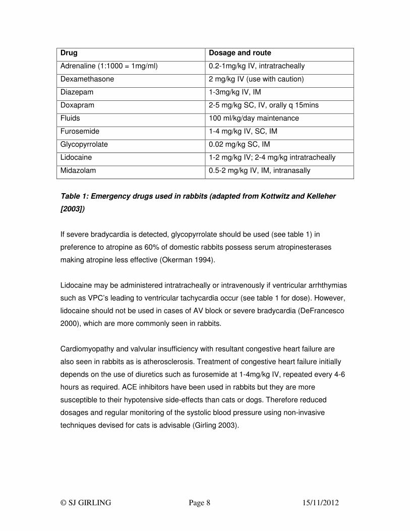
Drug Dosage and route
Adrenaline (1:1000 = 1mg/ml) 0.2-1mg/kg IV, intratracheally
Dexamethasone 2 mg/kg IV (use with caution)
Diazepam 1-3mg/kg IV, IM
Doxapram 2-5 mg/kg SC, IV, orally q 15mins
Fluids 100 ml/kg/day maintenance
Furosemide 1-4 mg/kg IV, SC, IM
Glycopyrrolate 0.02 mg/kg SC, IM
Lidocaine 1-2 mg/kg IV; 2-4 mg/kg intratracheally
Midazolam 0.5-2 mg/kg IV, IM, intranasally
Table 1: Emergency drugs used in rabbits (adapted from Kottwitz and Kelleher
[2003])
If severe bradycardia is detected, glycopyrrolate should be used (see table 1) in
preference to atropine as 60% of domestic rabbits possess serum atropinesterases
making atropine less effective (Okerman 1994).
Lidocaine may be administered intratracheally or intravenously if ventricular arrhthymias
such as VPC’s leading to ventricular tachycardia occur (see table 1 for dose). However,
lidocaine should not be used in cases of AV block or severe bradycardia (DeFrancesco
2000), which are more commonly seen in rabbits.
Cardiomyopathy and valvular insufficiency with resultant congestive heart failure are
also seen in rabbits as is atherosclerosis. Treatment of congestive heart failure initially
depends on the use of diuretics such as furosemide at 1-4mg/kg IV, repeated every 4-6
hours as required. ACE inhibitors have been used in rabbits but they are more
susceptible to their hypotensive side-effects than cats or dogs. Therefore reduced
dosages and regular monitoring of the systolic blood pressure using non-invasive
techniques devised for cats is advisable (Girling 2003).
© SJ GIRLING Page 9 15/11/2012
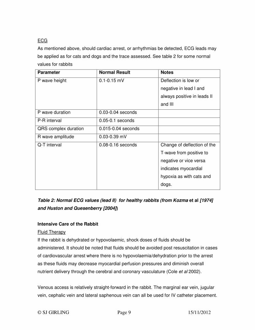
ECG
As mentioned above, should cardiac arrest, or arrhythmias be detected, ECG leads may
be applied as for cats and dogs and the trace assessed. See table 2 for some normal
values for rabbits
Parameter Normal Result Notes
P wave height 0.1-0.15 mV Deflection is low or
negative in lead I and
always positive in leads II
and III
P wave duration 0.03-0.04 seconds
P-R interval 0.05-0.1 seconds
QRS complex duration 0.015-0.04 seconds
R wave amplitude 0.03-0.39 mV
Q-T interval 0.08-0.16 seconds Change of deflection of the
T-wave from positive to
negative or vice versa
indicates myocardial
hypoxia as with cats and
dogs.
Table 2: Normal ECG values (lead II) for healthy rabbits (from Kozma et al [1974]
and Huston and Quesenberry [2004])
Intensive Care of the Rabbit
Fluid Therapy
If the rabbit is dehydrated or hypovolaemic, shock doses of fluids should be
administered. It should be noted that fluids should be avoided post resuscitation in cases
of cardiovascular arrest where there is no hypovolaemia/dehydration prior to the arrest
as these fluids may decrease myocardial perfusion pressures and diminish overall
nutrient delivery through the cerebral and coronary vasculature (Cole et al 2002).
Venous access is relatively straight-forward in the rabbit. The marginal ear vein, jugular
vein, cephalic vein and lateral saphenous vein can all be used for IV catheter placement.
© SJ GIRLING Page 10 15/11/2012
Long-term (>2-3days) use of the marginal ear vein may however cause sloughing of the
ear tip, although in the author’s experience this is relatively rare with careful
venipuncture technique. Use of a topical local anaesthetic cream is recommended prior
to placement. In addition, if a sedative such as Hypnorm® (VetaPharma UK Ltd.) is used
(a fentanyl/fluanisone combination neuroleptanalgesic drug) peripheral vasodilation is
common facilitating ear vein catheter placement. The fentanyl portion of this drug may
be reversed using the partial opioid agonists buprenorphine (0.01-0.05 mg/kg) or
butorphanol (0.1-0.5 mg/kg). The jugular vein may be difficult to access, particularly in
does where there is a pronounced ruff of skin and fat deposits. In addition it forms the
main venous drainage for the eye, and thrombus formation may lead to periocular
swelling. An Elizabethan collar can be used to prevent the rabbit from chewing or
removing the catheter although this will prevent caecotrophy and care should be taken to
ensure the rabbit is still managing to eat, otherwise assisted feeding (see below) should
be instituted.
Intraosseous catheters may be placed into the proximal femur, in the trochanteric fossa,
in a parallel direction to the long axis of the femur. Use a 18-23 gauge, 1-1.5 inch spinal
or hypodermic needle. Analgesia should be employed (see table 3) whenever placing an
intraosseous catheter as should prophylactic antibiosis such as enrofloxacin (Baytril
2.5% Bayer – licensed for use in rabbits).
Intraosseous and intravenous fluid administration should be accurately titrated using
syringe drivers rather than relying on drip sets, as even a small error in fluid
administration may be proportionally more significant considering the small size of many
rabbits.
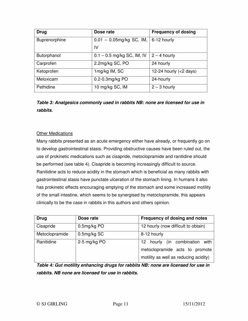
Analgesia
Analgesia is essential when performing any invasive procedure. In addition, many acute
emergency are associated with considerable pain e.g. fractures, intestinal obstructions,
pyelonephritis, renal calculi etc. The same care should be taken when using NSAIDs in
rabbits as is taken with their usage in cats and dogs, i.e. they should be avoided where
renal disease or gastric/intestinal ulceration/perforation is suspected.
© SJ GIRLING Page 11 15/11/2012
Drug Dose rate Frequency of dosing
Buprenorphine 0.01 – 0.05mg/kg SC, IM,
IV
6-12 hourly
Butorphanol 0.1 – 0.5 mg/kg SC, IM, IV 2 – 4 hourly
Carprofen 2.2mg/kg SC, PO 24 hourly
Ketoprofen 1mg/kg IM, SC 12-24 hourly (<2 days)
Meloxicam 0.2-0.3mg/kg PO 24-hourly
Pethidine 10 mg/kg SC, IM 2 – 3 hourly
Table 3: Analgesics commonly used in rabbits NB: none are licensed for use in
rabbits.
Other Medications
Many rabbits presented as an acute emergency either have already, or frequently go on
to develop gastrointestinal stasis. Providing obstructive causes have been ruled out, the
use of prokinetic medications such as cisapride, metoclopramide and ranitidine should
be performed (see table 4). Cisapride is becoming increasingly difficult to source.
Ranitidine acts to reduce acidity in the stomach which is beneficial as many rabbits with
gastrointestinal stasis have punctate ulceration of the stomach lining. In humans it also
has prokinetic effects encouraging emptying of the stomach and some increased motility
of the small intestine, which seems to be synergised by metoclopramide, this appears
clinically to be the case in rabbits in this authors and others opinion.
Drug Dose rate Frequency of dosing and notes
Cisapride 0.5mg/kg PO 12 hourly (now difficult to obtain)
Metoclopramide 0.5mg/kg SC 8-12 hourly
Ranitidine 2-5 mg/kg PO 12 hourly (in combination with
metoclopramide acts to promote
motility as well as reducing acidity)
Table 4: Gut motility enhancing drugs for rabbits NB: none are licensed for use in
rabbits. NB none are licensed for use in rabbits.
© SJ GIRLING Page 12 15/11/2012
Assisted feeding should be carried out in conjunction with the use of prokinetics. This
can start off with easily absorbed essential sugars and amino acids (e.g. Critical Care
Formula® Vetark Professional) either syringed into the mouth or delivered via a naso-
oesophageal tube. As the rabbit improves clinically this should be stepped up to use
proprietary critical feeding formulas such as Science Recovery® (Supreme Petfoods) or
Critical Care for Herbivores® (Oxbow Pet Products) or vegetable based baby foods
(lactose-free varieties). The disadvantage of the baby foods is that they do not contain
fibre and so have little or no prokinetic activity, although they do provide nutrients in an
easily digestible form.
The levels of energy required for a debilitated rabbit should approach that calculated for
growing to lactating rabbits using the formula MER = k x (wt[kg])0.75 where k=200 for
growth and 300 for lactation (Carpenter and Kolmstetter 2000). Therefore for debilitation,
the following daily energy requirement may be used.
MER = 250 x (wt[kg])0.75
To re-populate he intestinal flora, transfaunation of caecotrophs from a healthy rabbit
may aid the return of normal bowel function. The use of commercial probiotics designed
for rabbits has also been advocated, and reduces the risk of transferring potential
parasites and other agents to the debilitated patient.
© SJ GIRLING Page 13 15/11/2012
Anaesthesia of Rodents and Ferrets
Preanaesthetic Care
Preanaesthetic Management
Rodents – These do not need to be starved prior to anaesthesia as none can easily
vomit and starvation can enhance post op ileus. Instead a brief (1 hour) period of food
withdrawal can be instituted to prevent food being present in the mouth at the time of
induction.
Ferrets – Starvation should be performed as ferrets can easily vomit, but it should only
be for a short period (generally 2-4 hours) as insulinomas are commonplace in ferrets
>1.5 years and the metabolic rate of the ferret is high.
Preanaesthetic Premedications
Atropine
This is used in some species such as guinea pigs and chinchillas where oral secretions
are high, and intubation difficult. Doses of 0.05 mg/kg (Mason 1997) have been used
subcutaneously 30 minutes before induction. Atropine also acts to prevent excessive
bradycardia which often occurs during the induction phase.
Tranquillisers are frequently used to reduce the stress of induction, which plays a large
part in the risks of anaesthetising Lagomorphs and Hystricomorphs. These species will
breath-hold during gaseous induction, to the point where they become cyanotic.
Acepromazine (ACP)
This can be used at doses of 0.2 mg/kg in ferrets, and 0.5-1 mg/kg in rats, mice,
hamsters, chinchillas and guinea pigs (Mason 1997). In general it is a very safe
premedicant even in debilitated animals. However, it is advised not to use this in gerbils,
as acepromazine reduces the seizure threshold, and many gerbils suffer from hereditary
epilepsy. It may be combined with buprenorphine at 0.02mg/kg to provide a smoother
induction and analgesia.
Diazepam
This is useful in rodents at doses of 3 mg/kg (Mason 1997) particularly in gerbils.
© SJ GIRLING Page 14 15/11/2012
Fentanyl/fluanisone
This combination (known as Hypnorm®) may be used at varying doses as either a
premedicant, a sedative, or as part of an injectable full anaesthesia. As a premedicant
doses of 0.08 ml/kg for rats, 0.2 ml/kg for guinea pigs (one fifth the recommended
sedation doses) can produce sufficient sedation to prevent breath-holding and allow
gaseous induction. These doses are given intramuscularly 15-20 minutes before
induction. Hypnorm® is an irritant and large doses in one spot may cause post-
operative lameness. It can be reversed after the operation with butorphanol at
0.2 mg/kg intravenously, or buprenorphine at 0.05 mg/kg (Mason 1997).
Fluid therapy is also a vitally important pre-anaesthetic consideration and will be
mentioned below.
Induction and Maintenance Agents
Ketamine and its Combinations
Ferrets
Ketamine may be used alone for chemical restraint in the ferret at doses of 10-20 mg/kg
but as with cats and dogs, the muscle relaxation is poor, and salivation occurs. More
often ketamine is combined with other agents such as the alpha-2 drugs, xylazine and
medetomidine. In ferrets 10-30 mg/kg ketamine may be used with 1-2 mg/kg xylazine,
preferably giving the xylazine 5-10 minutes before the ketamine (Mason 1997). This
author has used ketamine at 10mg/kg with medetomidine at 0.15mg/kg and butorphanol
at 0.1mg/kg which will provide a light plane of anaesthesia.
Muridae/Cricetidae
Ketamine can be used at 90 mg/kg in combination with xylazine at 5 mg/kg
intramuscularly or intraperitoneally in rats, with mice and hamsters requiring 100-
150 mg/kg of ketamine and 5 mg/kg xylazine (Harkness and Wagner 1989). These
combinations provide 30 minutes or so of surgical anaesthesia.
In gerbils the dose of xylazine may be reduced to 2-3 mg/kg as they appear more
sensitive to the hypovolaemic effects of the alpha-2 drugs, with ketamine doses at
50 mg/kg.
© SJ GIRLING Page 15 15/11/2012
Ketamine may also be used in combination with medetomidine at doses of 0.5 mg/kg.
The advantages of the alpha 2 antagonists are that they produce good analgesia (which
the ketamine does not) and that they may be quickly reversed with atipamazole at 0.5-
1 mg/kg. Their disadvantages include their severe hypotensive effects, and that once
administered any injectable anaesthetic is always more difficult to control than a
gaseous one. They also increase diuresis and may exacerbate renal dysfunction.
Hystricomorphs
Ketamine at 40 mg/kg may be used in conjunction with xylazine at 5 mg/kg can be used
in guinea-pigs to produce a light plane of anaesthesia. Ketamine at 40 mg/kg may also
be used with medetomidine at 0.5 mg/kg for guinea pigs, or ketamine at 30 mg/kg with
medetomidine at 0.3 mg/kg for chinchillas (Mason 1997). Reversal with 1 mg/kg
atipamazole may be performed. Both of these may be improved after an acepromazine
premedication of 0.25 mg/kg.
Alternatively for chinchillas a ketamine (40 mg/kg) and acepromazine (0.5 mg/kg)
combination can be used. Induction with these drugs takes 5-10 minutes and typically
lasts for 45-60 minutes, but recovery may take 2-5 hours for the non reversable
acepromazine combination, hence reducing this drug and using the reversible alpha-2
antagonists may be beneficial, but should be weighed against the greater hypotensive
effects of the alpha-2 drugs.
In chinchillas this author prefers a premed dosage of 0.2mg/kg acepromazine with
0.1mg/kg atropine as a premedication SC. This is followed by a SC dosage of 2mg/kg
ketamine combined with 0.02mg/kg medetomidine 10-15 minutes later. This
combinations allows most dental procedures to be performed as well as radiography
etc., although it requires volatile (isoflurane or sevoflurane) gaseous anaesthesia
supplementation for surgical planes of anaesthesia.
© SJ GIRLING Page 16 15/11/2012
Fentanyl/Fluanisone (Hypnorm®)
Muridae
This may be used as sedation only on its own at a dose of 0.01 ml/30 gm bodyweight in
mice and 0.4 ml/kg in rats. Again this produces sedation and immobilisation for 30-60
minutes and may be reversed with buprenorphine or butorphanol as above.
Alternatively it may be combined with diazepam (mice 0.01 ml/30 gm Hypnorm® with
5 mg/kg diazepam intraperitoneally; rats 0.3 ml/kg Hypnorm® with 2.5 mg/kg diazepam
intraperitoneally) where the diazepam and Hypnorm® are given in separate syringes as
they do not mix, or with midazolam. Midazolam is miscible with Hypnorm® and for
rodents the recommendation is that each drug is mixed with an equal volume of sterile
water first and then mixed together. Of this stock solution, mice receive 10 ml/kg and
rats 2.7 ml/kg as a single intraperitoneal injection. These two combinations provide
anaesthesia for a period of 20-40 minutes.
Hystricomorphs
This may be used for sedation only on its own at a dose of 1 ml/kg intramuscularly. This
may be problematic in guinea pigs as large volumes are required and Hypnorm® is an
irritant and may cause lameness when the whole dose is placed in one spot –multiple
sites are therefore preferred.
Alternatively it may be combined as above with diazepam (1 ml/kg Hypnorm® and
2.5 mg/kg diazepam) in separate syringes intraperitoneally, or with midazolam by
making the stock solution as described in Muridae, and then administering 8 ml/kg of this
solution intraperitoneally. Hypnorm® may be reversed with the partial opioid agonists
buprenorphine and butorphanol, or with the full antagonist naloxone.
Isoflurane
This is now becoming the most widespread used gas for maintenance and indeed
induction. Usually a premedication is used with analgesia as it has no analgesic effects
post op and it is irritant to the mucus membranes. Its outweighing advantages though
are in its safety for the debilitated patient as <0.3% of the gas is metabolism hepatically,
the rest merely being exhaled for recovery to occur. Recovery is therefore rapid.
© SJ GIRLING Page 17 15/11/2012
Induction levels vary at 2.5-4% and maintenance usually is 1.5-2.5% assuming adequate
analgesia. Breath-holding still occurs, but the practice of supplying 100% oxygen to the
patient for 2 minutes prior to anaesthetic administration helps minimise hypoxia, and
then gradually introducing the isoflurane, first 0.5% for 2 minutes, then assuming regular
breathing, increase to 1% for 2 minutes and so on until anaesthetic levels are reached
usually allows a smooth induction.
Sevoflurane
This gas has an even lower solubility index than isoflurane and appears to be a safe
anaesthetic for most small mammals. They may be masked straight down with this gas
in 100% oxygen at a level of 4% sevoflurane (any higher levels still induce breath
holding) or premedications may be given and then gaseous induction performed. In this
and other author’s opinions it is difficult to maintain a stable level of surgical anaesthesia
in the guinea pig where isoflurane appears to be the preferred gaseous anaesthetic.
Intubation
This is potentially difficult in most rodents due to their small size. In hystricomorphs,
particularly the guinea pig this is further complicated by the palatal ostium, a physical
connection between the nasopharynx/soft palate and the larynx making viewing the
laryngeal opening via the mouth very difficult.
It may however be possible to intubate even small rodents using an otoscope, a guide
wire and cut-down urinary/feeding catheters with an ET portex connector attached.
Intermittent positive pressure ventilation
This may be necessary in some individuals who proceed to breath-hold during induction.
If intubation is not possible then three options are available:
1 To ensure a tight-fitting face mask and have an Ayres-T
piece/Mapleson C/Modified Bain circuit with half litre bag attached
which can be used to attempt ventilation.
2 To place a nasopharygeal tube via the medial meatus of the nose, into
the pharyngeal area. Then to supply 4 litres or so of oxygen (to
© SJ GIRLING Page 18 15/11/2012
combat the resistance of the small diameter tubing of 1-2 mm) via this
route. This requires a small mammal of small guinea-pig size and
upwards.
3 Perform an emergency tracheostomy with a 25/27 gauge needle
attached to the oxygen outlet.
Anaesthetic circuits used
Most of the small mammals described here are <2 kg in weight. For this reason an
Ayres T-piece, a modified Bain or Mapleson C circuit are the best ones to use so
removing as much of the dead space as is possible. For larger rabbits, an Ayres T piece
is usually sufficient.
Additional supportive therapy
Recumbancy
During most surgical procedures the method of restraint/recumbancy will be dependent
upon the area being operated on. Frequently this necessitates the patient being placed
in dorsal recumbancy. This brings some problems to small herbivoresas their abdomen
to thorax ratio is 2:1 instead of 1:2 as with cats and dogs. More gut-fill therefore means
when in dorsal recumbancy more weight on the diaphragm, and so more resistance to
inspiration. During lengthy surgical procedures this may lead to apnoea and hypoxia.
Placement therefore with the cranial end of the patient elevated above the caudal when
in dorsal recumbancy can help in this situation.
Maintenance of body temperature
Maintenance of core body temperature is vitally important in all patients to ensure
successful recovery from anaesthesia. In small mammals their increased surface area in
relation to their volume allows more heat to escape per gram of animal. To help
minimise this, the following actions may be taken:
1 Perform minimal surgical scrubbing of the site, and minimal clipping of
fur from the area. Do not use surgical spirit as this rapidly cools the
skin.
© SJ GIRLING Page 19 15/11/2012
2 Ensure the environmental room temperature is at the warm end of
comfortable (>18°C).
3 Place the patient onto either a water circulating heat pad, a Bair
Hugger® or use latex gloves/hot water bottles filled with warm water
around the patient (making sure that the patient does not directly
contact the containers as skin burns may ensue).
4 Administer warmed isotonic fluids subcutaneously/intravenously/
intraperitoneally during and prior to surgery.
It is also worth noting that anaesthetic gases have a rapidly cooling effect on the oral
and respiratory membranes, and so patients maintained on gaseous anaesthetics will
cool down quicker than those on injectable ones, and this will worsen as the length of
the anaesthesia increases.
It is worth noting however that hyperthermia may be as bad as hypothermia. Small
mammals generally have few or no sweat glands, and so heat cannot be lost via this
route. In addition very few actually pant to lose heat, so if over warmed, the core body
temperature rises and irreversible hyperthermia will occur. A rectal thermometer is
useful to monitor body temperature which should read as follows:
Rabbits – 37-39.4°C
Guinea-pigs – 37.2-39.5°C
Chinchillas – 38-39°C
Ferrets – 37.8-40°C
Rats – 38°C
Mice – 37.5°C
Gerbils – 37.4-39°C
Hamsters – 36.2-37.5°C
Chipmunks – 38°C
Fluid therapy
Intra, pre and post-operative fluid therapy is very important in small mammals, even for
routine surgery. Again, as with the issue of core body temperature maintenance, the
small size, high metabolic rates and relatively large body surface area in relation to
© SJ GIRLING Page 20 15/11/2012
volume of these patients means that they will also dehydrate much faster, gram for
gram, than a larger cat or dog. Studies have shown that the provision of maintenance
levels of fluids to small mammals during and immediately after routine surgery improved
anaesthetic safety levels by as much as 15% in some cases, with higher levels if the
surgery was being performed on severely debilitated animals.
It is therefore to be strongly recommended that all small mammal patients receive fluids
during and after an anaesthetic whether it be routine or not.
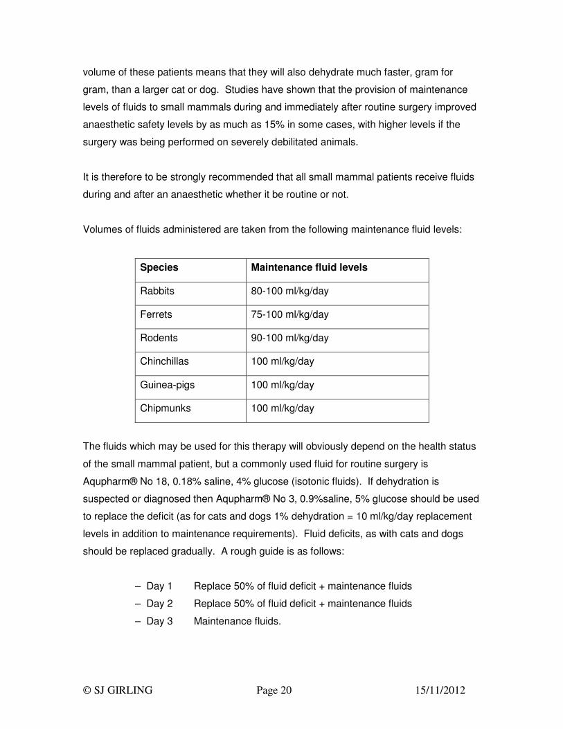
Volumes of fluids administered are taken from the following maintenance fluid levels:
Species Maintenance fluid levels
Rabbits 80-100 ml/kg/day
Ferrets 75-100 ml/kg/day
Rodents 90-100 ml/kg/day
Chinchillas 100 ml/kg/day
Guinea-pigs 100 ml/kg/day
Chipmunks 100 ml/kg/day
The fluids which may be used for this therapy will obviously depend on the health status
of the small mammal patient, but a commonly used fluid for routine surgery is
Aqupharm® No 18, 0.18% saline, 4% glucose (isotonic fluids). If dehydration is
suspected or diagnosed then Aqupharm® No 3, 0.9%saline, 5% glucose should be used
to replace the deficit (as for cats and dogs 1% dehydration = 10 ml/kg/day replacement
levels in addition to maintenance requirements). Fluid deficits, as with cats and dogs
should be replaced gradually. A rough guide is as follows:
– Day 1 Replace 50% of fluid deficit + maintenance fluids
– Day 2 Replace 50% of fluid deficit + maintenance fluids
– Day 3 Maintenance fluids.
© SJ GIRLING Page 21 15/11/2012
Colloidal fluids should be contemplated if intravenous access can be achieved when
haemorrhage occurs, and the use of lactated Ringer’s solution for conditions where
potassium loss and metabolic acidosis may occur (e.g. renal disease, chronic
diarrhoea).
The routes by which these fluids may be administered will depend on the species and
the degree of dehydration. If the fluids are required for routine post/intra-operative
requirements with minimal/no dehydration, then the subcutaneous route is satisfactory.
The scruff region or the lateral thoracic wall may be utilised in any small mammal
patient. For more severe dehydration the intraperitoneal route or intravenous routes (in
the larger patients) or intraosseous routes are required.
Intraperitoneal injections require the patient to be placed in dorsal recumbancy with the
head tilted downwards, so causing the abdominal viscera to move cranially and away
from the injection site which is in the right lower quadrant of the ventral abdominal wall.
The needle is angled between 20-40° in a cranial direction and advanced until it just
pops through the peritoneum.
In the larger species such as rabbits and ferrets and even guinea-pigs the intravenous
route may be used. The lateral saphenous or cephalic veins may be used in guinea-pigs
and ferrets, again preferably with 25-27 gauge butterfly catheters. In guinea-pigs,
chinchillas and ferrets the jugular veins may also be used in a cut-down procedure which
necessitates some form of sedation. In rats and mice the lateral tail veins may be used
to administer small volumes of fluid. A 27 gauge needle/butterfly catheter will be
required as well as warming the tail to improve vessel dilation.
For those species which have small or reduced peripheral circulation such as gerbils,
hamsters or shocked patients, the intraosseous route can be used. In the majority of
species the proximal femur is the site of choice. Because of the confining nature of the
bone marrow cavity, only small boluses of fluid can be administered, and so an infusion
device is important, such as a syringe driver. Asepsis must be strongly adhered to in the
case of intraosseous catheters as osteomyelitis can easily ensue in debilitated patients.
© SJ GIRLING Page 22 15/11/2012
Monitoring anaesthesia
This becomes more and more difficult as the size of the patient decreases. No one
factor (as with cats and dogs) will allow you to assess anaesthetic depth. Eye position
should not be used to assess depth of anaesthesia in small mammals. Instead a useful
method is to assess depth by the response to noxious stimuli such as pain.
Initially though the first reflex lost is usually the righting reflex. The next reflex to be lost
for example in rabbits and guinea-pigs is the swallow reflex, however this may be difficult
to assess. Palpebral reflexes are generally lost early on in the course of anaesthetic, but
rabbits may retain this reflex until well into the deeper planes of surgical anaesthesia.
The palpebral reflex is also altered by the anaesthetic agent chosen, with most inhalant
gaseous anaesthetics causing loss of the reflex early on, but it is maintained with
ketamine.
The pedal withdrawal reflex is useful in small mammals, with the leg being extended and
the toe firmly pinched. Loss of this reflex suggests surgical planes of anaesthesia, but
rabbits again will retain the pedal reflex in the forelimbs until much deeper (and often
dangerously deep!) planes of anaesthesia are reached. Other pain stimuli such as the
ear pinch in the guinea-pig and rabbit are useful, loss of this indicates a surgical plane,
as does the loss of the tail pinch reflex in rats and mice.
Monitoring of the heart and circulation may be performed in a conventional manner with
stethoscope and femoral pulse evaluation, or in the larger species using an oesophageal
stethoscope. As with cats and dogs the detection of increases in the respiratory and
heart rates can be used to indicate lightening of the plane of anaesthesia. More
sophisticated techniques may also be used with pulse oximetry to monitor heart rate and
haemoglobin saturation. As with cats and dogs, the aim is to achieve 100% saturation
and levels below 92% would indicate dangerous hypoxaemia and the initiation of
assisted ventilation. The ear artery is useful for this in rabbits using the clip-on probe,
conversely the linear probes may be used successfully on the ventral aspect of the tail in
most species (lateral tail in rats and mice). Other forms of cardiac monitoring include
ECG trace, which is frequently adapted to minimise trauma from the alligator forcep
attachments by substituting these for fine needle probes, or by blunting the alligator
teeth.
© SJ GIRLING Page 23 15/11/2012
An extremely useful monitoring device is the Doppler probe which can detect blood flow
in the smallest of vessels up to the heart itself.
Respiratory monitors may also be used if the patient is intubated and many pulse
oximeters have outlets for these allowing assessment of respiratory rates. It is important
to obtain an oximeter that can read high heart rates (e.g. VetOx by Heska). Even so-
many small rodents have heart rates too fast for any current affordable pulse oximeter.
© SJ GIRLING Page 24 15/11/2012
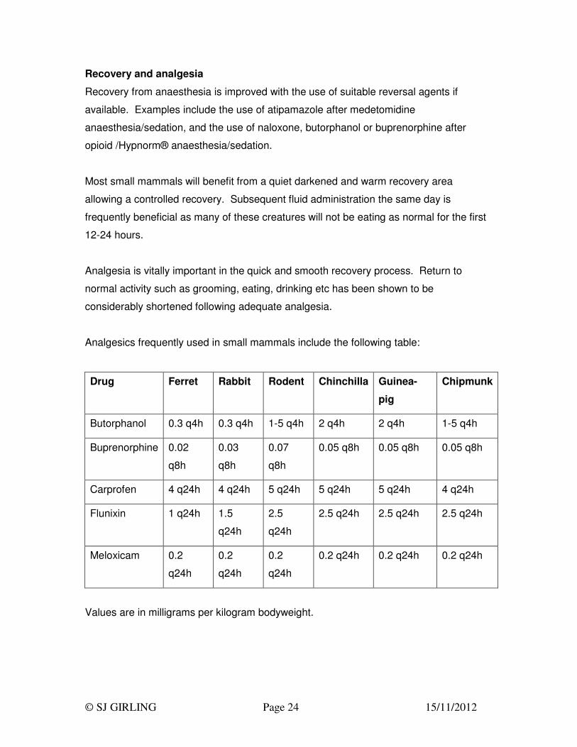
Recovery and analgesia
Recovery from anaesthesia is improved with the use of suitable reversal agents if
available. Examples include the use of atipamazole after medetomidine
anaesthesia/sedation, and the use of naloxone, butorphanol or buprenorphine after
opioid /Hypnorm® anaesthesia/sedation.
Most small mammals will benefit from a quiet darkened and warm recovery area
allowing a controlled recovery. Subsequent fluid administration the same day is
frequently beneficial as many of these creatures will not be eating as normal for the first
12-24 hours.
Analgesia is vitally important in the quick and smooth recovery process. Return to
normal activity such as grooming, eating, drinking etc has been shown to be
considerably shortened following adequate analgesia.
Analgesics frequently used in small mammals include the following table:
Drug Ferret Rabbit Rodent Chinchilla Guinea-
pig
Chipmunk
Butorphanol 0.3 q4h 0.3 q4h 1-5 q4h 2 q4h 2 q4h 1-5 q4h
Buprenorphine 0.02
q8h
0.03
q8h
0.07
q8h
0.05 q8h 0.05 q8h 0.05 q8h
Carprofen 4 q24h 4 q24h 5 q24h 5 q24h 5 q24h 4 q24h
Flunixin 1 q24h 1.5
q24h
2.5
q24h
2.5 q24h 2.5 q24h 2.5 q24h
Meloxicam 0.2
q24h
0.2
q24h
0.2
q24h
0.2 q24h 0.2 q24h 0.2 q24h
Values are in milligrams per kilogram bodyweight.

© SJ GIRLING Page 25 15/11/2012
As with cats and dogs and indeed humans, the administration of analgesia prior to the
onset of pain makes for the most effective control of pain. If this is to be considered,
then COX-2 inhibitors are preferred, specifically meloxicam or carprofen.
References
Carpenter, J.W. and Kolmstetter, C.M. (2000) Feeding small exotic animals In: Hill’s
Nutrition III,(Eds Lewis, L.D., Morris, M.L. and Hand, M.S.), Mark Mervis Institute,
Marceline, Missouri pp943-960.
Briscoe, J.A. and Syring, R. (2004) Techniques for emergency airway and vascular
access in special species Seminars in Avian and Exotic Pet Medicine 13(3):118-131.
Cole, S.G., Otto, C.M. and Hughes, D. (2002) Cardiopulmonary cerebral resuscitation in
small animals: A clinical practice review, Journal of Veterinary Emergency Critical Care
12:261-267.
Costello, M.F. (2004) Principals of cardiopulmonary cerebral resuscitation in special
species. Seminars in Avian and Exotic Pet Medicine 13(3):132-141.
DeFrancesco, T.C. (2000) Cardiac emergencies In Kirk and Bistner’s Handbook of
Veterinary Procedures and Emergency Treatment (Eds. Bistner, S.I., Ford, R.B. and
Raffe, M.R.), WB Saunders, Philadelphia, pp54-61.
Girling, S.J. (2003) Preliminary study into the possible use of benazepril in the
management of renal disease in rabbits Proceedings of the British Veterinary Zoological
Society November, Edinburgh, 44.
Harkness J E; Wagner J E. (1989) Anaesthesia, In: The Biology and Medicine of
Rabbits and Rodents, (3rd Edition), Lea and Febiger, Philadelphia pp 61-67.
Heard, D. (2004) Anesthesia, analgesia and sedation of small mammals In: Ferrets,
rabbits and rodents: Clinical medicine and surgery 2nd Edition (Eds. Quesenberry, K.E.
and Carpenter, J.W. ), WB Saunders, Philadelphia, pp356-369.
© SJ GIRLING Page 26 15/11/2012
Henrik, R.A. (1992) Basic life support and external cardiac compression in dogs and
cats. Journal of the American Veterinary Medical Association 200:1925-1931.
Huston, S.M. and Quesenberry, K.E. (2004) Cardiovascular and lymphoproliferative
diseases. In Ferrets rabbits and rodents: Clinical medicine and surgery 2nd Edition (Eds.
Quesenberry, K.E. and Carpenter, J.W.) WB Saunders, St Louis, pp211-220.
Kottwitz, J. and Kelleher, S. (2003) Emergency drugs: Quick reference chart for exotic
animals. Exotic DVM, 5.5(November):23-25.
Kozma, C., Macklin, W., Cummins, L.M. and Mauer, R. (1974) The anatomy, physiology
and biochemistry of the rabbit In. The Biology of the Laboratory Rabbit (Eds. Weisbroth,
S.H., Flatt, R.E. and Kraus, A.L.) Academic Press, London, pp50-69.
Mason D E. (1997) Anaesthesia, Analgesia, and Sedation for Small Mammals In
Ferrets, Rabbits and Rodents. Ed. Hillyer E V, Quesenberry K E, W B Saunders,
Philadelphia pp 378-391.
Okerman, L. (1994) Inherited conditions and congenital deformities In: Diseases of
domestic rabbits 2nd edition, Blackwell Publishing, Oxford, pp109-112.
Rush, J.E. and Wingfield, W.E. (1992) Recognition and frequency of dysrhythmias
during cardiopulmonary arrest Journal of the American Veterinary Medical Association
200:1932-1937.