Embed Size (px)
Citation preview
ANALYSIS OF FUNCTIONAL OUTCOME OF
ANTEROLATERAL PLATING IN TIBIAL
PILON FRACTURES.
Dissertation submitted for
M.S DEGREE EXAMINATION
BRANCH II-ORTHOPAEDIC SURGERY
INSTITUTE OF ORTHOPAEDICS AND TRAUMATOLOGY
MADRAS MEDICAL COLLEGE AND RAJIV GANDHI
GOVERNMENT GENERAL HOSPITAL
CHENNAI-600003
THE TAMILNADU DR.M.G.R MEDICAL UNIVERSITY
CHENNAI-600032
MARCH-2014
CERTIFICATE
This is to certify that this dissertation in “ANALYSIS OF
FUNCTIONAL OUTCOME OF ANTEROLATERAL PLATING IN
TIBIAL PILON FRACTURES” is a bonafide work done by
Dr.A.THIRUMURUGAN under my guidance during the period 2011–2014.
This has been submitted in partial fulfilment of the award of M.S. Degree
in Orthopedic Surgery (Branch–II) by The Tamilnadu Dr.M.G.R. Medical
University, Chennai.
PROFM.R.RAJASEKAR
DIRECTOR, INSTITUTE OF ORTHOPAEDICS AND TRUAMATOLOGY MADRAS MEDICAL COLLEGE & RAJIV GANDHI GOVT GEN. HOSPITAL CHENNAI – 3.
PROF.V.KANAGASABAI M.D., DEAN MADRAS MEDICAL COLLEGE& RAJIV GANDHI GOVT GEN. HOSPITAL CHENNAI-3.
CERTIFICATE
This is to certify that this dissertation in “ANALYSIS OF
FUNCTIONAL OUTCOME OF ANTEROLATERAL PLATING IN
TIBIAL PILON FRACTURES” is a bonafide work done by
Dr.A.THIRUMURUGAN under my guidance during the period 2011–2014.
This has been submitted in partial fulfilment of the award of M.S. Degree
in Orthopedic Surgery (Branch–II) by The Tamilnadu Dr.M.G.R. Medical
University, Chennai.
PROF.M.R.RAJASEKAR
DIRECTOR &PROFESSOR, INSTITUTE OF ORTHOPAEDICS & TRUAMATOLOGY MADRAS MEDICAL COLLEGE & RAJIV GANDHI GOVT GEN. HOSPITAL CHENNAI- 600003.
DECLARATION
I, Dr.A.THIRUMURUGAN, solemnly declare that the dissertation
titled “ ANALYSISOF FUNCTIONAL OUTCOME OF
ANTEROLATERAL PLATING IN TIBIAL PILON FRACTURES.”
was done by me at the Rajiv Gandhi Government General Hospital,
Chennai-3, during 2011-2014 under the guidance of my unit chief
Prof.M.R.RAJASEKAR, M.S(Ortho), D.Ortho.
The dissertation is submitted in partial fulfilment of requirement for the
award of M.S. Degree (Branch –II) in Orthopaedic Surgery to The Tamil
Nadu Dr.M.G.R.Medical University.
Place:
Date:
Dr.A.THIRUMURUGAN
ACKNOWLEDGEMENT
I express my thanks and gratitude to our respected Dean
Dr. KANAGASABAI M.D., Madras Medical College, Chennai – 3 for
having given permission for conducting this study and utilize the clinical
materials of this hospital.
I have great pleasure in thanking my respected teacher, Director and
HOD Prof .M.R.RAJASEKAR.M.S Ortho.,D.Ortho ., for his valuable
advice and guidance.
My sincere thanks and gratitude to Prof. N. DEEN MOHAMED
ISMAIL. M.S. Ortho., D.Ortho., Professor of Orthopaedics, Institute of
Orthopaedics and Traumatology, for his constant inspiration and guidance
throughout the study.
My sincere thanks and gratitude toProf. V. SINGARAVADIVELU
M.S.Ortho., D.Ortho., Professor, Institute of Orthopaedics and
Traumatology, for his constant advice and guidance provided throughout this
study.
My sincere thanks and gratitude to Prof. A. PANDIASELVAM.,
M.S.Ortho., D.Ortho., Professor, Institute of Orthopaedics and
Traumatology, for his valuable advice and guidance.

I am very much grateful to Prof. R. SUBBIAH M.S.Ortho.,
D.Ortho., for his unrestricted help and advice throughout the study period.
My sincere thanks and gratitude to Prof. NALLI R. UVARAJ
M.S.Ortho.,D.Ortho., for his constant advice and guidance provided
throughout this study.
My sincere thanks and gratitude to Dr.A.Shanmugasundaram M.S.
Ortho.,for his constant advice and guidance provided throughout this study.
I sincerely thank Dr. Manimaran, Dr.Velmurugan, Dr.Prabhakar,
Dr.Senthil Sailesh, Dr. Karunakaran, Dr. Muthukumar , Dr. Kannan,
Dr. Kingsly, Dr. Kailraj, Dr.Pazhani, Dr. Sameer, Dr.Muthazhagan,
Dr. Nalli R.Gopinath and Dr.Hemanth Kumar Assistant Professors of
this department for their valuable suggestions and help all during this study.
I thank all anaesthesiologists and staff members of the theatre for their
endurance during this study. I am very grateful to all my post graduate
colleagues for helping in this study. Last but not least, my sincere thanks to
all our patients, without whom this study would not have been possible.

CONTENTS
S.NO TITLE PAGE NO
1. INTRODUCTION 1
2. AIM AND OBJECTIVE 7
3. HISTORY AND REVIEW OF LITERATURE 8
4. DEVELOPMENT PRINCIPLE AND RULES OF
LCP
12
5. ANATOMY 23
6. CLASSIFICATION 29
7. MATERIALS AND METHODS 37
8. RESULTS 64
9. COMPLICATIONS 71
10. DISCUSSION 73
11. CONCLUSION 85
12. CASE ILLUSTRATIONS 88
13. BIBLIOGRAPHY 100
14. PROFORMA 107
15. ANNEXURE
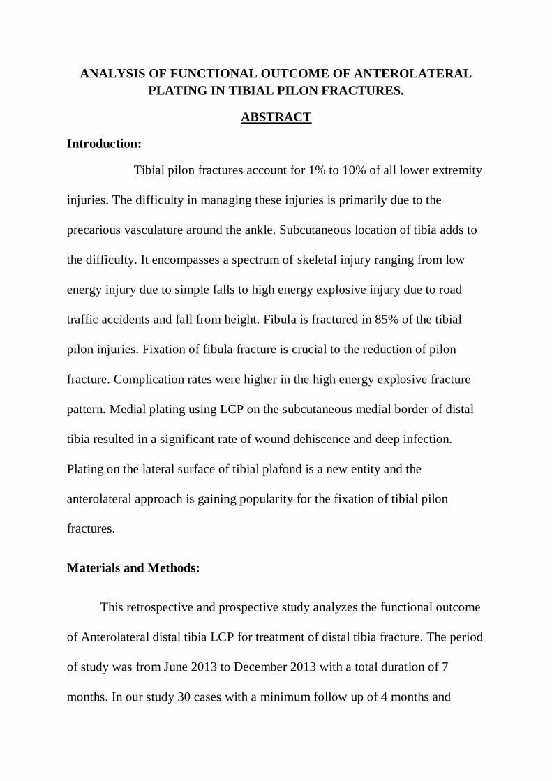
ANALYSIS OF FUNCTIONAL OUTCOME OF ANTEROLATERAL
PLATING IN TIBIAL PILON FRACTURES.
ABSTRACT
Introduction:
Tibial pilon fractures account for 1% to 10% of all lower extremity
injuries. The difficulty in managing these injuries is primarily due to the
precarious vasculature around the ankle. Subcutaneous location of tibia adds to
the difficulty. It encompasses a spectrum of skeletal injury ranging from low
energy injury due to simple falls to high energy explosive injury due to road
traffic accidents and fall from height. Fibula is fractured in 85% of the tibial
pilon injuries. Fixation of fibula fracture is crucial to the reduction of pilon
fracture. Complication rates were higher in the high energy explosive fracture
pattern. Medial plating using LCP on the subcutaneous medial border of distal
tibia resulted in a significant rate of wound dehiscence and deep infection.
Plating on the lateral surface of tibial plafond is a new entity and the
anterolateral approach is gaining popularity for the fixation of tibial pilon
fractures.
Materials and Methods:
This retrospective and prospective study analyzes the functional outcome
of Anterolateral distal tibia LCP for treatment of distal tibia fracture. The period
of study was from June 2013 to December 2013 with a total duration of 7
months. In our study 30 cases with a minimum follow up of 4 months and

maximum of 12 months with an average of 9 months was carried out. Fibula
fixation is performed initially to restore length and achieve indirect reduction of
tibia fracture using posterolateral approach. Anterolateral approach to ankle was
used to fix tibia. Anterolateral locking compression plates are placed through
the interval between the anterior and lateral compartments of leg. All cases were
assessed postoperatively using the Kaikkonen clinical ankle score and Teeny
wiss radiological score.
Results:
27 fractures united with a mean duration of 12 to 24 weeks. In our study we
were able to achieve anatomic reduction in 32% (7 cases) of the patients. Good
reduction was achieved in 50% (11 cases) of the patients. Fair reduction was
achieved in 18% (4 cases). There was no case of poor fracture reduction in our
study according to Teeny Wiss Score. In our study we had excellent functional
outcome in about 30% of cases, good functional outcome in 50% of cases fair
and poor outcome 10% of cases each based on Kaikkonen Clinical Ankle Score.
In our study the complication we met were 6 cases (20%) of wound dehiscence
and superficial infection which healed by secondary intention, 2 cases (7%)of
flap necrosis, 3 cases(10%) of nonunion, extensor tendon exposed in 1
case(3%), implant failure in one of the three non union cases. In our study we
had no deep infection (0%).
Conclusion:
Distal tibia fractures with high grade of soft tissue injury are to be
internally fixed after a delay of 21 days for the edema to settle down and the
wrinkle sign appears. The posterolateral incision to fibula provides a larger skin
bridge between this incision and the tibial incision. A 7 cm skin bridge between
two incisions is recommended to avoid wound complication. Restoration of the
articular surface and reestablishing its relationship to the tibial shaft is the
primary goal of treatment. Good functional result depends on reasonable
anatomic reduction of the articular surface and meticulous soft tissue handling.
From our study, 3.5mm Anterolateral Distal tibia Locking Compression Plating
for tibial pilon fractures were found to be safe and effective. For AO type A
fractures, can be fixed either using MIPPO or ORIF technique. For AO type C
fractures Open reduction of the articular fragment is mandatory and then
stabilize with locking compression plate for added up stability.
Keywords: dual incision technique, anterolateral approach for tibia, wound
problems, skin bridge

INTRODUCTION
1 | P a g e
INTRODUCTION
Tibial Pilon (Tibial Plafond) is a descriptive term for distal tibia
fracture suggesting that the talus acts as a hammer, impacting and injuring the
distal tibia1. Treatment of these fractures remains challenging for orthopaedic
surgeons2.Tibial pilon fractures account for 1% to 10% of all lower extremity
injuries2. The difficulty in managing these injuries is primarily due to the
precarious vasculature around the ankle. Subcutaneous location of tibia adds
to the difficulty.Tibial pilon encompasses a spectrum of skeletal injury
ranging from low energy injury due to simple falls to high energy explosive
injury due to road traffic accidents and fall fromheight3.
Low energy tibial pilon fractures occur in older age group and
mechanism of injury is usually rotation. It causes a spiral fracture of the distal
tibia with or without extension into ankle joint. Status of surrounding soft
tissue is good. As a result risk of post surgical wound healing problems and
chance of infection are minimal4.
High energy tibial pilon fractures occur in younger age group and the
usual mechanism of injury is axial loading. Here the hard dome of talus
impacts against the relatively soft tibial articular surface. As a result distal
tibial articular surface and the metaphyseal region crumbles. Foot position
during impact heavily influences the fracture pattern of the articular
2 | P a g e
surface1,4. This pattern is associated with severe soft tissue injury with edema
and skin blisters around the ankle 1,4.
Fibula is fractured in 85% of the tibial pilon injuries 3 . Fixation of
fibula fracture is crucial to the reduction of pilon fracture.
Decision making is tough only in the high energy pilon fractures. Some
complex questions arise, whether to treat the fracture conservatively or
operatively? If operative, whether immediate or delayed fixation? Fixation of
fibula? Internal or external fixation? Choice of approach? Implant choice? 2.
Answers to these questions and hence decision making should be made on an
individual basis.
Comorbid illness complicates this delicate situation. Peripheral
vascular disease, diabetes mellitus, smoking and alcoholism should be
considered3.
Internal fixation was considered gold standard in 1980 by Ruedi of the
AO group. It was the standard of care. The enthusiasm was soon lost due to
wound breakdown, deep osteomyelitis and sepsis associated with the open
reduction and plating technique. Complication rates were higher in the high
energy explosive fracture pattern1.
Because of the severity of the soft tissue injury and the reported soft
tissue complication rate attributed to extensive surgical exposures and bulky
3 | P a g e
internal fixation devices approximating 40% to 50%, external fixation
emerged as a successful technique for decreasing significant septic
complications that had been previously attributable to open surgical
management1 .
Minimal internal fixation to reconstruct the articular surface of tibia
supported with ankle spanning external fixators became the popular modality
of treatment in late 1990s. Fibula fractures were secured using one third
tubular or reconstruction plate to maintain length and axis alignment. Hybrid
fixators later replaced monolateral external fixators because of advantage of
stability and early weight bearing.Definitive management of pilon fracture by
external fixation has its own complications. They are pin tract infection,
secondary loss of reduction and ankle stiffness.
The lack of consistent results with the use of external fixation
techniques and an improved understanding of the associated soft tissue injury
with pilon fractures gave way to the reconsideration of open reduction and
internal fixation but after a period of soft tissue recovery1.
Role of non-surgical treatment should have a mention. It is possible for
stable fractures with minimal shortening to be treated by traction or plaster of
paris in a medically unfit patient , but malunion, shortening , limited range of
movement and early secondary osteoarthritis of ankle have all been reported
following conservative treatment of pilon fractures21,25,26. Complications of
4 | P a g e
recumbency such as deep vein thrombosis, pressure sores and pneumonia are
a problem.
Open Reduction and Internal Fixation has regained popularity largely
due to better assessment of soft tissue envelope. Tscherne soft tissue injury
classification was expanded by the AO group to create a more objective
system that evaluates and grades each component of soft tissue envelope,
including the skin, musculotendinous components and neurovascular tissue1.
With regards to closed intramedullary nail, a stable fixation with nail in
distal tibia may be difficult to achieve because the hourglass shape of the
intramedullary canal prevents a tight endosteal fit and compromises torsional
and angular stability. Secondary displacement of the fracture on insertion of
nail, breakage of nail and locking screws, limited applicability in severe intra
articular fracture and malunion of the tibia are potential risks.
Various modalities of internal fixation for tibial pilon by plating have
been described. These include conventional AO medial plating using medial
buttress plate, anterior plating using T plates, Cloverleaf plate or occasionally
simple dynamic compression plates5.AO medial plating has its own
drawbacks. One of the major disadvantages of this approach is the risk of
wound break down over the subcutaneous border, with the potential need for
flap cover. In addition this approach limits visualization of the lateral chaput
fragment7.
5 | P a g e
Locking compression plating (LCP) is the most widely used implant
for tibial pilon fractures at present. It can be performed by minimally invasive
technique or standard open reduction and internal fixation methods. These are
anatomically contoured to the bone surface to which it is applied. For pilon
fracture the gold standard was medial LCP, a low profile plate placed through
medial approach. The low profile nature tends to address the problem of
bulky implant used in standard AO plating techniques. Medial plating using
LCP on the subcutaneous medial border of distal tibia still resulted in a
significant rate of wound dehiscence and deep infection, although at a lower
rate compared to standard AO plates.
Surgeons preferred Minimally Invasive Percutaneous Plate
Osteosynthesis (MIPPO) technique to decrease the wound complication using
medial LCP. MIPPO technique achieved union rates ranging between 80%
and 100%. These techniques aim to reduce surgical trauma and to maintain a
more biologically favourable environment for fracture healing. Nevertheless,
complications such as angular deformities greater than 7°, articular mal
reduction, hardware failure and non-unions have been reported.
How to overcome this problem? Why not use the lateral surface of
tibia? Attempts were made in the past to treat pilon fractures by open
articular reduction and lateral buttress plating, but it failed. This was
attributed to the complex bony contour of the lateral surface of the distal tibia
6 | P a g e
as it blends with the tibial plafond distally. Contouring a semi tubular, one
third tubular or reconstruction plate to this surface was challenging and also
resulted in inadequate screw purchase. All this resulted in unstable fixation
and secondary loss of reduction.
Locking Compression Plates designed to contour the lateral surface of
distal tibia were developed recently. These plates were fixed using the
anterolateral approach to the distal tibia. Although anterolateral approach has
been described in the past, it was not popularized. Plating on the lateral
surface of tibial plafond is a new entity and the anterolateral approach is
gaining popularity for the fixation of tibial pilon fractures. Early studies have
shown it offers the benefit of improved soft tissue coverage and the potential
for a lower rate of wound healing complications7.
If it is so, then anterolateral plating for tibial pilon fractures could well
be a milestone in the management of pilon fracture.

AIM AND OBJECTIVE
7 | P a g e
AIM AND OBJECTIVE
Aim of our study is to analyze the functional outcome of Tibial pilon
fractures treated by Anterolateral Distal Tibial Locking Compression Plate in
the Institute of Orthopaedics and Traumatology, Madras Medical College and
Rajiv Gandhi Government General Hospital Chennai over a period of six
months from June 2013 to November 2013.
The primary objective of the study is to analyze the outcome in terms
of fracture union using radiographs and surgical wound healing.
The secondary objective of the study is to analyze the functional
outcome after surgery.

HISTORY AND
REVIEW OF LITERATURE
8 | P a g e
REVIEW OF LITERATURE
First described by French radiologist Destot in the year 1911, as ankle
fractures that involve the weight bearing distal tibial articular surfaces. The
term Pilon was coined by Etienne Descot to describe fractures occurring
within 5 cm of the ankle joint.
The term Pilonwas derived from French language and refers to a
pestle, which is a club shaped tool used for mashing or grinding substance in
mortar, or a large bar which is moved vertically to stamp.
In 1950 Bonin J.G. coined the term Plafond which means ceiling in
French. It describes the distal tibial articular surface to the roof of the ankle
joint.
In 1968 Ruedi performed early open reduction and internal fixation
for all tibial plafond fractures and demonstrated satisfactory results with few
complications.This was supported by subsequent studies performed by Heim
and later by Ovadia and Beals.
In 1980 and 1990s , early open reduction and internal fixation became
the standard of care for tibial pilon fractures.
Wrysch pointed out that Ruedi’s study included mostly low energy
spiral fracture pattern of tibial pilon. This resulted in superior results in early
open reduction. In late 1990s, studies showed early open reduction caused
9 | P a g e
severe wound complications and sepsis. This was attributed to more cases of
high velocity axial compression injury pattern in his series.
Because of this high rate of soft tissue complications due to open
reduction and bulky implants, external fixation emerged as treatment of
choice. Later Bone and colleagues, used combined minimal internal fixation
with external fixation.
Maisonneuve (1840), compares ankle with a mortise and tenon.
Sir Robert Jones described that, the most injured joint of the body
was that of ankle, but it was treated least.
In 1968 it was Reudi who published a paper on this topic, describing
the fracture, principles of treatment and a classification system. His
experience with immediate fixation of tibial fractures demonstrated durable
results and few complications41,42.
Kellam and Waddeli divided pilon fractures in two types based by the
mechanism of injury as either rotational or axial loading or both22.
Tornetta et.al described combined open stabilization of the articular
fractures and neutralization of the metaphyseal fractures with hybrid external
fixation without spanning the ankle joint withpotential benefit of allowing
cartilage nutrition through the use of early ankle range of motion.
10 | P a g e
Pugh and colleagues and Angen pointed out the complictaions of
external fixation such as malunion, nonunion and lower clinical scores and
slower return to function when they compared with their own ORIF group37.
Understanding the need for better managementof the soft tissue,
Schatzker and Tile in 1996 developed a distinction between the soft tissue
that is adequate for immediate fixation and the soft tissue that is not suitable
for surgery because of swelling. In the second group a delay of 7 to 10 days
was suggested prior to surgery, for the skin and soft tissues to return to a
reasonable state.
Mast et.al recommended that if the definitive surgery cannot be
performed within 8 to 12 hours, plan for a temporary treatment and definitive
procedure is delayed till the resolution of the swelling. He also recommended
that length stable injuries can be treated temporarily by casting and for
fractures with shortening calcaneal traction was applied to restore the length,
before any definitive procedures.
Sirkin and colleagues, and Patterson and Cole, in 1999, popularized
the staged management of tibial plafond injuries. They concluded that
performing immediate open reduction in the swollen and compromised soft
tissue envelope contributed to high rate of wound complication and sepsis.
11 | P a g e
This staged management protocol remains the foundation for the basis
of tibial pilon fracture management in current practice.
Helfet DL et.al in 2004 developed the minimally invasive
percutaneous plate osteosynthesis for distal tibia with low profile plate,
following better understanding of the osseous fracture anatomy.
12 | P a g e
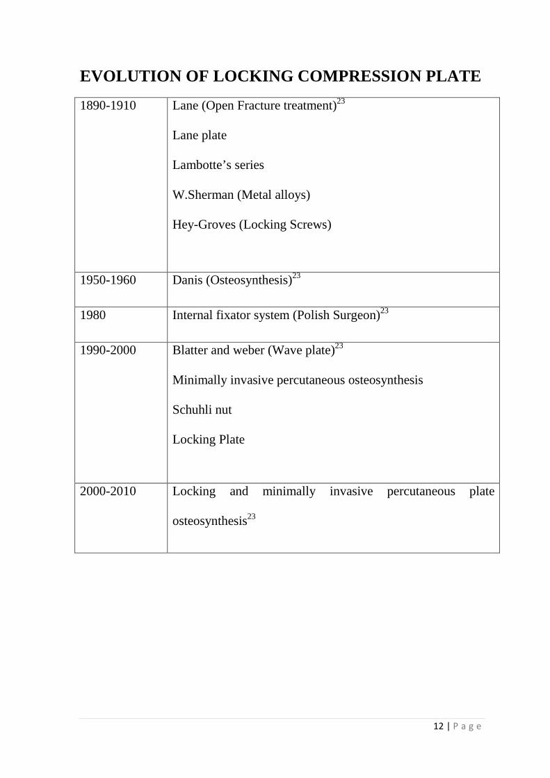
EVOLUTION OF LOCKING COMPRESSION PLATE
1890-1910 Lane (Open Fracture treatment)23
Lane plate
Lambotte’s series
W.Sherman (Metal alloys)
Hey-Groves (Locking Screws)
1950-1960 Danis (Osteosynthesis)23
1980 Internal fixator system (Polish Surgeon)23
1990-2000 Blatter and weber (Wave plate)23
Minimally invasive percutaneous osteosynthesis
Schuhli nut
Locking Plate
2000-2010 Locking and minimally invasive percutaneous plate
osteosynthesis23

DEVELOPMENT PRINCIPLE
AND RULES OF LCP
13 | P a g e
DEVELOPMENT OF LOCKING COMPRESSION PLATE
During the last decade, tremendous advances have been made in the
internal fixation of fractures by plating. The internal fixator system was first
developed by Polish surgeon in the 1980’s. They developed the ZESPOL
system. They based the design of their implant on a number of principles23.
1. The screw should be fixed to the plate.
2. Compression between the plate and the bone should be eliminated.
3. The number of screws necessary for stable fixation should be optimal.
4. Plate stability and Inter fragmentary compression should be preserved.
The following devices lead to the development of the so called locked
internal fixator.
1. Schuhli locked plate: This was developed by J. Mast.Schuhli, nuts
keep the plate away from the bone. It has three sharp projections. As it
makes less direct contact between the plate and bone it acts as a low
profile internal fixator. In addition, if in case of missing cortical bone,
Schuhli nuts can act as proximal cortices and bicortical fixation is
feasible.23
2. Point contact fixator (PC-FIX): These devices preserve the blood
supply of the periosteum by point contact. These fixators are secured
by unicortically inserted screws and hence have minimal contact. The
14 | P a g e
tapered head of the screw ensures that it lodges firmly in the plate hole
and provides the required angular stability. PC-FIX was the first plate
in which angular stability was achieved. PC-FIX was the basis for the
further development of LISS23.
3. The angled blade plate devised by AO is the strongest implant
providing that fixed angles gives improved stability.
4. Interlocking nail used in comminuted diaphyseal fracture proved that
open anatomical reduction of the fragment is not necessary and close
treatment of the comminuted fragments with splinting by
intramedullary nail produces abundant callus and solid healing. These
four developments, Schuhli nut, point contact plate, fixed angled blade
plate and locked intramedullary nail naturally lead to the development
of internal fixator by locked head plate23.
5. During the last decade, bridge plating and less invasive and minimally
invasive surgery developed.
6. Finally M. Wagner and R.Frigg developed the locking compression
plate (LCP), with combi holes and functionality of both locking and
conventional plate. This development revolutionized operative fracture
fixation16,17,18,53.

15 | P a g e
PRINCIPLES OF LCP
Locked plates rely on a different mechanical principle to provide
fracture fixation and in so doing they provide different biological
environments for healing.
In conventional plates the strength of fixation, depends on the frictional
force between the plate bone interface and the axial stiffness or pull out
strength of the screw bone interface of the single screw farthest away from
the fracture site during axial loading. Conventional plate creates contact
stress at bone plate interface, compromising the periosteal blood supply.
Under axial load, there is secondary loss of fixation post-operatively
due to toggling of the screws in the plate as screws are not locked to the
plate.In the locked plates, they behave biologically and mechanically
differently from that of conventional plate.
Single Beam Construct:
Locked plate is a single beam construct by design. Plate and screws act
as a single unit. Locked plate controls the axial orientation of the screw to the
plate, thereby enhancing the screw-plate-bone construct by creating a single
beam construct. In this construct there is no motion between the components
of the beam that is the plate or the screw or the bone. This construct is four
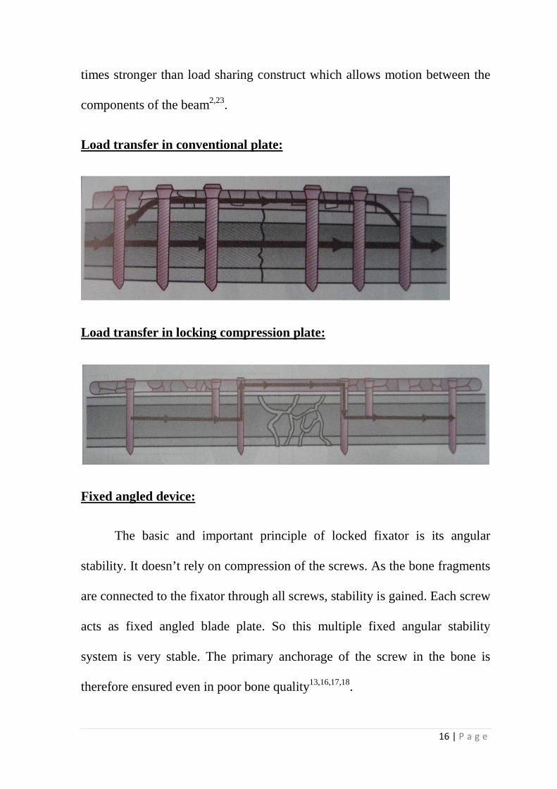
16 | P a g e
times stronger than load sharing construct which allows motion between the
components of the beam2,23.
Load transfer in conventional plate:
Load transfer in locking compression plate:
Fixed angled device:
The basic and important principle of locked fixator is its angular
stability. It doesn’t rely on compression of the screws. As the bone fragments
are connected to the fixator through all screws, stability is gained. Each screw
acts as fixed angled blade plate. So this multiple fixed angular stability
system is very stable. The primary anchorage of the screw in the bone is
therefore ensured even in poor bone quality13,16,17,18.
17 | P a g e
In the locked head plate load transfer from one fragment of bone
occurs through the locked screw head to the plate and from the plate to screw
of other fragment and finally to the opposite fragment without loading the
bone, not like that of the conventional plate.
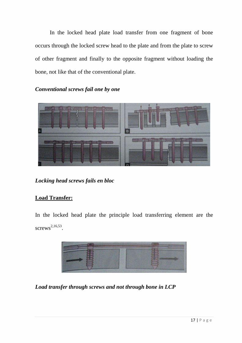
Conventional screws fail one by one
Locking head screws fails en bloc
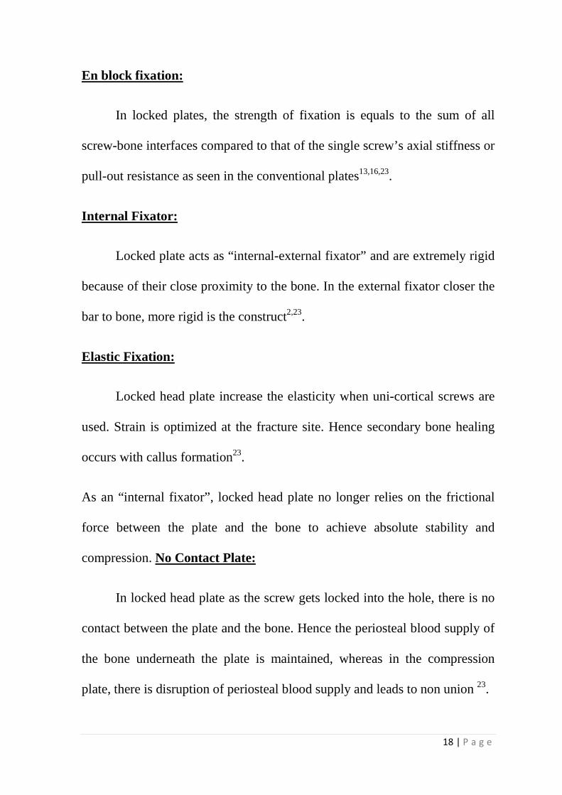
Load Transfer:
In the locked head plate the principle load transferring element are the
screws2,16,53.
Load transfer through screws and not through bone in LCP
18 | P a g e
En block fixation:
In locked plates, the strength of fixation is equals to the sum of all
screw-bone interfaces compared to that of the single screw’s axial stiffness or
pull-out resistance as seen in the conventional plates13,16,23.
Internal Fixator:
Locked plate acts as “internal-external fixator” and are extremely rigid
because of their close proximity to the bone. In the external fixator closer the
bar to bone, more rigid is the construct2,23.
Elastic Fixation:
Locked head plate increase the elasticity when uni-cortical screws are
used. Strain is optimized at the fracture site. Hence secondary bone healing
occurs with callus formation23.
As an “internal fixator”, locked head plate no longer relies on the frictional
force between the plate and the bone to achieve absolute stability and
compression. No Contact Plate:
In locked head plate as the screw gets locked into the hole, there is no
contact between the plate and the bone. Hence the periosteal blood supply of
the bone underneath the plate is maintained, whereas in the compression
plate, there is disruption of periosteal blood supply and leads to non union 23.
19 | P a g e
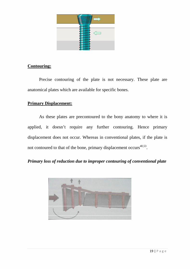
Contouring:
Precise contouring of the plate is not necessary. These plate are
anatomical plates which are available for specific bones.
Primary Displacement:
As these plates are precontoured to the bony anatomy to where it is
applied, it doesn’t require any further contouring. Hence primary
displacement does not occur. Whereas in conventional plates, if the plate is
not contoured to that of the bone, primary displacement occurs40,53.
Primary loss of reduction due to improper contouring of conventional plate
20 | P a g e
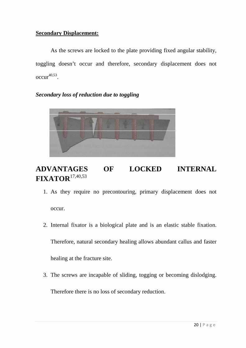
Secondary Displacement:
As the screws are locked to the plate providing fixed angular stability,
toggling doesn’t occur and therefore, secondary displacement does not
occur40,53.
Secondary loss of reduction due to toggling
ADVANTAGES OF LOCKED INTERNAL FIXATOR17,40,53
1. As they require no precontouring, primary displacement does not
occur.
2. Internal fixator is a biological plate and is an elastic stable fixation.
Therefore, natural secondary healing allows abundant callus and faster
healing at the fracture site.
3. The screws are incapable of sliding, togging or becoming dislodging.
Therefore there is no loss of secondary reduction.
21 | P a g e
4. Locking the screws ensures angular, as well as axial stability and
eliminates unwanted movement of the screws.
5. Blood supply to the bone is preserved as the plate is away from bone.
6. Ideally suited in osteoporotic bones, with less pull-out of screws.
7. Screws with multiple angular stability in the epiphyseal and
metaphyseal fragments make the construct very stable.
8. Locked internal fixators are noncontact plates, hence no disturbances
in periosteal blood supply, and therefore there is no risk of refracture
after removal of plate.
9. No need to contour the plate and also no need to the compress the plate
to bone.
10. Also there is no need for reconstruction of the opposite deficient
cortex.
11. Polyaxial screws have an advantage. It can be angled in any desired
direction.
22 | P a g e
RULES OF SCREW PLACEMENT IN A LOCKING
COMPRESSION PLATE17, 40, 53
1. Conventional screws are inserted before any locking screws.
2. Conventional screws will bring the plate closer to the bone.
3. Conventional screws can be used to lag fracture fragment through plate
or individually.
4. Locking screws will not reduce the bone to the plate.
5. Locking screws form a fixed angle construct with plate to increase the
stability in osteoporotic bone.
6. Lag before lock. After placing locking screws no additional
compression or reduction of the fragments are possible.
7. Locking screws should be placed as the final step of plate
osteosynthesis.

ANATOMY
23 | P a g e
ANATOMY
Tibia acts as the major weight bearing bone of the leg through which
neurovascular bundle to the foot also traverse. Subcutaneous location of tibia
in the anterior and medial aspect makes it more susceptible to injury and
especially vulnerable to open fractures.
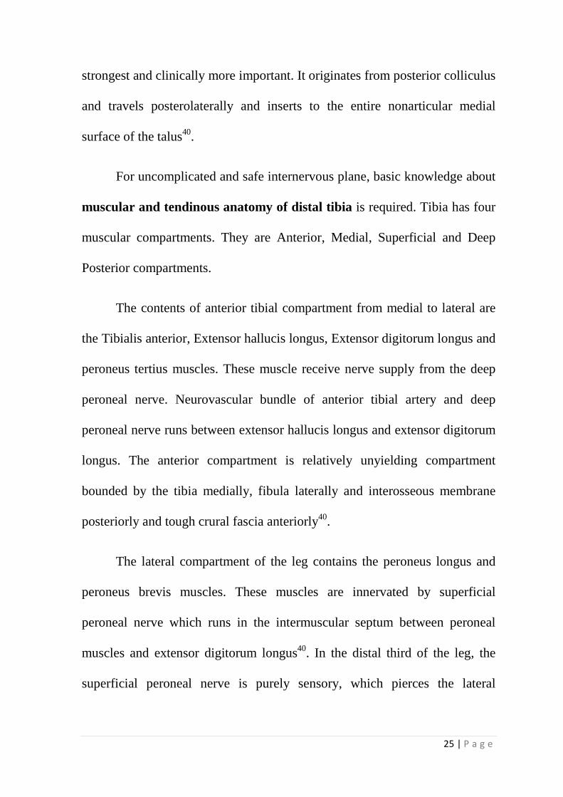
The Osseous anatomy40 of ankle joint includes the distal tibia, the
distal fibula and the talus. Ankle mortise is formed when the distal ends of
tibia and fibula meets the superior dome of the talus. The articular surface of
distal tibia is broader anteriorly and gradually narrows posteriorly. Also the
articular surface is more concave from anterior to posterior.
The medial malleolus is a bony projection from the medial end of
distal tibia. It extends more distal and slightly anterior. The articular surface
of medial malleolus is oriented perpendicular to tibial plafond and articulates
with medial articular portion of talar body40.
The distal end of fibula also known as the lateral malleolus articulates
with the lateral articular portion of the talar body. Ariculation of the distal
tibia with the distal fibula forms the tibio fibular syndesmosis distally. The
subchondral bone of the distal tibia represents the strongest cancellous bone
and provides secure screw purchase for fixation devices.
24 | P a g e
Regarding the anatomic axis of tibia, the tibial plafond is oriented in
slight valgus in the frontal plane (2 degrees), and the anatomic axis passes
just medial to midline of the talus. The tibial plafond is slightly extended in
sagittal plane (approximating 5 to 10 degrees) and the mid-diaphyseal line of
the tibia passes through the lateral process of the talus40.
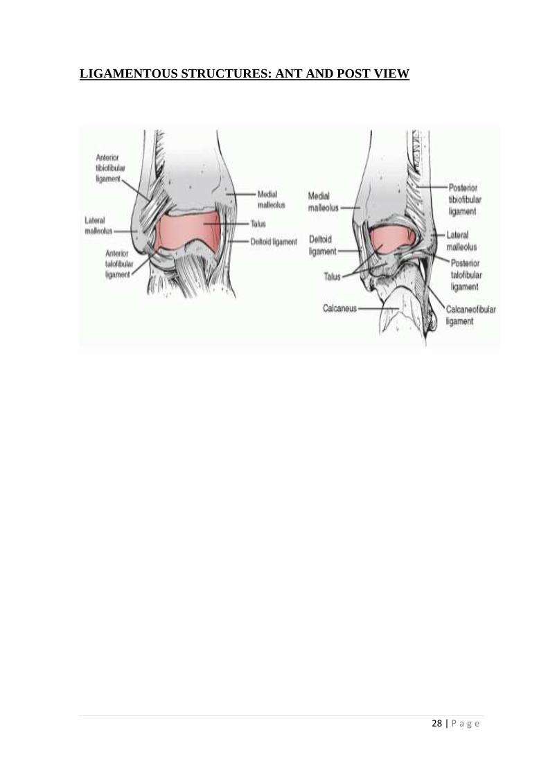
Knowledge about the ligamentous attachments at the ankle joint is
useful for understanding the fracture anatomy and displacement patterns40.
The irregular convex surface of the medial aspect of distal fibula meets
the irregular concave surface on the lateral aspect of tibia to form the distal
tibiofibular syndesmosis. The components of the distal tibio fibular
syndesmosis include anterior tibiofibular ligament, posterior tibiofibular
ligament and the strong interosseous tibiofibular ligament40.The posterior
tibiofibular ligament has superficial and deep component, the latter is called
as the transverse tibiofibular ligament. This ligament projects below the
margin of distal tibia to form a labral articulation for the posterolateral talus.
The deltoid ligament is also called the medial collateral ligament of the
ankle. It is a strong, flat broad triangular band composed of superficial and
deep layers. The superficial layer has three attachments distally. They are the
tibio calcaneal, tibio navicular and superficial tibio talar ligaments. The deep
portion of the deltoid ligament consists of deep anterior tibio talar ligament
and deep posterior tibio talar ligament. Deep posterior component is the
25 | P a g e
strongest and clinically more important. It originates from posterior colliculus
and travels posterolaterally and inserts to the entire nonarticular medial
surface of the talus40.
For uncomplicated and safe internervous plane, basic knowledge about
muscular and tendinous anatomy of distal tibia is required. Tibia has four
muscular compartments. They are Anterior, Medial, Superficial and Deep
Posterior compartments.
The contents of anterior tibial compartment from medial to lateral are
the Tibialis anterior, Extensor hallucis longus, Extensor digitorum longus and
peroneus tertius muscles. These muscle receive nerve supply from the deep
peroneal nerve. Neurovascular bundle of anterior tibial artery and deep
peroneal nerve runs between extensor hallucis longus and extensor digitorum
longus. The anterior compartment is relatively unyielding compartment
bounded by the tibia medially, fibula laterally and interosseous membrane
posteriorly and tough crural fascia anteriorly40.
The lateral compartment of the leg contains the peroneus longus and
peroneus brevis muscles. These muscles are innervated by superficial
peroneal nerve which runs in the intermuscular septum between peroneal
muscles and extensor digitorum longus40. In the distal third of the leg, the
superficial peroneal nerve is purely sensory, which pierces the lateral
26 | P a g e
compartmental fascia, and travels in the subcutaneous fascia from posterior to
anterior, typically encountered during the anterolateral surgical exposure40.
A posterior septum intervenes between the superficial and deep
posterior compartments. The superficial posterior compartment contains the
gastrocnemius, soleus and plantaris muscle. It also serves as a source for local
muscle flap for covering soft tissue defects which are encountered with
internal fixation of tibial pilon fractures. These muscles are innervated by the
tibial nerve40.
The deep posterior compartment is largely tendinous and includes
Tibialis posterior, Flexor digitorum longus and the Flexor hallucis longus
muscle. All these muscles are innervated by the tibial nerve40.
27 | P a g e
OSSEOUS ANATOMY40
ANTERIOR AND POSTERIOR VIEW
SUPERIOR VIEW UNDERSURFACE VIEW
28 | P a g e
LIGAMENTOUS STRUCTURES: ANT AND POST VIEW

CLASSIFICATION
29 | P a g e
CLASSIFICATION
There are many classification system followed from the early days
such as Mast, Speigl and Pappas, Bohler classification, Weber classification,
Ruedi and Allgower and AO/OTA types.
Of all the classification systems most commonly followed are Ruedi
and Allgower and AO/OTA classification.
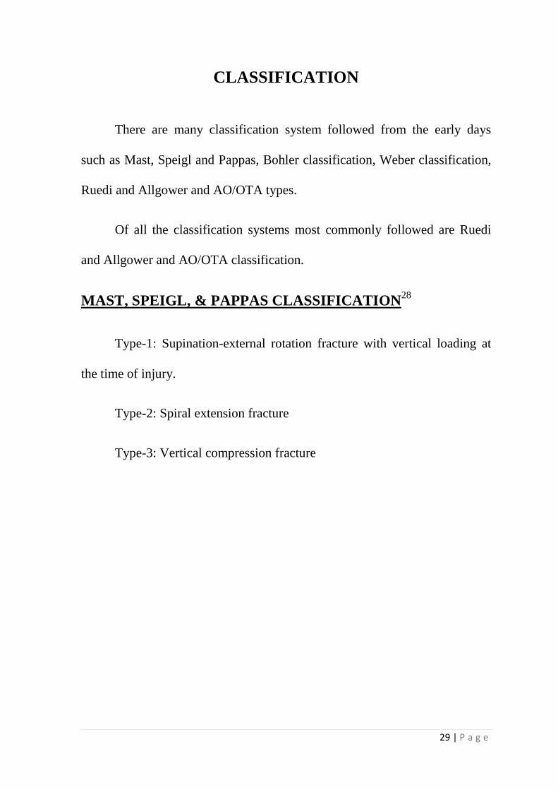
MAST, SPEIGL, & PAPPAS CLASSIFICATION28
Type-1: Supination-external rotation fracture with vertical loading at
the time of injury.
Type-2: Spiral extension fracture
Type-3: Vertical compression fracture
30 | P a g e
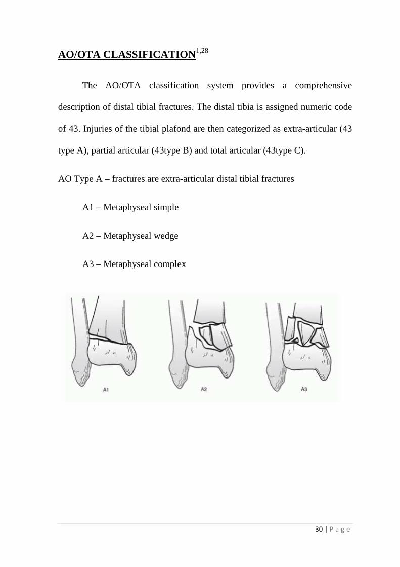
AO/OTA CLASSIFICATION1,28
The AO/OTA classification system provides a comprehensive
description of distal tibial fractures. The distal tibia is assigned numeric code
of 43. Injuries of the tibial plafond are then categorized as extra-articular (43
type A), partial articular (43type B) and total articular (43type C).
AO Type A – fractures are extra-articular distal tibial fractures
A1 – Metaphyseal simple
A2 – Metaphyseal wedge
A3 – Metaphyseal complex
31 | P a g e
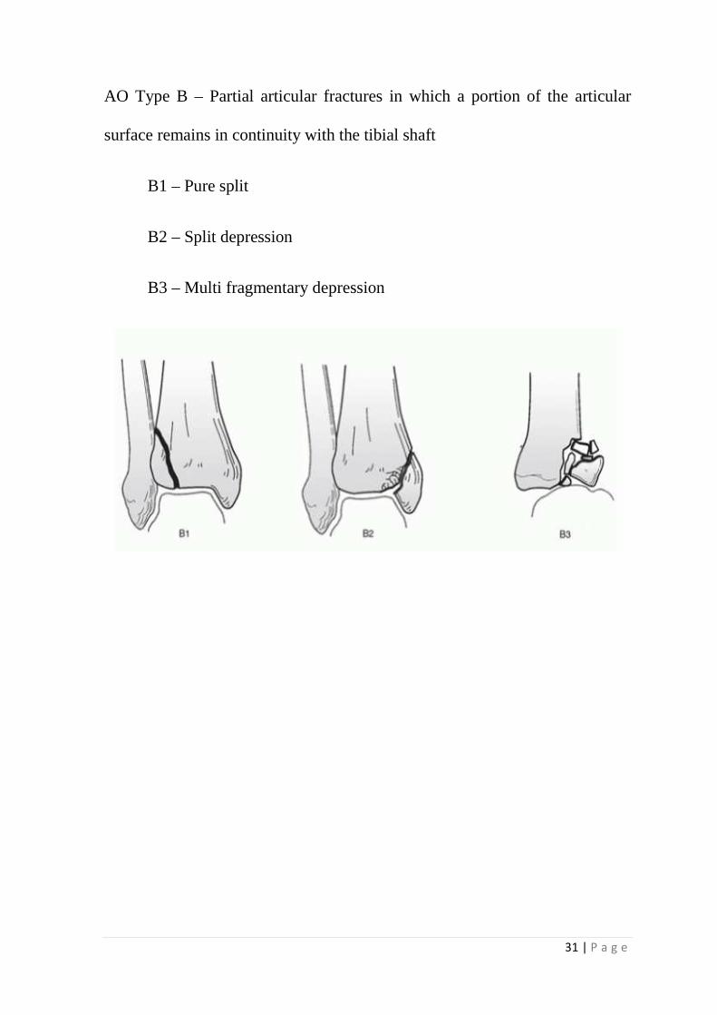
AO Type B – Partial articular fractures in which a portion of the articular
surface remains in continuity with the tibial shaft
B1 – Pure split
B2 – Split depression
B3 – Multi fragmentary depression
32 | P a g e
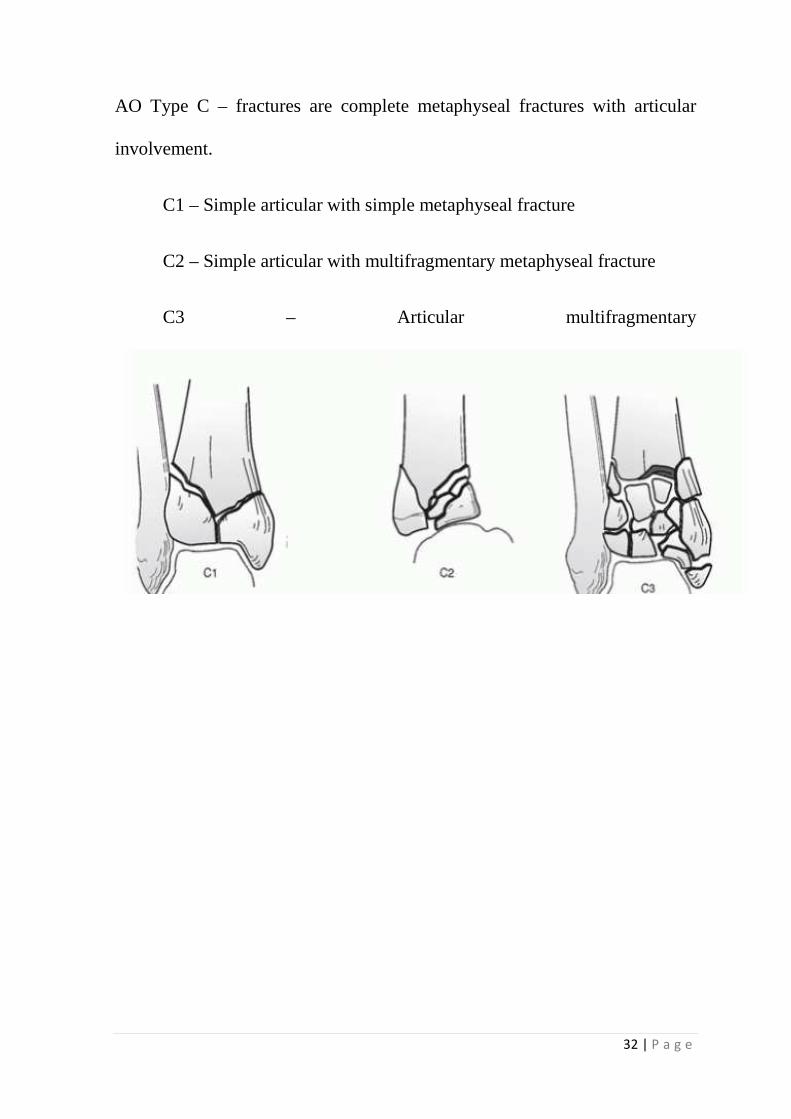
AO Type C – fractures are complete metaphyseal fractures with articular
involvement.
C1 – Simple articular with simple metaphyseal fracture
C2 – Simple articular with multifragmentary metaphyseal fracture
C3 – Articular multifragmentary
33 | P a g e
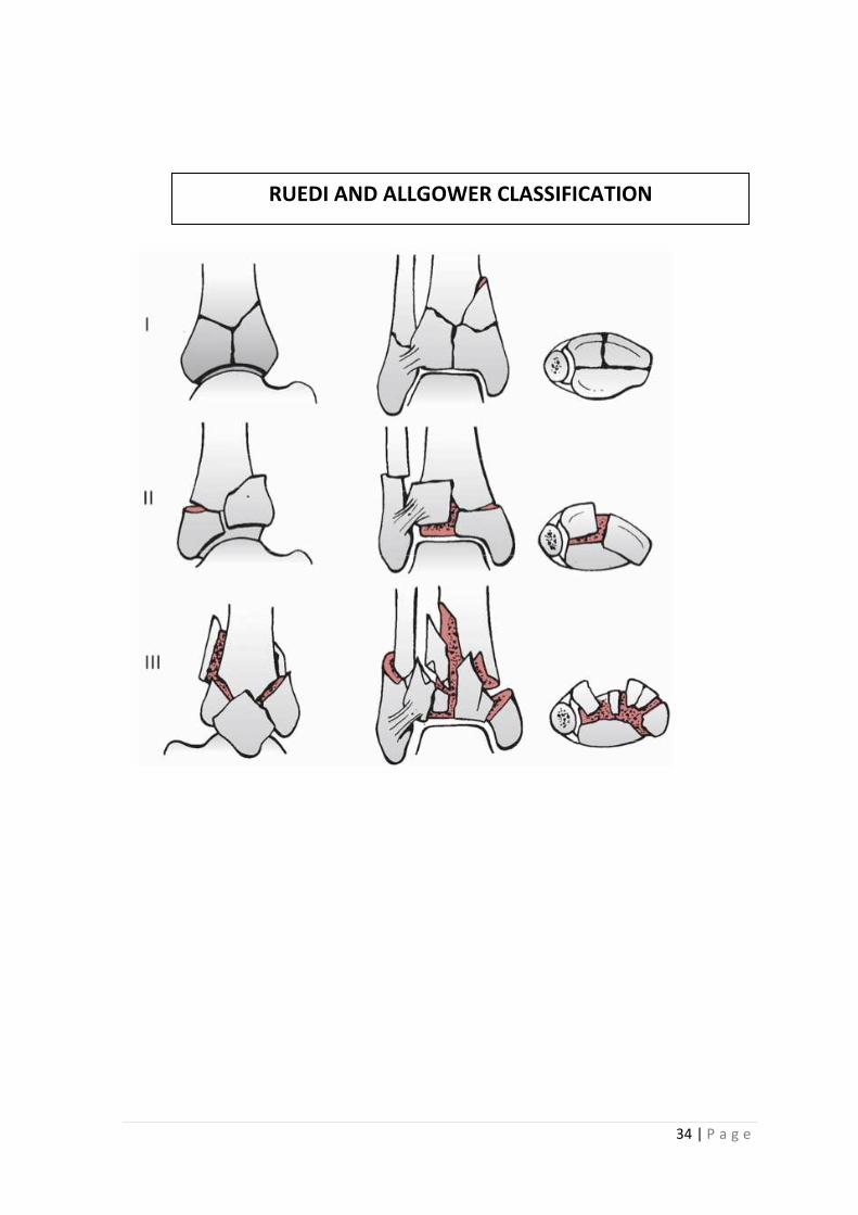
RUEDI AND ALLGOWER CLASSIFICATION1
Ruedi and Allgower, classified plafond fractures into three categories.
Type I: cleavage fracture of the distal tibia without significant
displacement of the articular surface.
Type II: significant fracture displacement of the articular surface
without comminution.
Type III: Impaction and comminution of the distal tibial articular
surface.
34 | P a g e
RUEDI AND ALLGOWER CLASSIFICATION
35 | P a g e
Reudi and Allgower Classification was modified by Ovadia and
Beals
Type I: Undisplaced articular fracture
Type II: Minimally displaced articular fracture
Type III: Displaced articular with large fragments
Type IV: Displaced articular fracture with multiple fragments and large
metaphyseal defect
Type V: Displaced articular fracture with severe communition.
36 | P a g e
Soft Tissue Injury Classification:
The recognition of the soft tissue injury associated with tibial plafond
fractures has resulted in the evolution of their surgical treatment. A thorough
evaluation of the soft tissue envelope and surgeon’s experience and judgment
remains the mainstay. The soft tissue injury classification system of Tscherne
and Goetzen is subjective and grades soft tissue injuries of closed fractures
into one of four categories, organized from 0 to 3.
Grade 0 - Closed fractures with no appreciable soft tissue injuryand
demonstrate an indirect injury simple fracture pattern.
Grade 1 - superficial abrasion or contusion of skin; simple or medium-
energy fracture patterns
Grade 2 - deep abrasions and local contused skin; medium to severe
fracture patterns
Grade 3 - extensive contusions or crushing, significant muscle
destruction and subcutaneous tissue degloving injury.
Compartmental syndrome, vascular injuries, and severe fracture
comminution are often identified.

MATERIALS AND METHODS

37 | P a g e
MATERIALS AND METHODS
This retrospective and prospective study analyzes the functional
outcome of Anterolateral distal tibia LCP for treatment of distal tibia fracture
depending on the type of fracture and to find out their prognosis.
The study included patients withdistal tibia fractures, who were treated
in Rajiv Gandhi Government General Hospital by Anterolateral distal tibia
Locking compression plate.
Period of Study:
The period of study was from June 2013 to December 2013 with a total
duration of 7 months.
In this period patients already treated by 3.5mm anterolateral distal
tibia LCP for distal tibia fracturesin our institution were identified and their
data collected retrospectively after obtaining informed consent. These
patients were followed up prospectively till fracture union and clinical
outcomes were recorded.
In the same period patients admitted with distal tibia fractures with or
without intra-articular extension were enrolled for this study after informed
consent. The mean duration from hospital admission to definitive surgery was
around 21days to 30 days in cases of closed fractures.
38 | P a g e
Inclusion Criteria
• Patients volunteering to participate in this study.
• Skeletally mature patients.
• Ruedi and Allgower type – I, II, III fractures.
• AO type 43 A, B and C fractures.
• Closed fractures.
• Minimum follow up of 4 months.
Exclusion Criteria
• Age below 18 years and above 80 years.
• Compound fractures
• Associated calcaneum fractures and talus fractures
• Severely mangled extremity and associated spinal and abdominal
injuries
The total number of patients in our study was30.
39 | P a g e
CLINICAL EVALUATION
Patient presenting with injury to lower extremity around the distal 1/3rd
of leg and ankle joint are suspected to have a tibial pilon fracture. After
confirming that the general condition of the patient is stable, examination of
the injured ankle is carried out. Thorough history is mandatory as it gives
vital clue to the mechanism of injury, thereby we can assess the velocity of
injury. Also history of comorbid illness should be elicited, as they are one of
the factors determining the outcome of operative intervention.
On physical examination, signs of fracture such as swelling,
tenderness, abnormal mobility, crepitus and deformity are noted. Systematic
palpation to localize tenderness is done in a less severely injured ankle. A
combination of tenderness, swelling or ecchymosis over the bone, ligament,
or joint line suggests an injury.
Evaluation of skin status is important. The ankle should be inspected
circumferentially for open wounds, soft tissue contusion and bruises.
Limb edema, palpation of the local skin temperature, development of
skin blisters should be looked for. Capillary refill of the involved extremity is
monitored periodically in the initial period of injury. Thorough neurovascular
40 | P a g e
examination is carried out. Functions of the extensor tendons crossing ankle
are assessed. It is necessary to assess the strength generated and not just the
apparent motion of the part.
Examination of the ipsilateral knee joint to rule out associated injuries
and also distal tibiofibular syndesmotic joint is necessary.
41 | P a g e
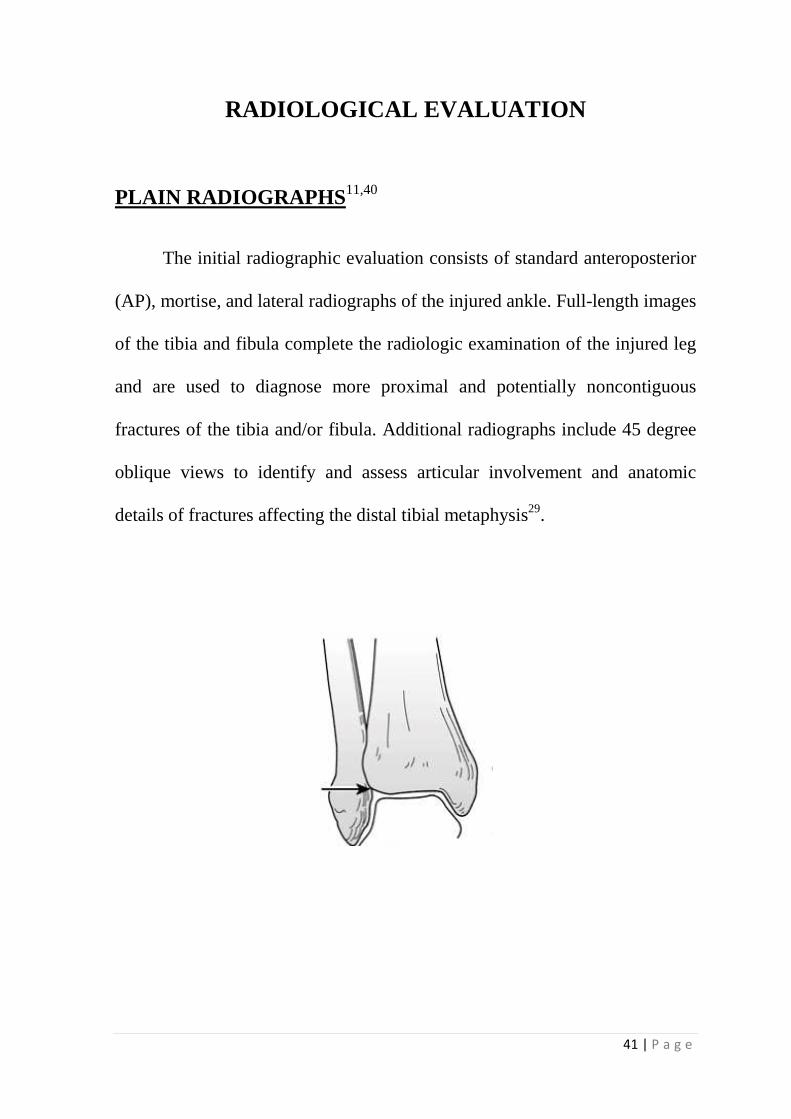
RADIOLOGICAL EVALUATION
PLAIN RADIOGRAPHS11,40
The initial radiographic evaluation consists of standard anteroposterior
(AP), mortise, and lateral radiographs of the injured ankle. Full-length images
of the tibia and fibula complete the radiologic examination of the injured leg
and are used to diagnose more proximal and potentially noncontiguous
fractures of the tibia and/or fibula. Additional radiographs include 45 degree
oblique views to identify and assess articular involvement and anatomic
details of fractures affecting the distal tibial metaphysis29.
42 | P a g e
MORTISE VIEW11,40
It is taken by internally rotating the leg up to 15 degrees, so that x-ray
beam passes perpendicular to the intermalleolar line. This view helps in
evaluating the lateral talar shift (the medial clear space), fibular shortening
and fibular rotation (tibiofibular line)29.
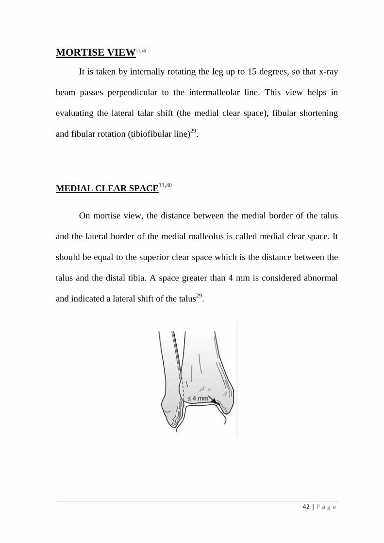
MEDIAL CLEAR SPACE11,40
On mortise view, the distance between the medial border of the talus
and the lateral border of the medial malleolus is called medial clear space. It
should be equal to the superior clear space which is the distance between the
talus and the distal tibia. A space greater than 4 mm is considered abnormal
and indicated a lateral shift of the talus29.
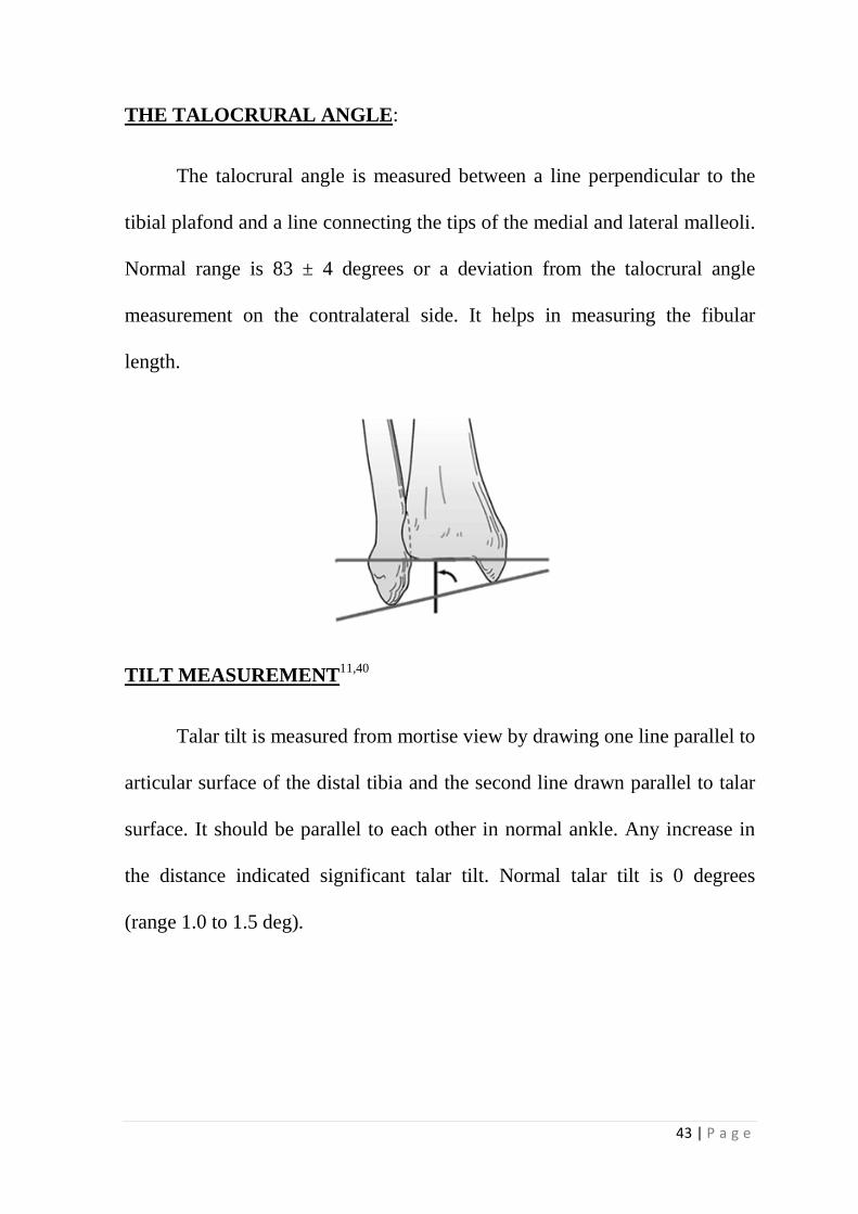
THE TALOCRURAL ANGLE
The talocrural angle is measured between a
tibial plafond and a line connecting the tips of the medial and lateral malleoli.
Normal range is 83 ± 4 degrees or a deviation from the talocrural angle
measurement on the contralateral side. It helps in measuring the fibular
length.
TILT MEASUREMENT
Talar tilt is measured from mortise view by drawing one line parallel to
articular surface of the distal tibia and the second line drawn parallel to talar
surface. It should be parallel to each other in normal ankle. Any incre
the distance indicated significant talar tilt. Normal talar tilt is 0 degrees
(range 1.0 to 1.5 deg).
THE TALOCRURAL ANGLE:
The talocrural angle is measured between a line perpendicular to the
tibial plafond and a line connecting the tips of the medial and lateral malleoli.
Normal range is 83 ± 4 degrees or a deviation from the talocrural angle
measurement on the contralateral side. It helps in measuring the fibular
TILT MEASUREMENT11,40
Talar tilt is measured from mortise view by drawing one line parallel to
articular surface of the distal tibia and the second line drawn parallel to talar
surface. It should be parallel to each other in normal ankle. Any incre
the distance indicated significant talar tilt. Normal talar tilt is 0 degrees
43 | P a g e
line perpendicular to the
tibial plafond and a line connecting the tips of the medial and lateral malleoli.
Normal range is 83 ± 4 degrees or a deviation from the talocrural angle
measurement on the contralateral side. It helps in measuring the fibular
Talar tilt is measured from mortise view by drawing one line parallel to
articular surface of the distal tibia and the second line drawn parallel to talar
surface. It should be parallel to each other in normal ankle. Any increase in
the distance indicated significant talar tilt. Normal talar tilt is 0 degrees
44 | P a g e
In AP view talar tilt is measured by the difference in width of superior
clear space between medial and lateral sides of joint and it should by <2 mm.
These are the static measurements. The talus may tilt up to 5 degrees with
respect to inversion stress.
TIBIOFIBULAR LINE:
It is formed by sub chondral surface of distal tibia and medial aspect of
the fibula. It should be continuous indicating that the articular surface of the
talus is congruous with that of distal fibula. Any disruption indicates
shortening, rotation and lateral displacement of the lateral malleolus and also
syndesmotic ligaments disruption.
EVALUATION OF SYNDESMOSIS:
The simplest method is to measure the distance between the medial
wall of the fibula and incisural surface of the tibia. This tibiofibular clear
space should be less than 6 mm on both AP and Mortise views.
45 | P a g e
CT SCAN40
Standard tomography is helpful in documenting articular surface
involvement, fracture communition and osteochondral lesion of the talus.
Computerized Tomography gives a clear cut idea of the number, size,
position and shape of the various fracture fragments. It is important in all
cases that are evaluated for open reduction and internal fixation. Three
dimensional reconstruction cuts adds to more details and helps in better
understanding of the fracture morphology.
MRI SCAN
This investigation provides excellent soft-tissue contrast resolution, has
proved to be superior to CT for evaluation of soft tissue structures around
ankle. The pathologic conditions of the ligaments and nerve entrapments are
demonstrated clearly, so that appropriate treatment can be planned.
46 | P a g e
METHODS OF TREATMENT
NON-OPERATIVE TREATMENT
Non operative treatment includes long leg plaster cast technique.
Casting is reserved for non-displaced fractures after thoroughly scrutinizing
radiographs in two views and for those patients with displaced fractures who
have relative or absolute contraindication to surgical management. Axially
loading fractures of the distal tibia with metaphyseal and articular
displacement are not indicated for this type of treatment.
OPERATIVE TREATMENT
The principles of operative treatment are anatomic restoration of
articular surface, maintain proper length and axial alignment, stable fixation
of fractures, early mobilization of joints. Displaced pilon fractures are
managed operatively, particularly those with displaced intra-articular fracture
fragments. The age, velocity of injury, condition of local soft tissue envelope,
fracture pattern and comorbid conditions are the major determinants for
proceeding with operative treatment4,6,8,10,32.
47 | P a g e
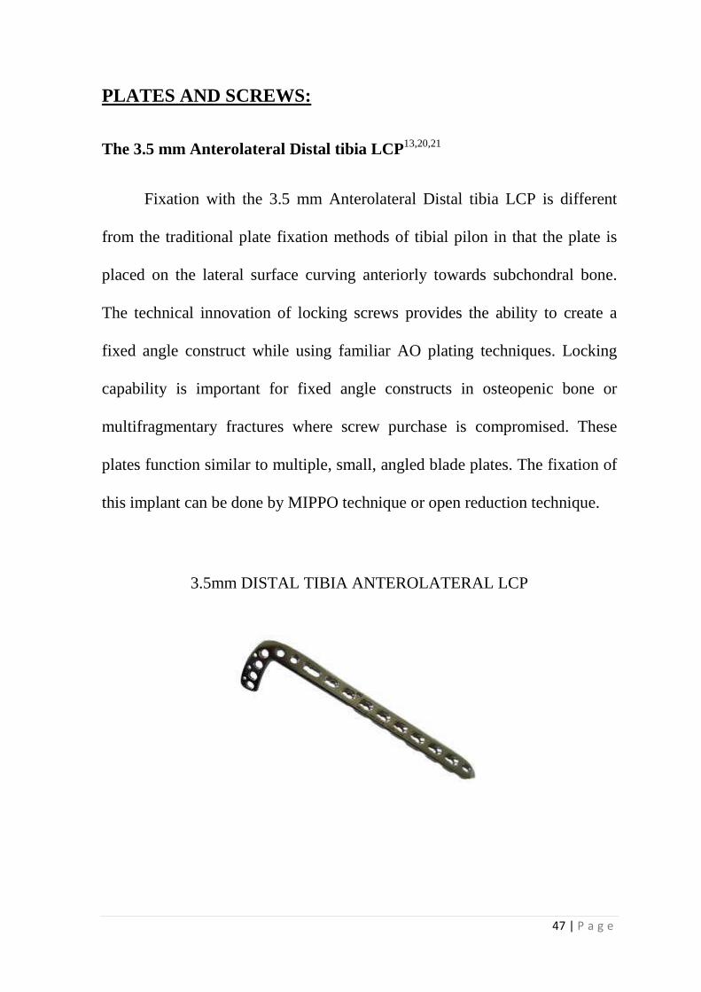
PLATES AND SCREWS:
The 3.5 mm Anterolateral Distal tibia LCP13,20,21
Fixation with the 3.5 mm Anterolateral Distal tibia LCP is different
from the traditional plate fixation methods of tibial pilon in that the plate is
placed on the lateral surface curving anteriorly towards subchondral bone.
The technical innovation of locking screws provides the ability to create a
fixed angle construct while using familiar AO plating techniques. Locking
capability is important for fixed angle constructs in osteopenic bone or
multifragmentary fractures where screw purchase is compromised. These
plates function similar to multiple, small, angled blade plates. The fixation of
this implant can be done by MIPPO technique or open reduction technique.
3.5mm DISTAL TIBIA ANTEROLATERAL LCP
48 | P a g e
SURGICAL APPROACHES46
Skin incisions and surgical approaches to tibial pilon are many and
they have been tested and modified over many decades in an attempt to
decrease the incidence of wound complications. Extensile incisions are
avoided in the distal tibia because of the precarious vascular supply and risk
of wound dehiscence
Although the indications to fix the fibula internally have been modified
in recent years, it is still an integral part of fixing tibial plafond fractures, so
that length and axial alignment can be maintained.
TIBIA
Surgical approaches40,46 include
1. Anteromedial
2. Anterolateral
3. Modified anteromedial
4. Posterolateral
5. Posteromedial
49 | P a g e
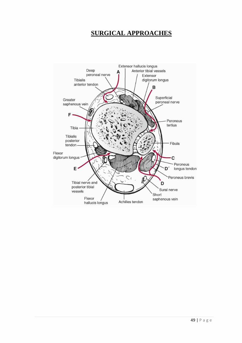
SURGICAL APPROACHES
50 | P a g e
SURGICAL TECHNIQUE
Positioning:
• Regional Anaesthesia.
• Supine position on an operating table with a radiolucent extension. A
small soft rolled towel is placed beneath the ipsilateral buttock to
decrease the tendency to externally rotate.
• Ipsilateral limb is painted from groin to foot.
Surgical Exposure:
Fibula fixation is performed initially to restore length and achieve indirect
reduction of tibia fracture. Fibula fractured is fixed by the following steps:
Fibula (Postero lateral approach of Henry):40,46
Skin incision begins 12 cm proximal to the tip of the lateral malleolus
and extended distally along the posterior margin of the fibula to the tip of the
lateral malleolus. It is curved anteriorly for 2.5 to 4 cm in the line of the
peroneal tendons.
Fibula, including the lateral malleolus is exposed subperiosteally, and
the sheaths of the peroneal retinacula and tendons are incised. This permits
51 | P a g e
the tendons to be displaced anteriorly. Fibula reduction and fixation is carried
out.
FIBULA FIXATION:
• Proximal and distal fracture ends are reached. Fragment ends are
freshened using curette.
• Reduction of fracture achieved by inline traction and varus force to
bring the overridden distal fragment beneath the proximal fragment.
• Reduction confirmed using image intensifier.
• One-third/Semi tubular plate secured to lateral surface of fibula using
two plate holding forceps.
• After confirming fracture reduction again and ruling out plate offset to
bone, three 3.5mm conventional cortical screws distally and , three
3.5mm conventional cortical screws proximally are inserted.
• Wound wash given and closed in layers over drain.
52 | P a g e
POSITION INCISION
EXPOSURE FIXATION
53 | P a g e
CLOSURE
This posterolateral incision provides a larger skin bridge between this
incision and the tibial incision. A 7 cm skin bridge was routinely
recommended. Howard recently demonstrated minimal soft tissue
complications with skin incision bridges between 5 and 6 cm when treating
tibial plafond fractures.
54 | P a g e
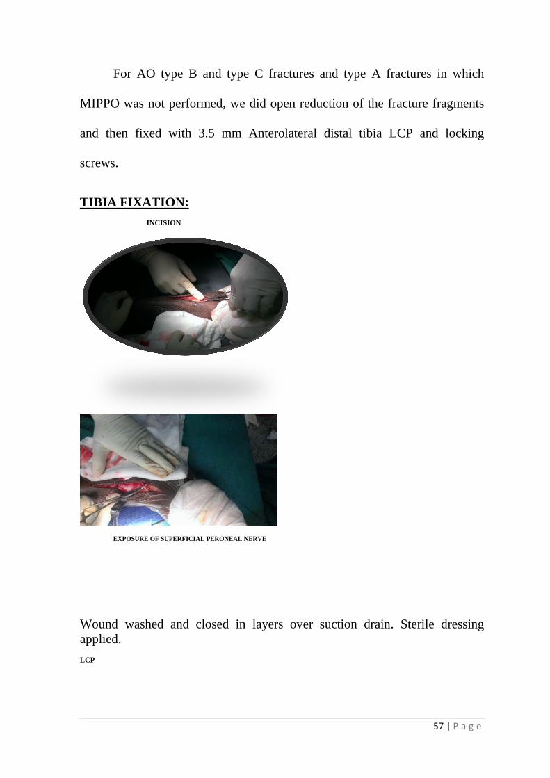
TIBIA FIXATION:
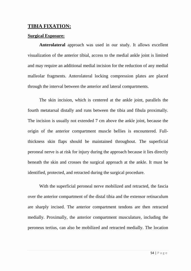
Surgical Exposure:
Anterolateral approach was used in our study. It allows excellent
visualization of the anterior tibial, access to the medial ankle joint is limited
and may require an additional medial incision for the reduction of any medial
malleolar fragments. Anterolateral locking compression plates are placed
through the interval between the anterior and lateral compartments.
The skin incision, which is centered at the ankle joint, parallels the
fourth metatarsal distally and runs between the tibia and fibula proximally.
The incision is usually not extended 7 cm above the ankle joint, because the
origin of the anterior compartment muscle bellies is encountered. Full-
thickness skin flaps should be maintained throughout. The superficial
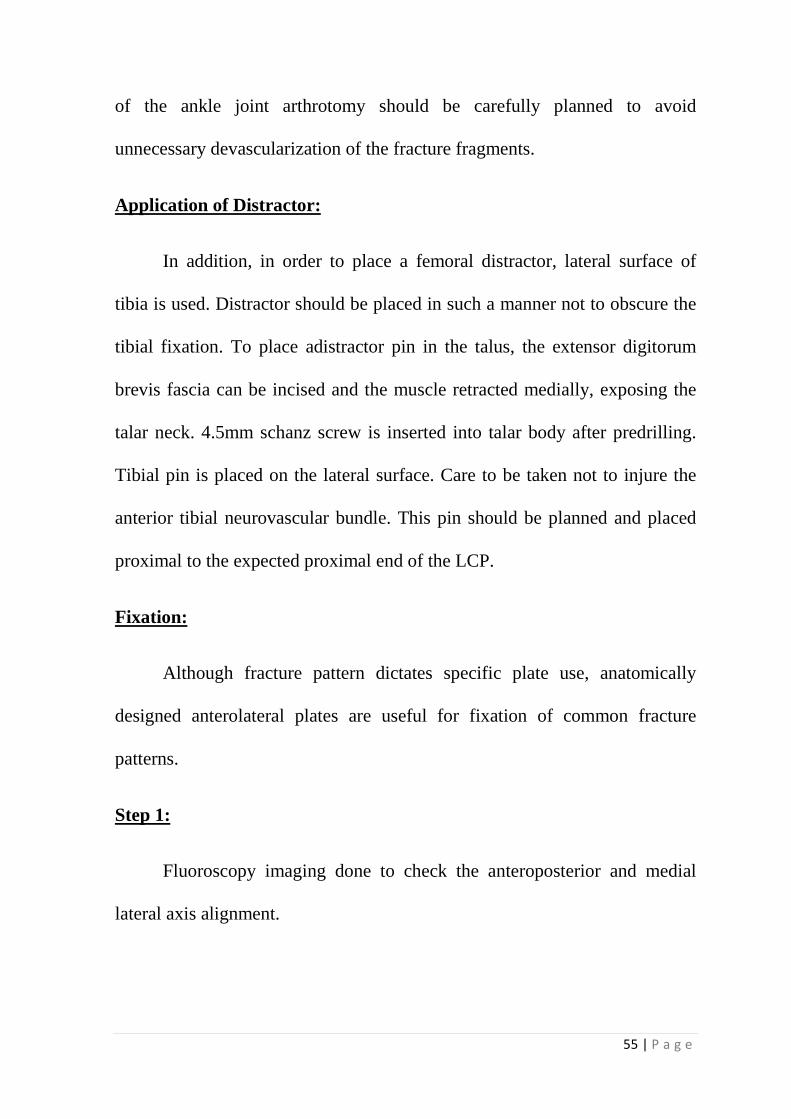
peroneal nerve is at risk for injury during the approach because it lies directly
beneath the skin and crosses the surgical approach at the ankle. It must be
identified, protected, and retracted during the surgical procedure.
With the superficial peroneal nerve mobilized and retracted, the fascia
over the anterior compartment of the distal tibia and the extensor retinaculum
are sharply incised. The anterior compartment tendons are then retracted
medially. Proximally, the anterior compartment musculature, including the
peroneus tertius, can also be mobilized and retracted medially. The location
55 | P a g e
of the ankle joint arthrotomy should be carefully planned to avoid
unnecessary devascularization of the fracture fragments.
Application of Distractor:
In addition, in order to place a femoral distractor, lateral surface of
tibia is used. Distractor should be placed in such a manner not to obscure the
tibial fixation. To place adistractor pin in the talus, the extensor digitorum
brevis fascia can be incised and the muscle retracted medially, exposing the
talar neck. 4.5mm schanz screw is inserted into talar body after predrilling.
Tibial pin is placed on the lateral surface. Care to be taken not to injure the
anterior tibial neurovascular bundle. This pin should be planned and placed
proximal to the expected proximal end of the LCP.
Fixation:
Although fracture pattern dictates specific plate use, anatomically
designed anterolateral plates are useful for fixation of common fracture
patterns.
Step 1:
Fluoroscopy imaging done to check the anteroposterior and medial
lateral axis alignment.
56 | P a g e
Step 2:
Articular fragment and main fracture fragments identified, reduced and
fixed with temporary k – wires. Order of reduction of articular fragments is
posterolateral--->posteromedial-->central-->anterior-->anterolateral.
Step 3:
Articular fragments reduced to proximal metaphyseal fragment and
locking compression plate is slid and positioned over the anterior subchondral
surface and lateral surface of tibia. Length of the plate is three times the span
of fractured segment.
Step 4:
Plate position and offset checked using fluoroscopy and then definitive
fixation carried out. First screw being 3.5mm conventional cortical screw
predrilled with 2.7mm drill bit proximally followed by locking 3.5mm
cortical screws proximally and distally.
For AO type A fractures we used the technique of MIPPO in six cases
successfully. Here the distal part of the approach was used and it is a
longitudinal incision. The fracture was reduced indirectly and the plate was
inserted through the incision. Through separate incision, screws were inserted
in the proximal fragment17,26.
For AO type B and type C fractures and type A fractures in which
MIPPO was not performed, we did open reduction of the fracture fragments
and then fixed with 3.5 mm Anterolateral dista
screws.
TIBIA FIXATION:
INCISION
EXPOSURE OF SUPERFICIAL PERONEAL NERVE
Wound washed and closed in layers over suction drain. Sterile dressing applied.
LCP
For AO type B and type C fractures and type A fractures in which
MIPPO was not performed, we did open reduction of the fracture fragments
and then fixed with 3.5 mm Anterolateral distal tibia LCP and locking
EXPOSURE OF SUPERFICIAL PERONEAL NERVE
Wound washed and closed in layers over suction drain. Sterile dressing
57 | P a g e
For AO type B and type C fractures and type A fractures in which
MIPPO was not performed, we did open reduction of the fracture fragments
l tibia LCP and locking
Wound washed and closed in layers over suction drain. Sterile dressing
58 | P a g e
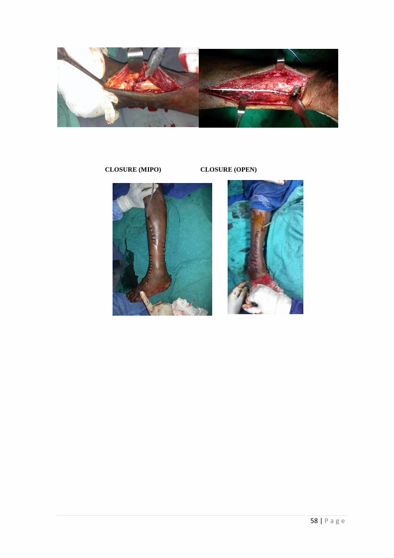
CLOSURE (MIPO) CLOSURE (OPEN)
59 | P a g e
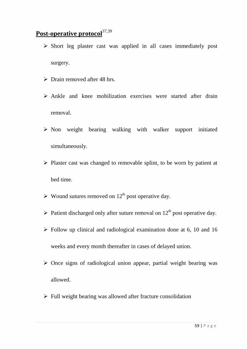
Post-operative protocol37,39
� Short leg plaster cast was applied in all cases immediately post
surgery.
� Drain removed after 48 hrs.
� Ankle and knee mobilization exercises were started after drain
removal.
� Non weight bearing walking with walker support initiated
simultaneously.
� Plaster cast was changed to removable splint, to be worn by patient at
bed time.
� Wound sutures removed on 12th post operative day.
� Patient discharged only after suture removal on 12th post operative day.
� Follow up clinical and radiological examination done at 6, 10 and 16
weeks and every month thereafter in cases of delayed union.
� Once signs of radiological union appear, partial weight bearing was
allowed.
� Full weight bearing was allowed after fracture consolidation
60 | P a g e
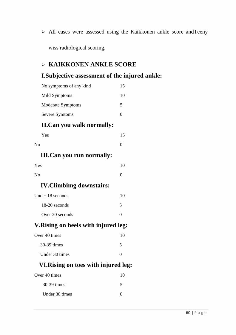
� All cases were assessed using the Kaikkonen ankle score andTeeny
wiss radiological scoring.
� KAIKKONEN ANKLE SCORE
I.Subjective assessment of the injured ankle:
No symptoms of any kind 15
Mild Symptoms 10
Moderate Symptoms 5
Severe Symtoms 0
II.Can you walk normally:
Yes 15
No 0
III.Can you run normally:
Yes 10
No 0
IV.Climbimg downstairs:
Under 18 seconds 10
18-20 seconds 5
Over 20 seconds 0
V.Rising on heels with injured leg:
Over 40 times 10
30-39 times 5
Under 30 times 0
VI.Rising on toes with injured leg:
Over 40 times 10
30-39 times 5
Under 30 times 0
61 | P a g e
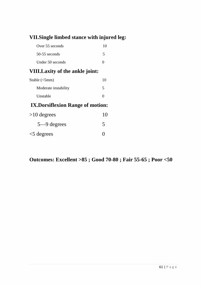
VII.Single limbed stance with injured leg:
Over 55 seconds 10
50-55 seconds 5
Under 50 seconds 0
VIII.Laxity of the ankle joint:
Stable (<5mm) 10
Moderate instability 5
Unstable 0
IX.Dorsiflexion Range of motion:
>10 degrees 10
5—9 degrees 5
<5 degrees 0
Outcomes: Excellent >85 ; Good 70-80 ; Fair 55-65 ; Poor <50
62 | P a g e
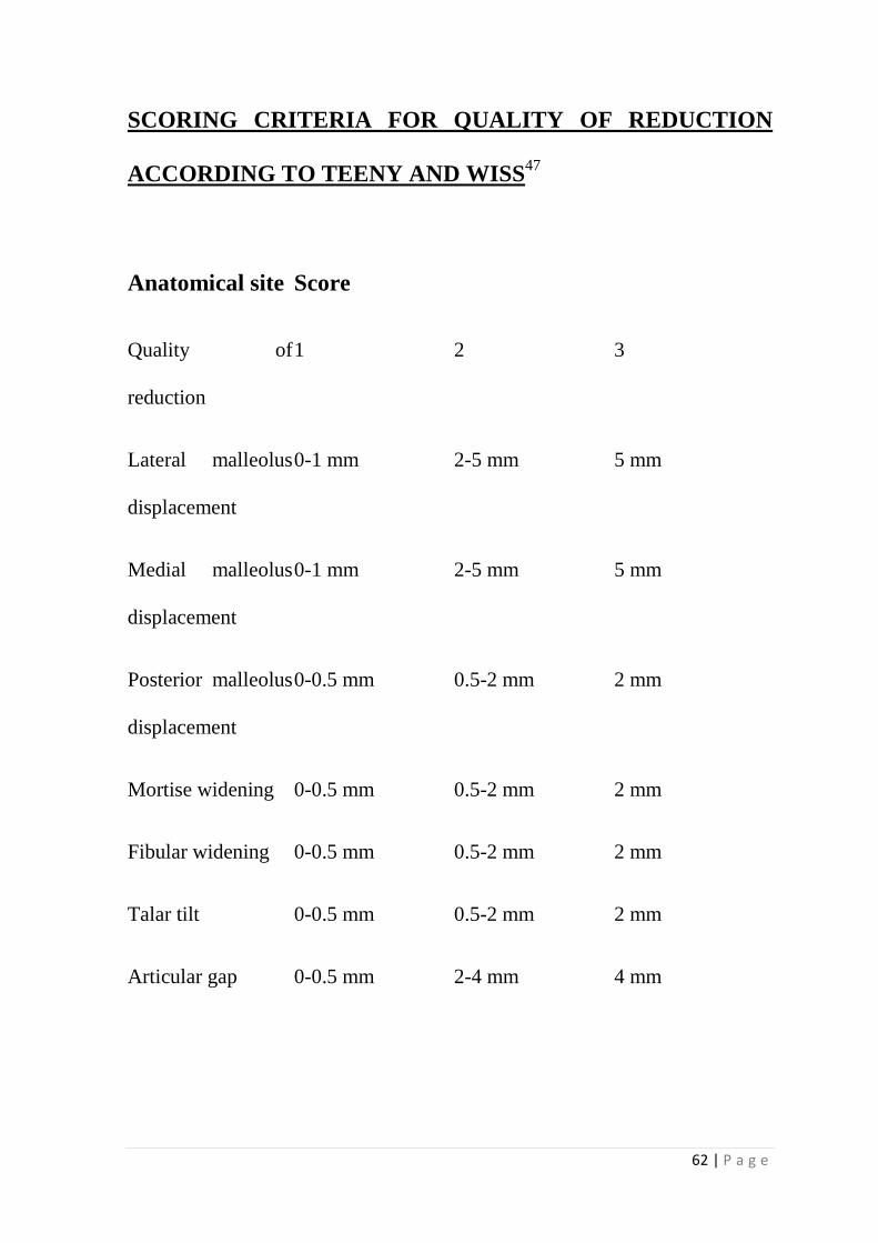
SCORING CRITERIA FOR QUALITY OF REDUCTION
ACCORDING TO TEENY AND WISS47
Anatomical site
Quality of
reduction
Score
1 2 3
Lateral malleolus
displacement
0-1 mm 2-5 mm 5 mm
Medial malleolus
displacement
0-1 mm 2-5 mm 5 mm
Posterior malleolus
displacement
0-0.5 mm 0.5-2 mm 2 mm
Mortise widening 0-0.5 mm 0.5-2 mm 2 mm
Fibular widening 0-0.5 mm 0.5-2 mm 2 mm
Talar tilt 0-0.5 mm 0.5-2 mm 2 mm
Articular gap 0-0.5 mm 2-4 mm 4 mm

63 | P a g e
Rating
Points
Anatomic 9
Good
10-12
Fair
13-16
Poor > 16

RESULTS
64 | P a g e
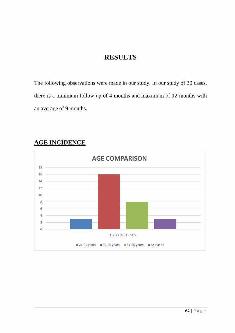
RESULTS
The following observations were made in our study. In our study of 30 cases,
there is a minimum follow up of 4 months and maximum of 12 months with
an average of 9 months.
AGE INCIDENCE
0
2
4
6
8
10
12
14
16
18
AGE COMPARISON
AGE COMPARISON
21-35 years 36-50 years 51-65 years Above 65

20%
0
2
4
6
8
10
12
14
16
18
20
SEX INCIDENCE:
MODE OF INJURY:
80%
20%
Sex Incidence
Mode of Injury
Self Fall
Road Traffic Accident
65 | P a g e
Male
Female
Self Fall
Road Traffic Accident
66 | P a g e
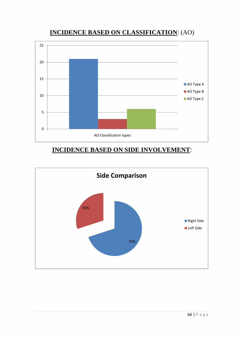
INCIDENCE BASED ON CLASSIFICATION: (AO)
INCIDENCE BASED ON SIDE INVOLVEMENT:
0
5
10
15
20
25
AO Classification types
AO Type A
AO Type B
AO Type C
70%
30%
Side Comparison
Right Side
Left Side
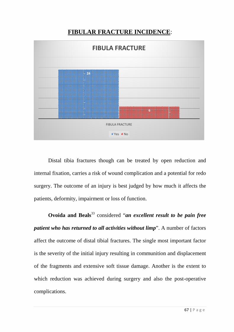
FIBULAR FRACTURE INCIDENCE
Distal tibia fractures though can be treated by
internal fixation, carries a
surgery. The outcome of an injury is
patients, deformity, impairment or loss of function.
Ovoida and Beals
patient who has returned to all activities without limp
affect the outcome of distal tibial fractures. The single most important factor
is the severity of the initial injury resulting in comm
of the fragments and extensive
which reduction was achieved
complications.
FIBULAR FRACTURE INCIDENCE
fractures though can be treated by open reduction and
, carries a risk of wound complication and a potential for redo
surgery. The outcome of an injury is best judged by how much it affects the
patients, deformity, impairment or loss of function.
Ovoida and Beals33 considered “an excellent result to be pain free
patient who has returned to all activities without limp”. A number of factors
affect the outcome of distal tibial fractures. The single most important factor
severity of the initial injury resulting in communition and
nd extensive soft tissue damage. Another is the extent
which reduction was achieved during surgery and also the post
24
6
FIBULA FRACTURE
FIBULA FRACTURE
Yes No
67 | P a g e
FIBULAR FRACTURE INCIDENCE:
open reduction and
complication and a potential for redo
best judged by how much it affects the
an excellent result to be pain free
”. A number of factors
affect the outcome of distal tibial fractures. The single most important factor
tion and displacement
soft tissue damage. Another is the extent to
and also the post-operative
68 | P a g e
FRACTURE UNION:
27 fractures united. Radiological union obtained in 27cases with a
mean duration of 12 to 24 weeks. Three patients went in for non union and
out of which one implant failure occured. All patients were started full weight
bearing after signs of radiological union.
FUNCTIONAL SCORE:
A variety of rating systems were proposed for subjective and objective
components. We have used Teeny and Wiss Score for radiological evaluation
of ankle and Kaikkonen ankle scoring for functional analysis. The mean
functional ankle scores were 72 with a maximum of 95 and minimum of 40.
Anatomic reduction was achieved in about 7 cases with good alignment.
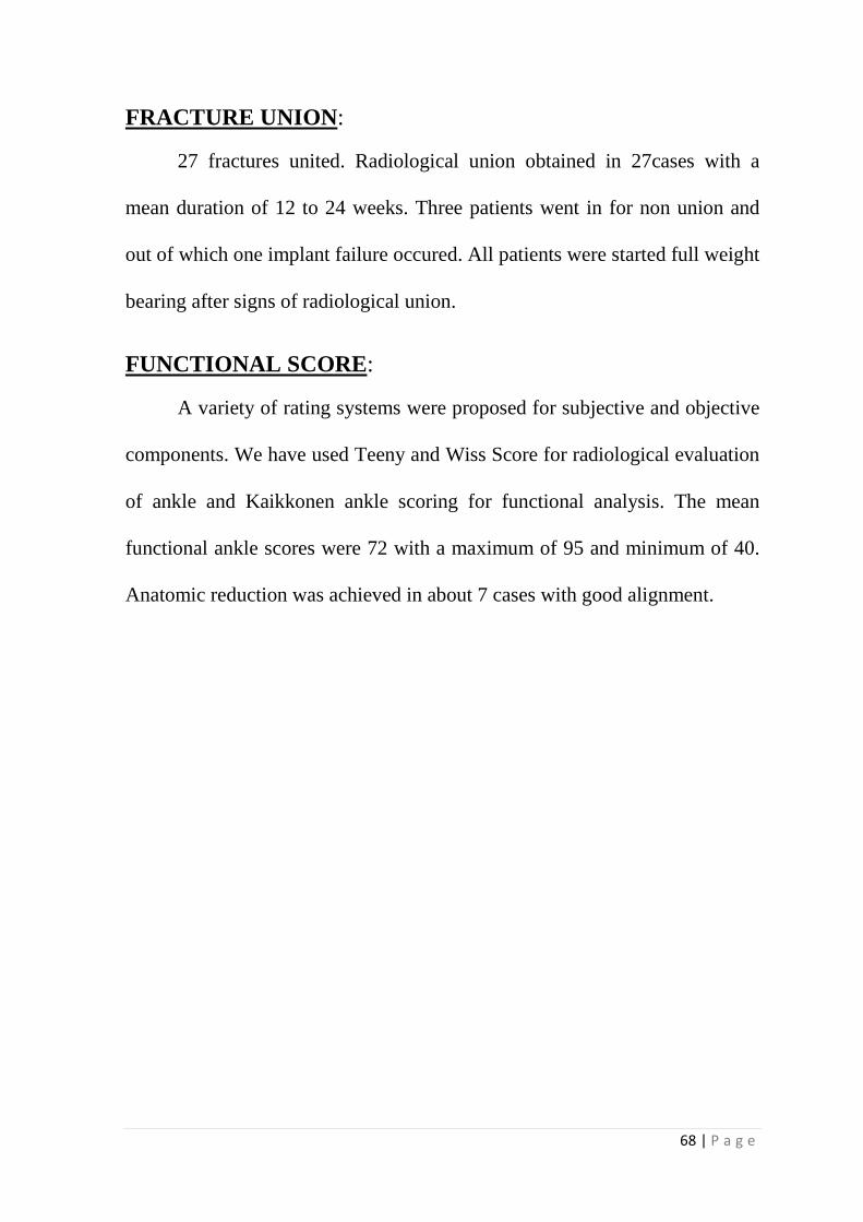
TEENY WISS RADIOLOGICAL SCORING
In our study we were able to achieve anatomic reduction in 32% (7
the patients. Good reduction was achieved in
Fair reduction was achieved in 18% (4 cases). There was no case
fracture reduction in our study.
Reduction AO Type A
Anatomic 6
Good 12
Fair 3
Poor 0
TEENY WISS RADIOLOGICAL SCORING
5
8
02
ANATOMIC
RADIOLOGICAL SCORING:
udy we were able to achieve anatomic reduction in 32% (7
Good reduction was achieved in 50%(11 cases) of the patients
Fair reduction was achieved in 18% (4 cases). There was no case
in our study.
AO Type A Type B
0
12 0
3
0
TEENY WISS RADIOLOGICAL SCORING
8
200
2
3
0
GOOD FAIR POOR
TEENY WISS SCORE
AO Type A AO Type B AO Type C
69 | P a g e
udy we were able to achieve anatomic reduction in 32% (7 cases) of
50%(11 cases) of the patients47.
Fair reduction was achieved in 18% (4 cases). There was no case of poor
Type C
3
3
0
0
0 0
POOR
KAIKKONEN ANKLE SCORE
Scoring Excellent
Type A 6
Type B 0
Type C 3
KAIKKONEN ANKLE SCORE
ANKLE SCORE :
Excellent Good Fair
12 0
0 3
3 0
ANKLE SCORE :
Excellent
32%
Good
50%
Fair
9%
Poor
9%
KAIKKONEN SCORE
70 | P a g e
Poor
3
0
0

COMPLICATIONS
71 | P a g e
COMPLICATIONS:
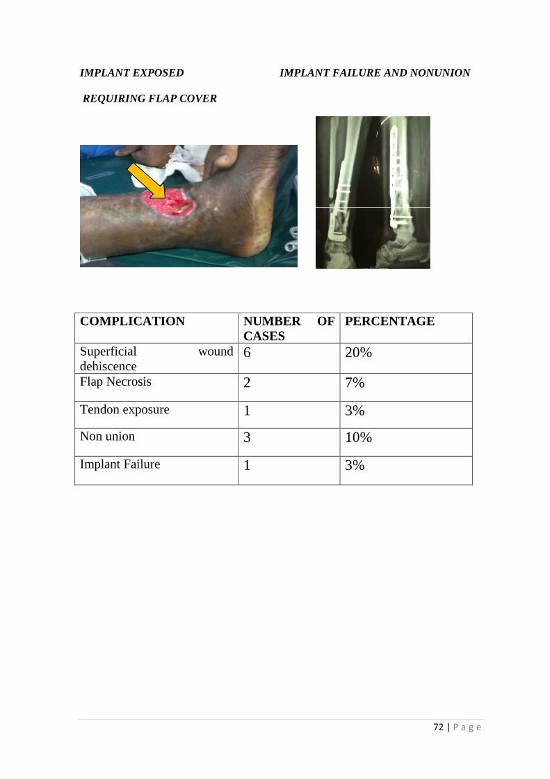
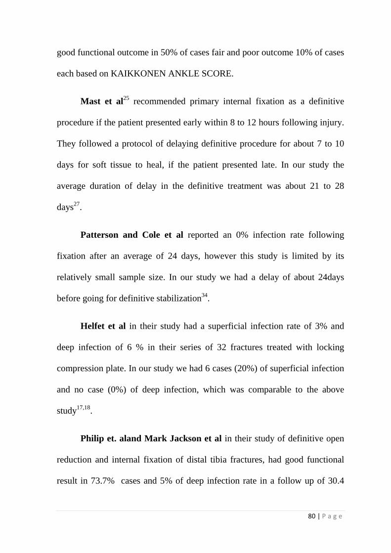
In our study the complication we met were 6 cases (20%) of wound
dehiscence and superficial infection which healed by secondary intention, 2
cases (7%)of flap necrosis, 3cases(10%) of nonunion, extensor tendon
exposed in 1 case(3%), implant failure in one of the three non union cases. In
our study we had no deep infection(0%).
SUPERFICIAL WOUND DEHISCENCE EXTENSOR TENDON EXPOSED
72 | P a g e
IMPLANT EXPOSED IMPLANT FAILURE AND NONUNION REQUIRING FLAP COVER
COMPLICATION NUMBER OF CASES
PERCENTAGE
Superficial wound dehiscence
6 20%
Flap Necrosis 2 7%
Tendon exposure 1 3%
Non union 3 10%
Implant Failure 1 3%

DISCUSSION
73 | P a g e
DISCUSSION
Distal tibia fractures result from low energy torsional or high energy
axial-loading mechanisms. High energy fractures are commonly associated
with severe soft tissue injury, comminution of metaphyseal and articular
fracture fragments of tibial plafond and comminuted distal fibula fractures.
Tibial pilon fractures account for <10% of lower extremity fractures and
occur in adults owing to fall from height or from road traffic accidents. The
optimal treatment for these fractures remains controversial. This is due tothe
associated significant soft tissue injury and precarious vascular supply of
distal tibia. The treatment of distal tibia fractures can be challenging because
of its subcutaneous location, poor vascularity and limited soft tissue3,4,12.
The main factor in treating these injuries is to estimate the degree of
associated soft tissue injury. Since only closed fractures were included in our
study, we used Tscherne soft tissue injury classification to assess and grade
the severity of soft tissue injury. Definitive fixation is advisable and
proceeded only when the soft tissue injury heals. This is indicated by the skin
wrinkle sign, once limb edema subsides. In our study, internal fixation was
carried out at an average of 3 to 4 weeks once wrinkle sign developed.
Minimally invasive plating techniques (MIPO) reduce the iatrogenic
soft tissue injury and damage to bone vascularity, and also preserve the
74 | P a g e
osteogenic fracture hematoma. But even MIPO techniques should be
performed after soft tissues heal. And with a delay of three weeks, MIPO is
not possible in some cases. This is why in our study too, MIPO couldnot be
carried out even in some AO type A fractures.
The key principles in the management of these fractures are –
1)Restoration of the length and limb axis by open reduction and internal
fixation of fibula fracture;
2) the anatomical reconstruction of the articular surface of tibial plafond; 3)
the filling of the defect resulting from impaction and the support of the lateral
side of tibia, by lateral plating to prevent the valgus deformity32.
In our study we used a single-stage direct internal fixation technique of
all distal tibia fractures. Average delay to fixation was 24 days. We used
3.5mm anterolateral distal tibia locking compression plate for all cases. This
plate is a low profile plate of 3.5 mm system. The 3.5mm anterolateral distal
tibia LCP is a pre-contoured plate to suit the lateral surface of tibial shaft and
anterior surface of the tibial plafond abutting the subchondral bone. This
design allows placement of the plate without disruption of fractures
fragments. The thread holes in the plate locks to that of the screw head and
minimize plate-bone interface and maintain the vascularity at the fracture
site.
75 | P a g e
Among 30 patients, 6 AO type A fractures were managed with MIPPO
technique. Rest of the AO type A, AO type B and AO type C fractures were
managed with open reduction and fixation with locking compression plate.
Our study included a total of 30 patients. The peak incidence in our
study was among the age group between 36-50 years. In our study we had
excellent functional outcome in about 30% of cases, good functional outcome
in 50% of cases fair and poor outcome 10% of cases each based on
KAIKKONEN ANKLE SCORE. As per TEENY WISS SCORE47 we had
anatomic reduction in 9 cases, good reduction in 15 cases, fair reduction in 6
cases.
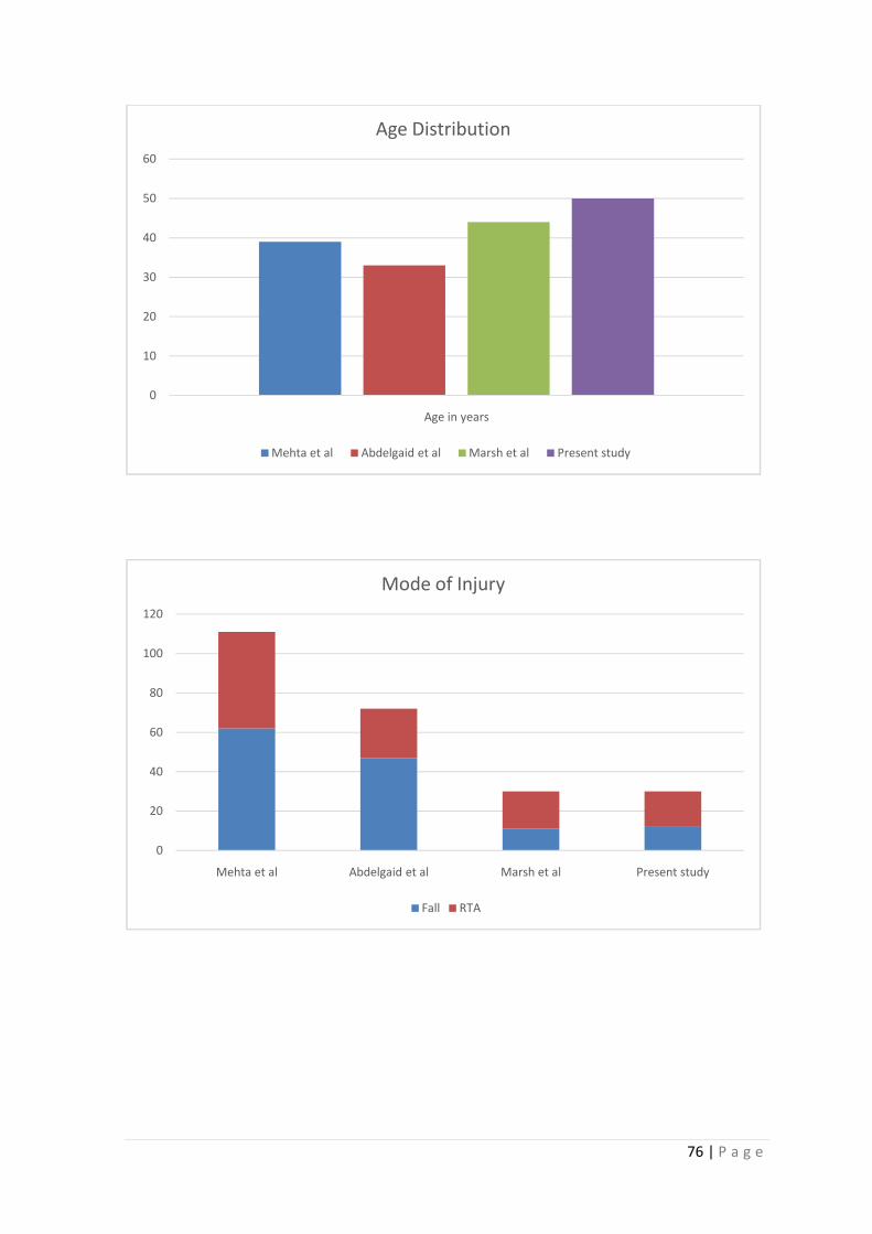
In the study by Mehta et al27of 131 patients, average age was 39 years
(compared to 50 years in present study), most injuries were due to fall
(compared to road traffic accident in our study), side involvement was almost
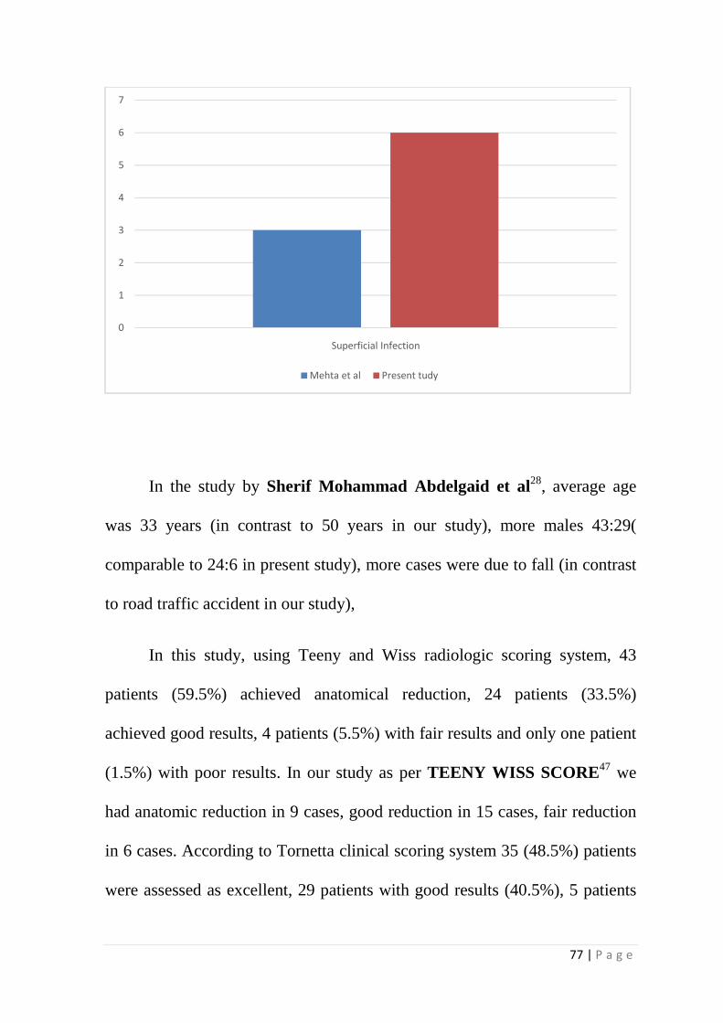
equal (right side predominant in our study). They reported superficial
infection in three patients (compared to 6 cases in present study). Superficial
infection rate was higher in our study. Average delay to surgery was 14 day
(compared to 24 days in present study).
76 | P a g e
0
10
20
30
40
50
60
Age in years
Age Distribution
Mehta et al Abdelgaid et al Marsh et al Present study
0
20
40
60
80
100
120
Mehta et al Abdelgaid et al Marsh et al Present study
Mode of Injury
Fall RTA
77 | P a g e
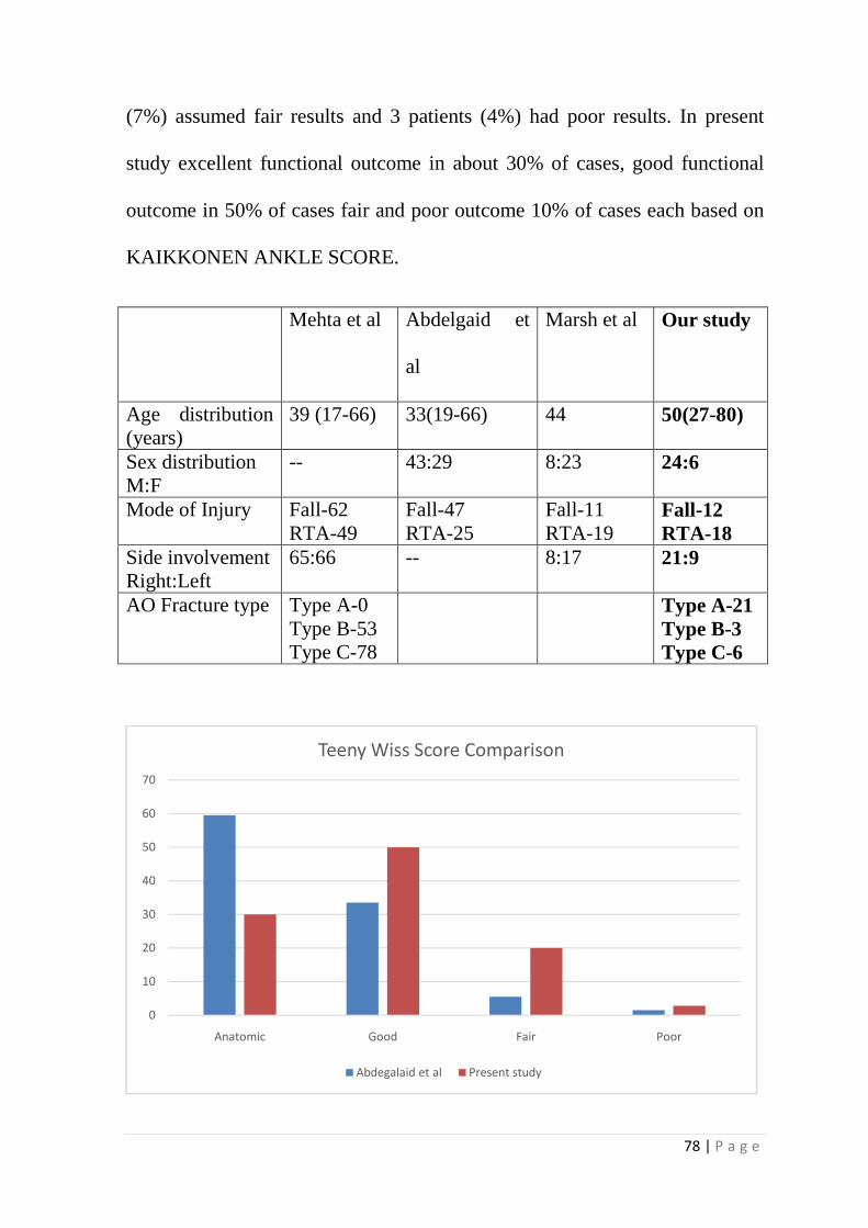
In the study by Sherif Mohammad Abdelgaid et al28, average age
was 33 years (in contrast to 50 years in our study), more males 43:29(
comparable to 24:6 in present study), more cases were due to fall (in contrast
to road traffic accident in our study),
In this study, using Teeny and Wiss radiologic scoring system, 43
patients (59.5%) achieved anatomical reduction, 24 patients (33.5%)
achieved good results, 4 patients (5.5%) with fair results and only one patient
(1.5%) with poor results. In our study as per TEENY WISS SCORE47 we
had anatomic reduction in 9 cases, good reduction in 15 cases, fair reduction
in 6 cases. According to Tornetta clinical scoring system 35 (48.5%) patients
were assessed as excellent, 29 patients with good results (40.5%), 5 patients
0
1
2
3
4
5
6
7
Superficial Infection
Mehta et al Present tudy
78 | P a g e
(7%) assumed fair results and 3 patients (4%) had poor results. In present
study excellent functional outcome in about 30% of cases, good functional
outcome in 50% of cases fair and poor outcome 10% of cases each based on
KAIKKONEN ANKLE SCORE.
Mehta et al Abdelgaid et
al
Marsh et al Our study
Age distribution (years)
39 (17-66) 33(19-66) 44 50(27-80)
Sex distribution M:F
-- 43:29 8:23 24:6
Mode of Injury Fall-62 RTA-49
Fall-47 RTA-25
Fall-11 RTA-19
Fall-12 RTA-18
Side involvement Right:Left
65:66 -- 8:17 21:9
AO Fracture type Type A-0 Type B-53 Type C-78
Type A-21 Type B-3 Type C-6
0
10
20
30
40
50
60
70
Anatomic Good Fair Poor
Teeny Wiss Score Comparison
Abdegalaid et al Present study
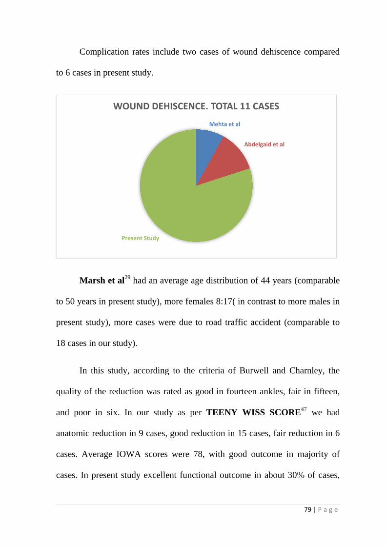
Complication rates include two cases of wound dehiscence compared
to 6 cases in present study.
Marsh et al29 had an average age distribution of 44 years (comparable
to 50 years in present study), more females 8:17( in contrast to more males in
present study), more cases were due to road traffic accident (comparable to
18 cases in our study).
In this study, accor
quality of the reduction was rated as good in fourteen ankles, fair in fifteen,
and poor in six. In our study
anatomic reduction in 9 cases, good reduction in 15 cases, fair
cases. Average IOWA scores were 78, with good outcome in majority of
cases. In present study excellent functional outcome in about 30% of cases,
Present Study
WOUND DEHISCENCE. TOTAL 11 CASES
rates include two cases of wound dehiscence compared
to 6 cases in present study.
had an average age distribution of 44 years (comparable
to 50 years in present study), more females 8:17( in contrast to more males in
present study), more cases were due to road traffic accident (comparable to
In this study, according to the criteria of Burwell and Charnley, the
quality of the reduction was rated as good in fourteen ankles, fair in fifteen,
and poor in six. In our study as per TEENY WISS SCORE
anatomic reduction in 9 cases, good reduction in 15 cases, fair
cases. Average IOWA scores were 78, with good outcome in majority of
cases. In present study excellent functional outcome in about 30% of cases,
Mehta et al
Abdelgaid et al
Present Study
WOUND DEHISCENCE. TOTAL 11 CASES
79 | P a g e
rates include two cases of wound dehiscence compared
had an average age distribution of 44 years (comparable
to 50 years in present study), more females 8:17( in contrast to more males in
present study), more cases were due to road traffic accident (comparable to
ding to the criteria of Burwell and Charnley, the
quality of the reduction was rated as good in fourteen ankles, fair in fifteen,
TEENY WISS SCORE47 we had
anatomic reduction in 9 cases, good reduction in 15 cases, fair reduction in 6
cases. Average IOWA scores were 78, with good outcome in majority of
cases. In present study excellent functional outcome in about 30% of cases,
Abdelgaid et al
80 | P a g e
good functional outcome in 50% of cases fair and poor outcome 10% of cases
each based on KAIKKONEN ANKLE SCORE.
Mast et al25 recommended primary internal fixation as a definitive
procedure if the patient presented early within 8 to 12 hours following injury.
They followed a protocol of delaying definitive procedure for about 7 to 10
days for soft tissue to heal, if the patient presented late. In our study the
average duration of delay in the definitive treatment was about 21 to 28
days27.
Patterson and Cole et al reported an 0% infection rate following
fixation after an average of 24 days, however this study is limited by its
relatively small sample size. In our study we had a delay of about 24days
before going for definitive stabilization34.
Helfet et al in their study had a superficial infection rate of 3% and
deep infection of 6 % in their series of 32 fractures treated with locking
compression plate. In our study we had 6 cases (20%) of superficial infection
and no case (0%) of deep infection, which was comparable to the above
study17,18.
Philip et. aland Mark Jackson et al in their study of definitive open
reduction and internal fixation of distal tibia fractures, had good functional
result in 73.7% cases and 5% of deep infection rate in a follow up of 30.4
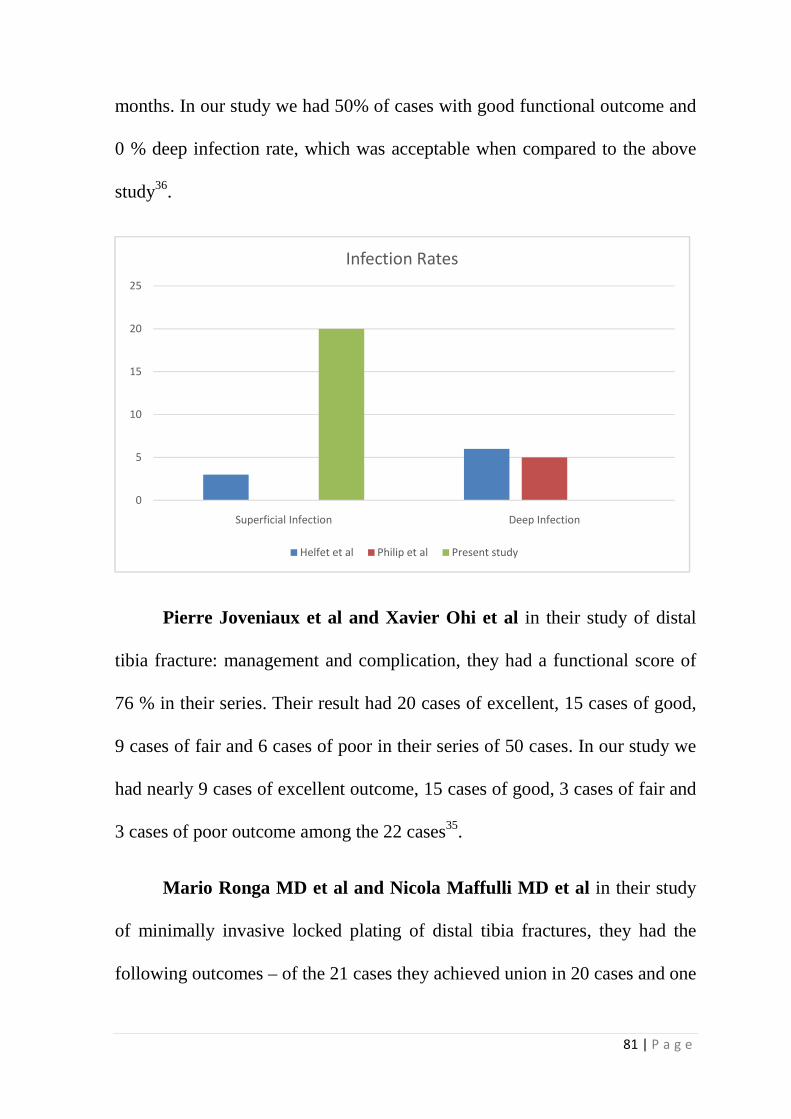
81 | P a g e
months. In our study we had 50% of cases with good functional outcome and
0 % deep infection rate, which was acceptable when compared to the above
study36.
Pierre Joveniaux et al and Xavier Ohi et al in their study of distal
tibia fracture: management and complication, they had a functional score of
76 % in their series. Their result had 20 cases of excellent, 15 cases of good,
9 cases of fair and 6 cases of poor in their series of 50 cases. In our study we
had nearly 9 cases of excellent outcome, 15 cases of good, 3 cases of fair and
3 cases of poor outcome among the 22 cases35.
Mario Ronga MD et al and Nicola Maffulli MD et al in their study
of minimally invasive locked plating of distal tibia fractures, they had the
following outcomes – of the 21 cases they achieved union in 20 cases and one
0
5
10
15
20
25
Superficial Infection Deep Infection
Infection Rates
Helfet et al Philip et al Present study
82 | P a g e
case went in for non-union. They had 3 cases of angular deformities all less
than 7° and no patient had a leg-length discrepancy. Compared to their study,
in our study we used MIPPO in about 6 cases, in which all cases went in for
union in about 10 to 14 weeks with no case of malunion or nonunion26.
Rakesh Gupta et al and Rajesh Kumar Rohllla et al in their study of
locking plate fixation in distal tibia fractures – series of 79 patients, had
reported about 88% of healing without malunion, 2.5% of malunion and 3.7%
of non-union. They used both MIPPO and ORIF for fixing these fractures.
They found good and early union rate in the MIPPO group. In our study also
we had good and early union in the MIPPO group and also we had three
cases of non-union.
Pugh and colleague evaluated 60 patients, 25 of whom were treated
with external fixators. They noted that they had more number of malunion in
the external fixator group compared to that of internal fixation. They met
most of their complication in the external fixator group. In our study also we
had good functional outcome in internal fixation group38.
The average follow up period our study was 9 months (range from 4 to
12 months). In type III fractures patients may develop late secondary
arthrosis, but it requires a longer follow up. In our series, the results of the
patients who had follow up period of upto 9 months had good clinical scores.
83 | P a g e
Arthrodesis was indicated when there was extensive communition and
also in severe type III fractures. In our study, type III injuries accounted for
20% of the cases and arthrodesis was not done in any of our patients,
compared to that of 23 % of cases in a study by M.Blauthet et al.
Marsh et al reported an arthrodesis rate of 13 % in their series of 40
ankle fractures after a minimum follow up of 5 years. Chen noted a 4.7%
arthrodesis rate in plating tibial plafond fractures when followed for a period
of 10 years. Since our study was a short term study, arthrodesis could not be
commented which might require a long term follow up.
Hence the outcome of surgically treated distal tibia fractures depends on the
severity of injury mechanism and assessing the grade of soft tissue injury,
delaying definitive fixation till soft tissue heals and accuracy of the articular
0
5
10
15
20
25
Arthrodesis rate
Ankle Arthrodesis
Marsh et al M.Blauthet et al Present study

84 | P a g e
reduction and fibula plating during definitive procedure. A correlation exists
between the severity of the fracture, overall outcome and the development of
secondary degenerative arthritis.

CONCLUSION
85 | P a g e
CONCLUSION
� A short series of result of our study were analyzed and the overall
results have encouraged us in preferring the surgical management of
distal tibia fractures over conservative methods.
� Distal tibia fractures with high grade of soft tissue injury are to be
internally fixed after a delay of 21 days for the edema to settle down
and the wrinkle sign appears.
� Respect the soft tissues: do not operate too early or through
compromised skin, instead wait till the soft tissues is amenable for
surgery.
� On the other hand, low energy fractures with insignificant soft tissue
injury can be addressed by definitive internal fixation immediately
� Restoration of the articular surface and reestablishing its relationship to
the tibial shaft is the primary goal of treatment.
� Good functional result depends on reasonable anatomic reduction of
the articular surface either by direct or indirect methods.
86 | P a g e
� Understand the fracture completely before planning any surgery
(obtain adequate radiographs, CT scan and radiographs of the
uninjured limb).
� Open reduction and internal fixation with anatomical restoration of the
articular surface is to be done in all cases AO type C fractures
otherwise it will lead to mal-alignment and secondary degenerative
osteoarthosis.
� Anatomic realignment of fibula indirectly reduces the talus beneath the
anatomic axis of tibia. Restoration of fibular length, alignment and
rotation has the substantial impact on the indirect realignment of
anterolateral and posterolateral tibial plafond from their attachment to
the anterior and posterior tibiofibular syndesmotic ligaments. Hence
fibula fixation is advocated wherever possible.
� Conservative management can be reserved in cases where fibula is
intact and type I fractures and also in those with associated medical
contraindication to surgery.
� Surgical reconstruction must be tailored to the personality of each
fractures and operative approaches dictated by the quality of the soft
tissues.
87 | P a g e
� From our study 3.5mm Anterolateral Distal tibia Locking Compression
Plating for tibial pilon fractures were found to be safe and effective.
For AO type A fractures, can be fixed either using MIPPO or ORIF
technique. For AO type C fractures Open reduction of the articular
fragment is mandatory and then stabilize with locking compression
plate for added up stability. Also the incision between the tibial and
fibula exposure must be atleast 7 cm.
� For highly comminuted injuries with unreconstructable articular
surface, primary ankle fusion is an alternative to ORIF.

CASE ILLUSTRATIONS
88 | P a g e
CASE ILLUSTRATIONS
Name : Kannan
Ip No. : 304
Age : 63/M
Occupation : Household
Mode of injury : History of Fall
Diagnosis : Distal tibia fracture
Classification : AO Type 43.A.3
Procedure : ORIF with 3.5mm anterolateral distal
tibia LCP
Approach : Anterolateral approach to ankle
Complication : None
Time of Union : 12 weeks
Ankle Range of Motion : Pain free and fully functional
Kaikkonen ankle Score : 90
Outcome : Excellent.
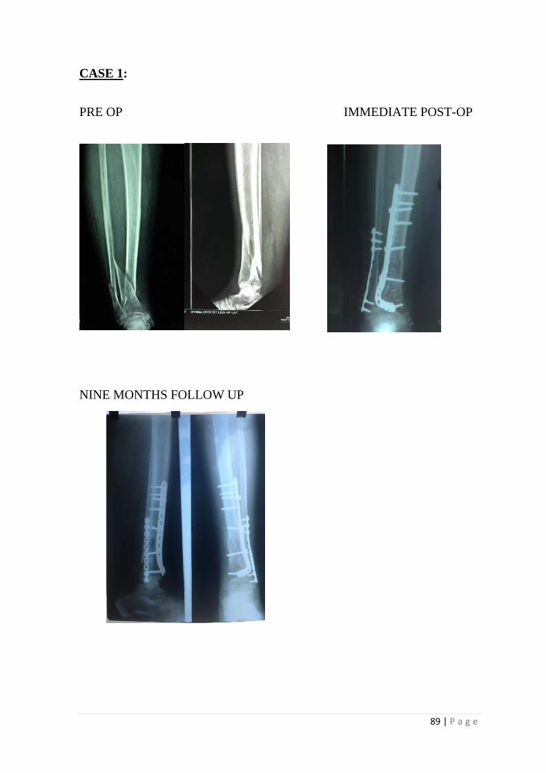
89 | P a g e
CASE 1:
PRE OP IMMEDIATE POST-OP
NINE MONTHS FOLLOW UP
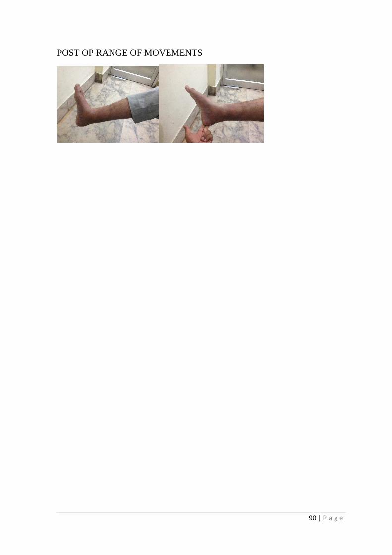
90 | P a g e
POST OP RANGE OF MOVEMENTS
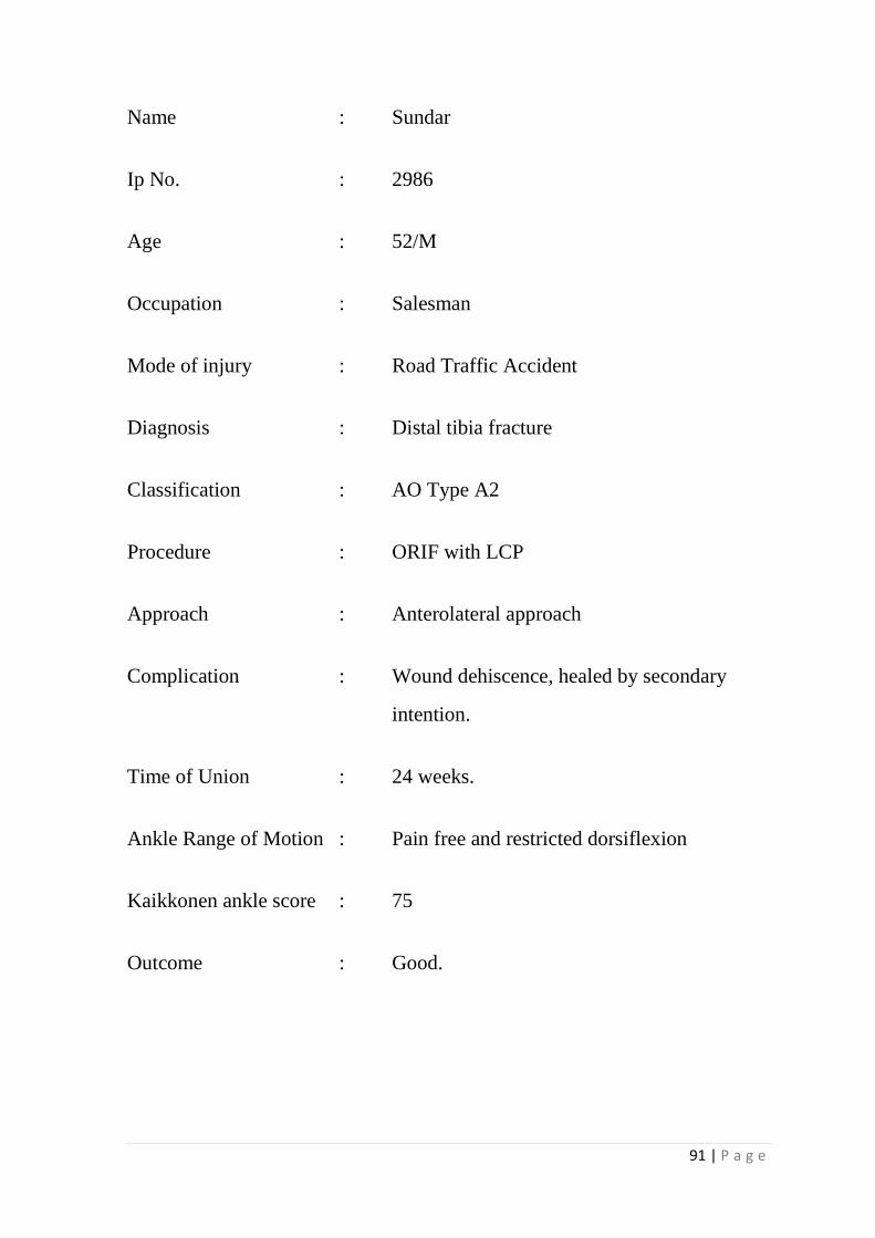
91 | P a g e
Name : Sundar
Ip No. : 2986
Age : 52/M
Occupation : Salesman
Mode of injury : Road Traffic Accident
Diagnosis : Distal tibia fracture
Classification : AO Type A2
Procedure : ORIF with LCP
Approach : Anterolateral approach
Complication : Wound dehiscence, healed by secondary
intention.
Time of Union : 24 weeks.
Ankle Range of Motion : Pain free and restricted dorsiflexion
Kaikkonen ankle score : 75
Outcome : Good.
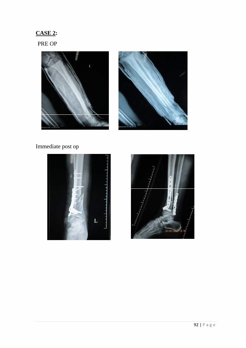
92 | P a g e
CASE 2:
PRE OP
Immediate post op
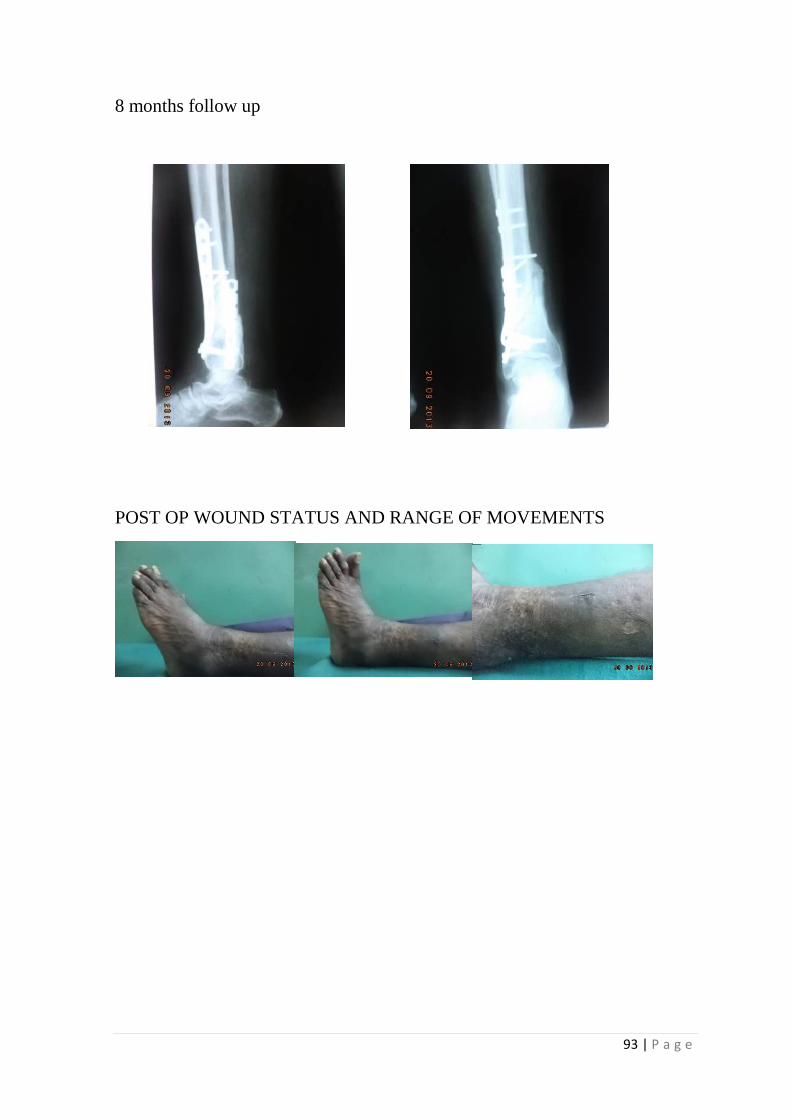
93 | P a g e
8 months follow up
POST OP WOUND STATUS AND RANGE OF MOVEMENTS
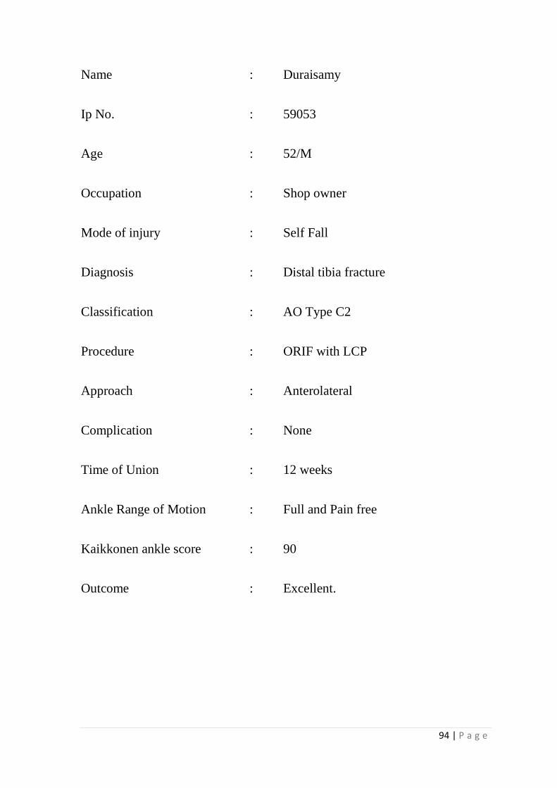
94 | P a g e
Name : Duraisamy
Ip No. : 59053
Age : 52/M
Occupation : Shop owner
Mode of injury : Self Fall
Diagnosis : Distal tibia fracture
Classification : AO Type C2
Procedure : ORIF with LCP
Approach : Anterolateral
Complication : None
Time of Union : 12 weeks
Ankle Range of Motion : Full and Pain free
Kaikkonen ankle score : 90
Outcome : Excellent.
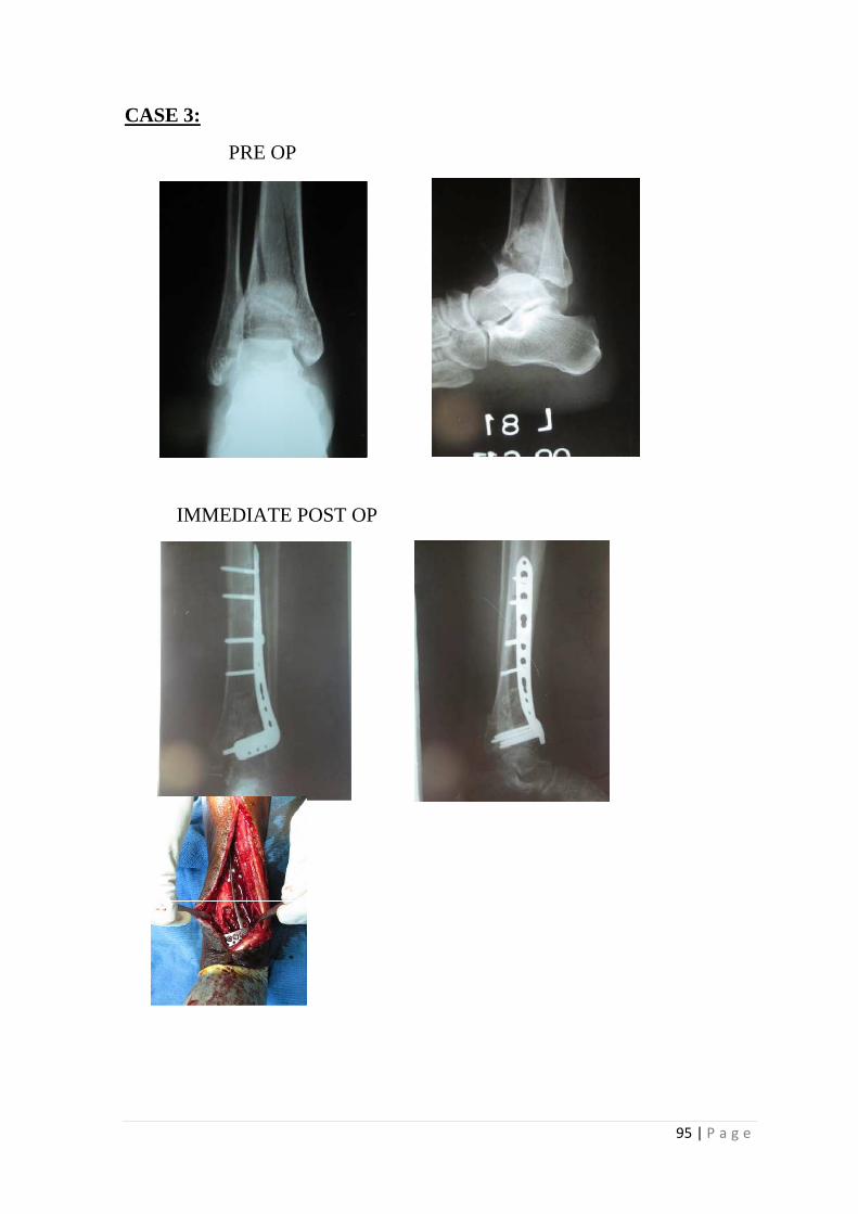
95 | P a g e
CASE 3:
PRE OP
IMMEDIATE POST OP
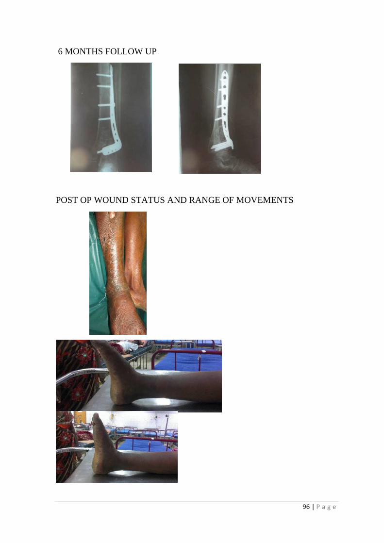
96 | P a g e
6 MONTHS FOLLOW UP
POST OP WOUND STATUS AND RANGE OF MOVEMENTS
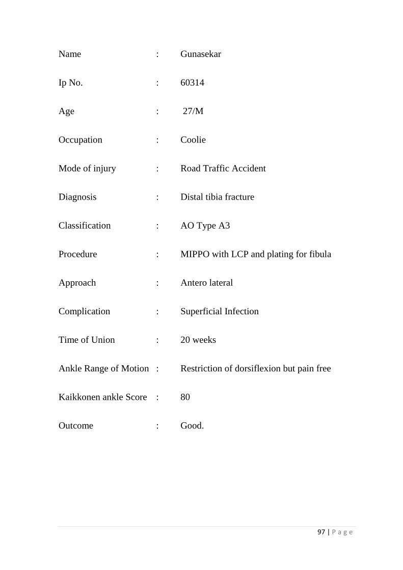
97 | P a g e
Name : Gunasekar
Ip No. : 60314
Age : 27/M
Occupation : Coolie
Mode of injury : Road Traffic Accident
Diagnosis : Distal tibia fracture
Classification : AO Type A3
Procedure : MIPPO with LCP and plating for fibula
Approach : Antero lateral
Complication : Superficial Infection
Time of Union : 20 weeks
Ankle Range of Motion : Restriction of dorsiflexion but pain free
Kaikkonen ankle Score : 80
Outcome : Good.
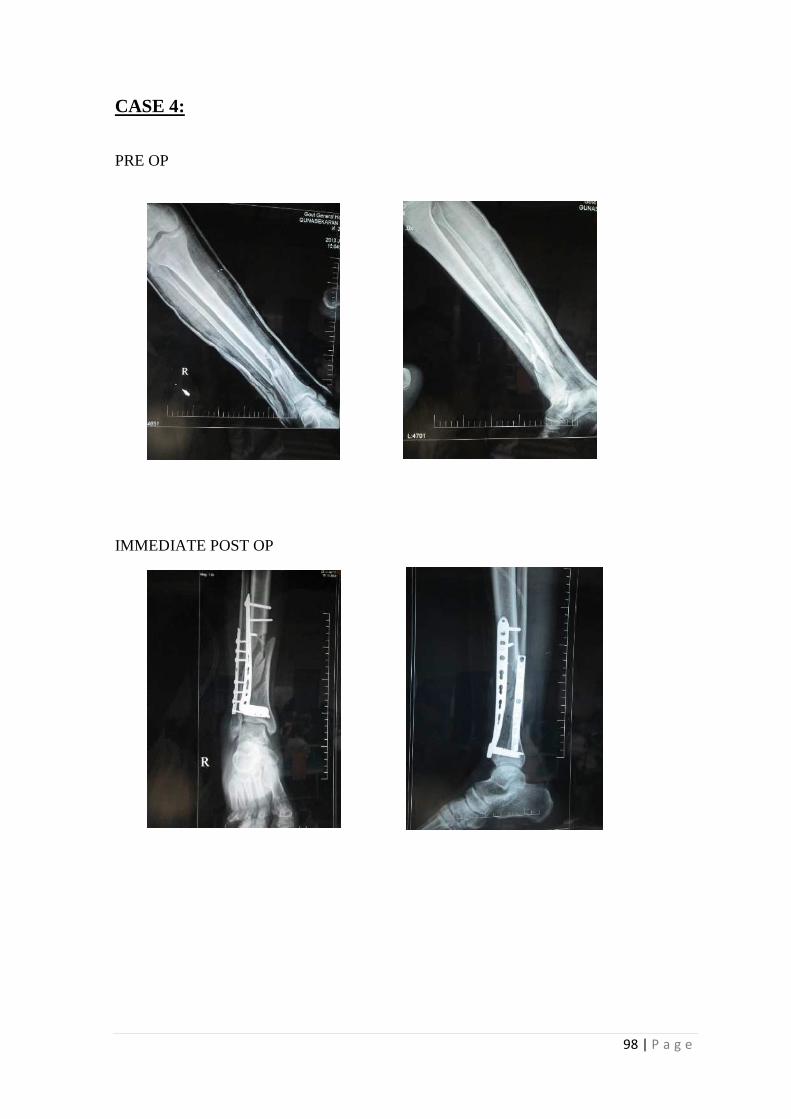
98 | P a g e
CASE 4:
PRE OP
IMMEDIATE POST OP
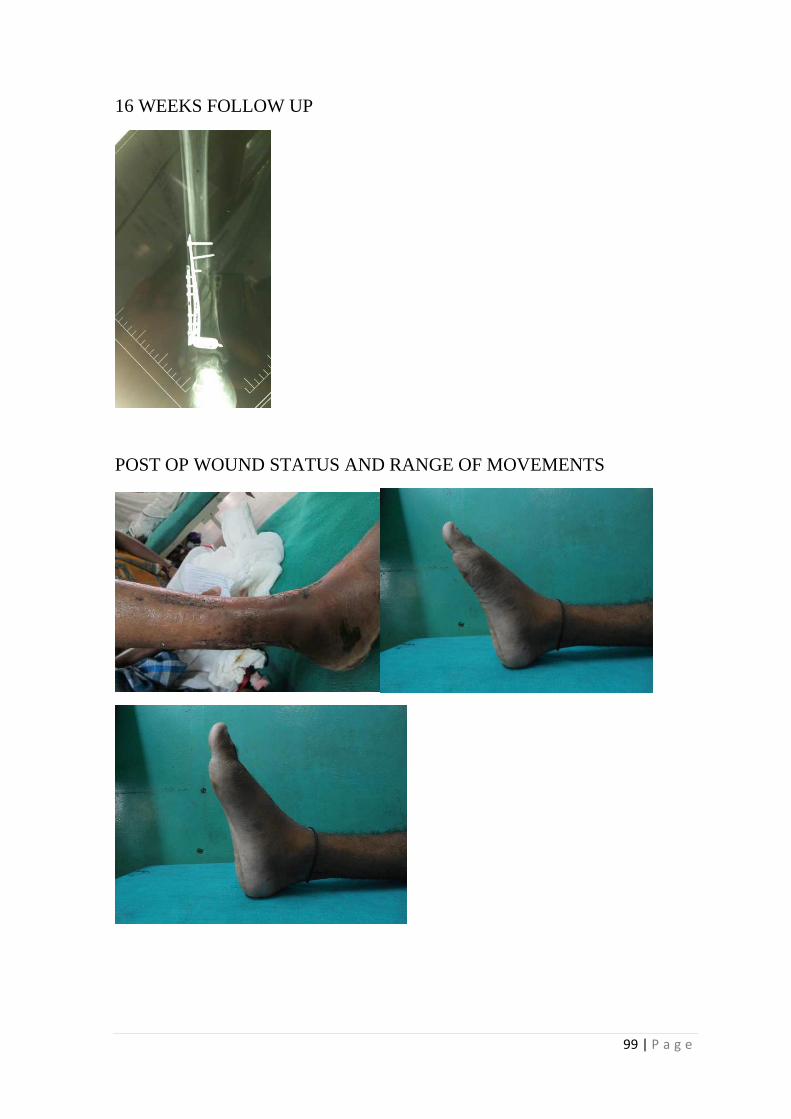
99 | P a g e
16 WEEKS FOLLOW UP
POST OP WOUND STATUS AND RANGE OF MOVEMENTS

BIBLIOGRAPHY
100 | P a g e
BIBILIOGRAPHY
1. Bucholz, Robert W.; Heckman, James D.; Court-Brown, Charles M.;
Tornetta, Paul, Rockwood and Green’s Fractures In Adults, 7th Edition
2. Michael S.Sirkin.; Plating of tibial pilon fractures; Am J Orthop.
2007;36(12 suppl):13-17
3. S.Terry Canale, James.H.Beaty ; Campbells operative orthopaedics
11th edition.
4. Michael.W.Chapman ; Chapman’s Orthopaedic Surgery.
5. M.E.Muller, M.Allgower,R Schneider, H.Willenegger ; Manual of
Internal Fixation.
6. Brent E. Fisher , Senthil T.Nathan, Robert D.Acland,Craig S.Roberts ;
The anterolateral incision for pilon fracture surgery : An anatomic
study of cutaneous blood supply ; Acta Orthop.Belg.,2011,77,355-361.
7. David J.Hak ; Anterolateral approach for Tibial Pilon Fractures;
Trauma Update; February 2012 | Volume 35 | Number 2.
8. Bonnevialle P, Lafossse JM et al. Distal leg fractures: How critical is
the fibular fracture and its fixation? Orthop Traumatol Surg Res. 2010
Oct;96(6):667-73.
9. Boren P, Richmond J et al Minimally invasive treatment of pilon
fractures with a low profile plate preliminary report 2006;8:230-235.
10. Decoster TA, Willis MC, Marsh JL et al. Rank order analysis of tibial
plafond fractures: does injury or reduction predict outcome? Foot
Ankle Int. 1999;20:44-49.
101 | P a g e
11. Egol KA, Kubiak EN, Fulkerson E, et al. Biomechanics of locked
plate and screws. J. Orthop Trauma 2004;18(8):483-93.
12. Egol KA, Wolinsky P, et al. Open reduction and internal fixation of
tibial pilon fractures. Foot Ankle Clin. 2000 Dec;5(4):873-85.
13. Etter C. Operative long term result of tibial pilon fractures treated with
open reduction and internal fixation Orthop Trauma 1991;189:1191-
1197.
14. Frigg R. Development of Locking Compression Plate. Injury
2003;34(supp.):B6-10.
15. Helfet DL, Suk M et al. Minimally invasive percutaneous plate
osteosynthesis of distal tibial fractures, Instr Course Lect 2004;3:471-
475.
16. Helfet DL, Koval K et al. Intraarticular pilon fractures of the tibia. Clin
Orthop Relat Res 1994(298):221-228.
17. Howard JJ, Barie et al. A prospective study of evaluating incision
placement and wound healing for tibial plafond fratures. J Orthop
trauma. 2008;5:250-255.
18. Hasenboehler E, Rikli D et al. Locking compression plate with
minimally invasive plate osteosynthesis in distal tibial fractures: a
retrospective study of 32 patients. Injury 2007, Mar;38(3):365-370.
19. John Scolaro, MD and Jaimo Ahn MD. In Brief: Pilon fractures. Clin
Orthop Relat Res 2011 February;469(2):621-623.
102 | P a g e
20. Kellam JF, Waddell JP. Fractures of the distal tibia metaphysis with
intra-articular extension: the distal tibial explosion fractures. J Trauma
1979;19:593-609
21. Kulkarni Text book of Orthopaedics – 2nd edition.
22. Lee YS, Chen SW, Chen SH et al. Stabilisation of the fractured fibula
plays an important role in the treatment of pilon fractures: a
retrospective comparison of fibular fixation methods. Int Orthop.
2009;695-699.
23. Rakesh K, Gupta, Rajesh Kumar Rohbilla et al. Locking plate fixation
in distal metaphyseal tibial fractures: series of 79 patients. Int Orthop
2010 December;34(8):1285-1290.
24. Mario Ronga MD, Umile Giuseppe Longo MD et al. Minimally
invasive locked plating of distal tibia fractures is safe and effective.
Clin Orthop Relat Res. 2010 April;468(4):975-982.
25. Mast J. A test of surgical judgement. In; Major Fractures of the pilon,
the Talus, and the Calcaneus, ed by Tscherne H, Schatzker J. Berlin,
Springer-Verlag, 1993,7-27.
26. Martin JS, Marsh JL, Bonar SK, et al. Assessment of the AO/ASIF
fracture classification for the distal tibia. J Orthop Trauma. 1997;11:
477-483.
27. Samir Mehta, MD, Michael J. Gardner, MD, David P. Barei, MD,
FRCS(C); Reduction Strategies Through the Anterolateral Exposure
103 | P a g e
for Fixation of Type B and C Pilon Fractures; J Orthop Trauma
2011;25:116–122.
28. Sherif Mohamed Abdelgaid, Mohamed Abdulghany Ahmed and Emad
Gaber Abdel-Mageed ; Minimally Invasive Treatment Protocol for
Closed Pilon Fractures; Clin Res Foot Ankle 2013, 1:2
29. J. Lawrence Marsh, Md, Dennis P. Weigel, Md, And Douglas R.
Dirschl, Md; Tibial Plafond Fractures.How Do These Ankles Function
Over Time?;
The Journal Of Bone & Joint Surgery · Jbjs.Org Volume 85-A ·
Number 2 · February 2003.
30. Muhr G, Breitfuss H. et al. Complications after plion fractures. In:
Major Fractures of the pilon, the talus and the calcaneus, ed by
Tscherne H, Schatzker J. Berlin, Springer-verlag, 1993, pp65-68.
31. Mu”ller ME, Allgo”wer M, Schneider R, et al. Manual of Internal
Fixation. Techniques recommended by the AO-ASIF group. Berlin,
Springer-verlag, 1991.
32. Ovadia DN, Beals RK. Fractures of the tibial plafond. J Bone Joint
Surg (Am) 1986;68:543-551.
33. Patterson MJ, Cole JE. Two staged delayed open reduction and internal
fixation of severe pilon fractures. J Orthop Trauma 1999;13:85-91.
34. Pierre Joveniaux, Xavier Ohl et al. Distal tibia fractures: management
and complications of 101 cases.
104 | P a g e
35. Philip A. McCann Mark Jackson et al. Complications of definitive
open reduction and internal fixation of pilon fractures of the distal
tibia. Int Orthop. 2011 March;35(3): 413-418.
36. Pollak AN, McCarthy ML, Bess RS et al. Outcomes after treatment of
high-energy tibial plafond fractures. J Bine Joint Surg Am.
2003;85:1893-900.
37. Pugh KJ, Wolinsky PR et al. Tibial Pilon fractures: a comparision of
treatment methodsJ Trauma 1999;47:937-941.
38. Resch H, Philander S, et al. Long term results of conservative and
surgical treatment for distal end of tibia 1986;16:117-123.
39. Rockwood and greens – Fractures in adult – seven edition.
40. Ruedi T. Fractures of the lower end of the tibia into the ankle joint:
result 9 years after open reduction and internal fixation. Injury
1973;5:130-134.
41. Reudi T, Matter P, Allgower M et al. Intra-articular fractures of the
distal tibial end. Helv Chir Acta. 1968;35:556-582.
42. Reudi TP, Allgower M et al. The operative treatment of intra-articular
fractures of the lower end of the tibia. Clin Orthop Relat Ras
1979(138):105-110.
43. Sands A, Byck et al. Clinical and functional outcomes of internal
fixation of displaced pilon fractures. Clin Orthop 1998;347:131-137.
105 | P a g e
44. Schatzker J, Tile M et al. Tibial pilon fracutes. Rationale of Operative
Fracture management. 2 ed. Berlin:Springer-Verlag;1996.
45. Syah Bahari, Brian Lenehan, et al. Minimally invasive percutaneous
plate fixation of distal tibia fractures. Acta Orthop, 2007,73;635-640.
46. Surgical exposures in Orthopaedics – Stanley Hoppenfield – fourth
edition.
47. Teeny S, Wiss D. Open reduction and internal fixation of tibial plafond
fractures. ClinOrthop 1993;292:108-117.
48. Topliss CJ, Jackson M, et al. Anatomy of pilon fractures of the distal
tibia. J Bone and Joint Surg Br. 2005;87:692-697.
49. Tornetta P, Weiner L, Bergman M, et al. Pilon fractures: treatment
with combined internal and external fixation. J Orthop Trauma
1993;7:489-496.
50. Wagner M. General principles for the clinical use of the LCP. Injury
2003 Nov;34 Suppl2;B31-42.
51. Watson JT, Moed BR Cramer KE et al. Pilon fractures – Treatment
protocol based on severity of soft tissue injury. Clin Orthop Relat Res
2000(375):78-90.
52. William TM, Nepola JV et al. Factors affecting the outcome in tibial
plafond fractures. Clin Orthop Relat Res 2004(423):93-98.

106 | P a g e
53. Wyrsch B, Mcferran MA, et al. Operative treatment of fractures of the
tibial plafond. A randomized prosepective study. J Bone Joint Surg
Am.1996;78:1646-1657.

PROFORMA
107 | P a g e
PROFORMA
Case No: Unit:
Name: Age/Sex:
I.P No: Occupation:
Address: Phone:
Date of Injury:
Date of Admission:
Date of Surgery:
Date of Discharge:
Mechanism of Injury:
• Road Traffic Accident
• Accidental Fall
• Industrial Accident
• Others
108 | P a g e
Severity of Injury
• High Velocity
• Moderate Velocity
• Trivial
General Condition:
Hemodynamic Status:
Side Involved: (Right/Left)
X-Ray findings:
Type of Fracture:
• Type A: Extra-articular
• Type B: Partial-articular
• Type C: Intra-articular
Associated other long bone injuries: (Yes/No)
Associated Head injury: (Yes/No)
Treatment elsewhere if any:
Treatment in our Institution:
• Time interval between injury and definitive management
• Procedure done
109 | P a g e
• Additional Procedures
• Bone grafting:(Yes/No)
Blood transfusion:(Yes/No)
Post Operative Events:
Complication:
Follow Up:
• No. of Weeks since Surgery
• Radiological Picture
• Scar Status
• Complications
• Ankle Range of motion
• Ankle Score
Assistant Professor Signature:

ANNEXURE
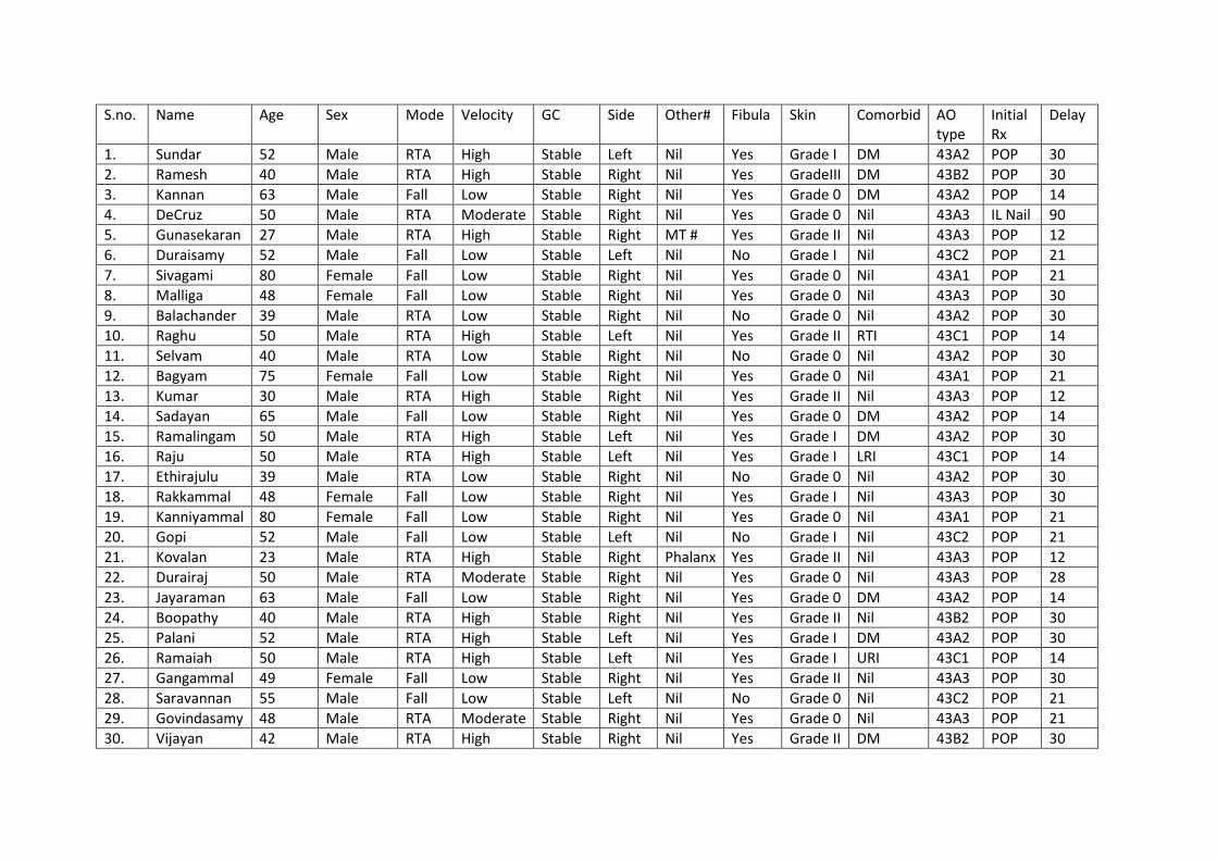
S.no. Name Age Sex Mode Velocity GC Side Other# Fibula Skin Comorbid AO
type
Initial
Rx
Delay
1. Sundar 52 Male RTA High Stable Left Nil Yes Grade I DM 43A2 POP 30
2. Ramesh 40 Male RTA High Stable Right Nil Yes GradeIII DM 43B2 POP 30
3. Kannan 63 Male Fall Low Stable Right Nil Yes Grade 0 DM 43A2 POP 14
4. DeCruz 50 Male RTA Moderate Stable Right Nil Yes Grade 0 Nil 43A3 IL Nail 90
5. Gunasekaran 27 Male RTA High Stable Right MT # Yes Grade II Nil 43A3 POP 12
6. Duraisamy 52 Male Fall Low Stable Left Nil No Grade I Nil 43C2 POP 21
7. Sivagami 80 Female Fall Low Stable Right Nil Yes Grade 0 Nil 43A1 POP 21
8. Malliga 48 Female Fall Low Stable Right Nil Yes Grade 0 Nil 43A3 POP 30
9. Balachander 39 Male RTA Low Stable Right Nil No Grade 0 Nil 43A2 POP 30
10. Raghu 50 Male RTA High Stable Left Nil Yes Grade II RTI 43C1 POP 14
11. Selvam 40 Male RTA Low Stable Right Nil No Grade 0 Nil 43A2 POP 30
12. Bagyam 75 Female Fall Low Stable Right Nil Yes Grade 0 Nil 43A1 POP 21
13. Kumar 30 Male RTA High Stable Right Nil Yes Grade II Nil 43A3 POP 12
14. Sadayan 65 Male Fall Low Stable Right Nil Yes Grade 0 DM 43A2 POP 14
15. Ramalingam 50 Male RTA High Stable Left Nil Yes Grade I DM 43A2 POP 30
16. Raju 50 Male RTA High Stable Left Nil Yes Grade I LRI 43C1 POP 14
17. Ethirajulu 39 Male RTA Low Stable Right Nil No Grade 0 Nil 43A2 POP 30
18. Rakkammal 48 Female Fall Low Stable Right Nil Yes Grade I Nil 43A3 POP 30
19. Kanniyammal 80 Female Fall Low Stable Right Nil Yes Grade 0 Nil 43A1 POP 21
20. Gopi 52 Male Fall Low Stable Left Nil No Grade I Nil 43C2 POP 21
21. Kovalan 23 Male RTA High Stable Right Phalanx Yes Grade II Nil 43A3 POP 12
22. Durairaj 50 Male RTA Moderate Stable Right Nil Yes Grade 0 Nil 43A3 POP 28
23. Jayaraman 63 Male Fall Low Stable Right Nil Yes Grade 0 DM 43A2 POP 14
24. Boopathy 40 Male RTA High Stable Right Nil Yes Grade II Nil 43B2 POP 30
25. Palani 52 Male RTA High Stable Left Nil Yes Grade I DM 43A2 POP 30
26. Ramaiah 50 Male RTA High Stable Left Nil Yes Grade I URI 43C1 POP 14
27. Gangammal 49 Female Fall Low Stable Right Nil Yes Grade II Nil 43A3 POP 30
28. Saravannan 55 Male Fall Low Stable Left Nil No Grade 0 Nil 43C2 POP 21
29. Govindasamy 48 Male RTA Moderate Stable Right Nil Yes Grade 0 Nil 43A3 POP 21
30. Vijayan 42 Male RTA High Stable Right Nil Yes Grade II DM 43B2 POP 30
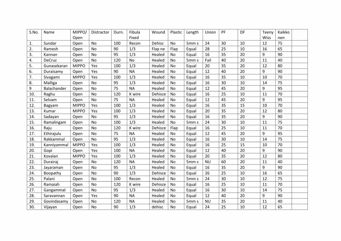
S.No. Name MIPPO/
Open
Distractor Durn. Fibula
Fixed
Wound Plastic Length Union PF DF Teeny
Wiss
Kaikko
nen
1. Sundar Open No 100 Recon Dehisc No 5mm s 24 30 10 12 75
2. Ramesh Open No 90 1/3 Flap ne Flap Equal 28 25 10 16 65
3. Kannan Open No 95 1/3 Healed No Equal 16 35 20 9 90
4. DeCruz Open No 120 No Healed No 5mm s Fail 40 20 11 40
5. Gunasekaran MIPPO Yes 100 1/3 Healed No Equal 20 35 20 12 80
6. Duraisamy Open Yes 90 NA Healed No Equal 12 40 20 9 90
7. Sivagami MIPPO Yes 100 1/3 Healed No Equal 16 35 10 10 70
8. Malliga Open No 95 1/3 Healed No Equal 16 30 10 14 75
9 Balachander Open No 75 NA Healed No Equal 12 45 20 9 95
10. Raghu Open No 120 K wire Dehisce No Equal 16 25 10 11 70
11. Selvam Open No 75 NA Healed No Equal 12 45 20 9 95
12. Bagyam MIPPO Yes 100 1/3 Healed No Equal 16 35 15 10 70
13. Kumar MIPPO Yes 100 1/3 Healed No Equal 20 35 20 12 80
14. Sadayan Open No 95 1/3 Healed No Equal 16 35 20 9 90
15. Ramalingam Open No 100 1/3 Healed No 5mm s 24 30 10 11 75
16. Raju Open No 120 K wire Dehisce Flap Equal 16 25 10 11 70
17. Ethirajulu Open No 75 NA Healed No Equal 12 45 20 9 95
18. Rakkammal Open No 95 1/3 Healed No Equal 16 30 10 14 75
19. Kanniyammal MIPPO Yes 100 1/3 Healed No Equal 16 25 15 10 70
20. Gopi Open Yes 100 NA Healed No Equal 12 40 20 9 90
21. Kovalan MIPPO Yes 100 1/3 Healed No Equal 20 35 20 12 80
22. Durairaj Open No 120 NA Healed No 5mm s NU 60 20 11 40
23. Jayaraman Open No 95 1/3 Healed No Equal 16 35 20 9 90
24. Boopathy Open No 90 1/3 Dehisce No Equal 26 25 10 16 65
25. Palani Open No 100 Recon Healed No 5mm s 24 30 10 12 75
26. Ramaiah Open No 120 K wire Dehisce No Equal 16 25 10 11 70
27. Gangammal Open No 95 1/3 Healed No Equal 16 30 10 14 75
28. Saravannan Open Yes 90 NA Healed No Equal 12 40 20 9 90
29. Govindasamy Open No 120 NA Healed No 5mm s NU 35 20 11 40
30. Vijayan Open No 90 1/3 dehisc No Equal 24 25 10 12 65
INFORMATION SHEET
Title : “ANALYSIS OF FUNCTIONAL OUTCOME OF ANTEROLATERAL PLATING IN TIBIAL PILON FRACTURES” Principal Investigator: Name of the Participant: Site :
We are conducting a study on “ANALYSIS OF FUNCTIONAL OUTCOME OF ANTEROLATERAL PLATING IN TIBIAL PILON FRACTURES” among patients attending the Institute of Orthopaedics & Traumatology, Rajiv Gandhi Government General Hospital, Chennai and for that your specimen may be valuable to us.
The purpose of this study is to evaluate and “ANALYSIS OF FUNCTIONAL OUTCOME OF ANTEROLATERAL PLATING IN TIBIAL PILON FRACTURES”
We are selecting certain cases and if you are found eligible, we may be using your radiographs of the spine to evaluate the outcome of surgery which in any way do not affect your final report or management.
The privacy of the patients in the research will be maintained throughout the study. In the event of any publication or presentation resulting from the research, no personally identifiable information will be shared.
Taking part in this study is voluntary. You are free to decide whether to participate in this study or to withdraw at any time; your decision will not result in any loss of benefits to which you are otherwise entitled.
The results of the special study may be intimated to you at the end of the study period or during the study if anything is found abnormal which may aid in the management or treatment. Signature of Investigator Signature of Participant Date : Place :
PATIENT CONSENT FORM
Study Detail : “ANALYSIS OF FUNCTIONAL OUTCOME OF ANTEROLATERAL PLATING IN TIBIAL PILON FRACTURES”
Study Centre : Rajiv Gandhi Government General Hospital, Chennai.
Patient’s Name :
Patient’s Age :
Identification Number :
Patient may check (√) these boxes
a) I confirm that I have understood the purpose of procedure for the above study. I have the opportunity to ask question and all my questions and doubts have been answered to my complete satisfaction. ❏
b) I understand that my participation in the study is voluntary and that I am free to withdraw at any time without giving reason, without my legal rights being affected. ❏
c) I understand that sponsor of the clinical study, others working on the sponsor’s behalf, the ethical committee and the regulatory authorities will not need my permission to look at my health records, both in respect of current study and any further research that may be conducted in relation to it, even if I withdraw from the study I agree to this access. However, I understand that my identity will not be revealed in any information released to third parties or published, unless as required under the law. I agree not to restrict the use of any data or results that arise from this study. ❏
d) I agree to take part in the above study and to comply with the instructions given during the study and faithfully cooperate with the study team and to immediately inform the study staff if I suffer from any deterioration in my health or well being or any unexpected or unusual symptoms. ❏
e) I understand that my identity will be kept confidential if my data are publicly presented ❏
f) I hereby give permission to undergo detailed clinical examination, Radiographs & blood investigations as required. ❏
g) ) I have had my questions answered to my satisfaction. ❏ h) ) I hereby consent to participate in this study. ❏
Signature/thumb impression Patient’s Name and Address:
Signature of Investigator
Study Investigator’s Name: Dr. THIRUMURUGAN.A