Embed Size (px)
Citation preview
Analysis of Linear Epitopes Recognised by thePrimary Human Antibody Response to a VariableRegion of the Attachment (G) Protein ofRespiratory Syncytial Virus
Patricia A. Cane*Department of Biological Sciences, University of Warwick, Coventry, United Kingdom
The sites of linear epitopes in a variable regionof the attachment (G) glycoprotein of respira-tory syncytial virus (RSV) that are recognised bythe human antibody response were examined.Two sets of overlapping 12mer peptides eachrepresenting the carboxy-terminal 84 or 85amino acids of the G protein of two group Aisolates of human RSV were synthesised. Thesepeptides were analysed using enzyme-linked im-munosorbant assays (ELISA) for their reactionswith sera obtained from infants with primaryRSV infection. Four pairs of overlapping pep-tides were found to react variously with the sera,the reactions depending on the infecting geno-type of RSV. Further 9mer peptides based onnatural variants in the epitope areas were thensynthesised to determine the specificity of thehuman antibody response and it was foundthat single amino acid changes could abrogaterecognition by these polyclonal sera. All thelinear epitopes found are involved in poten-tial N-glycosylation sites in at least some iso-lates of RSV. J. Med. Virol. 51:297–304, 1997.© 1997 Wiley-Liss, Inc.
KEY WORDS: linear epitopes; respiratory syn-cytial virus; G glycoprotein
INTRODUCTION
Respiratory syncytial virus (RSV) is a major cause oflower respiratory tract disease in infants and vulner-able adults. RSV is unusual in that it can frequentlyreinfect individuals and infects babies despite the pres-ence of maternal antibody. Isolates of RSV can be di-vided into two groups, A and B, on the basis of theirreactions with monoclonal antibodies and by nucleotidesequencing [Anderson et al., 1985; Mufson et al., 1985;Gimenez et al., 1986; Johnson and Collins, 1989; Caneand Pringle, 1991]. Each of the groups can be divided
further into a number of genotypes, again using nucleo-tide sequencing and to a lesser extent, monoclonal anti-bodies [Cane and Pringle, 1991, 1992, 1995; Garcia etal., 1994]. The gene that shows the greatest variabilityboth within and between the groups is that coding forthe attachment (G) protein, with isolates from the twogroups varying by 47% in amino acid sequence[Johnson et al., 1987] and within the groups by up to20% amino acid sequence for this protein [Cane et al.,1991; Sullender et al., 1991].
The G protein of human RSV is between 289 and 299amino acids in length depending on the strain and isoriented in the membrane with the carboxy-terminalthree fourths extracellular. The protein is heavily gly-cosylated with both N and O linked sugars and is richin serine, proline, and threonine: it is thus unusual fora viral glycoprotein and appears more similar to cellu-lar mucinous proteins [Wertz et al., 1985; reviewed bySullender and Wertz, 1991].
The variable regions of the protein lie in two parts ofthe ectodomain separated by a central highly con-served region [Johnson et al., 1987; Cane et al., 1991;Sullender et al., 1991]. Analysis of escape mutants re-sistant to murine monclonal antibodies which showvariable reactions with RSV isolates showed that mostof the mutants had amino acid changes in the variablecarboxy-terminal region of the protein indicating thatthis region includes antigenic areas [Garcia-Barreno etal., 1990; Rueda et al., 1991]. However, competitionenzyme-linked immunosorbant assays (ELISA) be-tween human convalescent sera and murine monoclo-nal antibodies failed to locate any reaction of the hu-man sera to this region [Palomo et al., 1991].
There is some evidence that there is progressive ac-cumulation of amino acid changes in the variable re-gions of the G protein of group A isolates [Cane andPringle, 1995]. Currently circulating group A isolates
*Correspondence to: Patricia A. Cane, Department of BiologicalSciences, University of Warwick, Coventry CV4 7AL, UK.
Accepted 25 October 1996
Journal of Medical Virology 51:297–304 (1997)
© 1997 WILEY-LISS, INC.
can be divided into a number of genotypes designatedSHL1/3/4, SHL2, SHL5, and SHL6 [Cane and Pringle,1992]. It has recently been demonstrated that there isan antibody response to the carboxy-terminal region ofthe G protein during primary infection and that thisresponse can be highly specific to the infecting geno-type of virus [Cane et al., 1996]. The experiments re-ported now were carried out to determine whetherthere are linear epitopes in this region that can be de-tected by the reaction of sera from infants experiencingtheir first infection with RSV, with peptides based onthe amino acid sequence of the G protein.
This approach has been used before: Norrby et al.[1987] looked at the reaction of human convalescentsera, murine monoclonal antibodies, and hyperimmunerabbit sera with overlapping 15mer peptides based onthe amino acid sequence of the ectodomain of the Gprotein of the prototype group A strain, A2. They foundthat human convalescent sera reacted only with theirpeptides 11, 12, and 15, corresponding to amino acids184–198, 174–188, and 144–158 respectively. Peptides11 and 12 lie in a highly conserved region of the pro-tein, and peptide 15 in a fairly conserved region. Theseauthors did not find any reaction of the human serawith the long comparatively hydrophilic carboxy-terminal part of G and thought that this region mightnot be immunogenic. However, the work was done be-fore it was appreciated that there was considerablevariability in this region of the G protein. In addition,Langedijk et al. [1996] carried out a similar pepscananalysis of bovine RSV G protein using polyclonal seraand again found antibody reactions only with the cen-tral conserved region of the protein: this study alsoused only prototype BRSV sequence for design of pep-tides.
The results described now show that there are in-deed linear epitopes recognised by the human antibodyresponse, present in the carboxy-terminal region of theG protein, but that their recognition is highly depen-dent both on the amino acid sequences of the peptidesused as targets in the tests, and on the infecting geno-type of virus.
MATERIALS AND METHODSPeptide Synthesis
Non-cleavable peptides were synthesised on polypro-pylene gears on stems using a multipin peptide synthe-sis kit (Chiron Mimotopes Peptide Systems, Clayton,Victoria, Australia), using Fmoc amino acids with theterminal amino group capped by acetylation. Firstly,two sets of peptides were synthesised each correspond-ing to the carboxy terminal 84 or 85 amino acids (i.e.,214–297/8) of the G protein of virus isolates RSB89-6256 and RSB89-6190 respectively [Cane et al., 1991].The genotypes of current group A isolates have beendesignated SHL1-6, although SHL1, 3, and 4 have beenfound to have very similar G gene sequences so are notdistinguished in this study [Cane and Pringle, 1992;Cane et al., 1996]. RSB89-6256 is genotype SHL3 andRSB89-6190 is genotype SHL2. Sequences from these
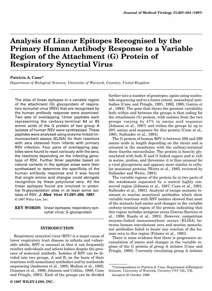
genotypes were chosen for the initial peptide synthesisas most of the human sera available (see below) camefrom babies infected with these genotypes of RSV. Thefirst set of peptides synthesised were 12mers, eachoverlapping by three amino acids, as illustrated in Fig-ure 1. In the case of peptides based on the amino acidsequence of RSB89-6190, the carboxy-terminal 12meroverlapped by one amino acid only. Initially, a totalof 51 RSV specific peptides were synthesised, togeth-er with two control peptides suggested by the Multi-pin kit manufacturers (PLAQGGGGGGGG andGLAQGGGGGGGG). Subsequently, a further set of9mer peptides based on the natural variants of aminoacids 250–258, 265–273, and 283–291 was also synthe-sised. These peptides are listed in Table I.
Sera
The patients from which the primary infection serawere obtained have been described previously [Cane etal., 1996]. These sera were made available through thegenerosity of Dr. H. Thomas, Southmead Hospital,Bristol, UK. Only sera that had been shown previouslyto react with the terminal 85/86 amino acids of the Gprotein as expressed as GST fusion proteins in E. coliwere selected for this study [Cane et al., 1996]. A mono-clonal antibody specific for the carboxy-terminal regionof the G protein under study in this report, 021/9G[Garcia et al., 1994], was kindly provided by Dr. J.A.Melero, Instituto de Salud Carlos III, Madrid, Spain.
Enzyme-Linked Immunosorbant Assay (ELISA)
All manipulations were carried out with the peptidesstill covalently linked to the pins. The pins carrying thepeptides were blocked using 5% dried milk in PBS with0.1% Tween 20 (PBS-T). All sera were reacted with thepeptides at a dilution of 1:1000 in blocking buffer. Afterwashing extensively with PBS-T, the pins were incu-bated with goat anti-human IgG (heavy and lightchain) conjugated with horseradish peroxidase,washed extensively, and developed using o-phenylene-diamine dihydrochloride. The reactions were stoppedby removing the pins and then adding 2.5 M sulphuricacid.
Antibody was removed from the pins by sonication in0.1 M phosphate buffer, pH 7.2 containing 1% SDS and0.1% 2-mercaptoethanol at 60°C, followed by washingin hot water and hot methanol.
RESULTSPeptide Synthesis
The positive control peptide, PLAQGGGGGGGG, re-acted with the control monoclonal antibody suppliedwith the Multipin kit while the negative control pep-tide, GLAQGGGGGGGG, did not, as expected from thepeptide synthesis kit protocol. In addition, 12mer pep-tides 8, 9, and 10 of both isolates reacted with mono-clonal 021/9G, for which escape mutant changes havebeen mapped to amino acid 244 (R-S) [Martinez, 1996].These results indicate that the peptide synthesis hadproceeded satisfactorily. However, it is unlikely that all
298 Cane
the peptides were synthesised with the same efficiencyso direct comparisons of degrees of reactions given bydifferent peptides are difficult.
Primary Response to Overlapping 12merPeptides Based on Amino Acids 214–298
Acute and convalescent primary RSV infection serafrom five babies, together with convalescent sera onlyfrom a further five babies, were reacted with the 5112mer peptides derived from the carboxy terminal 84/85 amino acids of two isolates (RSB89-6256 andRSB89-6190) of RSV. The peptides that the sera re-acted with are summarised in Table II, and some typi-cal results are illustrated in Figure 2. In most cases theELISA tests were repeated for each of the sera and theresults were very reproducible. However, it was notpossible to repeat all assays particularly those involv-ing the acute sera because of the extremely small quan-tities of sera available from most of the patients. Inaddition, again due to the scarcity of sera, only oneantibody dilution (1:1000) was used.
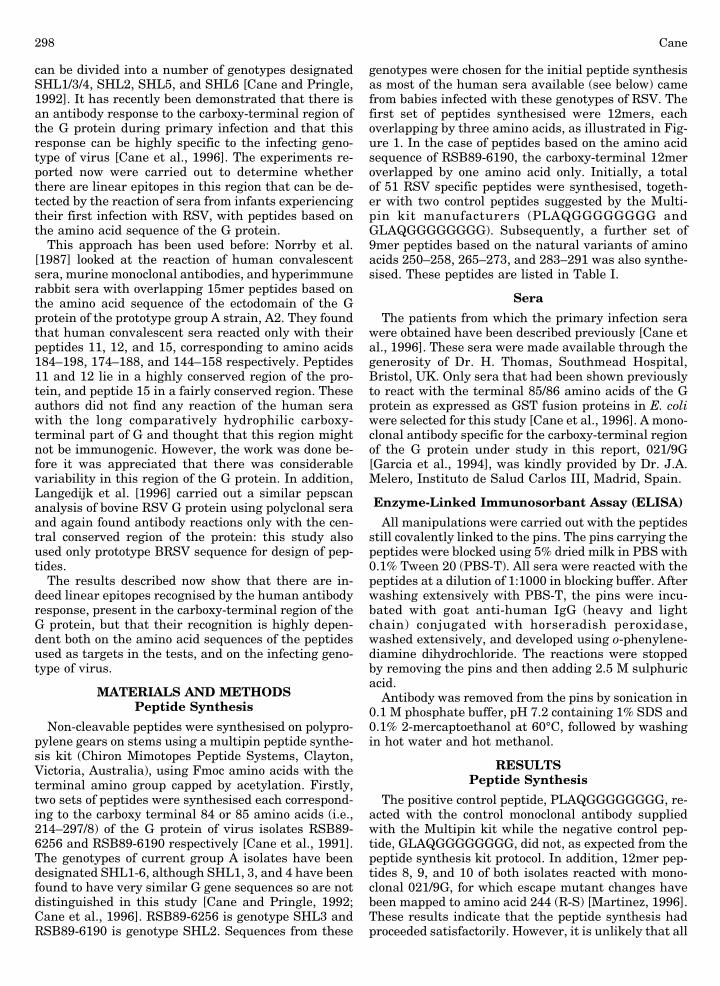
The acute serum from baby 25 reacted with only onepeptide: peptide 17 of RSB89-6190 (Fig. 2a). The con-valescent serum from this baby showed no detectablechange in reaction with this peptide, but displayedstrong reactions with peptides 17 and 18 of RSB89-6256 and a weaker reaction with peptide 18 of RSB89-6190 (Fig. 2b). This baby was infected with genotypeSHL6, which although having a distinct sequence oversome of the area covered by the peptides, differs fromRSB89-6256 only at position 270 (P-S) for peptides 17and 18.
Baby 47 was infected with RSV genotype A:SHL1/3/4, i.e., with a strain very similar to RSB89-6256. Theacute serum from this baby failed to react with any ofthe peptides (Fig. 2c), while the convalescent serumshowed strong reactions with peptides 17 and 18 fromboth peptide series (Fig. 2d). The acute serum of baby27, infected with this same genotype, reacted with pep-tide 17 of RSB89-6256, while the convalescent serumreacted strongly with peptides 17 and 18 from bothisolates (data not shown).
Fig. 1. Twelve amino acid long overlapping peptides representing the carboxy-terminal 84 or 85 amino acids of the G protein of RSV isolatesRSB89-6256 or RSB89-6190 respectively [Cane et al., 1991]. Sequence given in the top line is that of RSB89-6256, with changes in RSB89-6190shown in the lower line. Peptide 26 was synthesised for RSB89-6190 only. Positions of potential N-glycosylation sites in at least some RSVisolates (see text) are underlined.
RSV G Protein Variable Epitopes Recognised by Human Sera 299
Baby 109 was infected with RSV genotype A:SHL2.As with baby 47, the acute serum reacted with none ofthe peptides. The convalescent serum reacted with pep-tide 7 from isolate RSB89-6190 only and to a lesserextent with peptide 6 from the same isolate (data notshown).
Both acute and convalescent sera from baby 31 (in-fected with SHL1/3/4 genotype) serum reacted stronglywith peptides 23 and 24, and to a lesser extent withpeptide 22, of isolate RSB89-6256 only. The sera fromthis baby also showed some reactions with peptides 17and 18 from both isolates (Fig. 2e,f). The reason for thelack of change between the acute and convalescent serafrom this baby could be due to the acute serum beingcollected relatively late in the course of the infection.
Convalescent sera only from a further five infantswere reacted with this first series of peptides. Thesesera showed similar reactions to those observed for theother babies as shown in Table II: some sera (babies 8and 35) reacting with peptides 17 and 18 while others
(165 and 495) reacting with peptides 6 and 7 (Fig.2g,h). Serum from baby 706 also reacted with peptide12 and 17 of strain RSB89-6256 and peptide 7 of strainRSB89-6190.
It was clear that small differences between the pep-tides could abrogate recognition so failure to detect areaction with particular peptides could be due to dif-ferences in the sequence of the infecting strain of viruscompared to the test sequences. The specificity of thereaction with the appropriate peptides was thereforeexamined further.
Specificity of Response to Peptides
A set of 42 9mer peptides was synthesised based onthe naturally occurring variants of amino acids 250–258, 265–273, and 283–291 of the G protein of group ARSV isolates. These corresponded to the overlappingportions of peptides 12–13, 17–18, and 23–24 from thefirst series. Reactions with peptides 6–7 from the firstseries were not analysed further due to small numbers
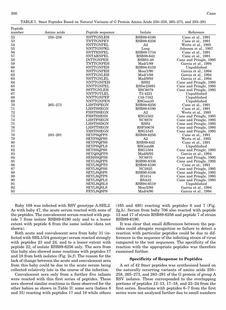
TABLE I. 9mer Peptides Based on Natural Variants of G Protein Amino Acids 250–258, 265–273, and 283–291
Peptidenumber Amino acids Peptide sequence Isolate Reference52 250–258 NSTTGNLEH RSB89-6190 Cane et al., 199153 TNTTGNPEY RSB89-6256 Cane et al., 199154 SNTTGNPEL A2 Wertz et al., 198555 NNTTGNPKL Long Johnson et al., 198756 SNTTRNPEL RSB89-1734 Cane et al., 199157 SNTARNPEL RSB89-642 Cane et al., 199158 INTTGNPEH RSB91-48 Cane and Pringle, 199559 TNTTGNPEH Mad/1/89 Garcia et al., 199460 SNTTGNPEH RSB94-6159 Unpublished61 NSTTGNPEH Mon/1/90 Garcia et al., 199462 SNTTGNLEH Mad/1/88 Garcia et al., 199463 NSTTGNLEL Mad/9/93 Garcia et al., 199464 NNTTGNPEH RSS2 Cane and Pringle, 199565 NNTTGNPEL RSSw23891 Cane and Pringle, 199566 SSTTGNLEH RSC6079 Cane and Pringle, 199567 NNTTGYLEL T2-4221 Unpublished68 NNTTGNPEF Ct8-7162 Unpublished69 NNTTGNPEN RSGam35 Unpublished70 265–273 LHSTSPEGN RSB89-6256 Cane et al., 199171 LHSTSSEGN RSB89-6190 Cane et al., 199172 FHSTSSEGN A2 Wertz et al., 198573 FHSTSSESN RSG1503 Cane and Pringle, 199574 LHSTFSEGN NC8870 Cane and Pringle, 199575 LHSTSSDGN RSS2 Cane and Pringle, 199576 LHSTTSEGN RSF50676 Cane and Pringle, 199577 YHSTSSEGN RSG1545 Cane and Pringle, 199578 283–291 SEYPSQPPS RSB89-6256 Cane et al., 199179 SEYPSQPSS A2 Wertz et al., 198580 SEYPPQPSS RSB89-642 Cane et al., 199181 SEYPSQPLS RSGam66 Unpublished82 SEYSSQPSS RSG1504 Cane and Pringle, 199583 SEYQSQPPS Mad/6/92 Garcia et al., 199484 SEHSSQPSS NC8870 Cane and Pringle, 199585 SEYLSQPPS RSB90-8336 Cane and Pringle, 199586 SEYLSQPTS RSB89-6190 Cane et al., 199187 SEYLSQPSS NC2825 Cane and Pringle, 199588 SEYLSQSPS RSB90-8106 Cane and Pringle, 199589 SEYLSQTPS H1414 Cane and Pringle, 199590 SEYLSQPLS RSA32 Cane and Pringle, 199591 SENLSQSLS RSB94-6519 Unpublished92 SEYLSQSLS Mon/2/88 Garcia et al., 199493 FEYLSQSPS Mad/4/90 Garcia et al., 1994
300 Cane
of sera reacting with these peptides. The peptide se-quences and their derivation are shown in Table I. Inall 18 different peptides were synthesised for aminoacids 250–258, 8 for amino acids 265–273 and 16 foramino acids 283–291. The reactions of the baby serawith these peptides are shown in Table II.
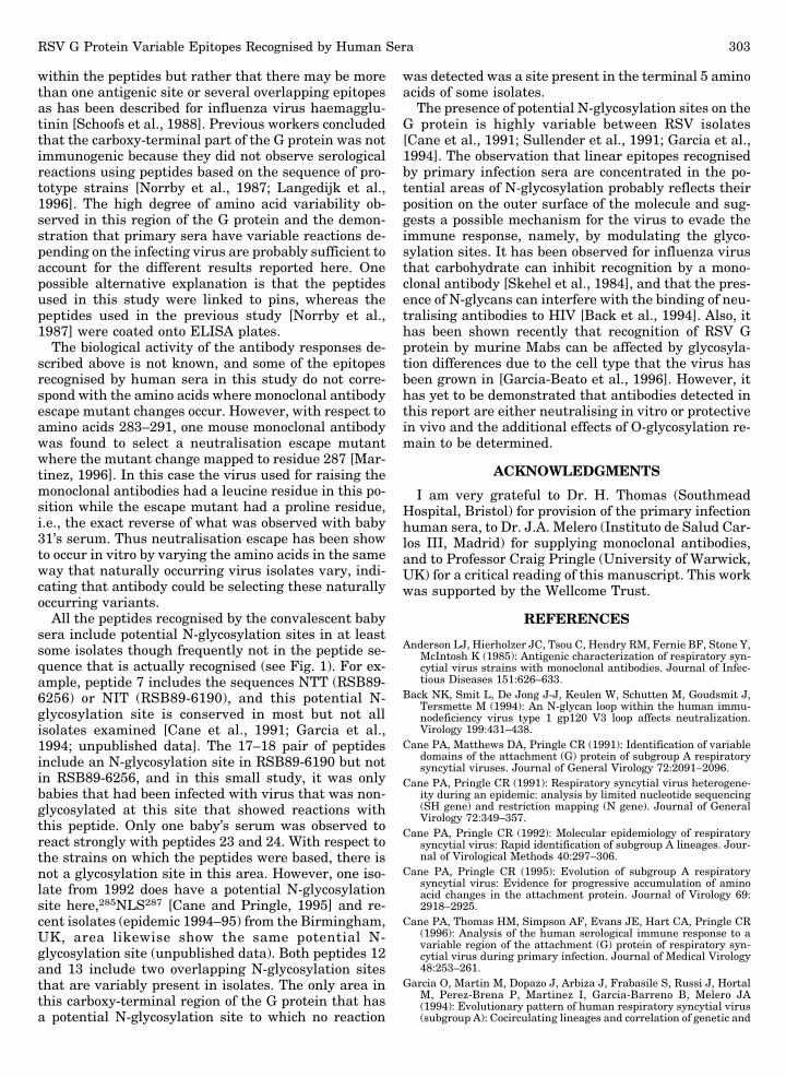
As illustrated above the convalescent baby sera werevariable as to which of the antigenic regions theyrecognised. Only two baby sera reacted clearly to pep-tides based on amino acids 250–258: Baby 706 serumreacted with peptide 58 together with weak reactionswith peptides 70–73, 75, and 77 (Fig. 3a), while baby E1reacted only with peptide 55 (data not shown). The se-quence of peptide 58 differs from those of peptides 59, 60,and 64 at only one position, namely the isoleucine residueat 250 is substituted with threonine, serine, or aspara-gine respectively and these single substitutions are suf-ficient to completely abrogate recognition of these pep-tides by this individual’s serum. The sequence of peptide55 differs from that of 65 with the substitution of a lysineresidue at position 257 with a glutamic acid residue.
Several of the baby sera reacted with some of the pep-tides derived from amino acids 265–273. Variations inresidue 265 (F/L/Y) and 271 (D/E) had little effect on rec-ognition by the infants’ antibody, but change of residue269 from serine to either threonine (peptide 76) or phe-nylalanine (peptide 74) completely abrogated recognition.Change of residue 272 from glycine to serine (peptide 73)reduced reaction with the sera considerably. The relativeintensity of reactions with these peptides varied betweenthe babies, with for example, baby 25’s serum reactingmarkedly more strongly with peptide 70 than with pep-tides 75 and 77 (Fig. 3b) while baby 47’s serum reactedequally strongly with peptides 70, 71, 72, 75, and 77 (Fig.3c): these results were reproducible between experi-ments. Pooled sera from mice vaccinated with a vaccinia
recombinant expressing the G protein from strain A2 re-acted with peptides 72, 73, and 77 (data not shown), in-dicating that perhaps in this instance the critical residuemay be amino acid 265 where change of the phenylala-nine to leucine but not to tyrosine affected recognition.
Serum from only one baby (31) reacted with peptidesderived from amino acids 283–291 (Fig. 2e,f). Baby 31’sserum reacted strongly with peptides derived from boththis region and from the 265–273 region. With respect tothe 283–291 region, this baby’s serum reacted stronglywith peptides 78, 79, 80, 81, and 82, weakly with peptide83 and failed to react with the other variant 283–291peptides (Fig. 3d). The key amino acids in this epitopeappear to be residues 286 and 287. The single change ofresidue 287 from proline to leucine abrogated all recog-nition. Change of this residue to serine had no effect un-less the tyrosine residue was also changed to histidine inwhich case no reaction of the serum with the peptideoccured. Change of the 287 proline residue to glutamineresulted in much reduced recognition.
DISCUSSION
The results demonstrate that there are specific anti-body responses during primary RSV infection to linearepitopes in the carboxy-terminal region of the G pro-tein. The sites recognised vary with the infecting geno-type of virus: serum from babies infected with A:SHL2reacted with peptides 6 and 7 (amino acids 229–243),while serum from babies infected with A:SHL1/3/4 re-acted with peptides 17 and 18 (amino acids 262–276)and, in one case, peptides 23 and 24 (amino acids 280–294). Sera from two babies also reacted peptides fromamino acids 247–258. It seems likely that these poly-clonal sera are not all recognising identical epitopes
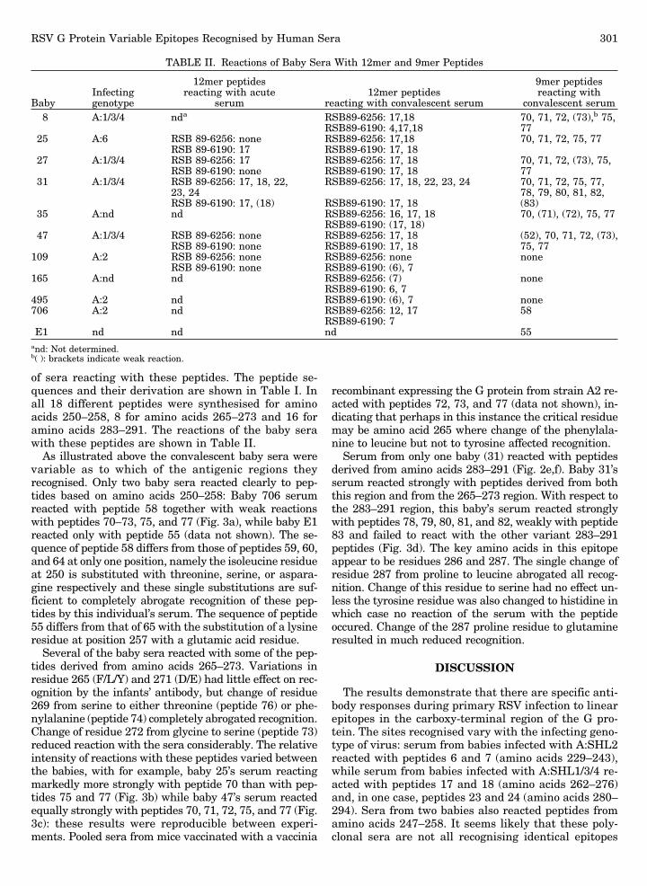
TABLE II. Reactions of Baby Sera With 12mer and 9mer Peptides
BabyInfectinggenotype
12mer peptidesreacting with acute
serum12mer peptides
reacting with convalescent serum
9mer peptidesreacting with
convalescent serum8 A:1/3/4 nda RSB89-6256: 17,18 70, 71, 72, (73),b 75,
RSB89-6190: 4,17,18 7725 A:6 RSB 89-6256: none RSB89-6256: 17,18 70, 71, 72, 75, 77
RSB 89-6190: 17 RSB89-6190: 17, 1827 A:1/3/4 RSB 89-6256: 17 RSB89-6256: 17, 18 70, 71, 72, (73), 75,
RSB 89-6190: none RSB89-6190: 17, 18 7731 A:1/3/4 RSB 89-6256: 17, 18, 22, RSB89-6256: 17, 18, 22, 23, 24 70, 71, 72, 75, 77,
23, 24 78, 79, 80, 81, 82,RSB 89-6190: 17, (18) RSB89-6190: 17, 18 (83)
35 A:nd nd RSB89-6256: 16, 17, 18 70, (71), (72), 75, 77RSB89-6190: (17, 18)
47 A:1/3/4 RSB 89-6256: none RSB89-6256: 17, 18 (52), 70, 71, 72, (73),RSB 89-6190: none RSB89-6190: 17, 18 75, 77
109 A:2 RSB 89-6256: none RSB89-6256: none noneRSB 89-6190: none RSB89-6190: (6), 7
165 A:nd nd RSB89-6256: (7) noneRSB89-6190: 6, 7
495 A:2 nd RSB89-6190: (6), 7 none706 A:2 nd RSB89-6256: 12, 17 58
RSB89-6190: 7E1 nd nd nd 55
and: Not determined.b( ): brackets indicate weak reaction.
RSV G Protein Variable Epitopes Recognised by Human Sera 301
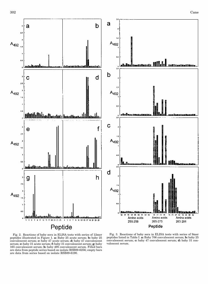
Fig. 2. Reactions of baby sera in ELISA tests with series of 12merpeptides illustrated in Figure 1. a: Baby 25 acute serum; b: baby 25convalescent serum; c: baby 47 acute serum; d: baby 47 convalescentserum; e: baby 31 acute serum; f: baby 31 convalescent serum; g: baby165 convalescent serum; h: baby 495 convalescent serum. Filled barsare data from peptide series based on isolate RSB89-6256; empty barsare data from series based on isolate RSB89-6190.
Fig. 3. Reactions of baby sera in ELISA tests with series of 9merpeptides listed in Table I. a: Baby 706 convalescent serum; b: baby 25convalescent serum; c: baby 47 convalescent serum; d: baby 31 con-valescent serum.
302 Cane
within the peptides but rather that there may be morethan one antigenic site or several overlapping epitopesas has been described for influenza virus haemagglu-tinin [Schoofs et al., 1988]. Previous workers concludedthat the carboxy-terminal part of the G protein was notimmunogenic because they did not observe serologicalreactions using peptides based on the sequence of pro-totype strains [Norrby et al., 1987; Langedijk et al.,1996]. The high degree of amino acid variability ob-served in this region of the G protein and the demon-stration that primary sera have variable reactions de-pending on the infecting virus are probably sufficient toaccount for the different results reported here. Onepossible alternative explanation is that the peptidesused in this study were linked to pins, whereas thepeptides used in the previous study [Norrby et al.,1987] were coated onto ELISA plates.
The biological activity of the antibody responses de-scribed above is not known, and some of the epitopesrecognised by human sera in this study do not corre-spond with the amino acids where monoclonal antibodyescape mutant changes occur. However, with respect toamino acids 283–291, one mouse monoclonal antibodywas found to select a neutralisation escape mutantwhere the mutant change mapped to residue 287 [Mar-tinez, 1996]. In this case the virus used for raising themonoclonal antibodies had a leucine residue in this po-sition while the escape mutant had a proline residue,i.e., the exact reverse of what was observed with baby31’s serum. Thus neutralisation escape has been showto occur in vitro by varying the amino acids in the sameway that naturally occurring virus isolates vary, indi-cating that antibody could be selecting these naturallyoccurring variants.
All the peptides recognised by the convalescent babysera include potential N-glycosylation sites in at leastsome isolates though frequently not in the peptide se-quence that is actually recognised (see Fig. 1). For ex-ample, peptide 7 includes the sequences NTT (RSB89-6256) or NIT (RSB89-6190), and this potential N-glycosylation site is conserved in most but not allisolates examined [Cane et al., 1991; Garcia et al.,1994; unpublished data]. The 17–18 pair of peptidesinclude an N-glycosylation site in RSB89-6190 but notin RSB89-6256, and in this small study, it was onlybabies that had been infected with virus that was non-glycosylated at this site that showed reactions withthis peptide. Only one baby’s serum was observed toreact strongly with peptides 23 and 24. With respect tothe strains on which the peptides were based, there isnot a glycosylation site in this area. However, one iso-late from 1992 does have a potential N-glycosylationsite here,285NLS287 [Cane and Pringle, 1995] and re-cent isolates (epidemic 1994–95) from the Birmingham,UK, area likewise show the same potential N-glycosylation site (unpublished data). Both peptides 12and 13 include two overlapping N-glycosylation sitesthat are variably present in isolates. The only area inthis carboxy-terminal region of the G protein that hasa potential N-glycosylation site to which no reaction
was detected was a site present in the terminal 5 aminoacids of some isolates.
The presence of potential N-glycosylation sites on theG protein is highly variable between RSV isolates[Cane et al., 1991; Sullender et al., 1991; Garcia et al.,1994]. The observation that linear epitopes recognisedby primary infection sera are concentrated in the po-tential areas of N-glycosylation probably reflects theirposition on the outer surface of the molecule and sug-gests a possible mechanism for the virus to evade theimmune response, namely, by modulating the glyco-sylation sites. It has been observed for influenza virusthat carbohydrate can inhibit recognition by a mono-clonal antibody [Skehel et al., 1984], and that the pres-ence of N-glycans can interfere with the binding of neu-tralising antibodies to HIV [Back et al., 1994]. Also, ithas been shown recently that recognition of RSV Gprotein by murine Mabs can be affected by glycosyla-tion differences due to the cell type that the virus hasbeen grown in [Garcia-Beato et al., 1996]. However, ithas yet to be demonstrated that antibodies detected inthis report are either neutralising in vitro or protectivein vivo and the additional effects of O-glycosylation re-main to be determined.
ACKNOWLEDGMENTS
I am very grateful to Dr. H. Thomas (SouthmeadHospital, Bristol) for provision of the primary infectionhuman sera, to Dr. J.A. Melero (Instituto de Salud Car-los III, Madrid) for supplying monoclonal antibodies,and to Professor Craig Pringle (University of Warwick,UK) for a critical reading of this manuscript. This workwas supported by the Wellcome Trust.
REFERENCES
Anderson LJ, Hierholzer JC, Tsou C, Hendry RM, Fernie BF, Stone Y,McIntosh K (1985): Antigenic characterization of respiratory syn-cytial virus strains with monoclonal antibodies. Journal of Infec-tious Diseases 151:626–633.
Back NK, Smit L, De Jong J-J, Keulen W, Schutten M, Goudsmit J,Tersmette M (1994): An N-glycan loop within the human immu-nodeficiency virus type 1 gp120 V3 loop affects neutralization.Virology 199:431–438.
Cane PA, Matthews DA, Pringle CR (1991): Identification of variabledomains of the attachment (G) protein of subgroup A respiratorysyncytial viruses. Journal of General Virology 72:2091–2096.
Cane PA, Pringle CR (1991): Respiratory syncytial virus heterogene-ity during an epidemic: analysis by limited nucleotide sequencing(SH gene) and restriction mapping (N gene). Journal of GeneralVirology 72:349–357.
Cane PA, Pringle CR (1992): Molecular epidemiology of respiratorysyncytial virus: Rapid identification of subgroup A lineages. Jour-nal of Virological Methods 40:297–306.
Cane PA, Pringle CR (1995): Evolution of subgroup A respiratorysyncytial virus: Evidence for progressive accumulation of aminoacid changes in the attachment protein. Journal of Virology 69:2918–2925.
Cane PA, Thomas HM, Simpson AF, Evans JE, Hart CA, Pringle CR(1996): Analysis of the human serological immune response to avariable region of the attachment (G) protein of respiratory syn-cytial virus during primary infection. Journal of Medical Virology48:253–261.
Garcia O, Martin M, Dopazo J, Arbiza J, Frabasile S, Russi J, HortalM, Perez-Brena P, Martinez I, Garcia-Barreno B, Melero JA(1994): Evolutionary pattern of human respiratory syncytial virus(subgroup A): Cocirculating lineages and correlation of genetic and
RSV G Protein Variable Epitopes Recognised by Human Sera 303
antigenic changes in the G glycoprotein. Journal of Virology 68:5448–5459.
Garcia-Barreno B, Portela A, Delgado T, Lopez JA, Melero JA (1990):Frame shift mutations as a novel mechanism for the generation ofneutralization resistant mutants of human respiratory syncytialvirus. EMBO Journal 9:4181–4187.
Garcia-Beato R, Martinez I, Franci C, Real FX, Garcia-Barreno B,Melero JA (1996): Host cell effect upon glycosylation and antige-nicity of human respiratory syncytial virus G glycoprotein. Virol-ogy 221:301–309.
Gimenez HB, Hardman N, Keir HM, Cash P (1986): Antigenic varia-tion between human respiratory syncytial virus isolates. Journalof General Virology 67:863–870.
Johnson PR, Collins PL (1989): The 1B (NS2), 1C (NS1) and N pro-teins of human respiratory syncytial virus (RSV) of antigenic sub-groups A and B: Sequence conservation and divergence withinRSV genomic RNA. Journal of General Virology 70:1539–1547.
Johnson PR, Spriggs MK, Olmsted RA, Collins PL (1987): The G gly-coprotein of human respiratory syncytial viruses of subgroups Aand B: Extensive sequence divergence between antigenically re-lated proteins. Proceedings of the National Academy of Science(USA) 84:5625–5629.
Langedijk JPM, Schaaper WMM, Meloen RH, van Oirschot JT (1996):Proposed three-dimensional model for the attachment protein G ofrespiratory syncytial virus. Journal of General Virology 77:1249–1257.
Martinez I (1996): PhD thesis, Universidad Autonoma de Madrid.Mufson MA, Orvell C, Rafnar B, Norrby E (1985): Two distinct sub-
types of human respiratory syncytial virus. Journal of GeneralVirology 66:2111–2124.
Norrby E, Mufson MA, Alexander H, Houghten RA, Lerner RA (1987):Site-directed serology with synthetic peptides representing the
large glycoprotein G of respiratory syncytial virus. Proceedings ofthe National Academy of Sciences (USA) 84:6572–6576.
Palomo C, Garcia-Barreno B, Penas C, Melero JA (1991): The G pro-tein of human respiratory syncytial virus: significance of carbohy-drate side chains and the C-terminal end to its antigenicity. Jour-nal of General Virology 72:669–675.
Rueda P, Delgado T, Portela A, Melero JA, Garcia-Barreno B (1991):Premature stop codons in the G glycoprotein of human respiratorysyncytial viruses resistant to neutralization by monoclonal anti-bodies. Journal of Virology 65:3374–3378.
Schoofs PG, Geysen HM, Jackson DC, Brown LE, Tang X-L, White DO(1988): Epitopes of an influenza viral peptide recognized by anti-body at single amino acid resolution. Journal of Immunology 140:611–616.
Skehel JJ, Stevens DJ, Daniels RS, Douglas AR, Knossow M, WilsonIA Wiley DC (1984): A carbohydrate side chain on hemagglutininsof Hong Kong influenza viruses inhibits recognition by a monoclo-nal antibody. Proceedings of the National Academy of Sciences(USA) 81:1779–1783.
Sullender WM, Mufson MA, Anderson LJ, Wertz GW (1991): Geneticdiversity of the attachment protein of subgroup B respiratory syn-cytial viruses. Journal of Virology 65:5425–5434.
Sullender WM, Wertz GW (1991): The unusual attachment glycopro-tein of the respiratory syncytial viruses. Kingsbury DW (ed): ‘‘TheParamyxoviruses.’’ New York and London: Plenum Press, pp 383–406.
Wertz GW, Collins PL, Huang Y, Gruber C, Levine S, Ball LA (1985):Nucleotide sequence of the G protein of human respiratory syncy-tial virus reveals an unusual type of viral membrane protein. Pro-ceedings of the National Academy of Sciences (USA) 82:4075–4079.
304 Cane


![A streamlined mass spectrometry-based proteomics workflow ... · [3,4]. Antibody-based histological methods are routinely performed but rely on the preservation of the relevant epitopes,](https://img.pdfslide.net/doc/110x75/5f0b51527e708231d42febe1/a-streamlined-mass-spectrometry-based-proteomics-workflow-34-antibody-based.jpg)