Embed Size (px)
Citation preview

Analysis of Osteogenic Property
in Dental Follicle Cells during Mouse
Molar Development
HEUI-JUNG HWANG
Department of Medical Science
The Graduate School, Yonsei University

Analysis of Osteogenic Property
in Dental Follicle Cells during Mouse
Molar Development
Directed by professor HAN-SUNG JUNG
The Master's Thesis submitted to the Department of
Medical Science, the Graduate School of Yonsei
University in Partial fulfillment of the requirement for
the degree of master of Medical Science
HEUI-JUNG HWANG
December 2004

This certifies that the Master's Thesis of
HEUI-JUNG HWANG is approved.
______________________________________________
HAN-SUNG JUNG
______________________________________________
YUN-JUNG YOO
______________________________________________
HYOUNG-WOO PARK
The Graduate School
Yonsei University
December 2004

ACKNOWLEDGMENTS
I would like to thank Professor Han-Sung Jung for invaluableadvice
and guidance.
My thanks also go to Professor Hee-Jin Kim, Dr. Hitoshi Yamamoto
at Nihon University for help me on several experiments.
It is great pleasure to thank Dr. Jae-Young Kim, Sung-Won Cho,
Jinglei Cai, Jong-Min Lee, Min-Jung Lee, Kyoung-Won Cho, Hyun-A
Lee, Kyung-Seok Hu, Hyun-Do Park, Hyun-Ho Kwak for letting me
collaborate on several experiments.
I am grateful to many people from the department of Oral Biology,
College of Dentistry, Yonsei University, who made my MS. course
enjoyable. Ju-Youn, Soo-Young, Hae, Hyun-Joo, Sun-Young,Du-Sik,
Jeong Hee, Su-Hyun.
Special thanks to Professor Syng-Il Lee, Professor Yun-Jung Yoo for
encouragement.
It is also great pleasure to thank E-Soon Kwon, Jin-il Kwon,
Kyung-Ran Park.
Finally, special thanks go to my family for supporting me always.
Heui-Jung Hwang

- i -
TABLE OF CONTENTS
LIST OF FIGURES
ABSTRACT............................................................................... 1
I. INTRODUCTION.................................................................. 3
II. MATERIAL AND METHODS.......................................... 9
1. Experimental Animals................................................ 9
2. Histology..................................................................... 9
3. Immunohistochemistry................................................ 9
4. Kidney transplantation............................................. 10
5. Cell culture............................................................... 11
6. Di.I application and Fate Mapping........................ 11
7. Beads implantation................................................... 12
8. Cell in situ hybridization........................................ 12
III. RESULT............................................................................ 14
1. Morphological finding.............................................. 14
2. Ossification of mandibular bone by
immunohistochemistry...............................................14

- ii -
3. Kidney transplantation.............................................. 18
4. Cell culture................................................................ 20
5. Alkaline phosphatase activity................................... 20
6. Gene expression of cultured dental follicle cells... 20
7. Bead and bone tissue implantation.............................. 23
IV. DISCUSSION.........................................................27
1. Differentiation of dental follicle cells.................... 27
2. Potential of dental mesenchymal cells................... 28
3. Runx2, Bsp Gene expression of cultured dental
follicle cells............................................................... 29
4. Osteogenic property of cultured dental
follicle cells............................................................... 29
V. CONCLUSION..........................................................31
REFERENCES...............................................................33
ABSTRACT (In KOREAN)...........................................38

- iii -
LIST OF FIGURES
Figure 1. Schematic diagram of tooth development..................... 4
Figure 2. Schematic diagram of dental follicle developmentfrom
E14 to PN8 in mouse embryos.......................................... 6
Figure 3. Histology of tooth morphogenesis of mouse first
Molar................................................................................ 15
Figure 4. The immunohistochemical expression of bone
sialoprotein (BSP) in the developing alveolar bone at E14,
E16, E18, PN2, PN8 and PN11........................................ 16
Figure 5. The immunohistochemical expression of bone
sialoprotein (BSP) in the developing alveolar bone at E14,
E16, E18, PN2, PN8 and PN11........................................ 17
Figure 6. Differentiation potential of dental follicle cells at
E14 in vivo............................................................ 19
Figure 7. Dental follicle cells in culture at E14.................. 21
Figure 8. Dental follicle cells in culture at E14 after 3 days

- iv -
and 7 days..................................................................... 22
Figure 9. Gene expression pattern after cultured dental
follicle cells for 3 days and 7 days at E14.............. 24
Figure 10. Number ofBsp-expressed colonies were examined
in aggregated cells after implanted of BMP4 protein
(100 / ) soaked beads for 48 hours....................... 25㎍㎖
Figure 11. Number of colonies expressingRunx-2 and Bsp
were counted in aggregated cells, which were cultured
with bone tissue for 3 weeks...................................... 26

- 1 -
Abstract
Analysis of Osteogenic Property in Dental Follicle Cells
during Mouse Molar Development
HEUI-JUNG HWANG
Department of Medical Science
The Graduate School, Yonsei University
(Directed by Professor HAN-SUNG JUNG)
In mice, dental mesenchymal cells comprise the dental papilla and dental
follicle in the developing mammalian tooth bud. It is thought that dental
follicle cells have the ability to differentiate into fibroblasts, cementoblasts,
and osteoblasts. However, cellular differentiation and the effects of
environmental factors are not known exactly.
In this study, the expression of BSP and OPN, using immunohistochemical
markers for hard tissue, was not detected at E14 dental follicle, and positive
BSP and OPN reactions were observed in the alveolar bone areaat PN8.
Despite the lack of hard tissue formation in the E14 dental follicles, dental
follicle cells showed the potential to form hard tissue in the following
experiment. In order to characterize this potential of dental follicle cells in
vitro, E14 dental follicle cells were separated into single cells,then cultured.
We assessed the expression of bothRunx2 and Bsp in the aggregated cells,

- 2 -
and determined to the ability of the cells to differentiate into osteoblasts and
their potential to form bone. Furthermore, after the addition of bone inducers
into the cultured dental follicle cells, including developing calvaria and
BMP4, Runx2 andBsp expressed in the aggregated cells showed the effects
of environmental factors on differentiation into osteoblasts. These results
suggest that dental follicle cells have the ability to differentiate into
osteoblasts as the result of interaction with environmental factors.
____________________________________________________________
Key words: tooth development, dental follicle cells, oseteoblast,differentiation, environmental factor

- 3 -
Analysis of Osteogenic Property in Dental Follicle Cells
during Mouse Molar Development
HEUI-JUNG HWANG
Department of Medical Science
The Graduate School, Yonsei University
(Directed by Professor HAN-SUNG JUNG)
I. INTRODUCTION
Early tooth development resembles, both morphologically and in terms of
the relevant molecules, the development of other ectodermal appendages
such as hair and glands. Interactions between the ectoderm and the
underlying mesenchyme constitute a central mechanism which regulates the
morphogenesis of all such organs.1 During the bud stage (Between
Embryonic day E9.0 and E11.5) in mouse, the dental epithelium initiates
tooth development, and the first morphological evidence oftooth
development is the appearance of the dental lamina. At cap stage (E13.5), the
epithelium has formed a bud, and the mesenchyme begins to condense. At
E14, the structure of the developing tooth is characterizedby the enamel
organ, dental papilla, and dental follicle. At bell stage (E15.5), tooth shape is
determined by epithelial folding. The dentin and enamel form the odontoblast

- 4 -
Journal of Cell Science 116, 1647-1648, 2003
Figure 1. Schematic diagram of tooth development. Tooth development
begins with initiation and morphogenesis, followed by differentiation and
mineralization. After the completion of crown formation, roots develop, and
the tooth erupts into the oral cavity. The blue box shows the specific region
for differentiation mechanism in control.
- 5 -
and ameloblast, respectively.2,3 The dental papilla differentiates into dentin
and pulp, while the dental follicle differentiates into thecementum,
periodontal ligament, and alveolar bone.4-6 The effects of various growth
factors on tooth development have been studied in mouse embryonic tooth
germs. Previously, the promotion of tooth morphogenesis and dental follicle
cell differentiation were thought to be predicated on the stimulation of cell
proliferation.7 Epithelial-mesenchymal tissue interactions, which are
ostensibly mediated by extracellular matrix molecules, constitute important
regulators of tooth morphogenesis and differentiation.8 It is generally
understood that the genes which are induced or expressed in developing
tissues can normally be considered good markers for cell determination and
differentiation. Bone sialoprotein and osteopontin are major non-collagenous
proteins in bone and other mineralized connective tissues,such as dentin,
cementum, and calcified cartilage tissue.9-13 Both bone sialoprotein and
osteopontin are prominent bone-matrix proteins, and are associated with the
formation and remodelling of the mineralized tissue matrix.14
Recent studies have demonstrated that bone sialoprotein mRNA is
expressed almost exclusively in differentiated osteoblasts, odontoblasts, and
cementoblasts.12,14,15 The expression of bone sialoprotein, osteopontin and
alkaline phosphatase in cells may reflect a specific trend toward osteoblastic
differentiation. Alkaline phosphatase is also expressed constitutively by
bone-forming cells, as well as some periodontal ligament cells, and is
recognized as an enzyme marker for bone differentiation.16-18 Runx2, a
member of the RUNX family of transcription factors, exhibits a highly
- 6 -
Periodontology, 24, 9-27, 2000
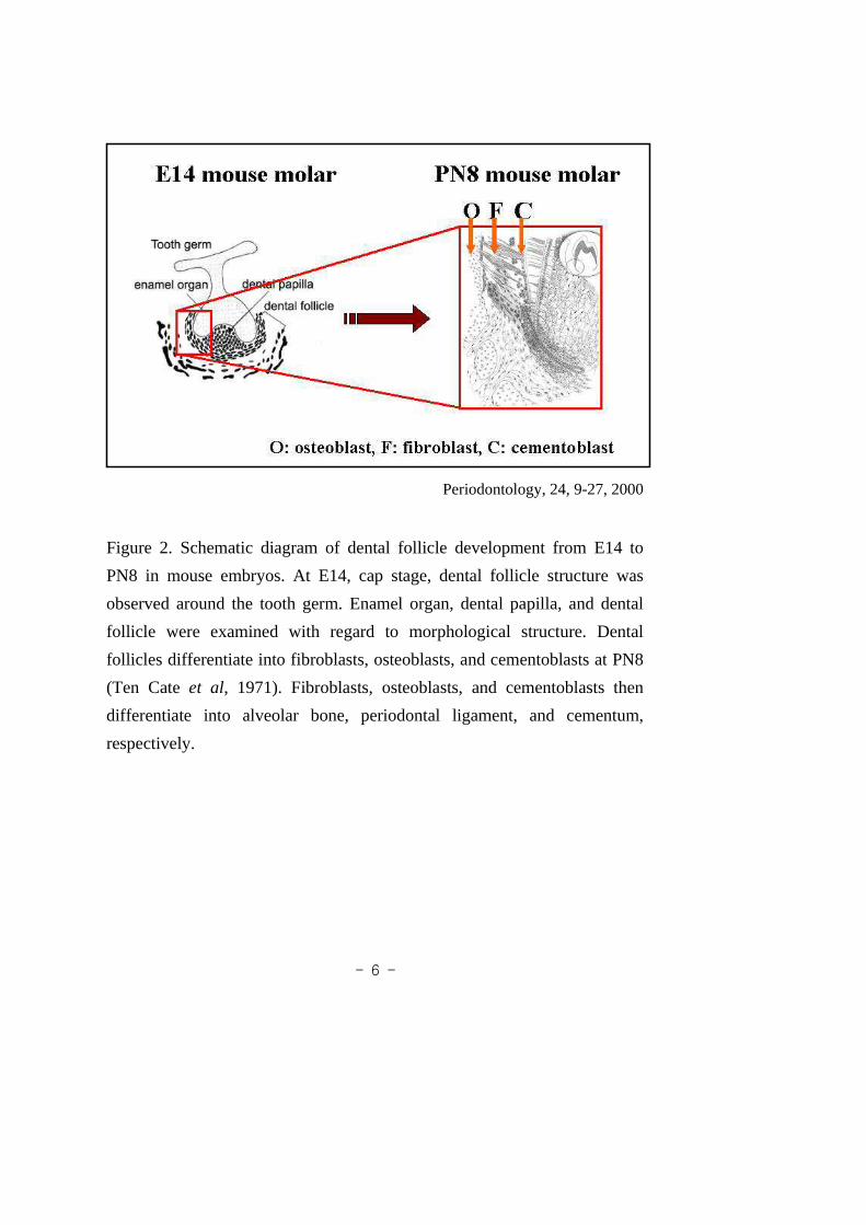
Figure 2. Schematic diagram of dental follicle developmentfrom E14 to
PN8 in mouse embryos. At E14, cap stage, dental follicle structure was
observed around the tooth germ. Enamel organ, dental papilla, and dental
follicle were examined with regard to morphological structure. Dental
follicles differentiate into fibroblasts, osteoblasts, and cementoblasts at PN8
(Ten Cateet al, 1971). Fibroblasts, osteoblasts, and cementoblasts then
differentiate into alveolar bone, periodontal ligament, and cementum,
respectively.

- 7 -
restricted tissue expression pattern in bone.Runx2 has been demonstrated to
regulate several osteoblast-specific genes.19-21 Runx2 is a runt domain
transcription factor that is essential for bone development and tooth
morphogenesis.22 Growth and differentiation factors (GDF) 5, 6, and 7 are
members of the bone morphogenetic protein (BMP) family, which
comprises a part of transforming growth factor (TGF-) superfamily.23 GDF
5, 6, and 7 are known to play in the formation of tendon and ligament
formation and are therefore probably involved in the formation of
periodontal ligament. GDF gene expression in the periodontal ligament was
first detected in cells associated with the initial processof periodontal
ligament fiber bundle formation.24 The developmental potency of the dental
follicle has been studied in a variety of tooth transplantation experiments.
However, the effects of environmental factors on cellular differentiation
have yet to be elucidated. Therefore, there are many intriguing facets of this
subject to be further explored at the tissue, cell and molecular levels.
In this study, the osteogenic property of dental follicle cells was examined
the expression of several osteoblast differentiation. To confirm osteoblast
differentiation during initial molar development in mouse, using the specific
bone-forming markers BSP and OPN, this study sought to characterize the
differentiation of both the mandibular bone and the dental follicle. In
addition, we attempted to analyse this potential in cultured dental follicle
cells in vitro. At E14, dental follicle cells were showed ability to
differentiate into osteoblasts, as well as the ability to differentiate into
alveolar bone. These activities were monitored according to the expression

- 8 -
of Runx2 and Bsp gene markers in aggregated cells, using cellin situ
hybridization. Our results indicate that dental follicle cells have the potential
to differentiate into osteoblasts via direct interaction with environmental
factors.

- 9 -
II. MATERIALS AND METHODS
1. Animals
Adult ICR mice were housed in a temperature-controlled room
(22±1 ) under artificial illumination (lights on from 05:00to 17:00 ), at 55%
relative humidity, with free access to food and water. Mouseembryos were
obtained from time-mated pregnant mice. The day on which a vaginal plug
was confirmed was designated as embryonic day 0 (E0).
2. Histology
Samples taken from mice at days E14, E16, and E18 of embryonic
development, and PN2, PN5, PN8, PN11 of the post-natal period, were
fixed with 4% paraformaldehyde (PFA) in PBS overnight at 4 , then
embedded in paraffin. Specimens were cut to a thickness of 7㎛, and
sections were then stained with both hematoxylin and eosin (H&E).
3. Immunohistochemistry
In order to conduct immunohistochemistry, the specimens were
fixed with 4% PFA at 4 and embedded in paraffin. Specimens were cut
to a thickness of 7㎛. The tissue sections were then deparaffinized and
rehydrated. In order to nonspecific background staining due to endogenous
- 10 -
peroxidase, slides were incubated in hydrogen peroxide blocks for 15
minutes. The specimens were then washed twice in buffer. When required,
the tissues were incubated in digestive enzymes. The specimens were then
washed an additional 4 times in buffer. Apply ultra v blick and incubated
for 60-70 minutes at room temperature in order to block reactive
immunoglobulins. The samples were washed an additional 4 times in
buffer. The specimens were then incubated for 10-15 minutesat room
temperature with biotinylated goat anti-mouse antibody, then washed4
times in buffer. The washed samples were incubated for a further 10-15
minutes at room temperature with streptavidin peroxidase,and rinsed 4
times in buffer. 1-2 drops DAB chromogen was then added to 1 mlof DAB
substrate, mixed by swirling, and applied to tissue. The tissues were then
incubated for an additional 5-15 minutes, according to the desired stain
intensity, and finally counter-stained and coverslipped using a permanent
mounting medium.
4. Kidney Transplantation
After 2 hours of incubation in a 37 incubator, the four particles of
divided dental follicle cells were carefully separated from the filter. Using a
male adult mouse as the host, the dental follicle cells were transplanted into
the kidney for 3 weeks. Thisin vivo culture method can result in the
formation of fully-calcified bone.

- 11 -
5. Cell culture
In order to culture the dental follicle cells, mouse dental follicles
were isolated from the first mandibular molars of E14 mice, then separated
into single cells. The dental follicle cells were then cultured in DMEM
(Dulbecco's minimum essential medium) containing 10% fetal bovine serum
at 37 in a humidified atmosphere containing 5% CO2. In general, dental
follicle cells were suitable for cell attachment onto 4-well Petri dishes(SPL
labware, Germany) after 3 days in culture.
6. Di.I. application and Fate Mapping
Di.I (1,19-dioctadecyl-3,3,39,39-tetramethyl indocar-bocyanine
perchlorate; Molecular Probes, Eugene, OR) was used as a cell tracer in the
observation of cell migration during mouse molar development. A 0.3%
w/v Di.I in DMSO (dimethyl sulfoxide) was washed for microinjection. The
Di.I injection was performed using 10 cm borosilicate capillary pipettes
(Sutter Instruments, BF120-94-10), pulled with a Sutter Instrument Flaming
Brown micropipette puller, filled by capillary action. Using an electrical
device, the lipophilic carbocyanine dye was introduced to the cell
membranes adjacent to the injection site. The exact position of the dye
could be determined using a fluorescent microscope (LEICA,MZ FL III).
Injections were directed into the dental follicle.

- 12 -
7. Beads implantation
Affigel Blue beads (BioRad) with 150㎛-diameters were dried, then
soaked in 0.5 mg/ ml of human recombinant NOGGIN (Regeneron). In
order to determine levels of osteogenic activity, single NOGGIN-soaked
beads were implanted into cultured dental follicles at E14.The cultured
dental follicle cells were used after bead implantation.
8. Cell in situ hybridization
Dental follicle cells in culture were then fixed in 4% PFA, and
washed three times in DEPC (dethyl pyrocarbonate)-PBS (phosphate
buffered saline). To 25 ml 0.1 M triethanolamine, pH 8.0, 62.5 ㎕ acetic
anhydride was added, and quickly mixed until completely dispersed. The
culture was incubated in this mixture for 10 minutes at room temperature.
The cultures were then washed in 1 x SSC for 5 minutes, and treated with 0.2
M HCl in DEPC-water for 10 minutes, and washed twice in DEPC-PBS
(dethyl pyrocarbonate-phosphate buffered saline) for 5 minutes.
Pre-hybridization solution was added, and this mixture wasincubated for 6
hours at room temperature. The pre-hybridization solutionwas removed, and
probes were added at a final concentration of between 1 and 2㎍/㎖. This
was allowed to hybridize overnight at 60 . The next day, the cultures were
rinsed in 0.2 x SSC, and washed in 0.2 x SSC at 60 for 1 hour. The
cultures were adjusted to room temperature in 0.2 x SSC for 5 minutes, then

- 13 -
blocked in 20% sheep serum in PBT for at least one hour at room
temperature. The culture was then incubated overnight withanti-digoxygenin
antibody (coupled to alkaline phosphatase) diluted to a final concentration of
1:1000 in 20% sheep serum in PBT, and rinsed three times in PBT. The
cultures were washed four times in PBT for 10 minutes, and then washed
twice in alkaline phosphatase buffer at room temperature for 10 minutes. For
every 10 ml of alkaline phosphatase buffer used, 4.5 ul of NBTand 3.5 ul of
BCIP was added, and developed in the dark, the duration of which depended
on the abundance of the RNA. When the reaction had proceeded far enough,
the sample was washed in PBT, and ultimately fixed in 4% formamide.

- 14 -
III. RESULTS
1. Morphological findings
In order to understand the precise development of the dentalfollicle
in mouse first molar tooth development, frontal wax sections of the E14 to
PN11 mouse teeth were stained with hematoxylin and eosin. AtE14 (cap
stage), the structure of tooth was observed to consist of theenamel organ,
dental papilla, and dental follicle (Fig. 3-A). At E16 (latecap stage), the
mandibular bone manifested around the dental follicle (Fig. 3-B). At E18
(bell stage), the mandibular bone was observed near the dental follicle (Fig.
3-C). Dental follicle cells thinned at PN2 (Fig. 3-D). At PN5, between the
dentin and mandibular bone, a cell layer developed (Fig. 3-E). At PN8, this
cell layer appeared as a thick line (Fig. 3-F). At PN11 and PN14, we
observed periodontal ligament- like and alveolar bone tissue (Fig. 3-G).
2. Ossification of the mandibular bone by immunohistochemistry
In order to determine the relationship between morphological
changes in the bone and the expression patterns of bone sialoprotein (BSP)
and osteopontin (OPN). BSP and OPN expression pattern during the
formation of the developing mandibular bone in the mouse first molar from
E14 to PN11 were examined immunohistochemistry after frontal section.
BSP and OPN are expressed in bone and other mineralized connective

- 15 -
Figure 3. Histology of tooth morphogenesis of mouse first molar. A,
B, C: Embryonic day 14 (E14) day cap stage frontal sections (A) E16
early bell stage frontal sections (B), E18 bell stage frontal sections (C).
D, E, F, G, H: Postnatal (PN) stage frontal sections. PN2 stage frontal
sections (D), PN5 stage frontal section (E), PN8 stage frontal sections
(F), PN11-14 stage frontal sections (G-H). (Scale bar: (A-H) 50 ㎛ )

- 16 -
Figure 4. The immunohistochemical expression of bone sialoprotein(BSP) in the developing alveolar bone at E14, E16, E18, PN2, PN8and PN11. To determine osteogenic activities, we examined theexpression of several osteoblast differentiation-specific markers includingBSP, using immunohistochemistry. The majority of bone-like tissuestained positive for BSP. At E14 (A),BSP was expressed on thebuccalside of the tooth germ. At E16 (B), BSP was expressed on both thelingual and buccal sides of the tooth germ. At E18 (C), BSP wasexpressed around the entirety of the tooth germ. At PN2, BSP wasexpressed adjacent to the dentin (D). At PN8, BSP was expressedadjacent to the dentin and root dentin (E). At PN11, BSP wasexpressed around the periodontal ligament (F). (Scale bar:(A-F) 50 ㎛)

- 17 -
Figure 5. The immunohistochemical expression of osetopontin (OPN) inthe developing alveolar bone at E14, E16, E18, PN2, PN8 and PN11.To determine osteogenic activities, we examined the expression ofseveral osteoblast differentiation-specific markers OPN, usingimmunohistochemistry. The majority of bone-like tissue stained positivefor OPN. At E14 (A), OPN was expressed on the buccal side of thetooth germ. At E16 (B), OPN was expressed on both the lingual andbuccal sides of the tooth germ. At E18 (C), OPN was expressedaround the entirety of the tooth germ. At PN2, BSP was expressedadjacent to the dentin (D). At PN8, OPN was expressed adjacent tothe dentin and root dentin (E). At PN11, OPN was expressed aroundthe periodontal ligament (F). (Scale bar: (A-F) 50㎛)

- 18 -
tissues. In general, the patterns of expression of BSP and OPN were similar.
BSP and OPN are known markers for osteoblasts, osteocytes and bone. At
E14, BSP and OPN were expressed on the buccal side of the toothgerm
(Fig. 4, 5-A). At E16, BSP and OPN were expressed on both the lingual and
buccal side of the tooth germ (Fig. 4, 5-B). At E18, BSP and OPNwere
expressed around whole the tooth germ (Fig. 4, 5-C). At PN2, BSP and
OPN were expressed adjacent to the dentin (Fig. 4, 5-D). At PN8, BSP and
OPN were expressed adjacent to both the dentin and root dentin (Fig. 4,
5-E). At PN11, BSP and OPN were expressed around the entiretyof the
periodontal ligament (Fig. 4, 5-F).
3. Kidney transplantation
To examine differentiation potential of the dental follicle cells, E14
mesenchymal cells were used in kidney transplantation forin vitro culture.
After micro-dissecting the tooth germ in an E14 mouse mandible, the tooth
germ was separated into the epithelium and mesenchyme. The remaining
mesenchymal cells were separated into lingual side, buccalside, dental
papilla and dental papilla with dental follicle, then transplanted into a kidney
capsule for 3 weeks. The lingual side cells, buccal side cells, dental papilla
cells, and dental papilla cells with dental follicle cells were calcified into
bone tissue (Figs. 6). Prior to E14, dental mesenchymal cells exhibit
osteogenic properties. As a result, dental mesenchymal cells maintain the
potential for differentiation at E14.

- 19 -
Figure 6. Differentiation potential of the dental folliclecells at E14 in
vivo. Calcified tissues were obtained after the transplantationof dental
follicle cells into a kidney capsule. A: lingual side, B: buccal side, C:
dental papilla, D: dental papilla + dental follicle.

- 20 -
4. Cell culture
Mouse dental follicles were isolated from the first mandibular molar
at E14. In order to conduct thein vitro culture, the E14 tooth germs were
micro-dissected using a tungsten needle. The tooth germ was separated into
the epithelium and the mesenchyme, and then the epithelium and dental
papilla were removed. After the epithelium was separated, we carefully
dissected out the dental papilla.
5. Alkaline phosphatase activity
In order to characterize bone differentiation, we assessedalkaline
phosphatase activity. 3 days (Fig. 8-A) and 7days (Fig. 8-B)after the
inception of cell culture, the cultures were stained for alkaline phosphatase
activity. Alkaline phosphatase activity is recognized as an enzyme marker
for bone differentiation. After 3 days (C) and 7 days (D), theculture
specimens exhibited increased alkaline phosphatase activity.
6. Gene expression of cultured dental follicle cells
In order to determine the expression patterns of signalling
molecules, E14 dental follicles were used to conduct another cell culture
experiment. After 3 days and 7 days of cell culture,in situ hybridization was

- 21 -
Fig 7. Dental follicle cells in culture at E14. For the culture of dental
follicle cells, mouse dental follicles were isolated from the first
mandibular molars of mice at E14, and then made into single cells.
The dental follicle cells were cultured in Dulbecco's minimum essential
medium, containing 10% fetal bovine serum at 37°C in a humidified
atmosphere containing 5% CO2.

- 22 -
Figure 8. Dental follicle cells in culture at E14 after 3 (A) and 7 days
(B). Alkaline phosphatase activity in cultured dental follicle cells at
E14 for 3 days (C) and 7 days (D). Cells at 7 days in culture showed
higher alkaline phosphatase activity than did cells at 3 days in culture.

- 23 -
carried out with the cultured cells.Runx2 andBsp expression were detected
in aggregated dental follicle cells (Fig. 9-A, B). These expression patterns
were higher than in the 3 day aggregated dental follicle cells.
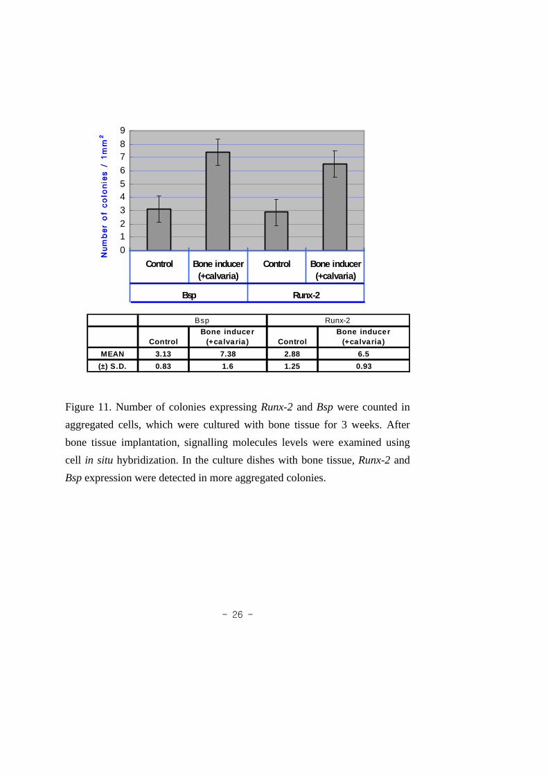
7. Bead and bone tissue implantation
In order to characterize the potential differentiation of dental follicle
cells, BMP4-soaked beads and developing calvaria (E14) were implanted on
a cultured dental follicle dish. PBS beads were implanted asa control. After
bead implantation,Bsp andRunx2 expression patterns were examined using
cell in situ hybridization. After BMP4 bead implantation,Bsp and Runx2
expression were found to be stronger around the bead-implanted dish (Fig.
10). In the developing calvaria sample,Bsp and Runx2 expressions were
detected after 3 weeks (Fig. 11). In general, bothBsp and Runx2 were
expressed in all samples, indicating that osteogenic potential could be
induced by surrounding factors.

- 24 -
Fig 9. Gene expression pattern after dental follicle cells were cultured
for 3 days (A) and 7 days (B) at E14.Runx2 and Bsp, expression
patterns were observed afterin situ hybridization on cultured cells.
These expression patterns appeared in aggregated cells at 7days. Gene
expression at 7 days (B) was higher than at 3 days.

- 25 -
Fig 10. Number ofBsp-expressed colonies were determined in aggregated
cells 48 hours after implantation of BMP4 protein (100㎍/㎖)-soaked
beads. After BMP4 bead implantation,Runx2 and Bsp expression stronger
near the bead-implanted dish.
- 26 -
0
12
3
45
6
78
9
Control Bone inducer(+calvaria)
Control Bone inducer(+calvaria)
Bsp Runx-2
Num
ber
of
co
lonie
s /
1m
mN
um
ber
of
co
lonie
s /
1m
mN
um
ber
of
co
lonie
s /
1m
mN
um
ber
of
co
lonie
s /
1m
m22 22
ControlBone inducer
(+ca lvaria) ControlBone inducer
(+ca lvaria)
MEAN 3.13 7.38 2.88 6.5
(±) S.D. 0.83 1.6 1.25 0.93
Bsp Runx-2
Figure 11. Number of colonies expressingRunx-2 andBsp were counted in
aggregated cells, which were cultured with bone tissue for 3weeks. After
bone tissue implantation, signalling molecules levels were examined using
cell in situ hybridization. In the culture dishes with bone tissue,Runx-2 and
Bsp expression were detected in more aggregated colonies.

- 27 -
DISCUSSION
1. Differentiation of dental follicle cells
These spacing patterns of teeth are developed by
epithelium-mesenchyme interactions, and the early development of the tooth
involves the development of other epithelial appendages, such as feather
buds and mammary glands.25,26Mesenchymal cells in the developing tooth,
so-called dental mesenchymal cells, are derived from two different origins:
the cranial neural crest (CNC) and the non-CNC.27 CNC-derived cells
migrate, proliferate and ultimately differentiate into odontoblasts,
cementoblasts, fibroblasts, osteoblasts and chondroblasts, etc.28-31Tooth bud
formation is one of the best examples of asynchronous development. From
the dental lamina to the individual tooth, each tooth establishes its own
identity during development in its proper position in relation to both the
maxilla and the mandible. At E18 (late bell stage), the dental follicle
surrounds each tooth germ, which is located between each tooth germ and
its bony compartment. At PN11, tooth structure have alreadyshown dentin,
enamel, cementum, alveolar bone, and periodontal ligaments. In addition,
immunohistochemistry for osteoblast was examined to studythe
differentiation of the mandibular bone. BSP and OPN are major
non-collagenous proteins occurring in bone and other mineralized
connective tissues.9-13 Both BSP and OPN are prominent bone-matrix
proteins that are related to function in the formation and remodeling of the
mineralized tissue matrix.14 In order to characterize distance changes during
tooth formation, we assessed the expression of the specificbone forming
markers, BSP and OPN. The expression patterns of BSP and OPN

- 28 -
expression were examined by using immunostaining (Figs. 4 and 5). During
embryonic development, distance changes in the dental follicle and
mandibular bones narrowed, as the tooth germ and mandibularbone grew.
At PN2, dental follicle cells and alveolar bone were almost attached. At
PN5, the dentin and alveolar bone begin to separate. Dental follicle cells,
however, could not be used to precisely determine differentiation time
during initial molar development in mice. In the case of H-E staining, dental
follicle cells did not shown for differentiation that how the cells differentiate
during tooth development. However, as a result, it seems that dental follicle
cell begin to differentiate first, as the result of interaction between the dental
follicle and mandibular bone.
2. Potential of dental mesenchymal cells
In order to determine the differentiation potential of dental
mesenchymal cells at E14in vitro, mesenchymal cells were transplanted into
a kidney capsule for 3 weeks (Fig 6). After 3 weeks, fully calcified tissues
were obtained. This indicates that calcified mesenchymal cells were have
differentiation potential of dental mesenchymal cells at E14 in vitro. In order
to determine the differentiative potential of dental follicle cells during mouse
initial molar development, we cultured dental follicle cells at E14 (Fig 7). At
E14, the distance between the mandible and the tooth germ waslargest
during embryogenesis, and thus the differentiation of dental follicle cells
could be clearly observed. In the construction of thein vitro culture, the E14
tooth germs were initially microdissected using a tungstenneedle. The dental
follicle cells, after 3 days of culturing, were suitable forcell attachment onto
4-well dishes, and the 7 day cultured dental follicle cells filled the dishes.

- 29 -
ALPase activity began to appear on the cultured dish from E14at 3 days (Fig
8). ALPase is constitutively expressed by bone-forming cells and some
periodontal ligament cells, and has been recognized as an enzyme marker for
bone differentiation.16-18 This result suggests that dental follicle cells cultured
for 3 days maintain the potential to differentiate into osteoblasts.
3. Runx2, Bsp expression in cultured dental follicle cells
Runx2 is a runt domain transcription factor which is essential for
bone development and tooth morphogenesis.22 In this study, the osteogenic
properties of dental follicle cells were assessed with regard to the expression
of the osteoblast differentiation-specific markers,Runx2 and Bsp, using in
situ hybridization. In order to characterize the potential of dental follicle
cells culturedin vitro, E14 dental follicle cells were evaluated in terms of
their potential to differentiate into osteoblasts, as wellas their potential to
differentiate into alveolar bone, using the monitoring ofRunx2 and Bsp
expression in aggregated cells (Fig 9). These results were consistent with
existing data suggesting that dental follicle cells maintain the ability to
differentiate into osteoblasts: however, the specific factors relevant to this
differentiation were not identified in prior studies.32,33
4. Osteogenic property of cultured dental follicle cells
In order to characterize the osteogenic property of dental follicle
cells, BMP4 soaked beads and calvaria were implanted into cultured dental
follicle dishes. BMPs are potent factors which regulate osteoblast
differentiation, and may be involved in terminal osteoblastic differentiation

- 30 -
and bone formation.34 The expression of the BMP gene was observed in the
mesenchymal cells, chondroblasts, and osteoblasts. Developing calvaria at
E14, has osteoblasts and an osteogenic front.35 This suggests active bone
formation. We predict that the gradients of this formation might be identical,
but the determination of gradation is outside the scope of the current study,
and so was not addressed. Calvaria are well known to be a modelsystem of
intramembranous ossification during embryogenesis, and calvaria exhibit the
ability to induce bone formation.35 In the experiment involving BMP4-soaked
beads, PBS beads were implanted as a control. After bead implantation,
signalling molecules were examined using cellin situ hybridization. After the
implantation of BMP4 beads,Runx2 and Bsp expression were found to be
more robust near the bead-implanted dish (Fig. 10). In the experiment adding
added calvaria, gene expression was detected in the cultured aggregated cells
with bone tissue for a period of 3 weeks (Fig. 11). These results suggest that
dental follicle cells have the potential to differentiate into osteoblasts as the
result of interaction with environmental factors.Bsp in dental follicle cells
activated using cellin situ hybridization and aggregated cells exhibits a
greater extent of osteogenic potential than doesRunx2.

- 31 -
CONCLUSION
In mice, dental mesenchymal cells consist of the dental papilla and
dental follicle in developing mammalian tooth buds. Dentalfollicle cells are
believed to have the ability to differentiate into fibroblasts, cementoblasts,
and osteoblasts. The role of the dental follicle was clarified by studies of the
development of tooth buds known to be unable to form mineralized tissue. I
examined to analyze the osteogenic properties of dental follicles in the molars
of mice. According to the results of immunohistochemical study of BSP and
OPN, the ossification of the mandible surrounding the dental follicle cells is
detected. This indicates that changes in the thickness of the dental follicle
cells might be fundamental to their own differentiation. When E14
mesenchymal cells and dental mesenchymal cells were transplanted into
kidney capsule, hard tissue formation was observed. Cytochemical
examination of the expression of alkaline phosphatase in the cultured
follicular cells revealed strong enzyme activity in the cultured dental follicle
cells. E14 dental follicle cells exhibited definite potential to differentiate into
osteoblasts, and their osteogenic properties were found toinvolve the
expression ofRunx2 andBsp in aggregated cells, thereby characterizing this
potential in cultured dental follicle cellsin vitro. After the addition of bone
inducers, including developing calvaria and BMP4-soaked beads, into the
cultured dental follicle cells,Runx2 andBsp were expressed in the aggregated
cells, demonstrating the effects of environmental factorson differentiation
into osteoblasts. This also indicates that the E14 cultureddental follicles

- 32 -
retained ossification potential. Therefore our results suggest that dental
follicle cells have osteogenic properties which are dependent on interactions
with environmental factors. Greater insight into the development of
periodontal tissue would lead to for a greater clinical understanding of
periodontal regeneration. Further study is, therefore, needed, in order to
obtain knowledge and understanding regarding dental follicle cells, and their
capacity in terms of specific differentiation.

- 33 -
REFERENCE
1. Thesleff I. Epithelial-mesenchymal signalling regulating tooth morphogenesis.
J of Cell Sci 2003;116:1647-1648
2. Butler PM. The ontogeny of molar pattern. Bio Rev 1956;31:30-70
3. Jernvall J, Kettunen P, Karavanova I, Martin LB, ThesleffI. Evidence for
the role of the enamel knot as a control center in mammalian tooth cusp
formation: non-dividing cells express growth stimulatingFgf-4 gene. Int J
Dev Bol 1994;38:463-469
4. Ten Cate A, Mills C, Solomon G. The development of the periodontium. A
transplantation and autoradiographic study. Anat Rec 1971;170(3):365-379
5. Ten Cate AR, Mills C. The development of the periodontium:the origin of
alveolar bone. Anat Rec 1972;173(1):69-77
6. Thesleff I. Does epidermal growth factor control tooth eruption. J Dent Child
1987;54:321-329
7. Zeichner-David M, Oishi K, Su Z, Zakatchenko V, Chen LS, Arzate H, Bringas P
Jr. Role of Hertwig's epithelial root sheath cells in tooth root development. Dev
Dyn 2003;228(4):651-663
8. Thesleff I. Interaction between the extracellular matrix and the cell surface

- 34 -
determine tooth morphogenesis and the cellular differentiation of the dental
mesenchyme. Ontogenez 1989;20(4):341-349
9. Fisher LW, McBride OW, Termine JD, Young MF. Human bone
sialoprotein. J Biol Chem 1990;26:2347-2351
10. Shapiro HS, Chen J, Wrana JL, Zhang Q, Blum M, Sodek J.
Characterization of porcine bone sialoprotein: primary structure and cellular
expression. Matrix 1993;13:431-440
11. Chen J, Shapiro HS, Wrana JL, Reimers S, Heersche JN, Sodek J.
Localization of bone sialoprotein (BSP) expression to sites of mineralized
tissue formation in fetal rat tissues by in situ hybridization. Matrix
1991;11:133-143
12. Chen J, Shapiro HS, Sodek J. Developmental expression ofbone
sialoprotein mRNA in rat mineralized conncetive tissue. J Bone Mineral
1992;8:987-997
13. Bianco P, Fisher LW, Young MF, Termine JD, Robey PG. Expression of
bone sialoprotein (BSP) in developing human tissues. Calcif Tissue Int
1991;49:421-426
14. Chen J, McCulloch CA, Sodek J. Bone sialoprotein in developing porcine
dental tissue: cellular expression and comparison of tissue localization with
osteopontin and osteonection. Arch Oral Biol 1993;38(3):241-249

- 35 -
15. Hultenby K, Reinholt FP, Norgard M, Oldberg A, Wendel M, Heinegard
D. Distribution and syn-thesis of bone sialoprotein in metaphyseal bone of
young rats show a distinctly different pattern from that of osteopontin. Eur
J Cell Biol 1994;63:230-239
16. Tenenbaum, Heersche. Differentiation of osteoblasts and formation of
mineralized bone in vitro. Calcif Tissue Int 1982;34(1):76-89
17. Arceo N, Sauk JJ, Moehring J, Foster RA, Someman MJ. Humanperiodontal
cells initiate mineral-like nodules in vitro. J Periodontol 1991;62(8):499-503
18. Hou LT, Yaeger JA. DNA content and alkaline phosphatase expression in
cells of different gingival overgrowths. J Oral Pathol Med
1995;24(3):97-102
19. Gitelman SE, Kirk M, Ye JQ, Filvaroff EH, Kahn AJ, DerynckR. Vgr-1/BMP-6
induces osteoblastic differentiation of pluripotential mesenchymal cells. Cell
Growth Differ 1995;6:827-836
20. Ducy P, Zhang R, Geoffroy V, Ridall AL, Karsenty G. Osf2/Cbfa1:a
transcriptional activator of osteoblast differentiation. Cell 1997;89:747-754
21. Komori T, Yagi H, Nomura S, Yamaguchi A, Sasaki K, DeguchiK et
al. Targeted disruption of Cbfa1 results in a complete lack of bone
formation owing to maturational arrest of osteoblasts. Cell 1997;89:755-764
22. Aberg T, Wang XP, Kim JH, Yamashiro T, Bei M, Rice R, Ryoo HM,
- 36 -
Thesleff I. Runx2 mediates FGF signaling from epithelium to mesenchyme
during tooth morphogenesis. Dev Biol 2004;270:76-93
23. Storm EE, Huynh TV, Copeland NG, Jenkins NA, Kingsley DM,Lee SJ.
Limb alterations in brachypodism mice due to mutations in a new member
of the TGFbeta-superfamily. Nature 1994;368(6472):639-643
24. Sena K, Morotome Y, Baba O, Terashima T, Takano Y, Ishikawa I. Gene
expression of growth differentiation factors in the developing periodontium of rat
molars. Journal of Dental Research. J Dent Res 2003;82:166-171
25. Hogan BL, Yingling JM. Epithleium/mesenchymal interactions and
branching morphogenesis of the lung. Cur Opin Dev 1998;8:481-486
26. Pispa J, Thesleff I. Mechanisms of ectodermal organogenesis. Dev Biol
2003;262:195-205
27. Chai Y, Jiang X, and Ito Y. Fate of the mammalian cranial neural crest
during tooth and mandibular morphogenesis. Development
2000;127:1671-1679
28. Noden DM. The role of the neural crest in patterning of avian cranial
skeletal, connective, and muscle tissue. Dev Biol 1983;96:144-165
29. Lumsden AG. Spatial organization of the epithelium and the role of
neural crest cells in the initiation of the mammalian tooth.Development
1988;103:155-169

- 37 -
30. Noden DM. Cell movements and control of patterned tissueassembly
during craniofacial development. J Craniofac Genet Dev Biol
1996;176:151-165
31. Imai H, Osumi-Yamashita N, Ninomiya Y, Kazuhiro E. Contribution of
early-emigrating midbrain crest cells to the dental mesenchyme of
mandibular molar teeth in rat embryos. Dev Biol 1991;11(4):192-213
32. Hou LT, Liu CM, Chen YI, Wong MY, Chen KC, Chen J, Thomas HF.
characterization of dental follicle cells in developing mouse molar. Arch
Oral Biol 1999;44:759-770
33. Arzate H, Aguilar-Mendoza ME, Esponda Aguilar C, Portilla Roverson J.
Bovine cementum extract influences murine dental folliclecells in vitro.
Arch Med Res 1997;28:470-413
34. Wozney JM, Rosen V, Celeste AJ, Mitsoc LM, Whitters MJ, Kriz RW, Hewick
RM, Wang EA. Novel regulators of bone formation: molecular clones and
activities. Science 1988;242:1528-1534
35. Rice DPC, Kim HJ, Thesleff I. Detection of gelatinase B expression reveals
osteoclastic bone resorption as a feature of early calvarial bone development.
Bone 1997;21:479-486

- 38 -
Abstract (IN KOREAN)
생쥐치아의발생동안치낭세포의골형성분석
지도교수 정한성( : )
연세대학교대학원
의과학과
황희정
마우스에서치아간엽세포는발생중인포유류의치아싹에서치유두와치아
주머니 세포로 구성되어 있다 치아주머니세포는 섬유모세포 시멘트모세포. , ,
뼈모세포로분화할수있는능력을가지고있는것으로알려져있다 그러나치.
아주머니의세포의세포분화와주위상호작용에대하여는정확하게알려져있
지않다.
면역화학적방법을 시행한 결과 경조직의 표지자로 알려진 와 이, BSP OPN
일된 흰쥐 배아의 치아주머니 세포에서 나타나지 않았고 생후 일된 흰쥐14 , 8
의 치조골로 예상되는 부분에서 관찰되었다 일된 흰쥐 배아의 치아주머니. 14
세포에서 경조직 형성되지 않았음에도 불구하고 이 세포들이 골 형성의 잠재
력을가지는것을다음과같은방법으로확인할수있었다 이러한치아주머니.
세포의 골형성잠재능력을 확인하기위해 일된마우스배아아래턱의 치아14
주머니 세포들을 단일세포로 만들어 체외배양을 하였다 뼈 형성과치아 발생.
에 필수적인 유전자 와 초기 시기 뼈 형성에 나타나는 이 모여 있는Runx2 BSP
치아주머니 세포들에서 반응이 나타나는 것으로 보아 뼈모세포로 어느정도

- 39 -
분화되기시작했고 뼈를형성하는잠재능력을가지고있다는것을알수있었,
다 치아주머니세포에주위상호인자로써발생중인머리덮개뼈와 첨가. BMP4
하여 Runx2 와 Bsp 로 확인하여 본 결과 모여진 세포들에서 반응이 나타나는
것을확인할수있었다 이러한결과들은주변환경의상호작용과함께치아주.
머니 세포들은 뼈모세포로 분화할 수 있는 잠재력을 가지고 있음을 나타내는
것이다.
___________________________________________________________________
핵심되는 말 치아발생 치낭세포 조골세포 분화 환경적 요인: , , , ,