Embed Size (px)
Citation preview
Introduction
The importance of selenium (Se) as an essential micronutrient in animals is illustrated by the fact that it is present in several of the key proteins found in plasma. Two selenoproteins which contain Se as selenocysteine (SeCys) in their primary structures, extracellular glutathione peroxidase (eGPx, GPx-3) and selenoprotein P (Sel P), have been detected in animal plasma.1-3 Other Se-containing proteins which have Se incorporated into their peptide sequence as selenomethionine (SeMet), are also detected because animals are unable to discriminate SeMet from methionine (Met).4 The most abundant Se-containing protein in human plasma is albumin.5 However, some studies have indicated that no or little Se-containing albumin is detected in the blood plasma of experimental animals compared to human plasma.6-8 This can be explained by the fact that humans ingest Se mainly as SeMet, whereas the major Se species in the feeds given to experimental animals is inorganic Se, such as selenite and selenate.
Analysis of selenoproteins in rat serum by Triple Quadrupole ICP-MS
Authors
Yasumi AnanYoshiko HatakeyamaMaki TokumotoYasumitsu Ogra
Laboratory of Chemical Toxicology and Environmental Health, Showa Pharmaceutical University, Machida, Tokyo 194-8543, Japan.
Application noteProteomics/Metallomics

HPLC-ICP-MS is often used for the determination of Se species because it provides high sensitivity and selectivity.9 Various studies report the separation of selenoproteins/Se-containing proteins in plasma/serum of human and experimental animals using HPLC-ICP-MS.4, 10, 11 Our research group has detected two major selenoproteins in rat serum with good separation on a multi-mode size-exclusion HPLC column.12
While ICP-MS is often used to determine Se, the three most abundant Se isotopes, 80Se (49.6%), 78Se (23.8%), and 76Se (9.36%), suffer from interference by several polyatomic ions originating from the Ar plasma, namely, 40Ar40Ar+, 40Ar38Ar+, and 38Ar38Ar+, respectively. In addition, 77Se is also subject to interference by 40Ar37Cl+ when chloride is present in the sample matrix, as is the case with biological samples. Thus, the less abundant 82Se isotope (8.73%) was frequently used for Se detection by ICP-MS. To overcome the polyatomic ion overlaps, an ICP-MS equipped with a collision/reaction cell (CRC) can be used. Several CRC methods have been used for the detection of Se.13 For instance, hydrogen (H2), helium (He), and methane (CH4) can be used individually or in combination as the collision/reaction gas.14-16 The use of H2 cell gas gives a dramatic reduction in the intensity of the Ar-based polyatomic ions that overlap the main Se isotopes, yielding single ng/L detection limits for Se. However, H2 reaction mode can lead to a new interference problem in the detection of Se in extracellular fl uid, such as plasma/serum and urine. Because extracellular fl uid contains substantial amounts of bromine (Br), newly generated polyatomic interferences may be formed from the combination of Br and the H2 reaction gas. These new polyatomic ions 79Br1H+ and 81Br1H+, adversely affect the detection of 80Se and 82Se, respectively. This causes a signifi cant problem in tracer studies using enriched Se isotopes, or isotope dilution (ID) studies for the accurate quantifi cation of Se containing compounds such as Se peptides and Se proteins. A previous study demonstrated the use of D2 to avoid the BrH interferences on 80Se and 82Se using ICP-MS17. However, a triple quadrupole mass spectrometer (ICP-QQQ) has been introduced, which offers some advantages over the single quadrupole instruments (ICP-QMS)18. Namely, ICP-QQQ can operate in mass shift mode using oxygen to shift the analyte ions for detection to M+16 amu e.g. 78Se+ is measured as 78Se16O+ at 94 amu; 80Se+ is measured at 96 amu; and 82Se+ is measured at 98 amu. Consequently ICP-QQQ is expected to give more precise detection for Se than ICP-QMS. The aim of this study is to evaluate the performance of ICP-QQQ for the speciation of Se in rat serum.
2
Experimental
ReagentsStandard Se solution (1000 µg/mL) was purchased from Kanto Chemicals (Tokyo, Japan) and diluted with 0.1 M nitric acid prior to use. Tris(hydroxymethyl)aminomethane (TRIZMA base and TRIZMA HCl) were purchased from Sigma (St. Louis, MO, USA). Deuterium gas (>99.6 atom %) was purchased from Showa Denko (Tokyo).
Animal experimentsAll animal experiments were carried out according to the “Principles of Laboratory Animal Care” (NIH version, revised 1996) and were approved by the Animal Investigation Committee, Showa Pharmaceutical University, Japan. Specifi c pathogen free (SPF) male Wistar rats (5 weeks of age; Sankyo Labo Service Corporation, Inc., Tokyo, Japan) were purchased. The animals were housed in a humidity-controlled room maintained at 22–25 °C with a 12 h light–dark cycle and fed a commercial diet and tap water ad libitum. After a one-week acclimatization period, blood was collected under light ether anesthesia and clotted blood was centrifuged at 1600 x g for 10 min to separate the serum. Serum samples were preserved at -30 °C prior to use.
HPLC-ICP-MS and HPLC-ICP-QQQ analysesAn Agilent 7500ce ICP-MS equipped with an Octopole Reaction System (ORS) and an Agilent 8800 Triple Quadrupole ICP-MS were used. The operating conditions are summarized in Table 1.
Table 1. Operating conditions for ICP-QMS and ICP-QQQ for the speciation of Se
Agilent 7500ce ICP-QMS
Agilent 8800 ICP-QQQ
Plasma setting RF power (W) 1450 1550 Nebulizer type Babington MicroMist Nebulizer gas fl ow (L/min) 1.15 0.90 Make-up gas fl ow (L/min) 0.11 0.25 Plasma gas fl ow (L/min) 15.0 14.0
Reaction/Collision cell D2 gas fl ow (mL/min) 3.0 - O2 gas fl ow (mL/min) - 0.3
Data acquisition m/z monitored 76 to 84
94 shifted from 7896 shifted from 8098 shifted from 82
3
ICP-QMS (D2 reaction) ICP-QQQ (O2 mass shift)
Retention time (min)Retention time (min)
Inte
nsity
(cps
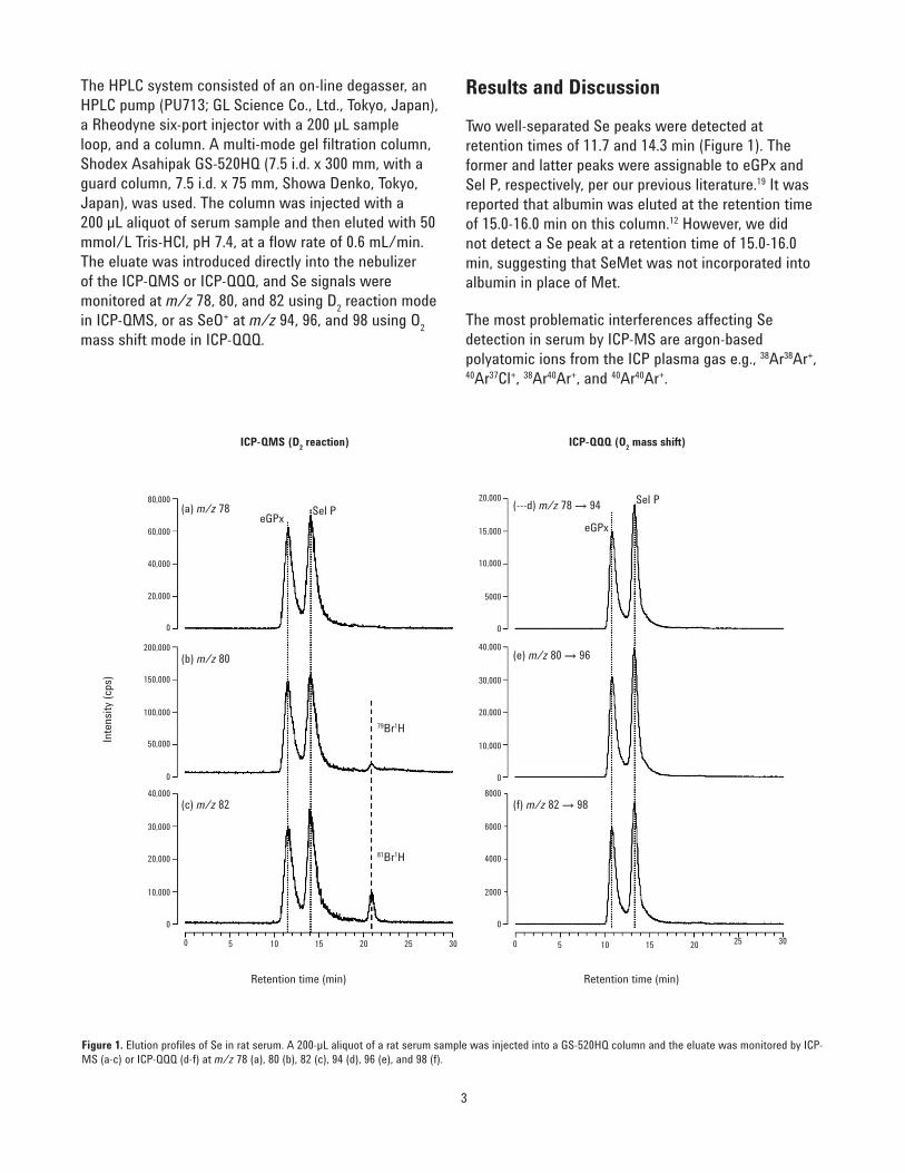
)The HPLC system consisted of an on-line degasser, an HPLC pump (PU713; GL Science Co., Ltd., Tokyo, Japan), a Rheodyne six-port injector with a 200 µL sample loop, and a column. A multi-mode gel fi ltration column, Shodex Asahipak GS-520HQ (7.5 i.d. x 300 mm, with a guard column, 7.5 i.d. x 75 mm, Showa Denko, Tokyo, Japan), was used. The column was injected with a 200 µL aliquot of serum sample and then eluted with 50 mmol/L Tris-HCl, pH 7.4, at a fl ow rate of 0.6 mL/min. The eluate was introduced directly into the nebulizer of the ICP-QMS or ICP-QQQ, and Se signals were monitored at m/z 78, 80, and 82 using D2 reaction mode in ICP-QMS, or as SeO+ at m/z 94, 96, and 98 using O2 mass shift mode in ICP-QQQ.
5 50 010 1015 1520 2025 2530 30
10,000
0
20,000
200,000
0
20,000
40,000
60,000
80,000
30,000
40,000
0
50,000
100,000
150,000
2000
0
4000
6000
8000
10,000
0
20,000
30,000
40,000
5000
0
10,000
15,000
20,000(a) m/z 78
(b) m/z 80
(c) m/z 82
(---d) m/z 78 → 94
(e) m/z 80 → 96
(f) m/z 82 → 98
eGPxeGPx
Sel PSel P
79Br1H
81Br1H
Figure 1. Elution profi les of Se in rat serum. A 200-µL aliquot of a rat serum sample was injected into a GS-520HQ column and the eluate was monitored by ICP-MS (a-c) or ICP-QQQ (d-f) at m/z 78 (a), 80 (b), 82 (c), 94 (d), 96 (e), and 98 (f).
Results and Discussion
Two well-separated Se peaks were detected at retention times of 11.7 and 14.3 min (Figure 1). The former and latter peaks were assignable to eGPx and Sel P, respectively, per our previous literature.19 It was reported that albumin was eluted at the retention time of 15.0-16.0 min on this column.12 However, we did not detect a Se peak at a retention time of 15.0-16.0 min, suggesting that SeMet was not incorporated into albumin in place of Met.
The most problematic interferences affecting Se detection in serum by ICP-MS are argon-based polyatomic ions from the ICP plasma gas e.g., 38Ar38Ar+, 40Ar37Cl+, 38Ar40Ar+, and 40Ar40Ar+.

4
Thus, 82Se is the least affected isotope in the detection by ICP-MS without a CRC (Figure 2a). H2 is frequently used as the reaction gas for Se detection. However, when Se in extracellular fl uid, such as serum, is analyzed, interferences originating from Br are formed (Figure 2b). Indeed, the quantifi cation and speciation of Se in urine and serum samples using multiple isotopically enriched tracers is not possible under H2 reaction mode due to severe interference from BrH. An alternative technique to avoid the interference is to use D2 in place of H2
17. When D2 is used as the reaction gas, the Br interference is reduced. However,
the commercially available D2 gas that we used contained substantial amounts of H2 (<0.5%); hence, the interference could not be excluded completely though the interference originating from BrH did not restrict Se speciation of serum. As shown in Figure 1b and 1c, the peak corresponding to BrH is well separated from the peaks of the two selenoproteins. There was a problem caused by D2 based interference (by SeD) as shown in Figure 2c, and this interference was unavoidable. Indeed, the signal at m/z 80 overlapped with the signal for 78Se2D+, and the signal at m/z 82 was more severely affected because 80Se has a larger abundance than 78Se (Figure 3a).
(a) ICP-QMS (D2 reaction) (b) ICP-QQQ (O2 mass shift)
2(a)
2(b)
2(c)
2(d)
m/z m/z
Relat
ive in
tens
ity (c
ps p
er
natu
ral a
bund
ance
1000200
0 078 80 82 989694
2000
400
3000
600
4000
800
1000
Q (m/z 82)82Se+
Ar+
Ar+
Br+ Ar+
H2
D2
O2
78Se+
80Se+
82Se+
78Se+
80Se+
82Se+
78Se16O+
79Br1H+
81Br1H+
76Se+D+
78Se+D+
80Se+D+
80Se16O+
82Se16O+
Q (m/z 78, 80, 82)
Detector
Detector
Detector
Detector
Q (m/z 78, 80, 82)
Q (m/z 78, 80, 82) Q (m/z 94, 96, 98)
Figure 2. Schematic diagram of the detection of Se in extracellular fl uid by ICP-MS under normal (a), H2 (b), and D2 (c) cell gas modes, and ICP-QQQ using O2 mass shift mode (d).
Figure 3. Effect of O2 mass shift mode by ICP-QQQ on the speciation of Se. Relative intensity was defi ned by counts per second (cps) of the peak heights of GPx (closed columns) and Sel P (open columns) divided by the isotope ratio of each Se isotope.

5
ICP-QQQ using O2 mass shift mode has been used successfully for the low level detection of sulfur and phosphorus19. In this study, we used the ICP-QQQ in O2 mass shift mode for Se speciation (Figure 2d). The fi rst quadrupole (Q1) was set to allow ions only at m/z 78, 80, and 82 to pass to the CRC thereby eliminating Br at m/z 81 and 83. No interference originating from BrH was detected in the elution profi les obtained by ICP-QQQ under O2 mass shift mode (Figures 1d-1f). In addition, the relative peak heights of the two serum selenoproteins were more consistent when measured using the ICP-QQQ method than by ICP-QMS (Figure 3b). Consequently, ICP-QQQ is a more powerful tool than ICP-QMS for the speciation of Se in biological samples.
Conclusions
Two major selenoproteins, eGPx and Sel P, in rat serum were well separated on an HPLC column. ICP-QQQ was a more accurate detector for the speciation of serum selenoproteins than ICP-QMS because it was completely free of interference originating from the plasma source Ar and the matrix elements.
Acknowledgements
We thank Mr. Naoki Sugiyama (Agilent Technologies International Japan, Ltd.) for facilitating the use of the ICP-QQQ. This study was supported by Grants-in-Aid from the Ministry of Education, Culture, Sports, Science and Technology, Japan (Nos. 23390032 and 24659022 to Y. O., and 25870739 to Y.A.), and the fi nancial support from Takeda Science Foundation, Japan.
References
1. M. Borglund, A. Akesson, and B. Akesson, Scand J Clin Lab Invest, 1988, 48, 27.
2. B. Akesson, and B. Martensson, J Inorg Biochem, 1988, 33, 257.
3. M. Persson-Moschos, W. Huang, T. S. Srikumar, B. Akesson, and S. Lindeberg, Analyst, 1995, 120, 833.
4. P. Jitaru, H. Goenaga-Infante, S. Vaslin-Reimann, and P. Fisicaro, Anal Chim Acta, 2010, 657, 100.
5. L. A. Daniels, Biol Trace Elem Res, 1996, 54, 185.
6. P. Òscar, and R. Łobiński, Talanta, 2007, 71, 1813.
7. Y. Kobayashi, Y. Ogra, and K. T. Suzuki, J. Chromatogr. B Biomed. Sci. Appl., 2001, 760, 73.
8. H. Koyama, Y. Kasanuma, C. Y. Kim, A. Ejima, C. Watanabe, H. Nakatsuka, and H. Satoh, Tohoku J Exp Med, 1996, 178, 17.
9. Y. Ogra, and Y. Anan, J. Anal. At. Spectrom., 2009, 24, 1477.
10. G. A. Jacobson, A. M. Featherstone, A. T. Townsend, R. Lord, and G. M. Peterson, Biol Trace Elem Res, 2005, 107, 213.
11. K. T. Suzuki, M. Itoh, and M. Ohmichi, J Chromatogr B Biomed Appl, 1995, 666, 13.
12. Y. Tsuji, T. Mikami, Y. Anan, and Y. Ogra, Metallomics, 2010, 2, 412.
13. J. J. Sloth, and E. H. Larsen, J. Anal. At. Spectrom., 2000, 15, 669.
14. J. J. Sloth, E. H. Larsen, S. H. Bügel, and S. Moesgaard, J. Anal. At. Spectrom., 2003, 18, 317.
15. J. Darrouzès, M. Bueno, G. Lespès, and M. Portin-Gautier, J. Anal. At. Spectrom., 2005, 20, 88.
16. W. Guo, S. Hu, Y. Wang, L. Zhang, Z. Hu, and J. Zhang, Microchem. J., 2013, 108, 106.
17. Y. Ogra, K. Ishiwata, and K. T. Suzuki, Anal. Chim. Acta, 2005, 554, 123.
18. S. Diez Fernandez, N. Sugishama, J. Ruiz Encinar, and A. Sanz-Medel, Anal Chem, 2012, 84, 5851.
19. Y. Anan, Y. Hatakeyama, M. Tokumoto, and Y. Ogra, Anal. Sci., 2013, 29, 787.

www.agilent.comAgilent shall not be liable for errors contained herein or for incidental or consequential damages in connection with the furnishing, performance or use of this material.
Information, descriptions, and specifi cations in this publication are subject to change without notice.© Agilent Technologies, Inc. 2013Published October 2, 2013Publication number: 5991-2750EN