Embed Size (px)
Citation preview

MOLECULAR AND CELLULAR BIOLOGY, Sept. 1992, p. 3665-36770270-7306/92/093665-13$02.00/0Copyright X 1992, American Society for Microbiology
Analysis of the Myogenin Promoter Reveals an IndirectPathway for Positive Autoregulation Mediated by the
Muscle-Specific Enhancer Factor MEF-2DIANE G. EDMONDSON, TSE-CHANG CHENG, PETER CSERJESI, TUSHAR CHAKRABORTY,
AND ERIC N. OLSON*
Department ofBiochemistry and Molecular Biology, Box 117, The University of Te-xas M. D. AndersonCancer Center, 1515 Holcombe Boulevard, Houston, Texas 77030
Received 4 March 1992/Returned for modification 31 March 1992/Accepted 25 May 1992
Transcriptional cascades that specify cell fate have been well described in invertebrates. In mammaliandevelopment, however, gene hierarchies involved in determination of cell lineage are not understood. With therecent cloning of the MyoD family of myogenic regulatory factors, a model system has become available withwhich to study the dynamics of muscle determination in mammalian development. Myogenin, along with othermembers of the MyoD gene family, possesses the apparent ability to redirect nonmuscle cells into the myogeniclineage. This ability appears to be due to the direct activation of an array of subordinate or downstream geneswhich are responsible for formation and function of the muscle contractile apparatus. Myogenin-directedtranscription has been shown to occur through interaction with a DNA consensus sequence known as an E box(CANNTG) present in the control regions of numerous downstream genes. In addition to activating thetranscription of subordinate genes, members of the MyoD family positively regulate their own expression andcross-activate one another's expression. These autoregulatory interactions have been suggested as a mechanismfor induction and maintenance of the myogenic phenotype, but the molecular details of the autoregulatorycircuits are undefined. Here we show that the myogenin promoter contains a binding site for the myocyte-specific enhancer-binding factor, MEF-2, which can function as an intermediary of myogenin autoactivation.Since MEF-2 can be induced by myogenin, these results suggest that myogenin and MEF-2 participate in atranscriptional cascade in which MEF-2, once induced by myogenin, acts to amplify and maintain the myogenicphenotype by acting as a positive regulator of myogenin expression.
The formation of skeletal muscle during vertebrate devel-opment involves the induction of mesoderm from primaryectoderm and the subsequent generation of proliferatingmyoblasts that ultimately terminally differentiate in responseto environmental cues. The recent discovery of a family ofrelated muscle-specific factors that can convert fibroblasts tomyoblasts has contributed to rapid progress toward under-standing the molecular events that underlie the establish-ment of the skeletal muscle phenotype (for reviews, seereferences 55 and 69). Members of this muscle regulatorygene family include MyoD (21), myogenin (25, 77), myf5 (9),and MRF4/herculin/myf6 (8, 48, 61), each of which canactivate myogenesis when introduced into a wide range ofnonmuscle cell types. Related myogenic factors have alsobeen identified in a variety of vertebrate and invertebratespecies (22, 30, 35, 39, 44, 47, 57, 65, 72). An additional gene,myd, that appears to be structurally unrelated to the MyoDfamily, has been identified as a genomic clone that caninduce myogenesis in 1OT1/2 cells, but it has not yet beencharacterized at the molecular level (59).Members of the MyoD family share extensive homology
within a basic region and adjacent helix-loop-helix (HLH)motif that mediate DNA binding and dimerization (11, 17,20, 41, 52, 73). Basic-HLH (bHLH) motifs have also beenidentified in members of the myc family of oncogenes,several proteins that mediate lineage decisions during Dro-sophila embryogenesis (1, 16), and a growing number oftranscription factors that are widely expressed (2, 29, 31, 36,
* Corresponding author.
37, 51; see also reference 3). All bHLH proteins that bindDNA recognize the consensus sequence CANNTG, referredto as an E box. However, individual HLH proteins showdistinct half-site preferences for binding that depend on thevariable nucleotides within and surrounding the invariantdyad symmetry of the CANNTG consensus (5). Binding ofmyogenic HLH proteins to DNA is enhanced in the presenceof the widely expressed E2A gene products (E12 and E47),with which they preferentially form hetero-oligomers (8, 14,18, 20, 52, 53).E boxes are present in the control regions of most muscle-
specific genes and are important for muscle-specific tran-scription (14, 15, 26, 41, 45, 58, 63, 74, 75). However, thereare examples of muscle-specific genes that do not contain Eboxes within their regulatory regions (46, 71). Whether thesegenes are controlled by other muscle-specific regulatoryfactors that act in parallel with, or are induced by, membersof the MyoD family remains to be determined. A candidatefor such factor is the myocyte-specific enhancer-bindingfactor MEF-2, which binds an A+T-rich element withinnumerous muscle-specific enhancers and promoters (28).MEF-2 is upregulated during myogenesis and can be inducedby myogenin and MyoD, suggesting it may function within a
regulatory pathway controlled by myogenic HLH proteins(19, 42).
In addition to activating downstream genes involved interminal differentiation, members of the MyoD family havethe ability to positively activate their own expression andcross-activate one another's expression in cultured cells (7,13, 48, 70). It is possible that autoregulation amplifies theexpression of these factors above a threshold necessary to
3665
Vol. 12, No. 9
on Novem
ber 26, 2018 by guesthttp://m
cb.asm.org/
Dow
nloaded from

3666 EDMONDSON ET AL.
activate myogenesis and provides stability to the myogenicphenotype. Similar autoregulatory activity has been de-scribed for a number of genes that regulate cell fate inDrosophila melanogaster (e.g., fushi tarazu, ultrabithorax,and deformed [4, 33, 40]). Although it has generally beenassumed that regulatory interactions among members of theMyoD family reflect direct binding of the correspondingproteins to their promoters, the regulatory regions thatcontrol transcription of these genes have not yet beencharacterized, leaving open the possibility that these inter-actions can be mediated indirectly through induction ofintermediate regulatory factors.
Despite their apparent ability to activate one another'sexpression in transfected cells in culture, members of theMyoD family show distinct temporal and spatial patterns ofexpression during embryogenesis (6, 32, 56, 64) and aredifferentially regulated during myogenesis in tissue culture(7, 13, 50, 61), suggesting that additional cellular factorsinfluence regulatory interactions among these genes. Myo-genin, for example, is first detected in replicating myogenicprecursors in the somite myotome at 8.5 days of mousedevelopment, several days prior to the appearance of MyoDor MRF4 (61, 64, 77). Myogenin expression is undetectableduring the migration of determined myogenic precursors intothe limb, where it again becomes expressed at high levels(64). In tissue culture, MyoD and myf5 are generally ex-pressed in proliferating myoblasts (7, 21), whereas myogenindoes not appear until differentiation has been triggered bywithdrawal of growth factors (25, 77). In contrast to MyoD,myf5, and MRF4, which are expressed only in subsets ofskeletal muscle cells, myogenin appears to be expressedduring differentiation of all skeletal muscle cell types, sug-gesting that the myogenin gene responds to a commonmyogenic regulatory pathway.To begin to define the early events that activate the muscle
differentiation program, we have analyzed the cis- andtrans-regulatory system required to induce myogenin tran-scription and to establish the myogenin autoregulatory cir-cuit during myogenesis. Here, we show that muscle speci-ficity and positive autoregulation of myogenin transcriptioncan be mediated by MEF-2, which binds a site in themyogenin promoter. The ability of MEF-2 to regulateexpression of myogenin, which in turn can induce MEF-2(19, 42), suggests that members of the MyoD family andMEF-2 function within a complex regulatory network oflineage-specific factors that cooperate to generate and main-tain the skeletal muscle phenotype.
MATERUILS AND METHODS
Cell culture. C2 (78) and 10T1/2 cells were maintained asdescribed previously (25). Differentiation of C2 cells wasinitiated by transferring cultures from growth medium (GM;Dulbecco's modified Eagle's medium with 20% fetal bovineserum) to differentiation medium (DM; Dulbecco's modifiedEagle's medium with 2% horse serum). Primary cultures ofchicken embryo muscle cells were prepared by modificationof a previously described procedure (26). Briefly, muscletissue from the breasts of 11- or 12-day-old chicken embryoswas dissected and dissociated with 0.05% trypsin. Cells werepreplated on 100-mm-diameter tissue culture dishes andwere sequentially transferred to new dishes coated withgelatin after incubations of 45 min to remove adherentfibroblasts. Where indicated, myoblasts were incubated with5-bromo-2'-deoxyuridine (BrdU; 5 ,ug/ml) for 4 days. Fibro-blasts were obtained from skin tissue of the same embryos
by 0.05% trypsin digestion and were plated into culturedishes.
Transfections and CAT assays. Transient transfectionswere performed by calcium phosphate precipitation as de-scribed previously (67). Forty-eight hours after transfection,cultures were transferred from GM to DM for an additional48 h. Cell extracts were then prepared, and levels of chlor-amphenicol acetyltransferase (CAT) activity were deter-mined by using aliquots of extract containing equivalentquantities of protein or aliquots normalized to 0-galactosi-dase activity generated from cotransfected RSV-lacZ, whichcontains the Rous sarcoma virus (RSV) long terminal repeatlinked to lacZ and is expressed constitutively. All transfec-tions were performed with at least two separate preparationsof each plasmid. Stable transfections were performed byusing 10 p,g of reporter plasmid and 500 ng of a neomycinresistance gene as described previously (25). Forty-eighthours after transfection, cells were transferred to GM withG418 (400 ,ug/ml) for 14 days.
Isolation of genomic clones. To isolate myogenin genomicclones, mouse genomic libraries, prepared by ligating anMboI partial digest of mouse genomic DNA into the BamHIsite of the lambda phage vector EMBL 3, were screened byhybridization to the full-length mouse myogenin cDNA (25).DNA was hybridized on duplicate filter lifts overnight at42°C in 50% formamide-5x SSC (0.75 M NaCl plus 0.075 Msodium citrate [pH 7.0])-5 x Denhardt's solution-0.05 MNa2HPO4 (pH 7.0)-5% dextran sulfate-100 p,g of salmonsperm DNA per ml. The filters were then washed twice for30 min in 2x SSC-0.1% sodium dodecyl sulfate (SDS) atroom temperature and once for 30 min in O.lx SSC-0.1%SDS at 65°C. Positive plaques were carried through threesuccessive rounds of screening. Four clones, designatedMG1, MG2, MG4, and MG6, remained positive after plaquepurification and were characterized further.
Subcloning and sequencing. Inserts from the phage clonesfor MG1, MG2, and MG6 were excised by digestion withSailI or XhoI and were subcloned into pUC19 or pBluescriptfor restriction mapping and sequencing. Subcloned genomicfragments were sequenced by using the dideoxy-chain ter-mination method in denatured double-stranded plasmid,using synthetic oligodeoxynucleotides for primers. Sequenc-ing primers were synthesized by the Macromolecular Syn-thesis Facility, M. D. Anderson Cancer Center. Sequenceanalysis was performed using the Beckman Microgenie andUniversity of Wisconsin Genetics Computer Group se-quence analysis software package.
S1 protection analysis. Total cellular RNA was isolatedfrom cell cultures according to the guanidinium isothiocya-nate procedure as described previously (25). To determinethe myogenin transcription start site, a fragment from MG2,which extended 1.8 kb upstream from within intron 1, wasdenatured by alkali treatment at 65°C, neutralized, ethanolprecipitated, and annealed to an oligonucleotide correspond-ing to nucleotides 177 through 191 on the antisense strand ofthe myogenin cDNA. Klenow fragment of DNA polymeraseI in the presence of unlabeled dATP, dGTP, and dTTP and32P-labeled dCTP was used to synthesize an antisense probeextending from nucleotide 191 several hundred bases intothe 5' flanking region of the gene. The labeled probe wasseparated on an alkaline denaturing gel and hybridized to 20p,g of total cellular RNA for 16 h at 55°C. RNA was thendigested with 50 U of S1 nuclease for 1 h at 37°C, anddigestion products were separated on a 5% polyacrylamidegel.Primer extension. Synthetic oligonucleotides correspond-
MOL. CELL. BIOL.
on Novem
ber 26, 2018 by guesthttp://m
cb.asm.org/
Dow
nloaded from

TRANSCRIPTIONAL CONTROL OF MYOGENIN 3667
ing to sequences within the myogenin cDNA or CAT wereend labeled with T4 kinase. The myogenin oligomer an-nealed to sequence located between nucleotides 50 and 84 ofthe myogenin sequence, and the CAT oligomer was a 20-merand annealed to a sequence 50 nucleotides downstream fromthe unique HindIII site in pSVOCAT. Primer extension wasperformed by adding 100,000 cpm of end-labeled product to20 to 40 ,ug of total cellular RNA. The mixture (in 10 mM Tris[pH 7.51-0.25 M KCl-1 mM EDTA, 20 ,ul in volume) washeated to 65°C for 60 min and then cooled slowly to roomtemperature. The reaction volume was adjusted to 67 ,u andfinal concentrations of 10 mM Tris (pH 7.5), 75 mM KCI, 10mM dithiothreitol, 3 mM MgCl2, 0.5 mM each deoxynucle-oside triphosphate (dNTP), and 50 ,ug of actinomycin D perml. Extension was performed at 37°C, using 400 U ofMoloney murine leukemia virus reverse transcriptase for 90min. Following ethanol precipitation, the products wereelectrophoresed on an 8% polyacrylamide gel. A dideoxysequencing reaction was performed by using the sameoligonucleotide primer, and samples were electrophoresed inadjacent lanes to determine more precisely the 3' end of theextended species.
Mutagenesis. Mutagenesis was performed by using single-stranded template DNAs as described previously (11). Allmutants were confirmed by sequencing, and mutagenicregions were subcloned into wild-type vectors to avoidextraneous mutations.Myogenin reporter gene constructs. The myogenin-CAT
reporter genes were constructed by cloning a HaeIII-PstIfragment that corresponded to nucleotides +18 to -184relative to the myogenin transcription start site. This frag-ment was inserted into the pCAThasic vector (Promega) toyield the construct pMY0184CAT. pCAThasic was pre-pared for cloning by digestion withXbaI, filling in of the endswith dNTPs in the presence of the Klenow fragment, anddigestion with PstI. Myogenin-CAT reporters were createdby cloning restriction fragments extending from the PstI siteat -184 to -3700 (PstI site), -1565 (XbaI), -1102 (SstI),or -335 (SmaI) into pMY0184CAT. These clones recreatethe exact sequence of the myogenin genomic clones.pMY015651acZ was created in the same way, using the lacZreporter plasmid pUCAUGbeta-gal (a gift from W. M. Perry,M. D. Anderson Cancer Center), which contains the lacZcoding sequences 3' of a multicloning site. For deletion of Ebox E-1, a restriction fragment was made by digesting theappropriate DNA with BanI, filling in the ends with Klenowpolymerase, and then digesting the DNA with PstI. Theresulting fragment was cloned into pCAThasic as describedabove and yielded constructs whose 3' ends correspond tothe myogenin sequence at -18. Unidirectional 5' deletionsof the promoter were performed by using an exonucleaseIII-mung bean nuclease kit as recommended by the manu-facturer (Stratagene).
In vitro transcription-translation and gel mobility shiftassays. Myogenin and E12 were prepared for gel mobilityshift assays by in vitro transcription-translation of the cor-responding cDNAs as described previously (11). Nuclearextracts were prepared from tissue culture cells as describedpreviously (28). Breast muscle from 13-day chicken embryoswas dissected, and nuclei isolated were as described by Marand Ordahl (46). Extracts were then prepared as for tissueculture cells. Five or ten micrograms of nuclear extract wasused in each gel mobility shift assay. Oligonucleotide probeswere synthesized by the Macromolecular Synthesis Facilityat the M. D. Anderson Cancer Center and were end labeledwith 32p.
Nucleotide sequence accession number. The nucleotidesequence of the mouse myogenin gene has been entered intothe GenBank data base under the accession number M95800.
RESULTS
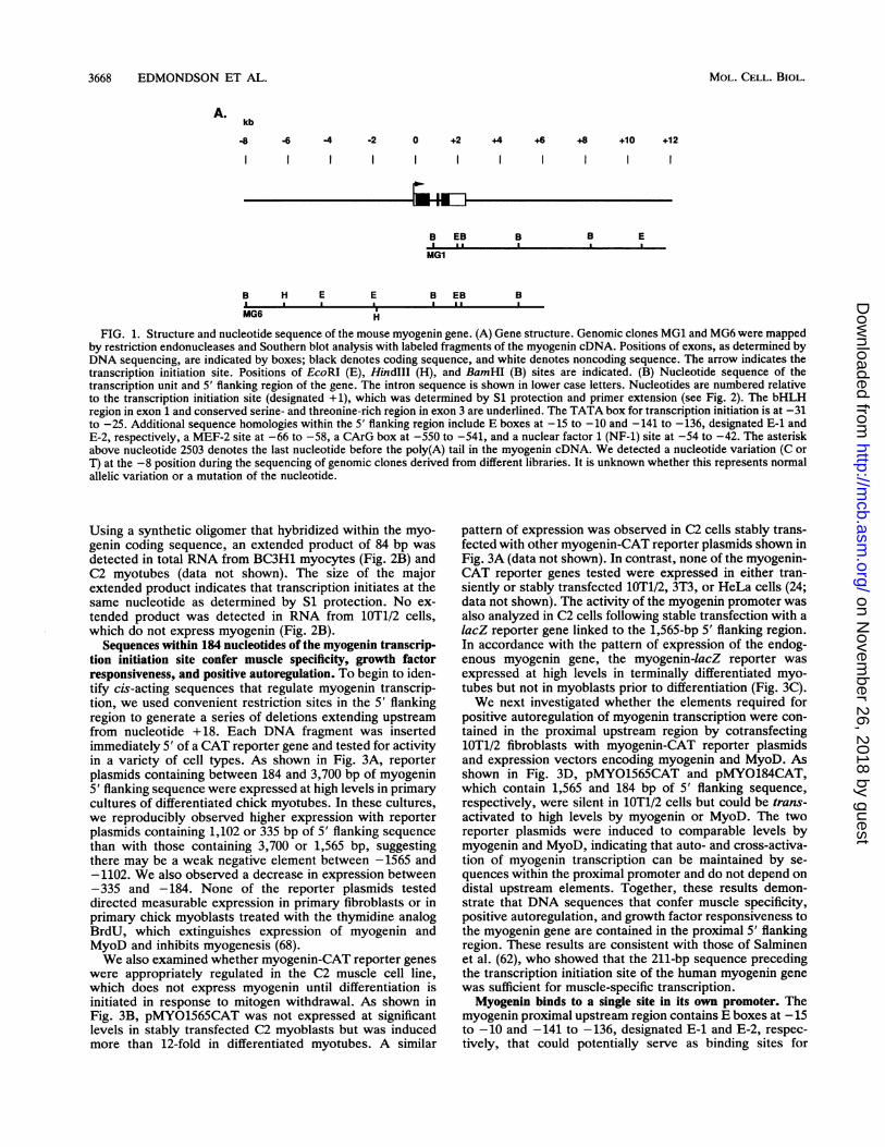
Structure of the mouse myogenin gene. To begin to definethe cis-acting elements that control myogenin transcription,we isolated myogenin genomic clones by screening a mousegenomic library with a full-length myogenin cDNA. Se-quence analysis of overlapping recombinant phage revealedthat the myogenin gene spans -2.5 kb and contains twointrons (Fig. 1). The sequence of human myogenin has alsobeen reported recently by Salminen et al. (62). The firstpotential initiation codon for myogenin translation is located50 nucleotides downstream of the transcription start site.The basic domain and HLH motif are contained within exon1. Exon 2 encodes 27 amino acids that comprise an essentialpart of the transcriptional activation domain of the myogeninprotein (66). Exon 3 is relatively rich in serine and threonineresidues and contains a segment of 12 amino acids that ishomologous to MyoD, myf5, and MRF4. The translationtermination codon and the entire 3' untranslated region arealso contained in exon 3. At the 3' end of the gene, there isa potential polyadenylation signal (AATAAA) 16 basesupstream of the poly(A) tail identified in the previouslypublished analysis of the cDNA (25). Both introns areflanked by consensus sequences for splice donors and ac-ceptors. The intron-exon organization of myogenin is thesame as that of the mouse MyoD (38) and herculin (18a, 48)genes. In both of the latter genes, the bHLH region isencoded by exon 1; exon 2 encodes between 27 and 29 aminoacids, and the conserved serine- and threonine-rich se-quence is contained in exon 3. The Drosophila and Cae-norhabditis elegans MyoD genes show a different organiza-tion (39, 47).We analyzed the 5' flanking region for sequence motifs
known to participate in transcriptional regulation of muscle-specific genes as well as of genes more widely expressed.Two A+T-rich sequences are located in the proximal 5'flanking region, -66 to -58 and -31 to -25; the lattersequence serves as the TATA box for transcription initiation(see below). The more distal sequence is similar to thebinding site for MEF-2 (19, 28), which binds the musclecreatine kinase (MCK) and myosin light-chain one-thirdenhancers. E boxes (CANNTG) are present at -15 to -10and -141 to -136. An element that resembles a CArG motif,CC(A+T)6GG, which has been implicated in transcription ofnumerous muscle-specific genes, is present at -550 to -541(49), and a sequence that corresponds to the binding site fornuclear factor 1 is located at -54 to -42 (23, 27). There isalso an A+C repeat between -604 and -560 that could havethe potential to form Z-DNA (54).
Determination of the transcription initiation site of myoge-nin. The transcription initiation site for myogenin was deter-mined by Si protection and primer extension. Using alabeled antisense probe that extended upstream from thefirst nucleotide of codon 48 in the open reading frame, wedetected a major Si-resistant product of 191 nucleotides withRNA from C2 myotubes (Fig. 2A). The length of theprotected species indicated that the major site for transcrip-tion initiation lies 25 bp downstream of the TTAAAT se-quence in the 5' proximal region (Fig. 1B). Si-resistantproducts were not generated by RNA from 3T3 fibroblasts(Fig. 2A), which do not express myogenin.Primer extension confirmed the results of Si analysis.
VOL. 12, 1992
on Novem
ber 26, 2018 by guesthttp://m
cb.asm.org/
Dow
nloaded from
3668 EDMONDSON ET AL.
A.kb
8 -6 -4 -2 0 +2 +4 +6 +8 +10 +12
IIIII
B EBI .I
MG1
B H E E B EBI I I I I I*.
MG6
B B EI I
B
H
FIG. 1. Structure and nucleotide sequence of the mouse myogenin gene. (A) Gene structure. Genomic clones MG1 and MG6 were mappedby restriction endonucleases and Southern blot analysis with labeled fragments of the myogenin cDNA. Positions of exons, as determined byDNA sequencing, are indicated by boxes; black denotes coding sequence, and white denotes noncoding sequence. The arrow indicates thetranscription initiation site. Positions of EcoRI (E), HindIlI (H), and BamHI (B) sites are indicated. (B) Nucleotide sequence of thetranscription unit and 5' flanking region of the gene. The intron sequence is shown in lower case letters. Nucleotides are numbered relativeto the transcription initiation site (designated +1), which was determined by S1 protection and primer extension (see Fig. 2). The bHLHregion in exon 1 and conserved serine- and threonine-rich region in exon 3 are underlined. The TATA box for transcription initiation is at -31to -25. Additional sequence homologies within the 5' flanking region include E boxes at -15 to -10 and -141 to -136, designated E-1 andE-2, respectively, a MEF-2 site at -66 to -58, a CArG box at -550 to -541, and a nuclear factor 1 (NF-1) site at -54 to -42. The asteriskabove nucleotide 2503 denotes the last nucleotide before the poly(A) tail in the myogenin cDNA. We detected a nucleotide variation (C orT) at the -8 position during the sequencing of genomic clones derived from different libraries. It is unknown whether this represents normalallelic variation or a mutation of the nucleotide.
Using a synthetic oligomer that hybridized within the myo-genin coding sequence, an extended product of 84 bp wasdetected in total RNA from BC3H1 myocytes (Fig. 2B) andC2 myotubes (data not shown). The size of the majorextended product indicates that transcription initiates at thesame nucleotide as determined by S1 protection. No ex-tended product was detected in RNA from 1OT1/2 cells,which do not express myogenin (Fig. 2B).
Sequences within 184 nucleotides of the myogenin transcrip-tion initiation site confer muscle specificity, growth factorresponsiveness, and positive autoregulation. To begin to iden-tify cis-acting sequences that regulate myogenin transcrip-tion, we used convenient restriction sites in the 5' flankingregion to generate a series of deletions extending upstreamfrom nucleotide +18. Each DNA fragment was insertedimmediately 5' of a CAT reporter gene and tested for activityin a variety of cell types. As shown in Fig. 3A, reporterplasmids containing between 184 and 3,700 bp of myogenin5' flanking sequence were expressed at high levels in primarycultures of differentiated chick myotubes. In these cultures,we reproducibly observed higher expression with reporterplasmids containing 1,102 or 335 bp of 5' flanking sequencethan with those containing 3,700 or 1,565 bp, suggestingthere may be a weak negative element between -1565 and-1102. We also observed a decrease in expression between-335 and -184. None of the reporter plasmids testeddirected measurable expression in primary fibroblasts or inprimary chick myoblasts treated with the thymidine analogBrdU, which extinguishes expression of myogenin andMyoD and inhibits myogenesis (68).We also examined whether myogenin-CAT reporter genes
were appropriately regulated in the C2 muscle cell line,which does not express myogenin until differentiation isinitiated in response to mitogen withdrawal. As shown inFig. 3B, pMYO1565CAT was not expressed at significantlevels in stably transfected C2 myoblasts but was inducedmore than 12-fold in differentiated myotubes. A similar
pattern of expression was observed in C2 cells stably trans-fected with other myogenin-CAT reporter plasmids shown inFig. 3A (data not shown). In contrast, none of the myogenin-CAT reporter genes tested were expressed in either tran-siently or stably transfected 1OT1/2, 3T3, or HeLa cells (24;data not shown). The activity of the myogenin promoter wasalso analyzed in C2 cells following stable transfection with alacZ reporter gene linked to the 1,565-bp 5' flanking region.In accordance with the pattern of expression of the endog-enous myogenin gene, the myogenin-lacZ reporter wasexpressed at high levels in terminally differentiated myo-tubes but not in myoblasts prior to differentiation (Fig. 3C).We next investigated whether the elements required for
positive autoregulation of myogenin transcription were con-tained in the proximal upstream region by cotransfecting1OT1/2 fibroblasts with myogenin-CAT reporter plasmidsand expression vectors encoding myogenin and MyoD. Asshown in Fig. 3D, pMYO1565CAT and pMYO184CAT,which contain 1,565 and 184 bp of 5' flanking sequence,respectively, were silent in 1OT1/2 cells but could be trans-activated to high levels by myogenin or MyoD. The tworeporter plasmids were induced to comparable levels bymyogenin and MyoD, indicating that auto- and cross-activa-tion of myogenin transcription can be maintained by se-quences within the proximal promoter and do not depend ondistal upstream elements. Together, these results demon-strate that DNA sequences that confer muscle specificity,positive autoregulation, and growth factor responsiveness tothe myogenin gene are contained in the proximal 5' flankingregion. These results are consistent with those of Salminenet al. (62), who showed that the 211-bp sequence precedingthe transcription initiation site of the human myogenin genewas sufficient for muscle-specific transcription.Myogenin binds to a single site in its own promoter. The
myogenin proximal upstream region contains E boxes at -15to -10 and -141 to -136, designated E-1 and E-2, respec-tively, that could potentially serve as binding sites for
MOL. CELL. BIOL.
on Novem
ber 26, 2018 by guesthttp://m
cb.asm.org/
Dow
nloaded from
VOL. 12, 1992 TRANSCRIPTIONAL CONTROL OF MYOGENIN 3669
BTCTAGAGTTG TATGACGCAG GCAAAGGTGA TGACTCAGGC AGGAAGGAAT AGAAGAGGCC AGCCTGGTGG CCCAGGACAG -1490
ACAAATGATG CAAAGGACTC TTTCTCCTTA TCGACCCTTC TACAGAAAGG AAAGAGTCAA MCGGTTCTA GTGCCAGAAG -1410
GCATTATTGA GGGGAAAGCA CAGAAGAGAT GATTAAGAGC ATCAGACAGG GTCCATCCCA TAATCAGCCA CCAAACACAG -1330
ACACTATTAC ATATGCCAGC AAGATTTTGC TGAAAGAACC CTGATATAGC TGCCTCTTGT GAGGCTATGC AGTGCCTGGC -1250
AAATACAGAA GTGGATGCTC ACAGTCAGCT ATTGGATGGA ACACAGGGCC CCCAATGGAG GAGCTAGAGA AAGTACCCAA -1170
GGAACTGAAG GGGTCTGCAA CCCTATAGGT GGAACAACAA TATGAACTAA CCAGTACCCC CAGAGCTCAT GTCTCTAGCT -1090
GCATATGTAG CAGAAGATGG CCTAGTCGGC CATCATTGGG AAGAGAGGCC CCTTGGTATT GCAAACTATA TGCCCCAGTA -1010
CAGGGGAACG CCAGGGCCAA GAAGTGGGAA TGAGTGGGTA GGGGAGCAGG GCGGGGGGAG GGGGGGGTAT AGGGAACTTT -930
TGGGATAGCA TTTGAAATGT AAATGAAGAA AATATCTAAT AAAAMTAAT TTAAAAAAGA GCGTCAGACA GGGGACTGAA -850
CAGCTCTTGC ACTAGGGGAG AAGAAGGCAA TGTAGAGTAG TCTGTGAGTT CTAATCCTTG CTAAACACTG ACTTCACCTG -770
ACCCCTACTA CTTAAGGCCC CCCCCCCTTA CTTAAGAAGT CCCTGTGTTC TCTTACTTCA ATCTACCCCC AACATCATGA -690
GACCTGGTCA AAGAAGCTGT AGAAACCCAA AAGTTGAATC CATTTGCCCT TCTGGGTTTC TGTCTTTGCC TCCATGGACG -610CArG
ATAGGACACA CACACACACA CACACACACA CACACACACA CACACACACA CGCCCCAAAT CTGGAGTGGT CCTGATGTGG -530
TAGTGGTAGG TCTTTAGGGG TCTCATGGGA CTGACATAGT ACGGTTTAAG GTGCTGCTGC TGAGCAGGAA AGAGAAGGCT -450
AAGTGGATTT TCAAGACCCC TTCCCGTCCG TCCAAGACAA CCCCTTTCTT GTTCCCTTCC TGCCCTGTCC ACCAGCTGCC -370
TTGGACCATG GAGGAGAGAG TAGGCAGGAG GCCCGGGTAG GAGTAATTGA AAGGAGCAGA TGAGACGGGG GAATGCACCC -290
ACCCCCACCT TCCCTGCCCC ACAGGNTGTG GAGAAATGAA AACTAATCAA ATTACAGCCG ACGGCCTCCC GACCCGTGCA -210E2
CAGGAGCCGC CTGGGCCAGG GGCAGGCCTG CAGGGTGGGG TGGGGGCAAA AGGAGAGGGA AGGGGAATCA CATGTAATCC -130MEF-2
ACTGGAAACG TCTTGATGTG CAGCAACAGC TTAGAGGGGG GCTCAGGTTT CTGTGGCGTT GGCTATATTT ATCTCTGGGT -50NF-i TATA E4 +1
TCATGCCAGC AGGGAGGGTT TMMTGGCAC CCAGCAGTTG GCGTGAGGGG CTGCGGGAGC TTGGGGGCCA GTGGCAGGAA 31
CAAGCCTTTT CCGACCTGAT GGAGCTGTAT GAGACATCCC CCTATTTCTA CCAGGAGCCC CACTTCTATG ATGGGGAAAA 111N E L Y E T S P Y F Y 0 E P H F Y D G E N
CTACCTTCCT GTCCACCTTC AGGGCTTCGA GCCCCCGGGC TATGAGCGGA CTGAGCTCAG CTTAAGCCCG GAAGCCCGAG 191Y L P V H L Q G F E P P G Y E R T E L S L S P E A R
GGCCCCTGGA AGAAAAGGGA CTGGGGACCC CTGAGCATTG TCCAGGCCAG TGCCTGCCGT GGGCATGTM GGTGTGTAAG 271G P L E E K G L G T P E H C P G Q C L P W A C K V C K
AGGAAGTCTG TGTCGGTGGA CCGGAGGAGG GCAGCCACAC TGAGGGAGAA GCGCAGGCTC AAGAAAGTGA ATGAGGCCTT 351R K S VS V D R R R A A T L R E K R R L K K V N E A F
CGAGGCCCTG AAGAGGAGCA CCCTGCTCAA CCCCAACCAG CGGCTGCCTA AAGTGGAGAT CCTGCGCAGC GCCATCCAGT 431E A L K R S T L L N P N 0 R L P K V E I L R S A I Q
ACATTGAGCG CCTACAGGCC TTGCTCAGCT CCCTCAACCA GGAGGAGCGC GATCTCCGCT ACAGAGGCGG GGGCGGGCCC 511Y I E R L Q A L L S S L N Q E E R D L R Y R G G G G P
CAGCCCATGg taagtggcta gtacaccaga tccagggata gggccagagg gaggtgcctg gtcagccccg ggccaccaga 591Q P Mgctagaacag gtgcaaagca gggcccttgg gatccctgga cccctttcct tgtcggctag actgtccagt ggccttctgg 671
gctcatactg tttccaaaat acagatgggg tagggtggac tggcacagga gaacagaagg ggcccggaaa caagcctgca 751
gtaagggctt tggtgagcct aggtgaaaat gagggaaaag tggcctaaga aacctgcttt gtccaagtcc acagccttca 831
tttttgacct gacgaaacac attagaatgc ccacaggcct gctcaggagc caccgtaaca aaatactgtg gtttatcacc 911
ctcaccgccg aaatgaaagc aaagttctca tcctagaagc taggaaagac ctagaagtag gatgtctatg gtaacattct 991
gcagtagact tgaccttgga ccttggcctt gtcttgcatg cagGTGCCCA GTGAATGCAA CTCCCACAGC GCCTCCTGCA 1071V P S E C N S H S A S C
GTCCGGAGTG GGGCAATGCA CTGGAGTTCG GTCCCAACCC AGGAGgtaag tgaatcctga ccctggtaac atggctcaaa 1151S P E W G N A L E F G P N P G
agggtaggca actgaccctg taacccaggc agagagaaat acctgagact gtcccttagt ccctacctag gctgggcttg 1231
tacccttacc ataagccctg tcccaatcac ccttcagcag cttttctagg aaattgctga cctgagggcc ccaaatggtg 1311
ggctggagac cctgtgcctc ccggtagcct tgaccagcta aagagtagtc agagcctcca gcaacttccc tgctggtctct 1392
gacctcagag agaagagcct gctcctgccc caggggtccc tggggatcac tcagtcagtg ttgtaaaact gtgccctgtgc 1473
cctaggatat cctggcttca cctagggata gccactttta aaaccccaca agcacacaca cagcatgggg agtactagtgt 1554
cttgggggcg gggaacactc ctgagtcttc cttacccatt ttgtcctgga ccagttgccc aggaaacatt ctcccactttt 1635
tcttgcagAT CATTTGCTCG CGGCTGACCC TACAGACGCC CACAATCTGC ACTCCCTTAC GTCCATCGTG GACAGCATCAC 1716D H L L A A D P T D A H N L H S L T S I V D S I T
GGTGGAGGAT ATGTCTGTTG CCTTCCCAGA CGAAACCATG CCCAACTGAG ATTGTCTGTC AGGCTGGGTG TGCATGTGAGC 1797V E D M S V A F P D E T N P N
CCCCAAGTTG GTGTCAAAAG CCATCACTTC TGTAGCAGGG GGCTTTTAAG TGGGGCTGTC CTGATGTCCA GAAAACAGCCC 1878
TGGGCTGCCA CAAGCCAGAC TCCCCACTCC CCATTCACAT AAGGCTAACA CCCAGCCCAG CGAGGGAATT TAGCTGACTCC 1959
TTAAAGCAGA GAGCATCCTC TTCTGAGGAG AGAAAGATGG AGTCCAGAGA GCCCCCTTGT TAATGTCCCT CAGTGGGGCAA 2040
ACTCAGGAGC TTCTTTTTTG TTTATCATAA TATGCCTCGA ATTCCACCCT CCACCTCCAA MTGAAACCG TTTGAGAGACA 2121
TGAGTGCCCT GACCTGGACA AGTGTGCACA TCTGTTCTAG TCTCTTCCTG AAGCCAGTGG CTGGGCTGGG CCTGCCCTGAG 2202
TTGAGAGAGA AGGGGGAGGA GCTATCCGGT TCCAAAGCCT CTGGGGGCCA AGCATTTGCA GTGGATCTTG GGAACCTTCCA 2283
GTGCTTTGTG TATTGTTTAT TGTTTTGTGT GTTGTTTGTA AAGCTGCCGT CTGCCAAGGT CTCCTGTGCT GATGATACCGG 2364
GAACAGGCAG GCAAGGGGGT GGGGGCTCTT GGGGTGACTT CTTTTGTTAA CTAAGCATTG TGTGGTTTTG CCAATTTTTTT 2445
TCTTTTGTAA TTCTTTTGCT AACTTATTTG GATTTCCTTT TTTAAAAAAT GAATAMGAC TGGTTGCTAT CAGAGCATCTG 2526
CAATGATATT ATCAAGANGG GGAGCCACCA AAACTCTTGA CACCTTTACC 2576
FIG. 1-Continued.
on Novem
ber 26, 2018 by guesthttp://m
cb.asm.org/
Dow
nloaded from

3670 EDMONDSON ET AL.
A. B.
<qnt
I' -240"IVI
=nt
sq -112
-86
-78'. '''
-157.
FIG. 2. Determination of the transcription initiation site formyogenin by Si protection and primer extension. (A) Si protection.The transcription initiation site for myogenin was determined by Siprotection as described in the text. The 3' end of the labeled probecorresponds to the first nucleotide of codon 48 in the open readingframe. The labeled probe was incubated with RNA from C2 myo-
tubes, 3T3 cells, or tRNA, as indicated. The protected species is 191nucleotides (nt) in length and is exactly the predicted size of a
transcript initiating 25 nucleotides downstream of the TATA-likeelement at -31 to -25. (B) Primer extension, performed as de-scribed in the text. The 3' end of the primer used for extensioncorresponds to nucleotide 84 of the myogenin cDNA. The primerwas annealed to RNA from BC3H1 myocytes, 1OT1/2 cells, or
tRNA, as indicated. The major extended product of 84 nucleotidesindicates that the primary transcription start site begins 25 nucleo-tides 3' of the TAAAT sequence, in exact agreement with Siprotection. A dideoxy sequencing reaction was performed by usingthe same primer used for the primer extension and was electro-phoresed in adjacent lanes to determine the exact extent of protec-tion (data not shown). (C) Positions of the S1 protection probe andprimer extension primer within the first exon of the myogenic gene.
myogenin, MyoD, or other myogenic HLH proteins. Todetermine whether myogenin was able to bind either of theseE boxes, we used oligonucleotide probes encompassing eachE box in gel mobility shift assays with in vitro-translatedmyogenin and E12. As shown in Fig. 4, the more 3' E box,E-1, strongly bound myogenin-E12 hetero-oligomers,whereas we did not detect significant binding to the more 5'E box, E-2 (compare lanes 2 and 7). Binding of myogenin-E12 to E-1 was sequence specific, as shown by competitionwith unlabeled E-1 oligomer but not with E-2 or an unrelatedsequence from another region of the promoter (Fig. 4, lanes2 to 5). The E-1 oligomer was also able to compete forbinding of myogenin-E12 to a labeled probe encompassingthe high-affinity right E box from the MCK enhancer, whichis the prototypical target sequence for myogenin and MyoD(Fig. 4, lane 13). The sequence of E-1 agrees with theconsensus sequence for DNA binding of MyoD-E12 andmyogenin-E12 hetero-oligomers, as determined by polymer-ase chain reaction-mediated binding site selection, whereasE-2 would not be predicted to bind these hetero-oligomersappreciably (5, 76).
It has been speculated that homo-oligomers of MyoDcould play a role in positive autoregulation through interac-
tion with the MyoD control region (3, 70). We thereforetested whether in vitro-translated myogenin or MyoD wouldbind E box E-1 or E-2 in the absence of E12. Under theconditions of these assays, we did not detect binding ofeither myogenic factor alone to E-1 or E-2 (data not shown).However, a bacterially expressed myogenin fusion protein,at concentrations significantly higher than can be achievedwith in vitro translation products, will bind to E-1 (data notshown). We also examined whether there might be crypticsites for binding of myogenin-E12 or MyoD-E12 within the184-bp promoter by performing gel mobility shift assays withoverlapping restriction fragments encompassing the entireregion; the only measurable binding was observed with Ebox E-1 (data not shown).The muscle-specific enhancer-binding factor MEF-2 binds
the myogenin promoter. The myogenin promoter also con-tains an A+T-rich element at -66 to -58 that resembles abinding site for the muscle-specific factor MEF-2, which wasoriginally identified as an MCK enhancer-binding factor thatis induced during the myoblast-to-myotube transition (28).Although the A+T-rich element in the myogenin promoterdeviates from the MCK MEF-2 site, it agrees with thepreliminary consensus sequence recently deduced forMEF-2 binding (19; see also reference 60). Therefore, todetermine whether MEF-2 could interact with this A+T-richelement, we used an oligomer encompassing this region as aprobe in gel mobility shift assays with nuclear extracts fromC2 myoblasts and myotubes. Figure 5A shows that thisprobe generated a complex specific to differentiated C2myotubes (lane 4) and that the migration of this complex wasindistinguishable from that of the complex generated with anoligomer corresponding to the MEF-2 site in the MCKenhancer (lane 1). Extracts from C2 myoblasts did notcontain detectable MEF-2 activity but generated a heteroge-neous series of more rapidly migrating complexes, one ofwhich represents a previously characterized binding activityreferred to as myoblast factor 1. Extracts from 1OT1/2 cellsdid not show significant binding to the labeled probes underthe conditions of these assays. All of the extracts showedequivalent binding activity with a labeled probe correspond-ing to the binding site for Oct-1, which is expressed ubiqui-tously (data not shown). Specificity of MEF-2 binding to themyogenin MEF-2 site was shown by competition by unla-beled MCK MEF-2 site and the lack of competition by anoligomer containing a single base substitution previouslyshown to abolish MEF-2 binding (19).Nuclear extract from embryonic chicken breast muscle
also contains the MEF-2 binding activity. Figure SB showsthat the MEF-2 complex produced with breast muscle ex-tract (lane 4) is identical to the complex generated by C2myotube extract (lane 1). This complex is competed for byexcess homologous oligomer (lanes 2 and 5) but not by anoligomer containing a mutant MEF-2 site (lanes 3 and 6).
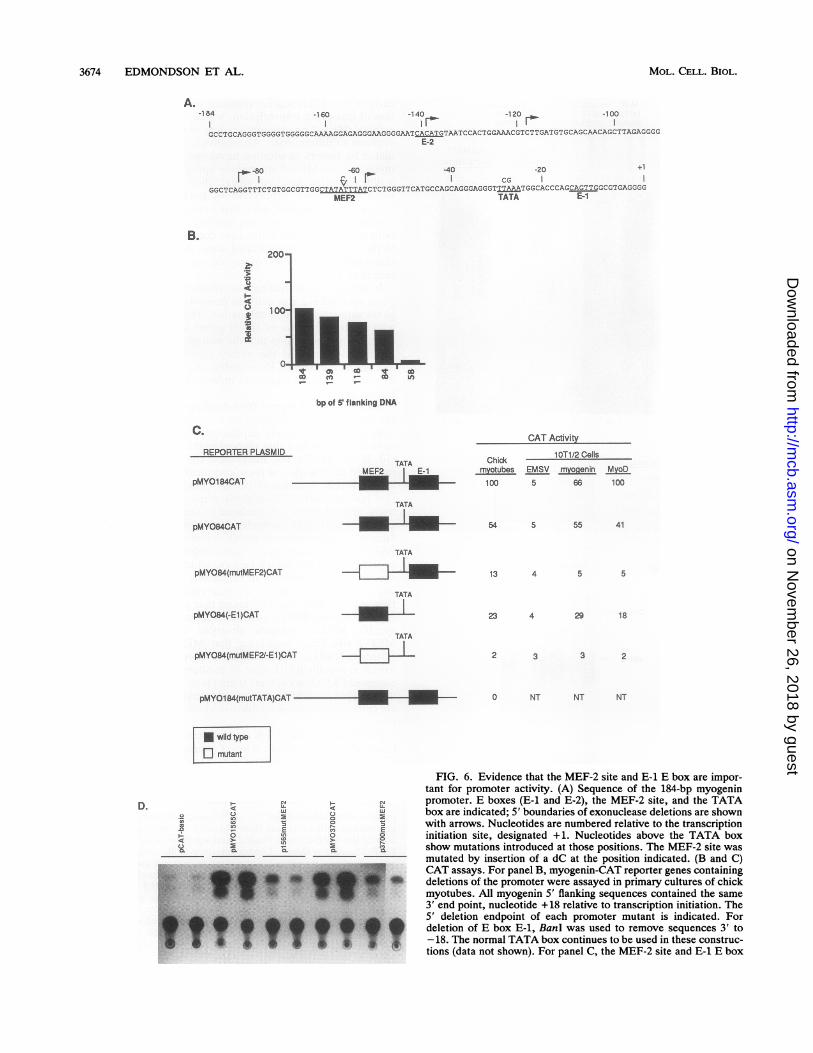
Muscle specificity of the myogenin promoter is E boxindependent. To further closely define the regulatory ele-ments responsible for myogenin transcription, we created aseries of exonuclease deletion mutants of the promoter,beginning at bp -184 (Fig. 6A). Deletion from -184 to -84bp had only a modest effect on promoter activity in chickprimary myotubes. However, deletion to -58 bp resulted ina greater than 20-fold decrease in activity (Fig. 6B), indicat-ing that the region of the promoter containing the MEF-2 siteis essential for transcriptional activity of the minimal pro-moter. The partial loss in activity when the region between-184 and -84 bp was deleted also suggests the presence ofregulatory elements in this region that contribute to the
MOL. CELL. BIOL.
on Novem
ber 26, 2018 by guesthttp://m
cb.asm.org/
Dow
nloaded from

TRANSCRIPTIONAL CONTROL OF MYOGENIN 3671
I-.~~~~~~~~~~-4c 4 49O 0o LO 04o Ua ot- U, vC.,o)- >- 2a. a. a.A.
chick myotubes
4 4c
In It
O 0U7 2o oX.~
._
A
>
CI r
*e*
% conversion: 8.4 6.4 20.5 17.5 4.8 0 70.7
chick myoblasts(BrdU)
% conversion: 0 0 0 0 0 0 8.6
chick tibroblasts
% conversion: 0 0 0 0 0 0 76.0
B. pMYO1565CAT
0 0as
o o
n
C.
pMYO15651acZ.1_
ac
o 0 o
E a E> >. >
cn 0 n en cnwi w w w w w
_ ~* 16
pMYO1 565C AT pMYO184C AT
FIG. 3. Muscle-specific expression and trans-activation of myogenin-CAT reporter genes. (A) CAT activity in primary cultures of chickmyotubes, myoblasts exposed to BrdU, or fibroblasts that were transiently transfected with the indicated reporter plasmids. CAT activity wasassayed as described in the text and was normalized to P-galactosidase activity generated with cotransfected RSV-lacZ. CAT activity inBrdU-treated myoblasts and fibroblasts was not significantly different from the activity observed with the promoterless CAT reporter. (B)CAT activity in C2 myoblasts (in GM) and myotubes (in DM) stably transfected with pMY01565CAT. (C) Assay of C2 cells stably transfectedwith pMY015651acZ and a neomycin resistance gene. After selection for G418 resistance, clones were stained for p-galactosidase activity.A representative clone, which contains intensely stained myotubes, is shown. (D) Assay of 1OT1/2 cells transfected with 5 p.g of EMSV (amammalian expression vector containing the Maloney sarcoma virus LTR), EMSV-myogenin, or EMSV-MyoD and 5 ,ug of the indicatedmyogenin-CAT reporter plasmids. After transfer to DM for 48 h, CAT activity was determined in cell extracts. Thin-layer chromatogramsfrom representative transfections are shown in panels A and B. Comparable results were obtained in at least five independent experiments.
overall level of transcription but are not essential for muscle-specific activity of the promoter.To further assess the importance of the MEF-2 site and the
E-1 E box to activity of the 84-bp promoter, we examinedthe effect of (i) a mutation in the MEF-2 site, (ii) deletion ofthe E-1 E box, and (iii) both (Fig. 6B). As shown in Fig. 6C,a single nucleotide insertion within the MEF-2 site, whichprevents MEF-2 binding [pMY084(mutMEF-2)CAT], re-sulted in an approximate fourfold loss in transcriptionalactivity in primary myotubes and abolished the ability of thepromoter to be trans-activated by myogenin and MyoD in
1OT1/2 cells. Deletion of E-1 [pMY084(-El)CAT] from the84-bp promoter also diminished promoter activity in chickmyotubes and reduced responsiveness to myogenin andMyoD, but the effect of the E-1 deletion was minimal relativeto that of the MEF-2 site mutation. The activity of thisreporter in muscle cells and in fibroblasts expressing exog-enous myogenin and MyoD implies the existence of amechanism for activation of muscle transcription indepen-dent of myogenic HLH proteins. Primer extension analysisof CAT RNA in transfected cells showed that deletion of E-1did not affect the site of transcription initiation; transcription
VOL. 12, 1992
Li.
on Novem
ber 26, 2018 by guesthttp://m
cb.asm.org/
Dow
nloaded from

3672 EDMONDSON ET AL.
E-1 probe E-2 probeQ myogenin/E12 s myogenin/E12° + competitors . + competitors
0CL LU LU C&O LU LU~
33:
MCK-E probe
@ myogenin/E1 2.2 +° + competitors.0LDUJDo c 7 cs C.)0*CL LU LUJ
w __
truncated myogenin promoter. As 5' flanking regions oftencontain redundant regulatory elements, we also examinedwhether the proximal MEF-2 site was essential for promoteractivity within the context of larger upstream regions. Asshown in Fig. 6D, mutation of the MEF-2 site within the1,565- or 3,700-bp 5' flanking regions, pMYO1565(mutMEF2)CAT and pMYO3700(mutMEF2)CAT, impaired tran-scriptional activity of the upstream region (between 2- and10-fold in different experiments) in primary myotubes, fur-ther confirming the importance of this site to myogenintranscription.
II
probes -_
1 2 3 4 5 6 7 8 9 10 11 12 13 14 15 16
probes
E-x: A G C A G T T G G C TATA
E-2: ATCAICATGITAE
MCK-E: A A C A C C T G C T
FIG. 4. Gel mobility shift assays using E boxes E-1 and E-2. Gelmobility shift assays were performed by using 32P-labeled oligonu-cleotide probes encompassing E boxes E-1 (lanes 1 to 5) and E-2(lanes 6 to 10) from the myogenin promoter and the high-affinityright E box from the MCK enhancer (lanes 11 to 16) and in vitrotranslation products of myogenin and E12. Sequences of the Eboxes within these probes and the positions of E boxes E-1 and E-2within the myogenin promoter are shown beneath the autoradio-gram. Unlabeled DNAs used as competitors are indicated above thelanes. NS denotes a nonspecific DNA competitor corresponding toa fragment of the myogenin promoter that does not contain E-1 orE-2. Unlabeled competitors were added in a 20-fold molar excessover labeled probes. The top arrow at the left indicates the positionof the major mobility-shifted complex.
initiated at exactly the same position relative to the TATAbox in the endogenous gene and in reporter genes withoutE-1 (data not shown). Mutation of MEF-2 combined withE-1 deletion [pMYO84(mutMEF-2/-E1)CAT] eliminated theresidual activity that remained with the single mutations andcompletely inactivated the promoter.Because the MEF-2 site in the myogenin promoter is close
to the transcription start site and resembles a TATA box, wealso mutated the TATA box to a sequence that will not bindTFIID to determine whether it was essential for transcrip-tion and whether the MEF-2 site could direct transcriptioninitiation. This reporter [pMYO(mutTATA)CAT] was de-void of transcriptional activity (Fig. 6C), indicating that theMEF-2 site in the myogenin promoter cannot mediate tran-scription initiation.From the results for these mutants, it can be concluded
that the MEF-2 site is essential for high-level activity of the
DISCUSSION
The myogenin gene serves as a target for diverse regula-tory cues during development. Myogenin transcription ismuscle specific and is positively regulated by myogenin itselfand by other myogenic HLH proteins (7, 13, 70). Myogeninis expressed early in embryogenesis in the somite myotomeat the time of lineage determination and later during myo-genesis in the developing limb (64, 77). The myogenin locusis also negatively regulated by growth factor and oncogenicsignals and is silenced in the presence of BrdU (7, 24, 25, 43,62, 68). It is unknown whether these different regulatoryinfluences are directed at a common control sequence or aremediated by multiple regulatory elements associated withthe myogenin gene. Our results show that muscle specificity,positive autoregulation by myogenin and MyoD, and growthfactor repression are conferred by sequences within themyogenin promoter and that MEF-2 plays an important rolein regulating this promoter.MEF-2 provides an indirect pathway for transcriptional
activation by myogenic bHLH proteins. The ability of myo-genin and other members of the MyoD family to inducemyogenin transcription (7, 13, 24, 70) suggested that themyogenin control region would contain one or more E boxesthat mediate these regulatory interactions. Indeed, there aretwo E boxes within 150 bp of the transcription start site ofthe myogenin gene. Our results show, however, that only theproximal E-1 exhibits significant affinity for myogenin andMyoD. Within the context of the 84-bp promoter, this site isrequired for maximal promoter activity, but it can be deletedwith only a partial loss in muscle-specific transcription ortrans-activation by myogenin and MyoD. The ability of theproximal promoter, lacking an E box, to direct muscle-specific transcription and trans-activation by the myogenicregulators reveals the existence of a mechanism for activa-tion of muscle transcription independent of direct binding ofmyogenic HLH proteins to the promoter (see below).An unanticipated outcome of these studies was the finding
that the myogenin promoter contains a MEF-2 site that isimportant for transcriptional activity. Myogenin and MyoDhave been shown to activate expression of MEF-2 in trans-fected 1OT1/2 and CV-1 cells (19, 42), suggesting that MEF-2lies downstream of the myogenic HLH proteins in a depen-dent regulatory pathway. Thus, it seems paradoxical thatMEF-2 participates in the regulation of myogenin transcrip-tion in differentiating muscle cells. How can these observa-tions be reconciled? One possibility is that MyoD or myf5,both of which are expressed in myoblasts prior to myogeninand MEF-2, may activate the expression of MEF-2, whichthen collaborates with these myogenic regulators to inducemyogenin transcription. Indeed, the rapid kinetics of MEF-2induction upon withdrawal of growth factors from myoblastsare consistent with the involvement of MEF-2 in the regu-lation of myogenin expression (28). Induction of MEF-2
MOL. CELL. BIOL.
on Novem
ber 26, 2018 by guesthttp://m
cb.asm.org/
Dow
nloaded from

TRANSCRIPTIONAL CONTROL OF MYOGENIN 3673
A
MEF2 -_
probes -
MCK myogeninMEF2 MEF2probe probe
D'4
E E E E
CM CMJ 0o\CNN0 0 OO0
myogeninMEF2probe
MCK MEF2competitor
00 0
0o c ) co
mut MEF2competitor
00 0
0 C) co co
1 2 3 4 5 6 7 8 9 10 1 11213 14
probes
MCK MEF2
msyogenin MEF2
MCK mut MEF2
consensus MEF2
B.
C T A A A A A T A A C C
A T A A A T A T A G C C
C T A A AEIA T A A C C
T A A A A A T A A CT T G
C2 ChickMyotube Muscle
N~ CMU- U-CQw ui4
c ~ -cW eUCompetitor: z E E z W 2
MEF-2 _ *_ $
Probe -_
FIG. 5. Binding of MEF-2 to the myogenin promoter. (A) Nu-clear extracts were prepared from C2 myoblasts, myotubes, and10T1/2 cells, as indicated. Five micrograms of nuclear extract fromeach cell type was used in gel mobility shift assays with labeledoligonucleotide probes encompassing the MEF-2 site from the MCKenhancer, the putative MEF-2 site from the myogenin promoter, ora mutant MCK MEF-2 site. In competition experiments (lanes 7through 14), unlabeled oligomers corresponding to the MCK MEF-2site or mutant MCK MEF-2 site were used to compete for MEF-2binding to the labeled myogenin MEF-2 probe. The molar excess ofcompetitor oligonucleotides relative to the labeled myogenin MEF-2
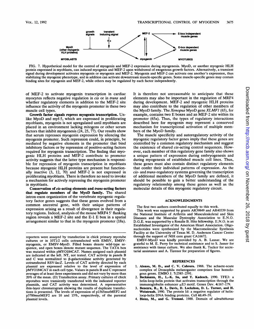
early in the differentiation program would lead to amplifica-tion of myogenin transcription, which would in turn furtheractivate MEF-2 expression, leading to reinforcement of thedecision to differentiate. Alternatively, MEF-2 may be reg-ulated by factors in addition to myogenic HLH proteins. Ifmyogenin and MEF-2 are initially upregulated in response toa differentiation inducing signal, their coexpression couldserve to stabilize expression of both. The presence offunctionally redundant sites that bind different factors thatregulate one another provides stability to the transcription ofboth genes and could in this case contribute to commitmentto the terminally differentiated state. A hypothetical modelillustrating these types of regulatory interactions is pre-sented in Fig. 7.
In addition to activating one another's expression, myo-genin and MEF-2 can activate downstream muscle-specificgenes separately or in combination. The ability of MEF-2 toactivate transcription of target genes lacking E boxes canexplain the observation that the mutant myogenin promoterlacking E-1 retained the ability to be trans-activated bymyogenin and MyoD. Presumably, the induction of MEF-2by these myogenic regulators indirectly activates myogenintranscription.MEF-2 is one of several DNA-binding activities recognizing
the MEF-2 site. In addition to the muscle-specific MEF-2complex, several widely expressed DNA-binding activitieshave been identified that recognize the MEF-2 site (Fig. 5)(10, 19, 28, 34, 53a, 60). Insight into the nature of the factorsinvolved in formation of these complexes has been providedby the recent isolation of cDNA clones encoding proteinsthat bind the MEF-2 consensus sequence (60). These pro-teins, termed RSRFs, for related to serum response factor,appear to represent a complex family of MEF-2 binding sitefactors, at least some of which are widely expressed. TheRSRFs and serum response factor belong to the MADS boxsuperfamily of regulatory factors, which regulate cell-type-specific gene expression in organisms ranging from plantsand yeasts to mammals (see reference 60 and referencestherein). We have recently found that polyclonal antibodiesdirected against RSRFs quantitatively supershift the muscle-specific MEF-2 complex observed with the myogeninMEF-2 site (25a), indicating that MEF-2 is antigenicallyrelated to RSRFs. Whether MEF-2 represents the product ofa muscle-specific RSRF gene or an alternate form of a widelyexpressed RSRF is unclear. It also remains to be determinedwhy ubiquitous RSRFs expressed in myoblasts and nonmus-cle cells are seemingly unable to induce myogenin transcrip-tion.
Paradoxically, MEF-2 is also expressed in cardiac muscle(53a), whereas myogenin is never expressed in the heart (25,77). We are presently testing whether the apparent inability
probe is shown above lanes 7 to 14. Sequences of probes used in thegel mobility shift assays are shown beneath the autoradiogram. Thecore of the MEF-2 site is shaded, and a preliminary consensus forMEF-2 binding is shown. Longer exposure to film or use of greateramounts of nuclear extract in the assay reveals the presence of theubiquitous MEF-2 binding site activity in 1OT1/2 nuclear extracts,but its abundance is very low relative to that of MEF-2 in extractsfrom highly differentiated myotube cultures. (B) Ten micrograms ofnuclear extract from C2 myotubes (lanes 1 to 3) or from chickenembryonic breast muscle (lanes 4 to 6) was incubated with a labeledoligonucleotide probe containing a MEF-2 site. For competitions,100-fold molar excesses of unlabeled competing oligomers wereadded to the binding reaction mixture.
VOL. 123, 1992
on Novem
ber 26, 2018 by guesthttp://m
cb.asm.org/
Dow
nloaded from
3674 EDMONDSON ET AL.
A.-184 -160 -140 -120 -100
IF o
GCCTGCAGGGTGGGG0TGGGGGCAAAAGGAGAGGGAAGGGGAATCACATGTAATCCACTGGAACGTCTTGATGTGCAGCACAGCTTAGArG'GGE-2
-80 -60 -40 -20 +1rol, -I|/ -I CG
GGCTCAGGTTTCTGTGGCGTTGGCTATATTTATCTCTGGGTTCATGCCAGCAGGGAGGGTTTAAATGGCACCCAGCAGTTGGCGTGAGGGGMEF2 TATA E-1
B.
.?.p
.g3a1--4cC)3a1
bp of S flanking DNA
C.CAT Activity
REPORTER PLASMID
pMY0184CAT
pMY084CAT
TATAMEF2 IE
TATA
Chick 1 OT1/2 Cells
myotubes EMSV myogenin MyoD100 5 66 100
54 55 41
pMY084(mutMEF2)CAT
pMY084(-E1 )CAT
pMYO04(mutMEF2/-E1)CAT
TATA
__TATA
TATA
_wLpMY0184(mutTATA)CAT -S 0 NT NT NT
* wild type
| g mutant
cli F- NU. < LL
Cn CDn
(3
Ps;~~~~~~~~~~~~~~i.v*. .S. . .^g._... 0 Q
F..... w.w T ws.T~~~~w C
.. L.^CLO
.79 .^.^ ^ ^ ^
FIG. 6. Evidence that the MEF-2 site and E-1 E box are impor-tant for promoter activity. (A) Sequence of the 184-bp myogeninpromoter. E boxes (E-1 and E-2), the MEF-2 site, and the TATAbox are indicated; 5' boundaries of exonuclease deletions are shownwith arrows. Nucleotides are numbered relative to the transcriptioninitiation site, designated +1. Nucleotides above the TATA boxshow mutations introduced at those positions. The MEF-2 site wasmutated by insertion of a dC at the position indicated. (B and C)CAT assays. For panel B, myogenin-CAT reporter genes containingdeletions of the promoter were assayed in primary cultures of chickmyotubes. All myogenin 5' flanking sequences contained the same3' end point, nucleotide +18 relative to transcription initiation. The5' deletion endpoint of each promoter mutant is indicated. Fordeletion of E box E-1, BanI was used to remove sequences 3' to-18. The normal TATA box continues to be used in these construc-tions (data not shown). For panel C, the MEF-2 site and E-1 E box
13
23 29 18
2 3 2
D.
MOL. CELL. BIOL.
on Novem
ber 26, 2018 by guesthttp://m
cb.asm.org/
Dow
nloaded from
TRANSCRIPTIONAL CONTROL OF MYOGENIN 3675
MyoD(other myogenicHLH proteins)
MYOBLASTS
MEF-2 x E-box Independente, p muscle genes
< ~~~~~E-box dependent
II / muscle genesmyogenin MYOTUBES
FIG. 7. Hypothetical model for the control of myogenin and MEF-2 expression during myogenesis. MyoD, or another myogenic HLHprotein expressed in myoblasts, can induced myogenin and MEF-2 upon withdrawal of exogenous growth factors. Alternatively, a transientsignal during development activates myogenin or myogenin and MEF-2. Myogenin and MEF-2 can activate one another's expression, thusstabilizing the myogenic phenotype, and in addition can activate downstream muscle-specific genes. Some muscle-specific genes may containbinding sites for myogenin and MEF-2, while others may be regulated by each factor independently.
of MEF-2 to activate myogenin transcription in cardiacmyocytes reflects negative regulation in cis or in trans andwhether regulatory elements in addition to the MEF-2 siteinfluence the activity of the myogenin promoter in these twomuscle cell types.Growth factor signals repress myogenin transcription. Un-
like MyoD and myf-5, which are expressed in proliferatingmyoblasts, myogenin is not upregulated until myoblasts areplaced in an environment lacking mitogens or other serumfactors that inhibit myogenesis (24, 25, 77). Our results showthat serum represses myogenin expression by silencing themyogenin promoter. Such repression could, in principle, bemediated by negative elements in the promoter that bindinhibitory factors or by repression of positive-acting factorsrequired for myogenin transcription. The finding that myo-genic HLH proteins and MEF-2 contribute to promoteractivity suggests that the latter type mechanism is responsi-ble for repression of myogenin transcription in myoblastsbecause myogenic HLH proteins are present but function-ally inactive (3, 12, 70) and MEF-2 is not expressed inproliferating myoblasts. There is therefore no need to invokea mechanism for actively repressing the myogenin promoterin myoblasts.
Conservation of cis-acting elements and trans-acting factorsthat regulate members of the MyoD family. The sharedintron-exon organization of the vertebrate myogenic regula-tory factor genes suggests that these genes evolved from acommon ancestral gene, with their unique patterns ofexpression arising as a result of divergence in their regula-tory regions. Indeed, analysis of the mouse MRF4 5' flankingregion reveals a MEF-2 site and the E-1 E box in a spatialarrangement similar to that in the myogenin promoter (18a).
reporters were assayed by transfection in chick primary myotubecultures or in 1OT1/2 cells cotransfected with EMSV, EMSV-myogenin, or EMSV-MyoD. Filled boxes denote wild-type se-
quence, and open boxes denote mutant sequence. The TATA boxwas mutated within pMYO184CAT. Names assigned each plasmidare indicated at the left. NT, not tested. CAT activity in panels Band C was normalized to 0-galactosidase activity generated bycotransfected RSV-lacZ. Levels of CAT activity directed by eachplasmid are expressed relative to the level of expression ofpMYO184CAT in each cell type. Values in panels B and C representaverages of at least three experiments and did not vary by more than20% of the mean. (D) Transfection assay. Primary cultures of chickmyotubes were transiently transfected with the indicated reporterplasmids, and CAT activity was determined. A representativethin-layer chromatogram showing the results of duplicate transfec-tions is presented. The levels of expression of pl565mutMEF2 andp3700mutMEF2 are 10 and 15%, respectively, of the parentalplasmid levels.
It is therefore not unreasonable to anticipate that theseelements may also be important in the regulation of MRF4during development. MEF-2 and myogenic HLH proteinsmay also contribute to the regulation of other members ofthe MyoD family. The Xenopus MyoD geneXLMF1 (65), forexample, contains two E boxes and an MEF-2 site within itspromoter (65a). Thus, the types of regulatory interactionsdescribed here for myogenin may represent a conservedmechanism for transcriptional activation of multiple mem-bers of the MyoD family.The muscle specificity and autoregulatory activity of the
myogenic regulatory factor genes imply that these genes arecontrolled by a common regulatory mechanism and suggestthe existence of shared cis-acting control sequences. How-ever, each member of this regulatory gene family also showsa unique pattern of expression during embryogenesis andduring myogenesis of established muscle cell lines. Thus,these genes must also contain distinct regulatory elementsthat confer their individual patterns of expression. As thecis- and trans-regulatory systems governing the transcriptionof additional members of the MyoD family are defined, itshould be possible to gain a better understanding of theregulatory relationship among these genes as well as themolecular details of this myogenic regulatory circuit.
ACKNOWLEDGMENTS
The first two authors contributed equally to this work.This work was supported by grants AR39849 and AR40339 from
the National Institute of Arthritis and Musculoskeletal and SkinDiseases and the Muscular Dystrophy Association to E.N.O.D.G.E. was supported by a Rosalie B. Hite fellowship. E.N.O. is anEstablished Investigator of the American Heart Association. Oligo-nucleotides were synthesized by the Macromolecular SynthesisFacility at the University of Texas M. D. Anderson Cancer Centerthrough the support of NIH core grant CA16672.EMSV-MyoD was kindly provided by A. B. Lassar. We are
grateful to M. E. Perry for technical assistance and to S. Jasser forassistance with tissue culture. We also thank K. Tucker for secre-tarial assistance and A. Tizenor for preparation of figures.
REFERENCES1. Alonso, M. D., and C. V. Cabrera. 1988. The achaete-scute
complex of Drosophila melanogaster comprises four homolo-gous genes. EMBO J. 7:2585-2591.
2. Beckmann, H., L.-K. Su, and T. Kadesch. 1990. TFE3: ahelix-loop-helix protein that activates transcription through theimmunoglobulin enhancer pE3 motif. Genes Dev. 4:167-179.
3. Benezra, R., R. L. Davis, D. Lockshon, D. L. Turner, and H.Weintraub. 1990. The protein Id: a negative regulator of helix-loop-helix DNA binding proteins. Cell 61:49-59.
4. Bienz, M., and G. Tremml. 1988. Domain of ultrabithorax
VOL. 12, 1992
on Novem
ber 26, 2018 by guesthttp://m
cb.asm.org/
Dow
nloaded from

3676 EDMONDSON ET AL.
expression in Drosophila visceral mesoderm from autoregula-tion and exclusion. Nature (London) 333:576-578.
5. Blackwell, K. T., and H. Weintraub. 1990. Differences andsimilarities in DNA-binding preferences of MyoD and E2Aprotein complexes revealed by binding site selection. Science250:1104-1110.
6. Bober, E., G. E. Lyons, T. Braun, G. Cossu, M. Buckingham,and H.-H. Arnold. 1991. The muscle regulatory gene, Myf-6, hasa biphasic pattern of expression during early mouse develop-ment. J. Cell Biol. 113:1255-1265.
7. Braun, T., E. Bober, G. Buschhausen-Denker, S. Kotz, K.Grzeschik, and H. H. Arnold. 1989. Differential expression ofmyogenic determination genes in muscle cells: possible autoac-tivation by the Myf gene products. EMBO J. 8:3617-3625.
8. Braun, T., E. Bober, B. Winter, N. Rosenthal, and H. H. Arnold.1990. Myf-6, a new member of the human gene family ofmyogenic determination factors: evidence for a gene cluster onchromosome 12. EMBO J. 9:821-831.
9. Braun, T., G. Buschhausen-Denker, E. Bober, E. Tannich, andH. H. Arnold. 1989. A novel human muscle factor related to butdistinct from MyoDl induces myogenic conversion in 1OT1/2fibroblasts. EMBO J. 8:701-709.
10. Braun, T., E. Tannich, D. G. Buschhausen, and H. H. Arnold.1989. Promoter upstream elements of the chicken cardiac myo-sin light-chain 2-A gene interact with trans-acting regulatoryfactors for muscle-specific transcription. Mol. Cell. Biol.9:2513-2525.
11. Brennan, T. J., T. Chakraborty, and E. N. Olson. 1991. Muta-genesis of the myogenin basic region identifies an ancientprotein motif critical for activation of myogenesis. Proc. Natl.Acad. Sci. USA 88:5675-5679.
12. Brennan, T., D. G. Edmondson, L. Li, and E. N. Olson. 1991.TGF-0 represses the actions of myogenin through a mechanismindependent of DNA binding. Proc. Natl. Acad. Sci. USA88:3822-3826.
13. Brennan, T. J., D. G. Edmondson, and E. N. Olson. 1990.Aberrant regulation of MyoDl contributes to the partiallydefective myogenic phenotype of BC3H1 cells. J. Cell Biol.110:929-937.
14. Brennan, T. J., and E. N. Olson. 1990. Myogenin resides in thenucleus and acquires high affinity for a conserved enhancerelement on heterodimerization. Genes Dev. 4:582-595.
15. Buskin, J. N., and S. D. Hauschka. 1989. Identification of amyocyte nuclear factor which binds to the muscle-specificenhancer of the mouse muscle creatine kinase gene. Mol. Cell.Biol. 9:2627-2640.
16. Caudy, M., H. Vassin, M. Branc, R. Tuma, L. Y. Jan, and Y. N.Jan. 1988. daughterless, a Drosophila gene essential for bothneurogenesis and sex determination, has sequence similaritiesto myc and the achaete-scute complex. Cell 55:1061-1067.
17. Chakraborty, T., T. J. Brennan, L. Li, D. G. Edmondson, andE. N. Olson. 1991. Inefficient homooligomerization contributesto the dependence of myogenin on E2A products for efficientDNA binding. Mol. Cell. Biol. 11:3633-3641.
18. Chakraborty, T., T. J. Brennan, and E. N. Olson. 1991. Differ-ential trans-activation of a muscle-specific enhancer by myo-genic helix-loop-helix proteins is separable from DNA binding.J. Biol. Chem. 266:2878-2882.
18a.Cheng, T. C., and E. Olson. Unpublished data.19. Cserjesi, P., and E. N. Olson. 1991. Myogenin induces the
myocyte-specific enhancer-binding factor MEF-2 independentlyof other muscle-specific gene products. Mol. Cell. Biol. 11:4854-4862.
20. Davis, R. L., P.-F. Cheng, A. B. Lassar, and H. Weintraub.1990. The MyoD DNA binding domain contains a recognitioncode for muscle-specific gene activation. Cell 60:733-746.
21. Davis, R. L., H. Weintraub, and A. B. Lassar. 1987. Expressionof a single transfected cDNA converts fibroblasts to myoblasts.Cell 51:987-1000.
22. de la Brousse, C. F., and C. P. Emerson, Jr. 1990. Localizedexpression of a myogenic regulatory gene, qmfl, in the somitedermatome of avian embryos. Genes Dev. 4:567-581.
23. DeVries, E., W. VanDriel, S. J. L. vanden Heuvel, and P. C. van
der Vliet. 1987. Contact point analysis of the HeLa nuclearfactor 1 recognition site reveals symmetrical binding at one sideof the DNA helix. EMBO J. 6:161-168.
24. Edmondson, D. G., T. J. Brennan, and E. N. Olson. 1991.Mitogenic repression of myogenin autoregulation. J. Biol.Chem. 266:21343-21346.
25. Edmondson, D. G., and E. N. Olson. 1989. A gene with homol-ogy to the myc similarity region of MyoDl is expressed duringmyogenesis and is sufficient to activate the muscle differentia-tion program. Genes Dev. 3:628-640.
25a.Edmondson, D. G., and E. N. Olson. Unpublished data.26. French, B. A., K.-L. Chow, E. N. Olson, and R. J. Schwartz.
1991. Heterodimers of myogenic helix-loop-helix regulatoryfactors and 12 bind a complex element governing myogenicinduction of the avian cardiac ox-actin promoter. Mol. Cell. Biol.11:2439-2450.
27. Gonostajski, R. M., S. Adhya, D. Nagata, R. A. Guggenheimer,and J. Hurwitz. 1985. Site-specific DNA binding of nuclearfactor 1: analysis of cellular binding sites. Mol. Cell. Biol.5:964-971.
28. Gossett, L. A., D. J. Kelvin, E. A. Sternberg, and E. N. Olson.1989. A new myocyte-specific enhancer-binding factor thatrecognizes a conserved element associated with multiple mus-cle-specific genes. Mol. Cell. Biol. 9:5022-5033.
29. Gregor, P. D., M. Sawadogo, and R. G. Roeder. 1990. Theadenovirus major late transcription factor USF is a member ofthe helix-loop-helix group of regulatory proteins and binds toDNA as a dimer. Genes Dev. 4:1730-1740.
30. Harvey, R. P. 1990. The Xenopus MyoD gene: an unlocalizedmRNA predates lineage restricted expression in the early em-bryo. Development 108:669-680.
31. Henthorn, P., M. Kiledjian, and T. Kadesch. 1990. Two distincttranscription factors that bind the immunoglobulin enhancer,uE5/KE2 motif. Science 247:467-470.
32. Hinterberger, T. J., D. A. Sassoon, S. J. Rhodes, and S. F.Konieczny. 1991. Expression of the muscle regulatory factorMRF4 during somite and skeletal myofiber development. Dev.Biol. 147:144-156.
33. Hiromi, Y., and W. J. Gehring. 1987. Regulation and function ofthe Drosophila segmentation genefushi tarazu. Cell 50:963-974.
34. Horlick, R. A., G. M. Hobson, J. H. Patterson, M. T. Mitchell,and P. A. Benfield. 1990. Brain and muscle creatine kinase genescontain common TA-rich recognition protein-binding regulatoryelements. Mol. Cell. Biol. 10:4826-4836.
35. Hopwood, N. D., A. Pluck, and J. B. Gurdon. 1989. MyoDexpression in the forming somites is an early response tomesoderm induction in Xenopus embryos. EMBO J. 8:3409-3417.
36. Hu, J.-S., E. N. Olson, and R. E. Kingston. 1992. HEB: ahelix-loop-helix protein related to E2A and ITF2 that canmodulate the DNA-binding ability of myogenic regulatory fac-tors. Mol. Cell. Biol. 12:1031-1042.
37. Hu, Y.-F., B. Luscher, A. Admon, N. Mermod, and R. Tjian.1990. Transcription factor AP-4 contains multiple dimerizationdomains that regulate dimer specificity. Genes Dev. 4:1741-1752.
38. Jones, R. A., M. J. Wolkowicz, W. M. Rideout III, F. A.Gonzales, C. M. Marziasz, G. A. Coetzee, and S. J. Tapscott.1990. De novo methylation of the MyoDI CpG island during theestablishment of immortal cell lines. Proc. Natl. Acad. Sci.USA 87:6117-6121.
39. Kraus, M., A. Fire, S. W. Harrison, J. Priess, and H. Weintraub.1990. CeMyoD accumulation defines the body wall muscle cellfate during C. elegans embryogenesis. Cell 63:907-919.
40. Kuziori, M. A., and W. McGinnis. 1988. Autoregulation of aDrosophila homeotic selector gene. Cell 55:477-485.
41. Lassar, A. B., J. N. Buskin, D. Lockshon, R. L. Davis, S. Apone,S. D. Hauschka, and H. Weintraub. 1989. MyoD is a sequence-specific DNA binding protein requiring a region of myc homol-ogy to bind to the muscle creatine kinase enhancer. Cell58:823-831.
42. Lassar, A. B., R. L. Davis, W. E. Wright, T. Kadesch, C. Murre,A. Voronova, D. Baltimore, and H. Weintraub. 1991. Functional
MOL. CELL. BIOL.
on Novem
ber 26, 2018 by guesthttp://m
cb.asm.org/
Dow
nloaded from

TRANSCRIPTIONAL CONTROL OF MYOGENIN 3677
activity of myogenic HLH proteins requires hetero-oligomeriza-tion with E12/E47-like proteins in vivo. Cell 66:305-315.
43. Lassar, A. B., M. J. Thayer, R. W. Overell, and H. Weintraub.1989. Transformation by activated RAS or FOS prevents myo-genesis by inhibiting expression of MyoDl. Cell 58:659-667.
44. Lin, A.-Y., C. A. Dechesne, J. Eldridge, and B. M. Paterson.1989. An avian muscle factor related to MyoDl activatesmuscle-specific promoters in nonmuscle cells of different germ-layer origin and in BrdU-treated myoblasts. Genes Dev. 3:986-996.
45. Lin, H., K. Yutzey, and S. F. Konieczny. 1991. Muscle-specificexpression of the troponin I gene requires interactions betweenhelix-loop-helix muscle regulatory factors and ubiquitous tran-scription factors. Mol. Cell. Biol. 11:267-280.
46. Mar, J. H., and C. P. Ordahl. 1990. M-CAT binding factor, anovel trans-acting factor governing muscle-specific transcrip-tion. Mol. Cell. Biol. 10:4271-4283.
47. Michelson, A. M., S. M. Abmayr, M. Bate, A. Martinez, and T.Maniatis. 1990. Expression of a MyoD family member prefig-ures muscle pattern in Drosophila embryos. Genes Dev. 4:2086-2097.
48. Miner, J. H., and B. Wold. 1990. Herculin, a fourth member ofthe MyoD family of myogenic regulatory genes. Proc. Natl.Acad. Sci. USA 87:1089-1093.
49. Minty, A., and L. Kedes. 1986. Upstream regions of the humancardiac actin gene that modulate its transcription in musclecells: presence of an evolutionarily conserved repeated motif.Mol. Cell. Biol. 6:2125-2136.
50. Montarras, D., C. Pinset, J. Chelly, A. Kahn, and F. Gros. 1989.Expression of MyoDi coincides with terminal differentiation indetermined but inducible muscle cells. EMBO J. 8:2203-2207.
51. Murre, C., P. S. McCaw, and D. Baltimore. 1989. A new DNAbinding and dimerization motif in immunoglobulin enhancerbinding, daughterless, MyoD, and myc proteins. Cell 56:777-783.
52. Murre, C., P. S. McCaw, H. Vaessin, M. Caudy, L. Y. Jan, Y. N.Jan, C. V. Cabrera, J. N. Buskin, S. D. Hauschka, A. B. Lassar,H. Weintraub, and D. Baltimore. 1989. Interactions betweenheterologous helix-loop-helix proteins generate complexes thatbind specifically to a common DNA sequence. Cell 58:537-544.
53. Murre, C., A. Voronova, and D. Baltimore. 1990. B cell andmyocyte-specific E2-box binding factors contain E12/E47-likesubunits. Mol. Cell. Biol. 11:1156-1160.
53a.Navankasattusas, S., H. Zhu, A. V. Garcia, S. M. Evans, andK. R. Chien. 1992. A ubiquitous factor (HF-la) and a distinctmuscle (HF-lb/MEF-2) form an E-box-independent pathway forcardiac muscle gene expression. Mol. Cell. Biol. 12:1469-1479.
54. Nordheim, A., and A. Rich. 1983. The sequence (dC-dA)sbn(dG-dT)n forms left-handed Z-DNA in negatively supercoiledplasmids. Proc. Natl. Acad. Sci. USA 80:1821-1825.
55. Olson, E. N. 1990. The MyoD family: a paradigm for develop-ment? Genes Dev. 4:1454-1461.
56. Ott, M.-O., E. Bober, G. Lyons, H. H. Arnold, and M. Buck-ingham. 1991. Early expression of the myogenic regulatorygene, myf5, in precursor cells of skeletal muscle in the mouseembryo. Development 111:1097-1107.
57. Paterson, B. M., U. Walldorf, J. Eldridge, A. Dubendorfer, M.Frasch, and W. J. Gehnng. 1991. The Drosophila homologue ofvertebrate myogenic-determination genes encodes a transientlyexpressed nuclear protein marking primary myogenic cells.Proc. Natl. Acad. Sci. USA 88:3782-3786.
58. Piette, J., J.-L. Bessereau, M. Huchet, and J.-P. Changeux. 1990.Two adjacent MyoDl-binding sites regulate expression of theacetyl choline receptor ot-subunit. Nature (London) 345:353-355.
59. Pinney, D. F., D. F. Pearson-White, S. F. Konieczny, K. E.Latham, and C. P. Emerson, Jr. 1988. Myogenic lineage deter-mination and differentiation: evidence for a regulatory genepathway. Cell 53:781-793.
60. Pollock, R., and R. Treismann. 1991. Human SRF-related pro-
teins: DNA-binding properties and potential regulatory targets.Genes Dev. 5:2327-2341.
61. Rhodes, S. J., and S. F. Konieczny. 1989. Identification ofMRF4: a new member of the muscle regulatory factor genefamily. Genes Dev. 3:2050-2061.
62. Salminen, A., T. Braun, A. Buchberger, S. Jurs, B. Winter, andH. H. Arnold. 1991. Transcription of the muscle regulatory geneMYF4 is regulated by serum components, peptide growth fac-tors and signaling pathways involving G proteins. J. Cell Biol.115:905-917.
63. Sartorelli, V., K. A. Webster, and L. Kedes. 1990. Muscle-specific expression of the cardiac a-actin gene requires MyoDl,CArG-box binding factor, and Spl. Genes Dev. 4:1811-1822.
64. Sassoon, D., G. Lyons, W. E. Wright, V. Lin, A. Lassar, H.Weintraub, and M. Buckingham. 1989. Expression of two myo-genic regulatory factors myogenin and MyoDl during mouseembryogenesis. Nature (London) 341:303-307.
65. Scales, J., E. N. Olson, and W. M. Perry. 1990. Two distinctXenopus genes with homology to MyoDl are expressed beforesomite formation in early embryogenesis. Mol. Cell. Biol.10:1516-11524.
65a.Scales, J., and W. M. Perry. Personal communication.66. Schwarz, J. J., T. Chakraborty, J. Martin, J. Zhou, and E. N.
Olson. 1992. The basic region of myogenin cooperates with twotranscription activation domains to induce muscle-specific tran-scription. Mol. Cell. Biol. 12:266-275.
67. Sternberg, E. A., G. Spizz, W. M. Perry, D. Vizard, T. Weil, andE. N. Olson. 1988. Identification of upstream and intragenicregulatory elements that confer cell-type-restricted and differ-entiation-specific expression on the muscle creatine kinasegene. Mol. Cell. Biol. 8:2896-2909.
68. Tapscott, S. J., A. B. Lassar, R. L. Davis, and H. Weintraub.1989. 5-bromo-2'-deoxyuridine blocks myogenesis by extin-guishing expression of myoDl. Science 245:532-536.
69. Tapscott, S. J., and H. Weintraub. 1991. MyoD and the regula-tion of myogenesis by helix-loop-helix proteins. J. Clin. Invest.87:1133-1138.
70. Thayer, M. J., S. J. Tapscott, R L. Davis, W. E. Wright, A. B.Lassar, and H. Weintraub. 1989. Positive autoregulation of themyogenic determination gene MyoDl. Cell 58:241-248.
71. Thompson, W. R., B. Nadal-Ginard, and V. Mahdavi. 1991. AMyoDl-independent muscle-specific enhancer controls theexpression of the ,B-myosin heavy chain gene in skeletal andcardiac muscle cells. J. Biol. Chem. 266:22678-22688.
72. Venuti, J. M., L. Goldberg, T. Chakraborty, E. N. Olson, andW. Klein. 1991. A myogenic factor from sea urchin embryoscapable of programming muscle differentiation in mammaliancells. Proc. Natl. Acad. Sci. USA 88:6219-6223.
73. Voronova, A., and D. Baltimore. 1990. Mutations that disruptDNA binding and dimer formation in the E47 helix-loop-helixprotein map to distinct domains. Proc. Natl. Acad. Sci. USA87:4722-4726.
74. Weintraub, H., R. Davis, D. Lockshon, and A. Lassar. 1990.MyoD binds cooperatively to two sites in a target enhancersequence: occupancy of two sites is required for activation.Proc. Natl. Acad. Sci. USA 87:5623-5627.
75. Wentworth, B. M., M. Donoghue, J. C. Engert, E. B. Berglund,and N. Rosenthal. 1991. Paired MyoD binding sites regulatemyosin light chain gene expression. Proc. Natl. Acad. Sci. USA88:1242-1246.
76. Wright, W. E., M. Binder, and W. Funk 1991. Cyclic amplifi-cation and selection of targets (CASTing) for the myogeninconsensus binding site. Mol. Cell. Biol. 11:41044110.
77. Wright, W. E., D. A. Sassoon, and V. K. Lin. 1989. Myogenin,a factor regulating myogenesis, has a domain homologous toMyoDI. Cell 56:607-617.
78. Yaffe, D., and 0. Saxel. 1977. Serial passaging and differentia-tion of myogenic cells isolated from dystrophic mouse muscle.Nature (London) 270:725-727.
VOL. 12, 1992
on Novem
ber 26, 2018 by guesthttp://m
cb.asm.org/
Dow
nloaded from


![BIOCHIMICA ET BIOPHYSICA ACTA ii!i,, · 18 D.M. Mazzuca, T. C. Y. Lo / Biochimica et Biophysica A cta 1414 (1998) 16-30 the myogenin promoter [36]. The PGK-myogenin con- struct and](https://img.pdfslide.net/doc/110x75/5ebe218ebd2e88479e3be038/biochimica-et-biophysica-acta-iii-18-dm-mazzuca-t-c-y-lo-biochimica-et.jpg)