Embed Size (px)
Citation preview
1
ANALYSIS OF THE OUTCOME OF MANDIBLE
FRACTURE MANAGEMENT
A dissertation submitted to
THE TAMIL NADU M.G.R. MEDICAL UNIVERSITY
In partial fulfillment of the Regulations of the award of degree of
M.Ch.
(PLASTIC AND RECONSTRUCTIVE SURGERY)
Branch – III
AUGUST 2015
DEPARTMENT OF PLASTIC AND
RECONSTRUCTIVE SURGERY
COIMBATORE MEDICAL COLLEGE HOSPITAL
COIMBATORE – 641 018
2
CERTIFICATE
This is to certify that this dissertation titled " ANALYSIS OF
THE OUTCOME OF MANDIBLE FRACTURE MANAGEMENT "
submitted by Dr.A.KAVITHA PRIYA to the faculty of Plastic Surgery,
The Tamil Nadu Dr.M.G.R. Medical University, Chennai in partial
fulfillment of the requirement for the award of MASTER OF
CHIRURGIE IN PLASTIC AND RECONSTRUCTIVE SURGERY
Branch – III for the August 2015 Examination is a bonafide research
work carried out by her under our direct supervision and guidance.
Prof.Dr.B.Asokan, M.S. (Gen), M.Ch., (Plastic) Prof.Dr.A.Edwin Joe, M.D., BL Prof and Head of the Department, The Dean, Department of Plastic Surgery, Coimbatore Medical College, Coimbatore Medical College, Coimbatore. Coimbatore.
3
DECLARATION
I Dr.A.KAVITHA PRIYA, solemnly declare that the
dissertation titled "ANALYSIS OF THE OUTCOME OF
MANDIBLE FRACTURE MANAGEMENT" is a bonafide research
work done by me at Coimbatore Medical College, during 2012-2015
under the guidance and supervision of Prof.Dr.B.Asokan, M.S. (Gen),
M.Ch., (Plastic). This dissertation is submitted to The Tamil Nadu Dr.
M. G. R. Medical University, towards partial fulfillment of the
University regulations for the award of M.Ch., Degree (Branch III) in
plastic and reconstructive surgery.
Place : Coimbatore Dr.A.KAVITHA PRIYA, Date :
4
5
ACKNOWLEDGEMENT
I owe my thanks to Prof. Dr.A.EDWIN JOE, M.D., BL, The DEAN,
COIMBATORE MEDICAL COLLEGE, for allowing me to avail the
facilities needed for my dissertation work.
I am greatly indebted to Prof. Dr.B.ASOKAN, M.S. (Gen), M.Ch.,
(Plastic) Professor and Head, Department of Plastic and Reconstructive
surgery, for his valuable guidance in completing this dissertation.
I would like to thank my Associate Prof. Dr.N.SEKAR, M.S.(Ortho),
M.Ch. (Plastic) for his guidance in completing this dissertation.
I am also deeply thankful to our Assistant Professors, Dr.V.P.Ramanan,
Dr.R.Senthil Kumar, Dr.S.Prakash and Dr.M.Sundararaj for their valuable
suggestions and inputs in preparing this dissertation.
I also thank our college Ethics Committee for their certification of
approval. I also thank my co-post graduates for their help in preparing this
dissertation.
Finally, I will be failing in my duty if I don't thank my patients who
have been my greatest source of inspiration in my work.
Dr. A.KAVITHA PRIYA
6
7
8
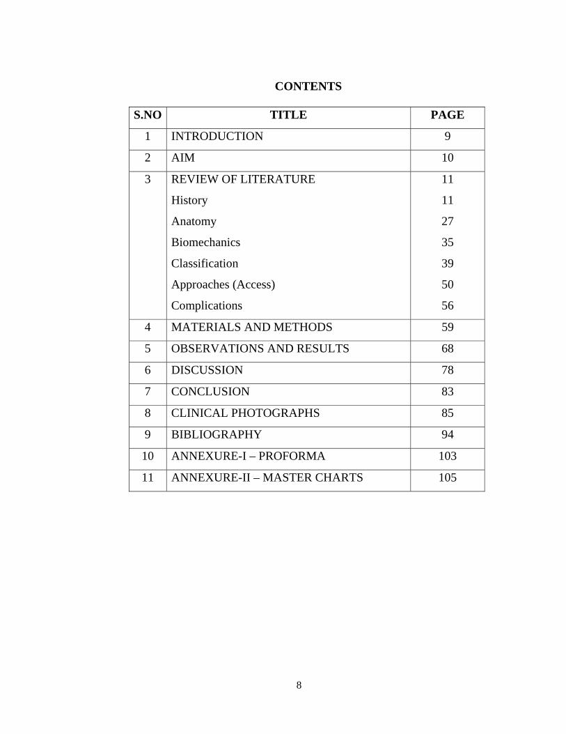
CONTENTS
S.NO TITLE PAGE
1 INTRODUCTION 9
2 AIM 10
3 REVIEW OF LITERATURE
History
Anatomy
Biomechanics
Classification
Approaches (Access)
Complications
11
11
27
35
39
50
56
4 MATERIALS AND METHODS 59
5 OBSERVATIONS AND RESULTS 68
6 DISCUSSION 78
7 CONCLUSION 83
8 CLINICAL PHOTOGRAPHS 85
9 BIBLIOGRAPHY 94
10 ANNEXURE-I – PROFORMA 103
11 ANNEXURE-II – MASTER CHARTS 105
9
INTRODUCTION
Face is the most admirable part of our body. Facial injury is the most
common cause of disfigurement and affects the personality of the individual very
much.
The most frequently injured facial bone is mandible after nasal bone because
it is the most mobile and prominent facial bone .The mandibular fractures
outnumbers zygomatic and maxillary fractures by a ratio of 6:2:1 respectively.
Fractures of mandible invariably produce malocclusion if not treated properly.
Knowledge of the dentition is thus an absolute prerequisite for the proper treatment of
jaw fractures.
Various techniques that are advocated in the literature to manage mandibular
fractures vary ranging from bandages and external appliances, extra oral and
intraoral appliances, mono maxillary wiring, intermaxillary wiring, plates and
screws.
Restoration of the occlusion usually indicates anatomic reduction and proper
positioning of the mandible and facial bones. Our goal should be restoration of the
function without any morbidity at the earliest .
10
AIM
To analyze the outcome of mandibular fracture fixation with eyelets, arch bars,
miniplates and screws and assess the
1. Stability of the fixation
2. Occlusion after fixation
3. Comparing the jaw dysfunction (chewing ability) before and after treatment.
4. Post operative sequalae – such as post operative pain, bony and soft tissue
infections, nonunion, nerve injury, osteomyelitis, malocclusion, and
malunion, .
11
REVIEW OF LITERATURE
HISTORY
The first description of mandibular fracture was as early as 1650 BC, when an
Egyptian papyrus described the examination, diagnosis, and treatment of mandible
fractures.
Hippocrates describe circumdental wires and external bandaging for
reapproximation and immobilization.
Surgeons such as Ceisus (Rome) , Avicenna (Islam) and Sushruta
(India), described treating the jaw fractures conservatively between 25 B.C. and 11
century AD.
Sushruta recommended complicated bandaging , manual manipulation and heat
to treat fractures of mandible
.
The importance of occlusion during the treatment of fractures was studied by
Avicenna (980 to 1037A.D). This concept is followed still today nearly 1000years after
his description.
Salerno of Italy, in 1180 wrote in a textbook the importance of establishing
proper occlusion.
Maxillomandibular fixation was first described in 1492, in an edition of the
book Cirugia printed in Lyons.
12
Dental prosthetic devices were used to immobilize fracture fragments.77 by
Chopart and Desault.
Until 19th century fracture mandible was treated with wrap, external
bandages and sometimes with bridle wire.
Gilmer reformed the treatment of fracture mandible by using fixed full arch
bars on the maxilla and the mandible.
Numerous splints were devised in the 19th century, the most important was
Gunning (1866 ) and Bean (1865). This period was known as “Prosthetic era”
in fracture management.
Mandibular fracture stabilization by means of a screw plate system was
described by Hausmann in 1886.
Solid steel plate held by 4 screws for fixation.54 was first used by Schede in
1888.
Angle described many methods of Intermaxillary fixation that used
bands and other orthodontic techniques. Angle criteria of occlusion is still
followed.
Skeletal fixation became popular during the world war I and II.
13
Roger Anderson (1936) improvised the method and produced an appliance by
which fracture fragments could be held and maintained by means of pins or screws
inserted through skin.
Circumferential wiring was popularised by G.V. Black and Ivy (1921).
Kazanjian used transosseous wiring placed through an intraoral approach
into the alveolar part of the bone during the first World war.
Cole in 1917 used silver plates and screws on either side of fracture and
attached silver wires to the plates for immobilize .
Vorschutz (1934) introduced two large screws through the skin into the
bone, reduced the fracture and held the screws in position with use of Plaster of
Paris bandage.
Rigid compression system to the bony cortex using plate and screw was
designed by Danis in 1949.
Open reduction with Internal fixation was described by Kruger (1964) as
a definite method . The need for adding Maxillomandibular fixation to the internal
fixation was stressed by him.
The technique of rigid internal fixation was developed and popularized by
Arbeitsgemeinschaft fur Osteosynthesefragen/Association for the Study of Internal
14
Fixation (AO/ASIF) in Europe in the 1970s. The basic principles of the AO, outlined
by Spiessl, call for primary bone healing under conditions of absolute stability.17
Rigid internal fixation must neutralize the forces (compression, torsion,
tension, shearing) developed during functional loading of the mandible to allow for
immediate function,. this was accomplished by inter fragmentary compression plates.
Use of superior border plate or arch bars to counter traction or tension forces at the
superior border.11 and the use of an inferior border plate to counter compression
forces.
AO reconstruction plates has created an impacted in the management of
infected and comminuted mandibular fractures. There was 7.5% infection rate in
treatment of mandibular angle fractures with an AO reconstruction plate without
intermaxillary fixation (IMF) as reported by Ellis .
During the same time Spiessl was expounding the AO doctrine, Champy et
al in France 15 were developing the concept of adaptive osteosynthesis.
Champy in 1978 advocated transoral placement of small, malleable, thin,
stainless steel miniplates with monocortical screws along an ideal line of
osteosynthesis .
Champy believed that compression plates were unnecessary because of
masticatory forces produce a natural strain of compression along the inferior border.
15
These 2 changes of AO rigid internal fixation and the Champy method of
monocortical miniplates revolutionized the treatment approach to mandibular
fractures. Many fractures previously treated with closed reduction or open reduction
with wire osteosynthesis are now commonly treated with open reduction with plate
and screw fixation.
Wagner W F et al 79 (1979) studied the extraoral open reduction of
mandibular fracture and the associated morbidity and concluded that open
reduction of mandibular angle fracture associated with removal of teeth from
fracture line resulted in the greatest incidence of complication.
The effects of plating on bone blood supply of mandible was studied by
Grunst 33 (1980) . Plates provide excellent stability of the fragments and allow
early restoration of medullary blood supply ,but the cortical blood supply is very
much interfered by the plates.
K.E. Kahn Berg et al 42 (1980) advocated an intraoral bone plate
method for mandibular fractures, emphasized the need for Maxillomandibular
fixation and therefore limiting the period of patient disability.
Monty et al 53 (1983) evaluated bone repair in the mandible by a
histological and biometric comparison between rigid and semi rigid fixation and
concluded that healing by primary intention required rigid internal fixation and
results in superior healing.
16
Wald. R.M et a1 80 (1988) prospectively evaluated the efficacy of non-
compression miniplates selected mandibular fracture.
Brown. J.S et al 12 (1989) retrospectively studied the fate of miniplates
and concluded that there is no advantage in removal of the plate after fracture
union.
Mitchell M. Rubin et al 51 (1990) retrospectively analyzed and concluded
thatthere had been no difference in the complications when the third molar teeth
in line of fracture was extracted or retained.
J.S. Brown et al 39 (1991) demonstrated the post operative functional
improvement and the weight gain after internal fixation when compared with
intermaxillary fixation . Patient treated with intermaxillary fixation had restricted
airway.
Smith.W.P 72 (1991) did a retrospective study on delay in surgery beyond 24
hours with miniplate osteosynthesis and surgery within 24 hours showed no
difference and he demonstrated that the incision lines were more important in
preventing wound infection and dehiscence rather than the implants. Stainless steel
appears effective in short term use.
Jeffrey.C.Posnick et al 41 (1991) in their retrospective analysis of pediatric
facial fractures reviewed 137 patients and found that mandibular (34%) and orbital
(23%) fractures predominated. Fewer mid face fractures (7%) were sustained than
17
would be expected in similar adult patients. Most fractures resulted from traffic
related accidents(50%), falls (23%) and sports (15%) Closed reduction with
Maxillomandibular fixation was frequently chosen for children with Condyle
fractures and open reduction (35%) for other regions of face.
Sindet et al 69 (1992) compared the treatment of fractures of mandible treated
with or without Maxillomandibular fixation and found that the miniplates gave good
stabilization of mandibular fractures and allowed treatment without post reduction
Maxillomandibular fixation.
Andres.J.J.Gonzalez et al 05(1992) analyzed retrospectively and found that
mandibular fractures when Champy’s plates were used , patients had experienced a
shorter period of trismus than using closed reduction with Maxillomandibular
fixation.
J. P. Hayter et al 38 (1993) analyzed the fractures of facial skeleton
result in discontinuity of facial bones. He concluded that Osteosynthesis of the
bones are needed to allow stable and uneventful healing.
The effects of screw length and number on tension bands were evaluated by
Richard. H.Haug 65 (1993). He concluded that the maximum resistance to vertical
force was achieved with three screws per segment and no additional benefit of
placing fourth screw.
18
Edward Ellis et al 20 (1993) used two 2.4 mm DCP through a trans oral
incision using trans buccal trochar instrumentation with mandibular angle fracture.
He said that the technique was easy but resulted in high rate of infection.
Fordyce A.M. et a1 29 (1994) in his retrospective study concluded that
the avoidance of pre and post operative Maxillomandibular fixation was safe and
economical for the patient.
Ellis E. et al 21 (1994) studied the effectiveness of two 2mm non
compression miniplates for fractures of angle of mandible and found that the use
of two plates was easy, but resulted in high of infection rate.
Edwards TJ et al 23 (1994) studied for relationship between fracture
severity and complication rate in miniplate fixation. He found that there is a strong
relationship between complication rate and fracture severity.
Nakamura. S et al 55 (1994) analyzed the complications of miniplate
osteosynthesis and found that malocclusion 3,6%, exposure of miniplates 3%,
delayed union1,8% and infections1.0% .
Y.V. Tuavinen et al 84 (1994) analyzed retrospectively 269 patients with
mandible fractures treated with miniplates (Titanium ) using Champy’s principles
and concluded that semi rigid fixation of mandible fracture with miniplate was
an ideal procedure in the management of these injuries.
19
Gregory.S.Tate et al 32 (1994) recorded voluntary bite forces in patients who
were treated with rigid internal fixation for mandibular angle fracture and controls. He
observed that there was less molar bite forces on the side of fracture when compared
with controls.
R.A. Loukota et al 60 (1995) in their mechanical analysis of maxillofacial
miniplates and found the by repeated bending the plate will result in decrease in
stiffness .
JI Cawood 13(1995) compared 50 cases of mandibular fractures treated
by mini plate osteosynthesis with Maxillomandibular fixation. He
concluded that the plates have good recovery rate of normal jaw function and
body weight when compared with Maxillomandibular fixation.
Vivek Shetty et al 78 (1995) found that compressive fixation system (DCP,
Lag screw, locking plate,) are bio-mechanically superior to adaptive system (Champy
miniplate, Mennen clamp plate) and provided immediate functional stability.
Valentino et al 77(1995) analyzed the use of Maxillomandibular fixation
with miniplate osteosynthesis and without supplemental Maxillomandibular for a 5
year period. Their rates of major complications were 11% and 9% with and
without Maxillomandibular fixation, while total rate of complications were 17%
with supplemental Maxillomandibular fixation and 20% without supplemental
Maxillomandibular fixation and concluded that there was no statistical significance
with the use of Maxillomandibular fixation.
20
Richard H. Haug et al 65 (1995) compared the use of superior border
wiring with open reduction and microplate screw technique for angle fracture
and concluded that with minimal effort, more convenient access, less stripping
of periosteum, monocortical screw placement and less chance of neurovascular
injury this technique is far superior than the other.
Nicholas Gerard et al 56 (1995) modified the technique for using a
mandibular angle superior border plate by burring the oblique ridge to place the
plate.
J. Tams et al 36 (1996) performed a three dimensional study of loads across
the fracture sites of the mandible and concluded that fixation devices for fractures
should be strong enough to withstand the loads across the fracture. The miniplates and
the dynamic compression plate system give good clinical results which are influenced
by mechanics at fracture site and mechanical properties of the implants. Fracture
characteristics such as direction shape and serration play important role by
neutralizing the loads across the fracture. The mechanical properties of the implant
such as strength and stiffness play an important role in stabilizing the loads across the
fracture.
Walz et al 81 (1996) retrospectively analyzed 300 patients treated
mandibular fracture with miniplate under LA and found fewer complications
than in general Anesthesia. They recommended this technique for simple cases
and in cases were general anesthesia is contraindicated.
21
Gerbino.G et al 30 (1997) retrospectively analyzed the results and
complication of mandibular fracture with a tooth in the line of fracture treated using
miniplates in fixation. Complication rates were higher when the tooth was extracted.
So they recommended retaining the teeth in the line of fracture, unless there is an
absolute indication of removing the teeth.
J.Tams et al 37 (1997) in his three dimensional study of bending and torsional
movements for different fracture sites in the mandible. An in-vitro study, stated that
the Symphysis fracture is usually treated by two bone plates, but as bending and
torsion movements were in same range as for body fracture, indicate that symphyseal
fracture and body fracture, treatment with one bone plate should be sufficient.
Schierie HP et al 67 (1997) in a prospective study treated mandibular
angle fracture with 2mm miniplate concluded that two plate fixation may not
contribute any advantages over single plate fixation.
T. Kawai et al 45 (1997) undertook radiological follow up to remove fixation
materials after treatment of mandible fractures. They observed union in 85% of case
in 3 months. So they recommended follow up radiological examination during the 5th
week in patients less than 18 years and 9th week for older patients and recommended
that fixation materials can be removed after 5 months after injury.
Robert A.Rudman et a1 66 (1997) conducted a study to reassess
Champy’s findings which were instrumental in justifying the theory of tension
band plating for mandibular Angle fracture. They used mandible which were
22
fabricated with photo elastic resin for their study. They found that stress fringes
were present surrounding the outer screws, indicating that these screws were
subjected to pull out forces. They concluded that there is greater force on the
outer screws that may contribute to fixation failure, and that the theory of
tension band plating for mandibular angle fracture is accurate but Champy’s
model is over simplified.
Bjorn et al 11 (1998) in their study on miniplate osteosynthesis in infected
mandibular fractures, found that, by using miniplates the surgical trauma could be
kept minimal and the periosteal blood supply could also be preserved by using an
intraoral approach.
James.W.Sikes et al 40 (1998) compared the fixation strengths of locking
head and conventional screws in fracture and reconstruction model. Due to the
increased resistance to displacement with the locking head screws only two screws
per segment were used in the reconstruction model. When four screws were used
there was no significant difference between locking head and conventional screw
types.
Alan S. Herford et al (1998) analyzed the use of a locking
reconstruction plate system for fractures of mandible with defects and found
them to be simple and advantageous over conventional bone plates by not
requiring the plate to be compressed to the bone to provide additional stability.
23
Jose Moreno et al (2000) compared the complication rates associated with
mandibular fracture managed with, 2mm miniplate, 2.5mm AO plates , 2.7mm A0
plates and Maxillomandibular fixation. The complications were directly related to the
fracture severity rather than to the type of treatment used.
Mathod 49 (2000) in their study concluded that body of mandible was the
common site of nonunion. Osteomyelitis ,unstable fixation and reduction, failure to
provide antibiotics, teeth in fracture line, delay in treatment alcohol and drug
abuse, inexperience of surgeons and lack of patient compliance are the other
causes of nonunion .
Wolfgang Heideman et a1 82 (2001) found that the drill free screws were
superior to self tapping screws.
K.U.Fuller et al (2002) in their experimental study on combination of micro
plate and miniplate for osteosynthesis of mandibular fractures found that the damage
to dental roots or nerve when using two plates is high in the mental foramen region.
With the use of microplates,the risk of injuring a dental root or mandibular nerve is
reduced by 25%. The disadvantage of micro plate is that plates and screws are
expensive.
Reza Bolourian et al 63 (2002) conducted a study to evaluate the efficacy
of 2.0mm miniplates in mandibular fracture and Maxillomandibular fixation for 2
weeks was a viable treatment option.
24
Fuselier et al 29 (2002) evaluated the risk of mandibular angle fractures
due to third molars. Patients with 3rd molar present had a 2.1 times greater
chance of an angle fracture than did patients without third molars . There was a
statistically significant variation in the risk of an angle fracture depending on 3rd
molar position according to Pell and Gregory classification.
Ellis III et al 22 (2002) conducted a study to evaluate the use of a 2mm
locking plate-screw system in 59 patients and found to be a stable fixation.
Feller. K et al 26 (2002) studied the combination of miniplate and micro plate
for osteosynthesis of mandible fracture in the mental foramen region concluded that
this combination of micro plate and miniplate was stable enough for early
mobilization.
Dimitrolis G 19 (2002) in his retrospective clinical study compared the
management of unilateral angle fracture of the mandible using the traditional
approach of open reduction and internal fixation and intermaxillary fixation with the
technique of open reduction and internal fixation without intermaxillary fixation. The
use of intermaxillary fixation for the management of angle fracture is unnecessary,
provided the skilled assistant was present to help manual reduction of the fracture site
for plating. Without the use of intermaxillary fixation, it improved patient comfort but
also reduces the operating time by up to one hour and accelerates discharge time by
one to half days.
25
Marisa et al 48(2003) concluded that rigid internal fixation of mandibular
fractures eliminates the need for inter-maxillary reduction while reducing the risk of
postoperative displacement of fractured segments, allowing immediate return to
function.
Ellis III et a1 25 (2003) assessed the methods of treatment used and outcomes
for 196 patients with comminuted mandibular fracture. They showed that the use of
open reduction and internal fixation is associated with a low complication rate.
However not all comminuted fractures are amenable to this treatment and in those
alternatives such as closed reduction with Maxillomandibular fixation or the
application of external pin fixation may be necessary.
Ralf Gutwald et al 61 (2003) studied the principle and stability of locking
plates and concluded that, in miniplate fixation, increase torsion and gapping of bone
fragments occurred during screw tightening when the plates were pressed onto the
bone. When using conventional miniplates, it is essential to contour the plate precisely
to the bone surface. Otherwise incongruence between bone surface and plate will be
transferred to the mobile bone fragments during tightening of screws resulting in more
extended gaps and torsion and will lead to primary loss of reduction. More torsional
movements are expected in less rigid miniplates than the DCP or reconstruction plates
and therefore miniplates are not recommended for comminuted and infected fracture.
Ayman Chritah et al 06 (2005) performed a prospective study on transoral
2.0mm locking miniplate fixation of mandibular fractures plus 1 week of
Maxillomandibular fixation and the use of single 2.0mm locking titanium miniplates
26
in non-comminuted, non-infected mandible fracture plus one week of
Maxillomandibular fixation was evaluated. The locking miniplate system has
demonstrated higher stability across a fracture and the osteotomy site compared with
conventional non-locking 2mm miniplates in-vitro studies.
Thomas. A. Chiodo et al 76 (2006) performed a laboratory study comparing
the performance of locking versus non-locking 2mm mandibular fixation plates and
their failure strengths on bovine ribs. They concluded that no significant difference
was found between the 2 types of mandibular plates, it also suggested that the type
and degree of failure were related to bone quality and surgical technique when using
the 2mm mandibular plate.
In 2007, Vural E published his results of 16 patients who underwent
manually provided intra-operative temporary Maxillomandibular fixation for open
reduction and internal fixation. Of the 16 patients, only one patient had malocclusion.
In 2007, David Wilson studied mandibular angle fractures managed by open
reduction and internal fixation. He divided the study group in to three groups based on
the intra operative MMF utilized- group 1- Erich arch bar, group 2- 24 gauge inter
dental wires and group 3- manual reduction. He found no significant difference in the
outcome and complication in the three groups.
In 2009, Mathieu Laurentjoye 46 reviewed 184 patients who had manual
reduction and semi rigid mini plate osteosynthesis for fracture mandible. The
functional result was similar to that reported in literature.
27
ANATOMY
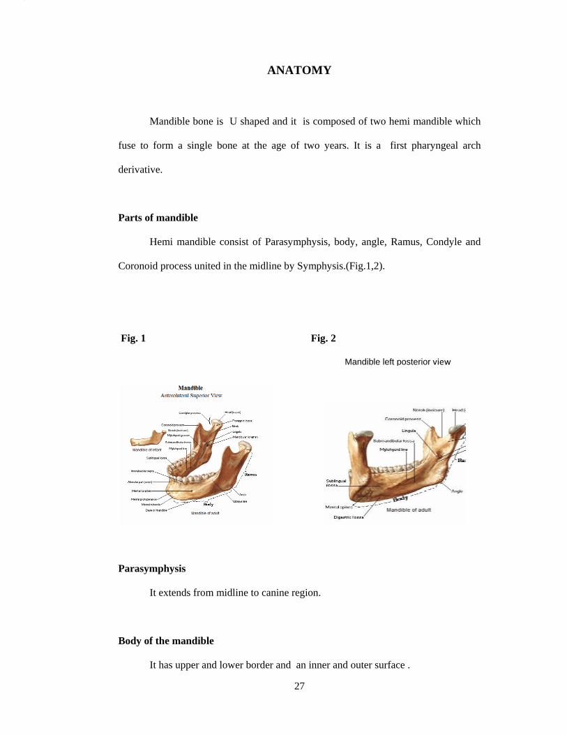
Mandible bone is U shaped and it is composed of two hemi mandible which
fuse to form a single bone at the age of two years. It is a first pharyngeal arch
derivative.
Parts of mandible
Hemi mandible consist of Parasymphysis, body, angle, Ramus, Condyle and
Coronoid process united in the midline by Symphysis.(Fig.1,2).
Fig. 1 Fig. 2
Parasymphysis
It extends from midline to canine region.
Body of the mandible
It has upper and lower border and an inner and outer surface .
Mandible left posterior view
28
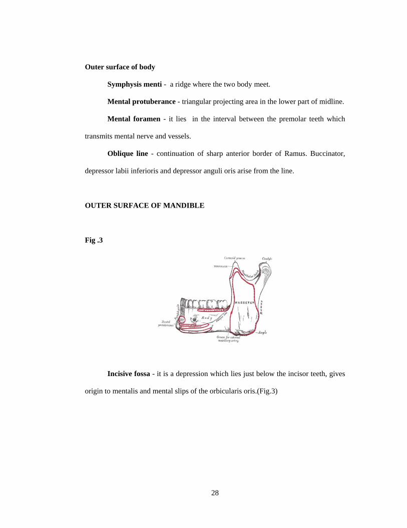
Outer surface of body
Symphysis menti - a ridge where the two body meet.
Mental protuberance - triangular projecting area in the lower part of midline.
Mental foramen - it lies in the interval between the premolar teeth which
transmits mental nerve and vessels.
Oblique line - continuation of sharp anterior border of Ramus. Buccinator,
depressor labii inferioris and depressor anguli oris arise from the line.
OUTER SURFACE OF MANDIBLE
Fig .3
Incisive fossa - it is a depression which lies just below the incisor teeth, gives
origin to mentalis and mental slips of the orbicularis oris.(Fig.3)
29
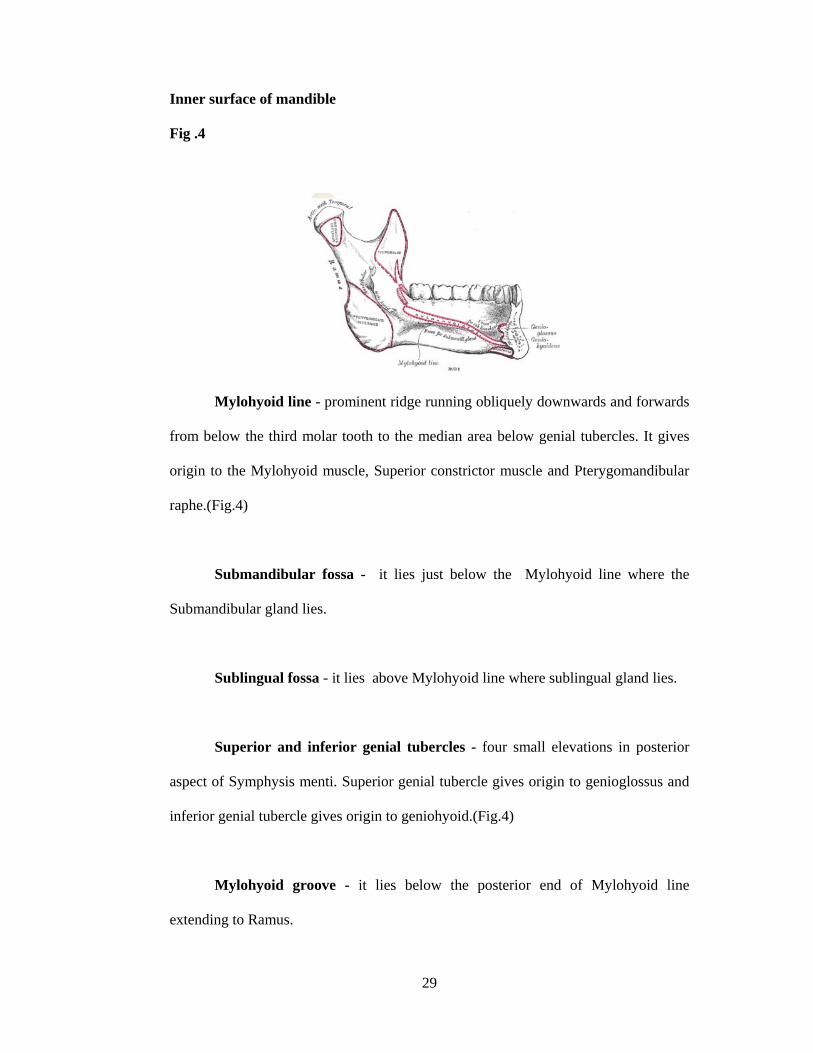
Inner surface of mandible
Fig .4
Mylohyoid line - prominent ridge running obliquely downwards and forwards
from below the third molar tooth to the median area below genial tubercles. It gives
origin to the Mylohyoid muscle, Superior constrictor muscle and Pterygomandibular
raphe.(Fig.4)
Submandibular fossa - it lies just below the Mylohyoid line where the
Submandibular gland lies.
Sublingual fossa - it lies above Mylohyoid line where sublingual gland lies.
Superior and inferior genial tubercles - four small elevations in posterior
aspect of Symphysis menti. Superior genial tubercle gives origin to genioglossus and
inferior genial tubercle gives origin to geniohyoid.(Fig.4)
Mylohyoid groove - it lies below the posterior end of Mylohyoid line
extending to Ramus.
30
Upper border - it holds the sockets of teeth.
Lower border - near the midline an oval depression called digastric fossa,
anterior belly of digastric muscle arises from the fossa. Platysma is inserted in this
border.
Ramus of the mandible
Quadrilateral in shape and has upper, lower, anterior and posterior borders,
lateral and medial surfaces.
Lateral surface
It is flat and having number of oblique ridges, Masseter inserted into it.
Medial surface
Mandibular foramen
It lies above the centre of Ramus leads to mandibular canal and descends into
body of mandible and opens at mental foramen. Mandibular canal gives entry to
Inferior alveolar nerve and vessels through mandibular foramen.(Fig.4)
Lingula - anterior margin of mandibular foramen. It gives attachment to
Sphenomandibular ligament.
Mylohyoid groove - it lies below the mandibular foramen. Medial pterygoid
muscle is inserted into the groove . In this groove, Mylohyoid nerve and vessels lie.
31
Upper border
Thin and curving downwards forming mandibular notch.
Lower border
Continuation of lower border of body. Posterior border is thicker than anterior
border.
Coronoid process - flat triangular projection in the anterosuperior part of
Ramus. Temporalis is inserted into it.
Condyle - an upward projection from poster superior part of Ramus. Fibro
cartilage covers the head and articulates with temporal bone forming
temporomandibular joint. Neck is the constriction below the head.
A depression in the anterior surface is called pterygoid fossa where lateral
pterygoid muscle is inserted.
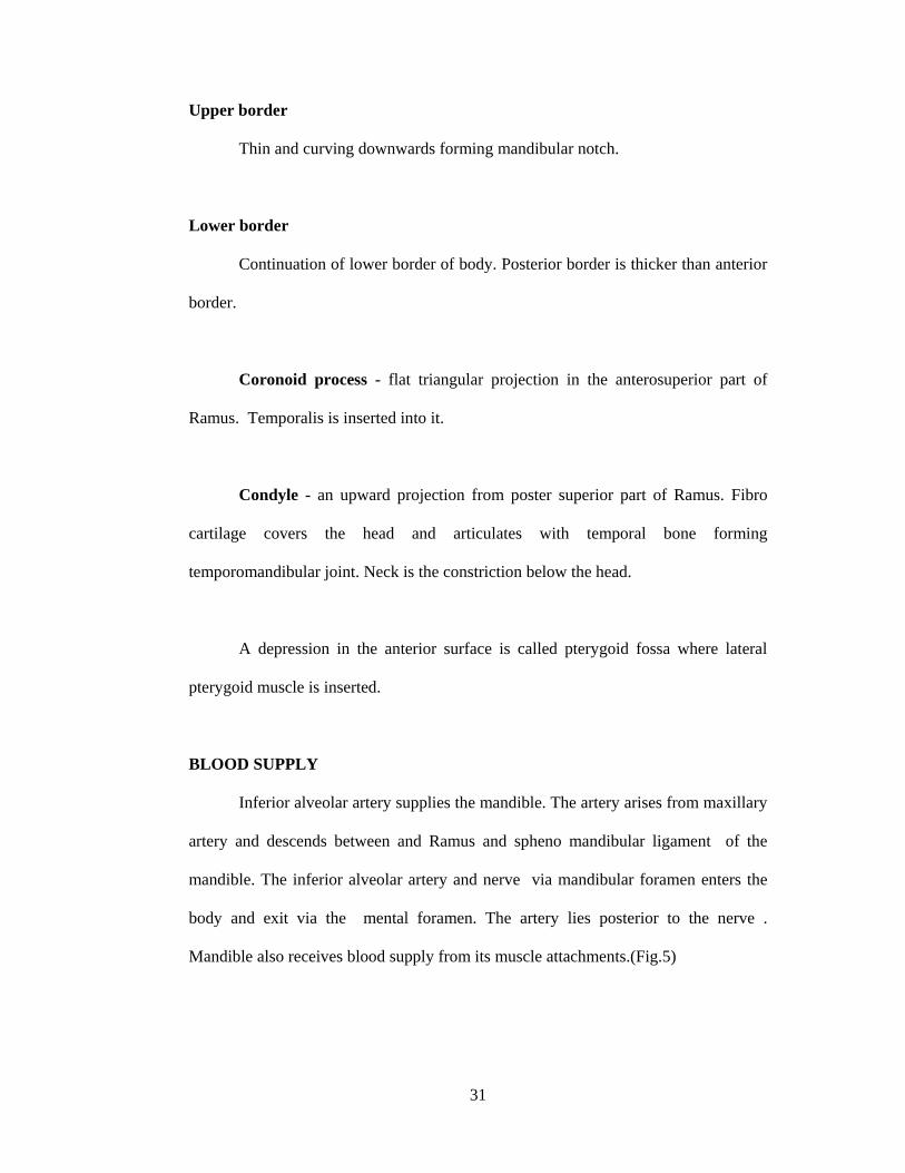
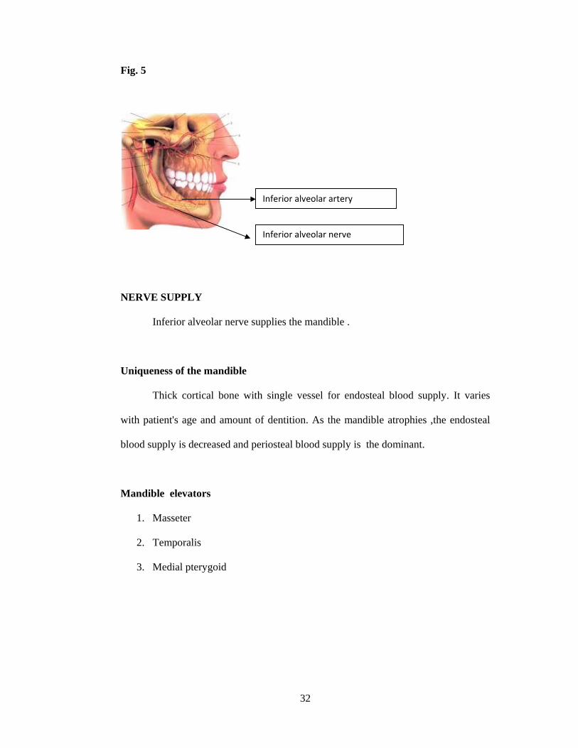
BLOOD SUPPLY
Inferior alveolar artery supplies the mandible. The artery arises from maxillary
artery and descends between and Ramus and spheno mandibular ligament of the
mandible. The inferior alveolar artery and nerve via mandibular foramen enters the
body and exit via the mental foramen. The artery lies posterior to the nerve .
Mandible also receives blood supply from its muscle attachments.(Fig.5)
F
N
U
w
bl
M
Fig. 5
NERVE SUP
Inferio
Uniqueness o
Thick
with patient's
lood supply
Mandible el
1. Masse
2. Temp
3. Media
PPLY
or alveolar n
of the mand
k cortical bo
s age and am
is decreased
levators
eter
oralis
al pterygoid
nerve supplie
dible
one with sin
mount of den
d and periost
Infe
Infe
32
es the mandi
ngle vessel f
ntition. As t
teal blood su
rior alveolar a
rior alveolar n
ible .
for endostea
the mandible
upply is the
artery
nerve
al blood sup
e atrophies ,
dominant.
pply. It varie
,the endoste
es
al
33
Mandible depressors
1. Lateral pterygoid
2. Mylohyoid
3. Digastric
4. Geniohyoid
All the muscles of mastication are supplied by mandibular branch (V3) of
trigeminal nerve .
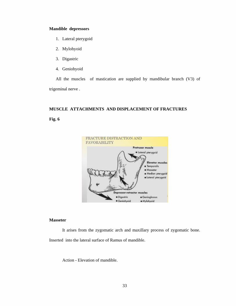
MUSCLE ATTACHMENTS AND DISPLACEMENT OF FRACTURES
Fig. 6
Masseter
It arises from the zygomatic arch and maxillary process of zygomatic bone.
Inserted into the lateral surface of Ramus of mandible.
Action - Elevation of mandible.
34
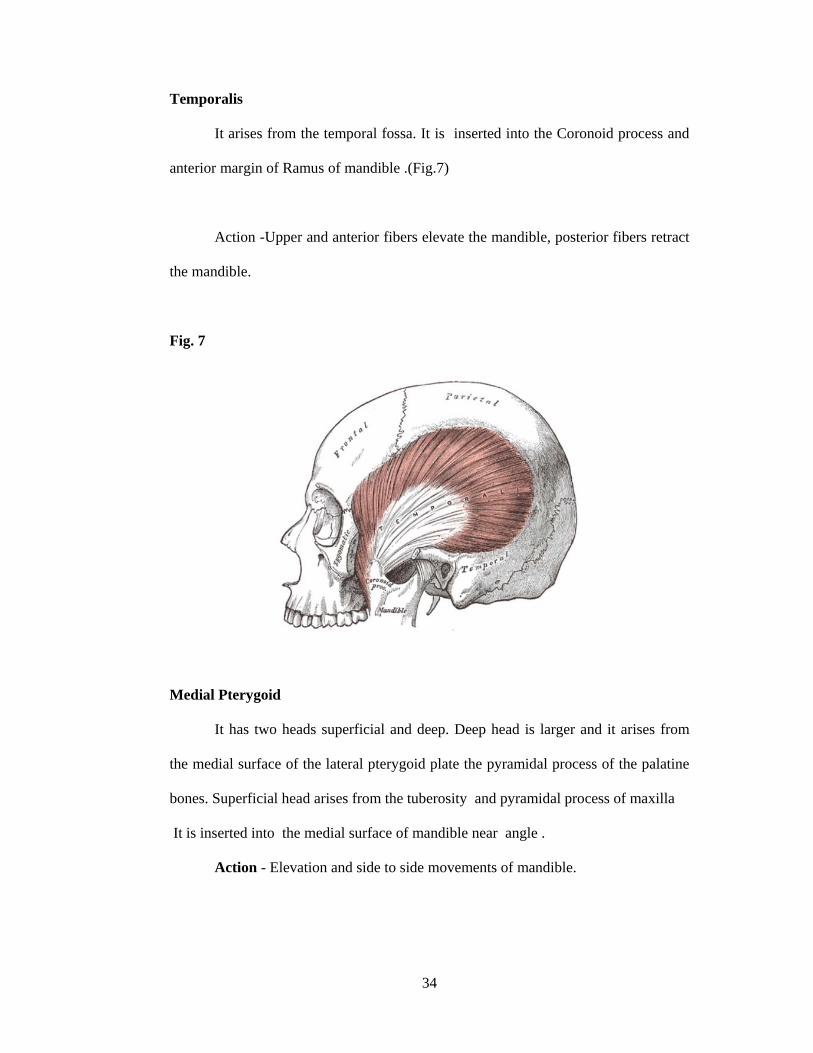
Temporalis
It arises from the temporal fossa. It is inserted into the Coronoid process and
anterior margin of Ramus of mandible .(Fig.7)
Action -Upper and anterior fibers elevate the mandible, posterior fibers retract
the mandible.
Fig. 7
Medial Pterygoid
It has two heads superficial and deep. Deep head is larger and it arises from
the medial surface of the lateral pterygoid plate the pyramidal process of the palatine
bones. Superficial head arises from the tuberosity and pyramidal process of maxilla
It is inserted into the medial surface of mandible near angle .
Action - Elevation and side to side movements of mandible.

35
Fig. 8
Lateral pterygoid
It has two heads. Superior head arises from the infratemporal fossa. Inferior
head arises from the lateral surface of the lateral pterygoid plate and both fuse into a
short thick tendon that inserts into pterygoid fovea in the neck of mandible and to the
capsule of temporomandibular joint.(Fig.8)
Action - Side to side movement and protrusion of mandible.
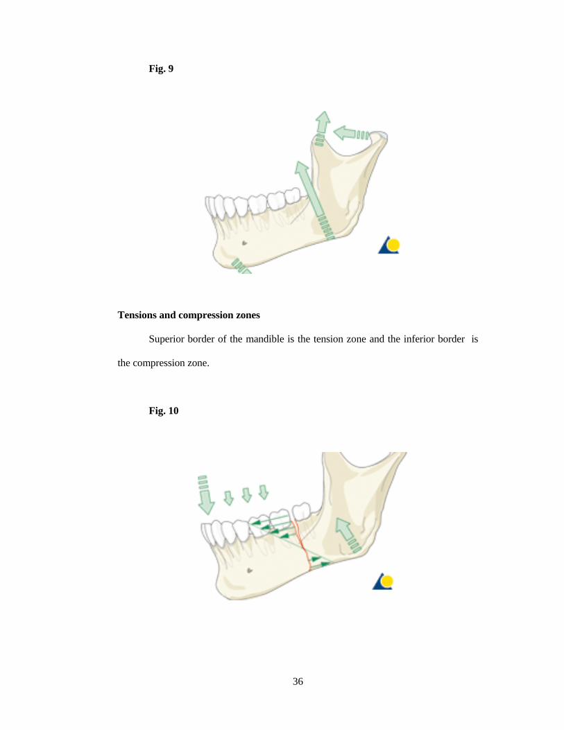
BIOMECHANICS OF MANDIBLE
Biomechanics of Mandible is a complex one. The forces applied to the
mandible cause varying zones of tension and compression depending on where the bite
force is located.
Muscle forces
Mandible is a hoop of bone that deforms with movement based on the origin
and insertion of the muscles of mastication.
36
Fig. 9
Tensions and compression zones
Superior border of the mandible is the tension zone and the inferior border is
the compression zone.
Fig. 10
37
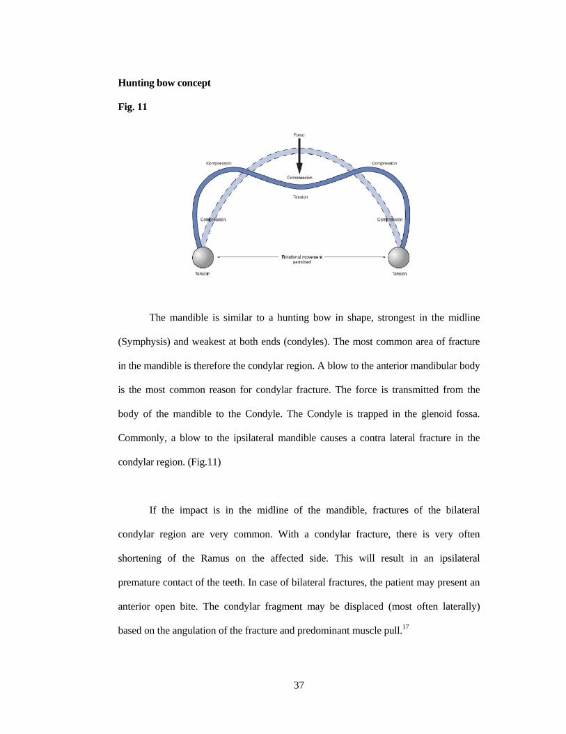
Hunting bow concept
Fig. 11
The mandible is similar to a hunting bow in shape, strongest in the midline
(Symphysis) and weakest at both ends (condyles). The most common area of fracture
in the mandible is therefore the condylar region. A blow to the anterior mandibular body
is the most common reason for condylar fracture. The force is transmitted from the
body of the mandible to the Condyle. The Condyle is trapped in the glenoid fossa.
Commonly, a blow to the ipsilateral mandible causes a contra lateral fracture in the
condylar region. (Fig.11)
If the impact is in the midline of the mandible, fractures of the bilateral
condylar region are very common. With a condylar fracture, there is very often
shortening of the Ramus on the affected side. This will result in an ipsilateral
premature contact of the teeth. In case of bilateral fractures, the patient may present an
anterior open bite. The condylar fragment may be displaced (most often laterally)
based on the angulation of the fracture and predominant muscle pull.17
38
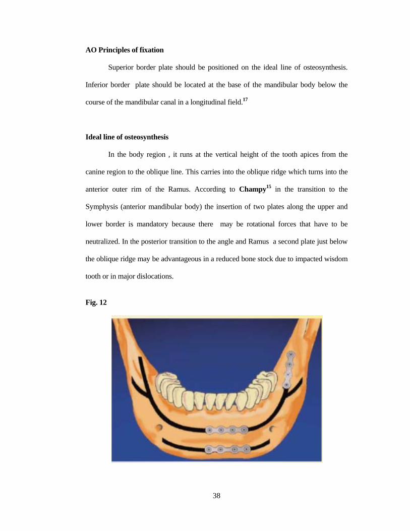
AO Principles of fixation
Superior border plate should be positioned on the ideal line of osteosynthesis.
Inferior border plate should be located at the base of the mandibular body below the
course of the mandibular canal in a longitudinal field.17
Ideal line of osteosynthesis
In the body region , it runs at the vertical height of the tooth apices from the
canine region to the oblique line. This carries into the oblique ridge which turns into the
anterior outer rim of the Ramus. According to Champy15 in the transition to the
Symphysis (anterior mandibular body) the insertion of two plates along the upper and
lower border is mandatory because there may be rotational forces that have to be
neutralized. In the posterior transition to the angle and Ramus a second plate just below
the oblique ridge may be advantageous in a reduced bone stock due to impacted wisdom
tooth or in major dislocations.
Fig. 12
39
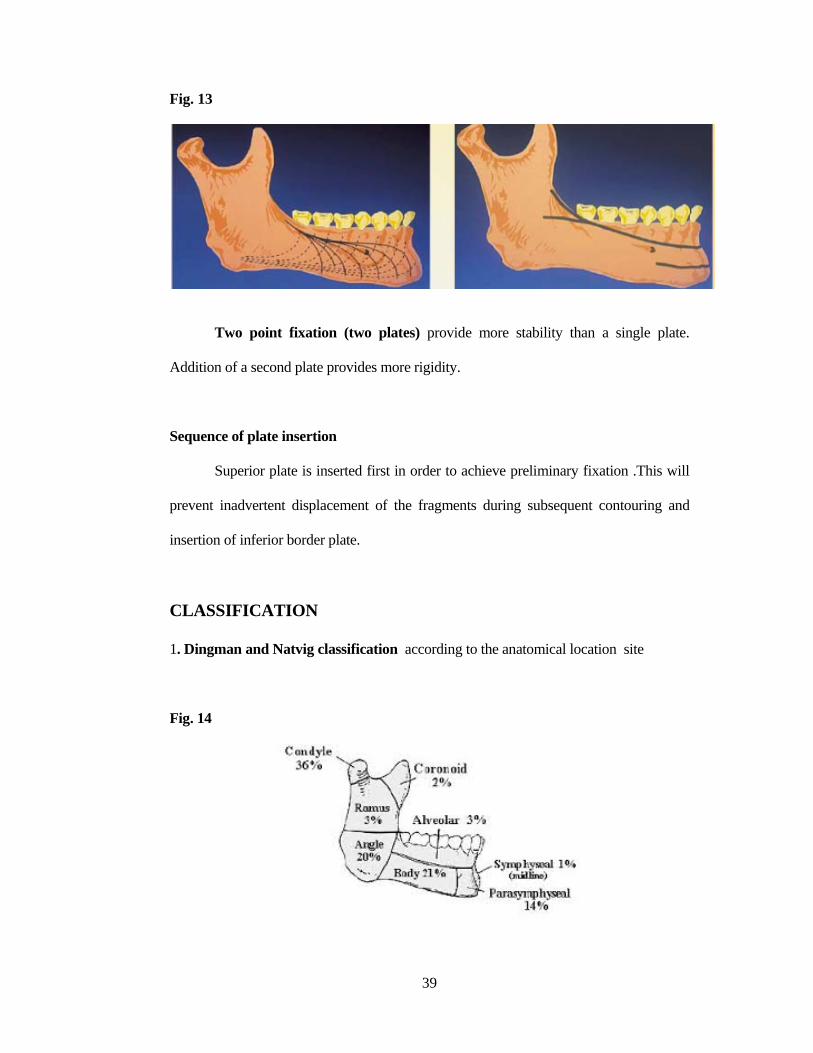
Fig. 13
Two point fixation (two plates) provide more stability than a single plate.
Addition of a second plate provides more rigidity.
Sequence of plate insertion
Superior plate is inserted first in order to achieve preliminary fixation .This will
prevent inadvertent displacement of the fragments during subsequent contouring and
insertion of inferior border plate.
CLASSIFICATION
1. Dingman and Natvig classification according to the anatomical location site
Fig. 14
2
F
3
a.
(e
b
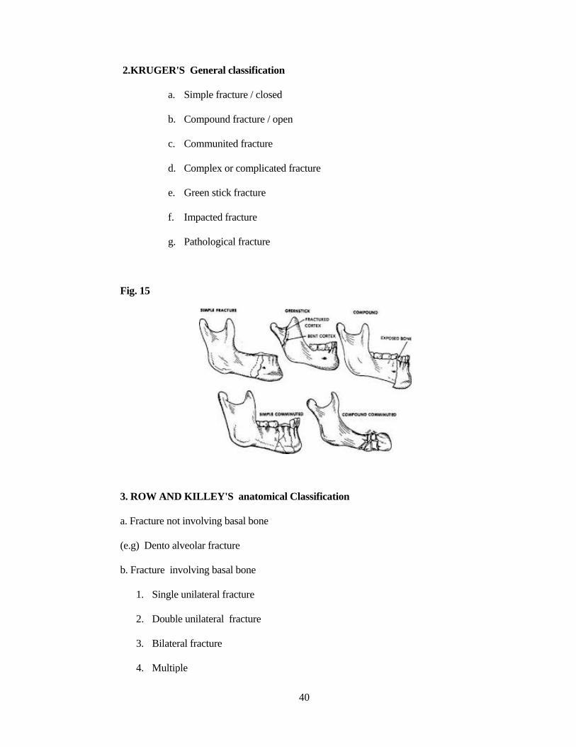
2.KRUGER
a.
b.
c.
d.
e.
f.
g.
Fig. 15
. ROW AND
. Fracture no
e.g) Dento a
. Fracture in
1. Single
2. Doubl
3. Bilate
4. Multip
'S General
Simple fra
Compound
Communit
Complex o
Green stick
Impacted f
Pathologic
D KILLEY'
ot involving b
alveolar fractu
nvolving basa
e unilateral fr
le unilateral
eral fracture
ple
classificatio
acture / closed
d fracture / op
ted fracture
or complicate
k fracture
fracture
cal fracture
S anatomic
basal bone
ure
al bone
racture
fracture
40
on
d
pen
ed fracture
cal Classificaation
41
4. Completeness of fracture
Complete fracture
Incomplete fracture
5. According to the presence or absent of tooth in relation to fracture line
Kazanjian and Converse
Class I - when the teeth are present on both sides of the fracture line
Class II - when the teeth are present on one side of the fracture line
Class III - when teeth are absent on both sides of the fracture line
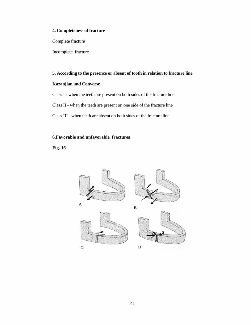
6.Favorable and unfavorable fractures
Fig. 16
42
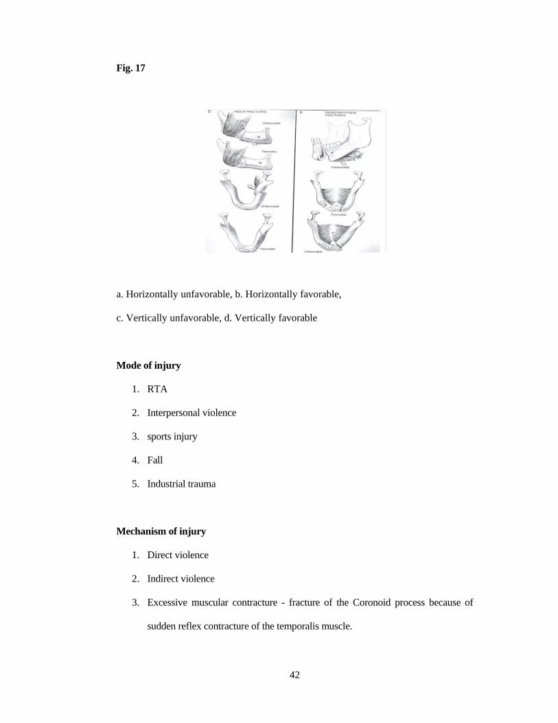
Fig. 17
a. Horizontally unfavorable, b. Horizontally favorable,
c. Vertically unfavorable, d. Vertically favorable
Mode of injury
1. RTA
2. Interpersonal violence
3. sports injury
4. Fall
5. Industrial trauma
Mechanism of injury
1. Direct violence
2. Indirect violence
3. Excessive muscular contracture - fracture of the Coronoid process because of
sudden reflex contracture of the temporalis muscle.
43
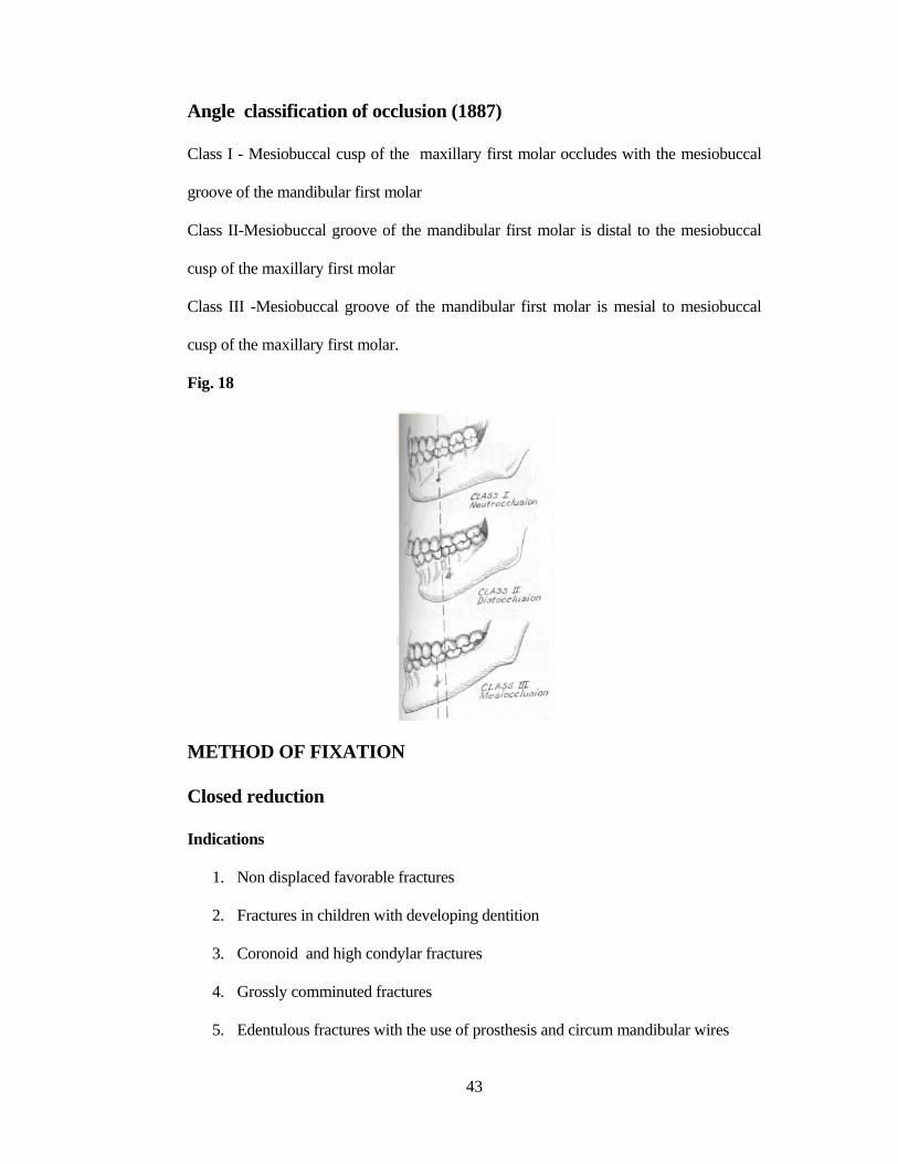
Angle classification of occlusion (1887)
Class I - Mesiobuccal cusp of the maxillary first molar occludes with the mesiobuccal
groove of the mandibular first molar
Class II-Mesiobuccal groove of the mandibular first molar is distal to the mesiobuccal
cusp of the maxillary first molar
Class III -Mesiobuccal groove of the mandibular first molar is mesial to mesiobuccal
cusp of the maxillary first molar.
Fig. 18
METHOD OF FIXATION
Closed reduction
Indications
1. Non displaced favorable fractures
2. Fractures in children with developing dentition
3. Coronoid and high condylar fractures
4. Grossly comminuted fractures
5. Edentulous fractures with the use of prosthesis and circum mandibular wires
44
Splints
1. Gunning splints
2. Lingual splints
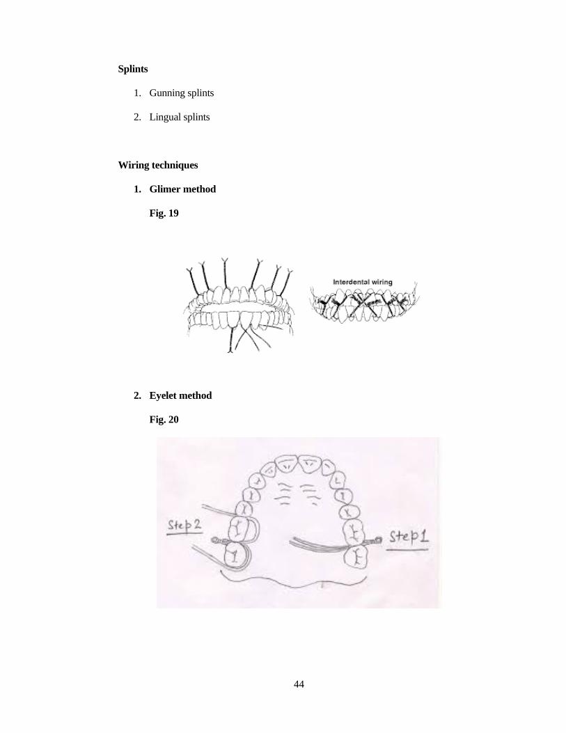
Wiring techniques
1. Glimer method
Fig. 19
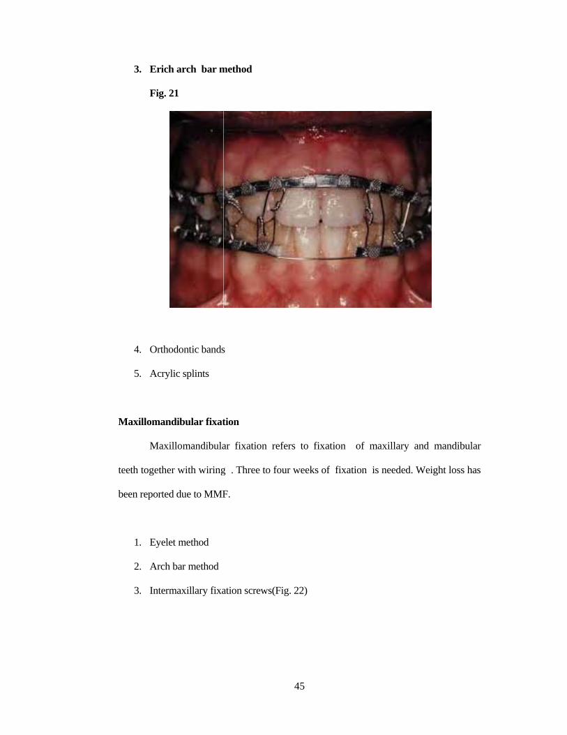
2. Eyelet method
Fig. 20
M
te
be
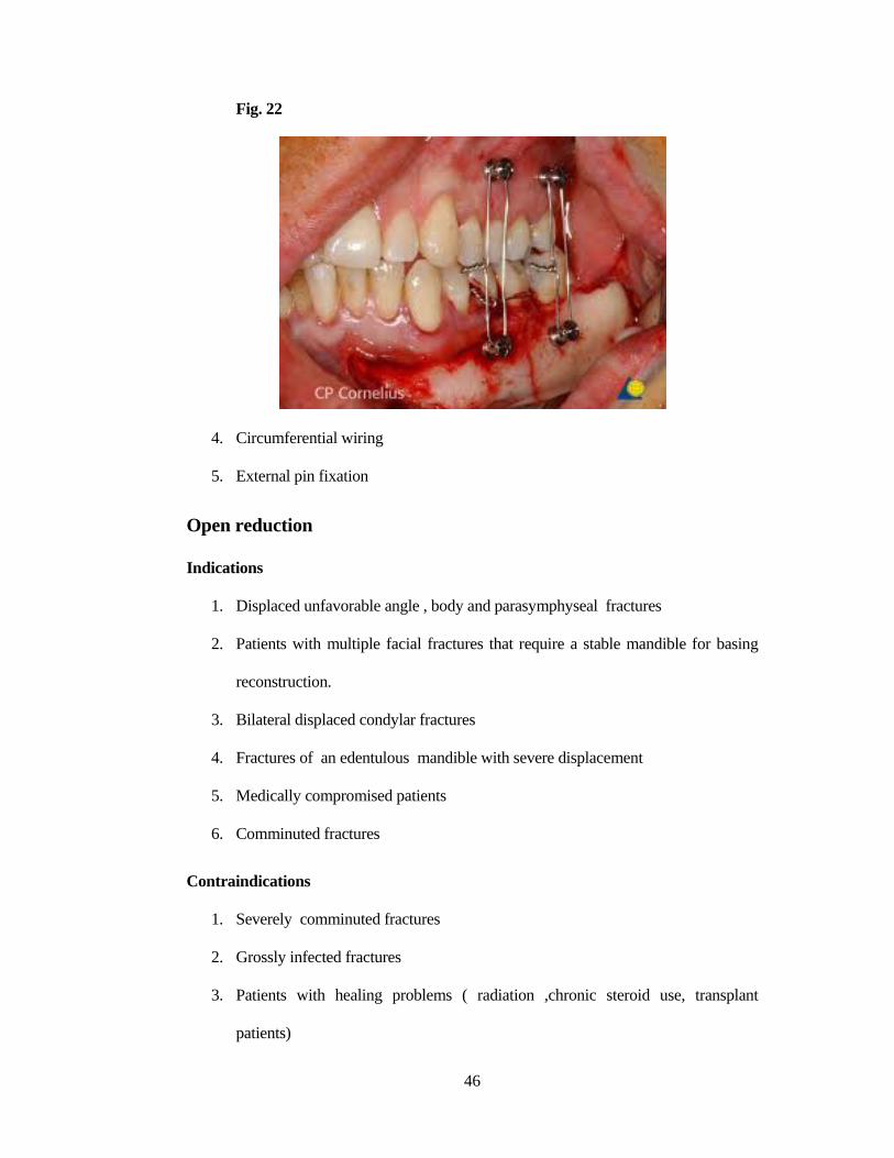
3. Erich
Fig. 2
4. Ortho
5. Acryli
Maxillomand
Maxil
eeth together
een reported
1. Eyelet
2. Arch b
3. Interm
h arch bar m
1
dontic bands
ic splints
dibular fixat
llomandibula
r with wiring
d due to MMF
t method
bar method
maxillary fixa
method
s
tion
ar fixation r
g . Three to
F.
ation screws(
45
refers to fix
four weeks o
(Fig. 22)
xation of m
of fixation i
maxillary an
is needed. W
nd mandibul
Weight loss ha
ar
as
46
Fig. 22
4. Circumferential wiring
5. External pin fixation
Open reduction
Indications
1. Displaced unfavorable angle , body and parasymphyseal fractures
2. Patients with multiple facial fractures that require a stable mandible for basing
reconstruction.
3. Bilateral displaced condylar fractures
4. Fractures of an edentulous mandible with severe displacement
5. Medically compromised patients
6. Comminuted fractures
Contraindications
1. Severely comminuted fractures
2. Grossly infected fractures
3. Patients with healing problems ( radiation ,chronic steroid use, transplant
patients)
47
Methods of fixation
1. Dynamic compression plates
In plates with compression holes ,as the screw is tightened the screw -bone unit
is moved towards the fracture site impacting against the bone on the opposite side of the
fracture. Screws inserted bicortically This promotes primary bone healing.
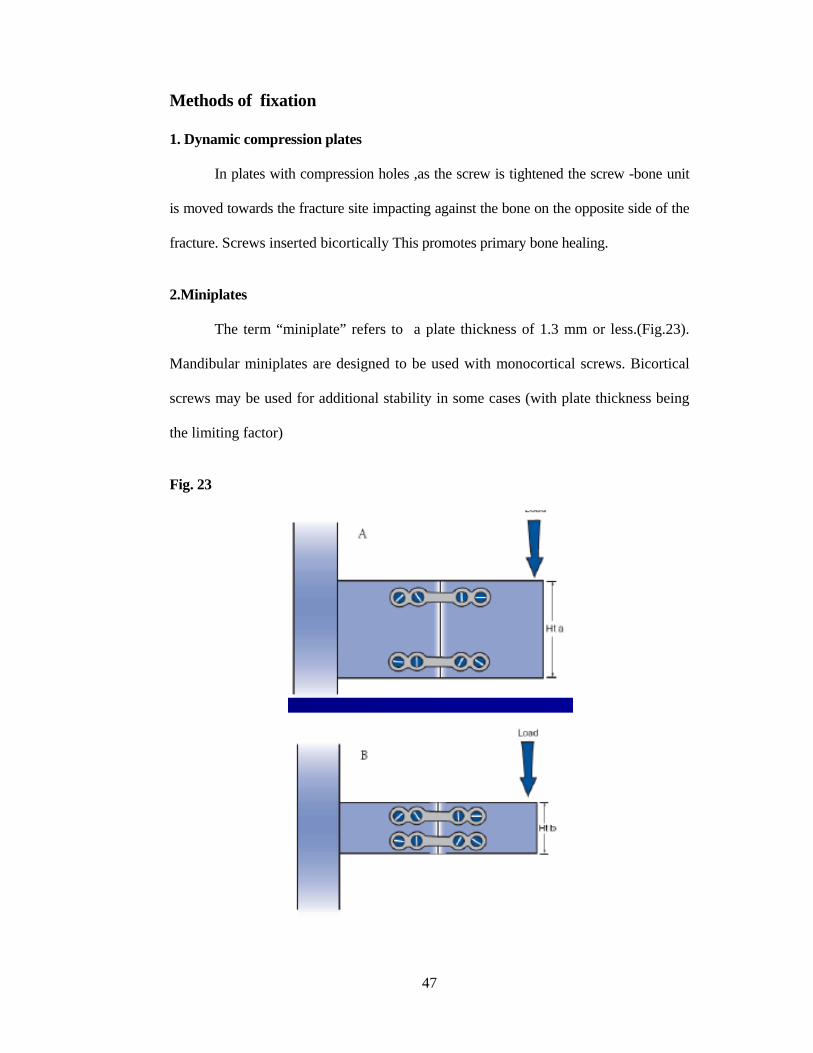
2.Miniplates
The term “miniplate” refers to a plate thickness of 1.3 mm or less.(Fig.23).
Mandibular miniplates are designed to be used with monocortical screws. Bicortical
screws may be used for additional stability in some cases (with plate thickness being
the limiting factor)
Fig. 23
48
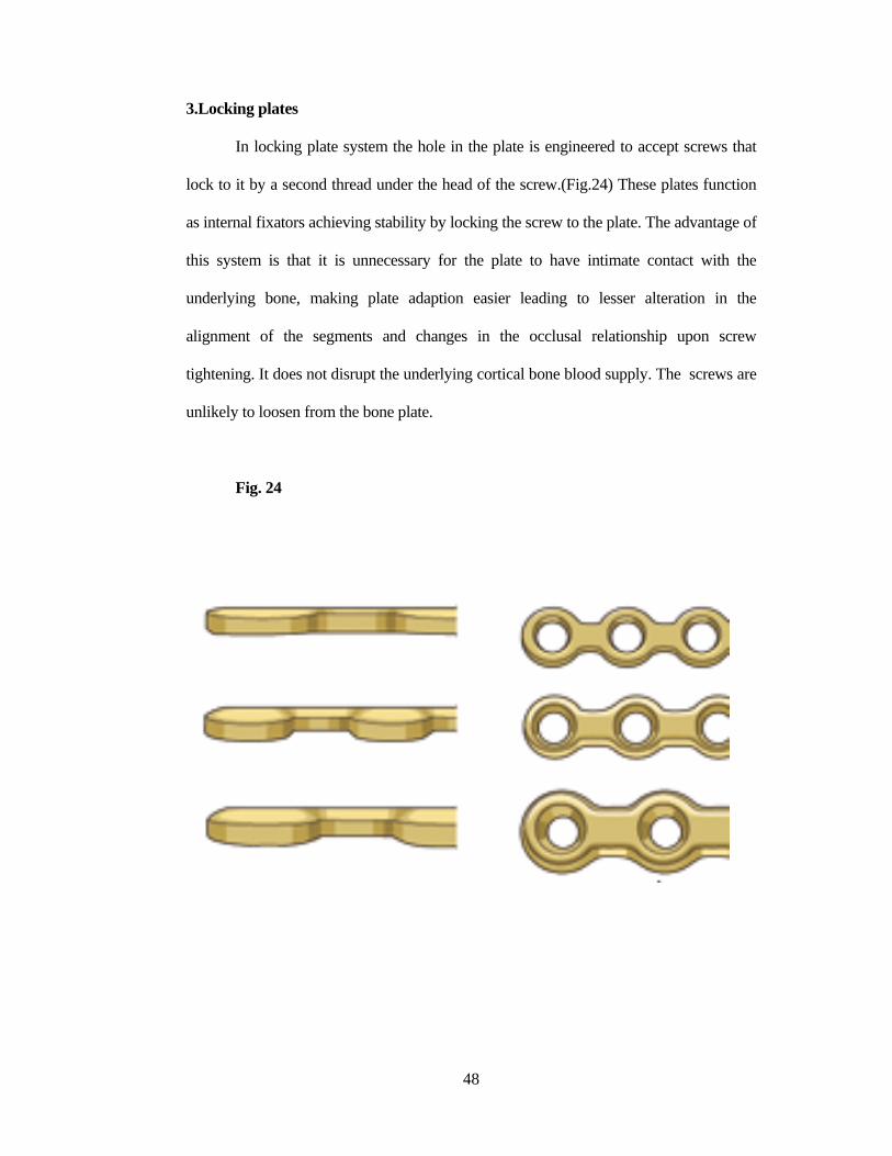
3.Locking plates
In locking plate system the hole in the plate is engineered to accept screws that
lock to it by a second thread under the head of the screw.(Fig.24) These plates function
as internal fixators achieving stability by locking the screw to the plate. The advantage of
this system is that it is unnecessary for the plate to have intimate contact with the
underlying bone, making plate adaption easier leading to lesser alteration in the
alignment of the segments and changes in the occlusal relationship upon screw
tightening. It does not disrupt the underlying cortical bone blood supply. The screws are
unlikely to loosen from the bone plate.
Fig. 24
49
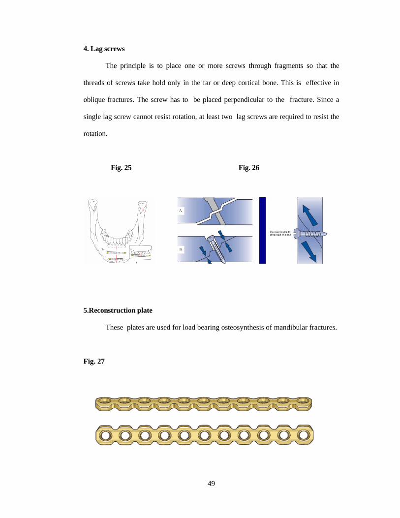
4. Lag screws
The principle is to place one or more screws through fragments so that the
threads of screws take hold only in the far or deep cortical bone. This is effective in
oblique fractures. The screw has to be placed perpendicular to the fracture. Since a
single lag screw cannot resist rotation, at least two lag screws are required to resist the
rotation.
Fig. 25 Fig. 26
5.Reconstruction plate
These plates are used for load bearing osteosynthesis of mandibular fractures.
Fig. 27
50
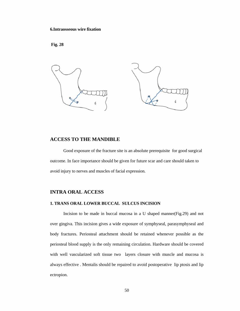
6.Intraosseous wire fixation
Fig. 28
ACCESS TO THE MANDIBLE
Good exposure of the fracture site is an absolute prerequisite for good surgical
outcome. In face importance should be given for future scar and care should taken to
avoid injury to nerves and muscles of facial expression.
INTRA ORAL ACCESS
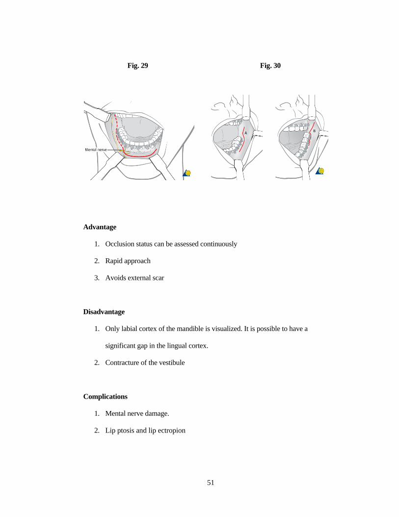
1. TRANS ORAL LOWER BUCCAL SULCUS INCISION
Incision to be made in buccal mucosa in a U shaped manner(Fig.29) and not
over gingiva. This incision gives a wide exposure of symphyseal, parasymphyseal and
body fractures. Periosteal attachment should be retained whenever possible as the
periosteal blood supply is the only remaining circulation. Hardware should be covered
with well vascularized soft tissue two layers closure with muscle and mucosa is
always effective . Mentalis should be repaired to avoid postoperative lip ptosis and lip
ectropion.
51
Fig. 29 Fig. 30
Advantage
1. Occlusion status can be assessed continuously
2. Rapid approach
3. Avoids external scar
Disadvantage
1. Only labial cortex of the mandible is visualized. It is possible to have a
significant gap in the lingual cortex.
2. Contracture of the vestibule
Complications
1. Mental nerve damage.
2. Lip ptosis and lip ectropion
52
2. TRANSBUCCAL ACCESS (TROCAR TECHNIQUE)
It is a combination of both intraoral and extra oral access. Trocar and specific
instruments are used to place the screw. This access is used for body and angle
fractures.
EXTRA ORAL ACCESS
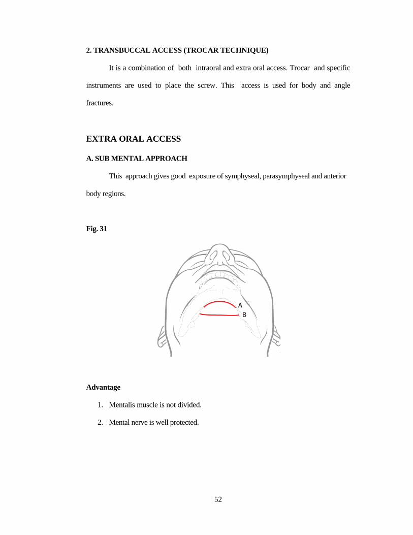
A. SUB MENTAL APPROACH
This approach gives good exposure of symphyseal, parasymphyseal and anterior
body regions.
Fig. 31
Advantage
1. Mentalis muscle is not divided.
2. Mental nerve is well protected.
53
Disadvantage
External scar is present
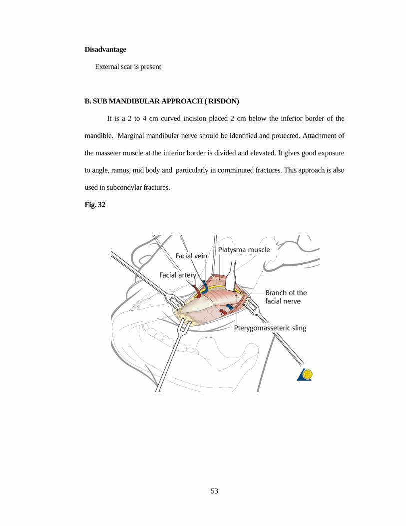
B. SUB MANDIBULAR APPROACH ( RISDON)
It is a 2 to 4 cm curved incision placed 2 cm below the inferior border of the
mandible. Marginal mandibular nerve should be identified and protected. Attachment of
the masseter muscle at the inferior border is divided and elevated. It gives good exposure
to angle, ramus, mid body and particularly in comminuted fractures. This approach is also
used in subcondylar fractures.
Fig. 32
54
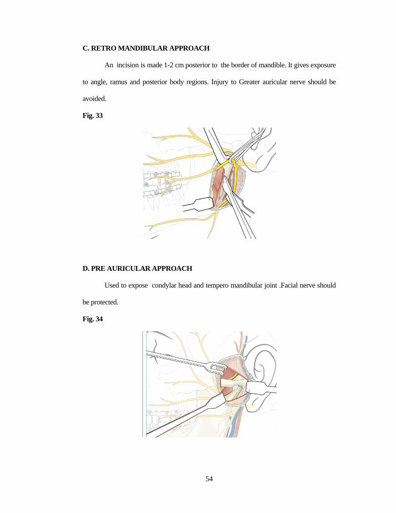
C. RETRO MANDIBULAR APPROACH
An incision is made 1-2 cm posterior to the border of mandible. It gives exposure
to angle, ramus and posterior body regions. Injury to Greater auricular nerve should be
avoided.
Fig. 33
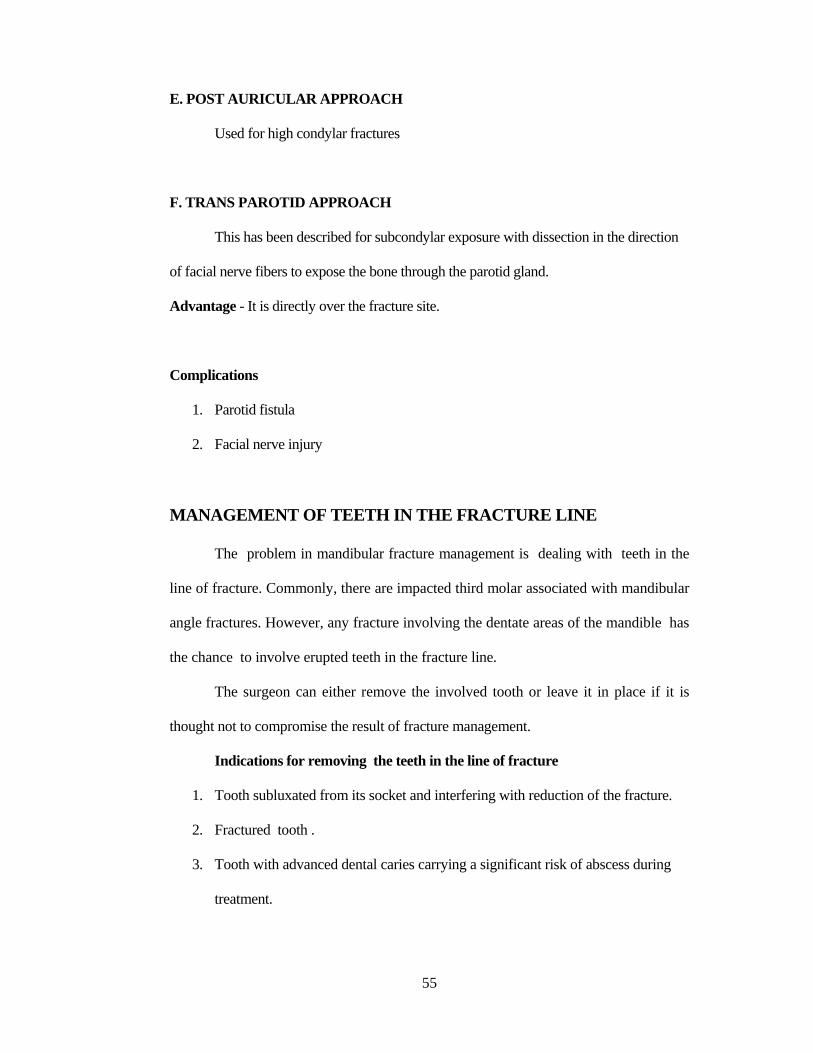
D. PRE AURICULAR APPROACH
Used to expose condylar head and tempero mandibular joint .Facial nerve should
be protected.
Fig. 34
55
E. POST AURICULAR APPROACH
Used for high condylar fractures
F. TRANS PAROTID APPROACH
This has been described for subcondylar exposure with dissection in the direction
of facial nerve fibers to expose the bone through the parotid gland.
Advantage - It is directly over the fracture site.
Complications
1. Parotid fistula
2. Facial nerve injury
MANAGEMENT OF TEETH IN THE FRACTURE LINE
The problem in mandibular fracture management is dealing with teeth in the
line of fracture. Commonly, there are impacted third molar associated with mandibular
angle fractures. However, any fracture involving the dentate areas of the mandible has
the chance to involve erupted teeth in the fracture line.
The surgeon can either remove the involved tooth or leave it in place if it is
thought not to compromise the result of fracture management.
Indications for removing the teeth in the line of fracture
1. Tooth subluxated from its socket and interfering with reduction of the fracture.
2. Fractured tooth .
3. Tooth with advanced dental caries carrying a significant risk of abscess during
treatment.
56
4. Tooth with advanced periodontal disease with mobility which could not
contribute to the establishment of stable occlusion.
5. Tooth with existing pathology such as cyst or pericoronitis.
Indications for leaving the teeth in the line of fracture
1. Tooth not interfering with the reduction and fixation of fracture.
2. If tooth removal requiring removal of excessive amount of bone, it will lead to
compromise in the fracture fixation.
3. Tooth that is in good condition and assists in establishing occlusion and
reducing the fracture.
COMPLICATIONS
1. MALOCCLUSION
Malocclusion is the most common complication and functional problem.
Causes of malocclusion are
1. Inaccurate alignment in initial reduction (poorly applied MMF)
2. severe comminution
3. Patient's non compliance.
Minor malocclusion can be corrected with occlusal splints. Severe malocclusion has to
be corrected by refracturing or osteotomy and plates osteosynthesis with MMF.
57
2. DELAYED OR NON UNION
Delayed union is more commonly due to inadequate reduction and fixation. If a
fibrous union is present , the fracture will heal with bony consolidation over a period of
time .
Non union occurs due to
1. Infection
2. Inadequate opposition of bone.
3. Severe comminution with gap
4. teeth in the fracture line
It has to be treated by re exploration and fixation with bone grafts.
3.MALUNION
Bone heels in abnormal position due to inadequate reduction and fixation.
Malunion has to be treated with osteotomy and re fixation with bone graft.
4.INFECTION
Infection is seen in compound fractures, excessive periosteal stripping, unstable
fracture fixation and poor oral hygiene. It is treated with culture specific antibiotic, re
exploration, removal of devitalized bony fragments and if the fixation is loose do a stable
rigid fixation and bone grafting of mandible. If the primary fixation is stable allow it till
fracture union.
58
5. EXPOSED OR LOOSE HARDWARE
Hardware get exposed when there is infection, wound contracture and when a
dental prosthesis is worn over hardware. Minor exposure is managed conservatively till
fracture union whereas major exposure requires hardware removal and more stable
fixation.
6. SENSORIMOTOR DISTURBANCES
Sensory disturbances of inferior alveolar nerve and mental nerve can occur.
Motor disturbances due to injury to marginal mandibular nerve and facial nerve have been
reported.
7.EXACERBATION OF DENTAL DISEASE
If oral hygiene is not maintained there can be exacerbation of existing dental
disease like caries .
8.TEMPOROMANDIBULAR JOINT DYSFUNCTION
Prolonged immobilization with MMF can lead to TMJ dysfunction. Simple jaw
exercises and mechanical exercises can improve the condition. Myositis ossificans can
occur when hematoma in the muscle organises and ossifies. The myositis has to be
excised, but there is a chance of recurrence.
9. SCARS
Unsightly scar can occur in compound fractures. It can be managed initially
with scar massaging followed by scar revision .
59
MATERIALS AND METHODS
This study was conducted in the Department of Plastic and Reconstructive
Surgery, Coimbatore Medical college and Hospital ,Coimbatore on 67 patients who
reported to the trauma ward and the department of plastic and reconstructive surgery
for the treatment of fracture mandible from December 2012 to December 2014.
Before the start of the study, ethical clearance was obtained from the Ethical
committee of the Coimbatore Medical College and Hospital, Coimbatore.
Informations were collected from the clinical and surgical notes of each of the
patients in a standardized and systematic pattern. The demographic variables such as
age, gender, and residence were assessed. Clinical information included diagnosis,
etiology, and anatomical distribution of mandibular fractures was assessed.
The mandibular fractures were classified according to the sites such as ramus,
Condyle, Coronoid Symphysis, body, Parasymphysis and angle.
INCLUSION CRITERIA
1. All adult patients between 25 to 55 years.
2. Patients reporting within first 7-10 days from the day of trauma.
3. Dentulous / partially dentulous patients
4. Patients giving consent for a follow up period of 3 months post
operatively.
60
EXCLUSION CRITERIA
1. Compound fractures
2. Patients with other facial bone fractures.
3. Patients with systemic / debilitating diseases
4. Patients with head injury
CLINICAL EVALUATION
1. History of incident
2. Inspection- swelling , laceration ,malocclusion, sublingual hematoma,
deformity and trismus
3. Palpation-step deformity/tenderness
4. Paresthesia / dysaesthesia/ anesthesia of mental nerve.
5. TMJ examination- to find any Condyle fracture.
All patients with suspected mandible fracture were subjected to OPG
(Orthopantomogram) & CT facial bones .The mandibular fractures were classified
according to the site such as Ramus, Condyle, Symphysis, Body, Parasymphysis and
Angle. All these patients were transferred to the Plastic surgery ward.
Out of 67 patients ,15 patients who had undisplaced fractures , Condylar &
Subcondylar fractures were treated conservatively with arch bars , eyelets and
Maxillomandibular fixation (MMF) for 4 -6weeks .They were done under mandibular
nerve block in our ward within 24-48 hours. Post MMF OPG was taken to assess the
reduction .These patients were started on liquid diet soon after the MMF and
61
encouraged to maintain oral hygiene The remaining 52 displaced ,unfavorable and
Communited fractures were treated surgically .
Arch bars and MMF were done preoperatively for all the cases to achieve
conclusion. Extra oral approach (Risdon) was used for the angle fracture . Intra oral
approach (gingivobuccal sulcal approach) was used for the Symphysis,
Parasymphysis and body the fractures.
Surgical technique
All the 52 patients who were taken up for surgery were treated according to
the principles outlined by Champy. Conventional non locking miniplates and
screws were used.
Taking into account the anatomy of the mandible, with the location of
the dental apices and the thickness of the cortical layer, Champy et al determined
an ideal line of osteosynthesis which corresponds to the course of a line of
tension at the base of the alveolar process.15,16
As Champy recommended , one plate was applied behind the mental
foramen, just below the dental roots and above the inferior alveolar nerve,
in order to neutralize the higher torsion forces between the canines. A second
plate was applied near the lower border of the mandible in addition to the sub-
apical plate. In the miniplate system unicortical fixation was done.
62
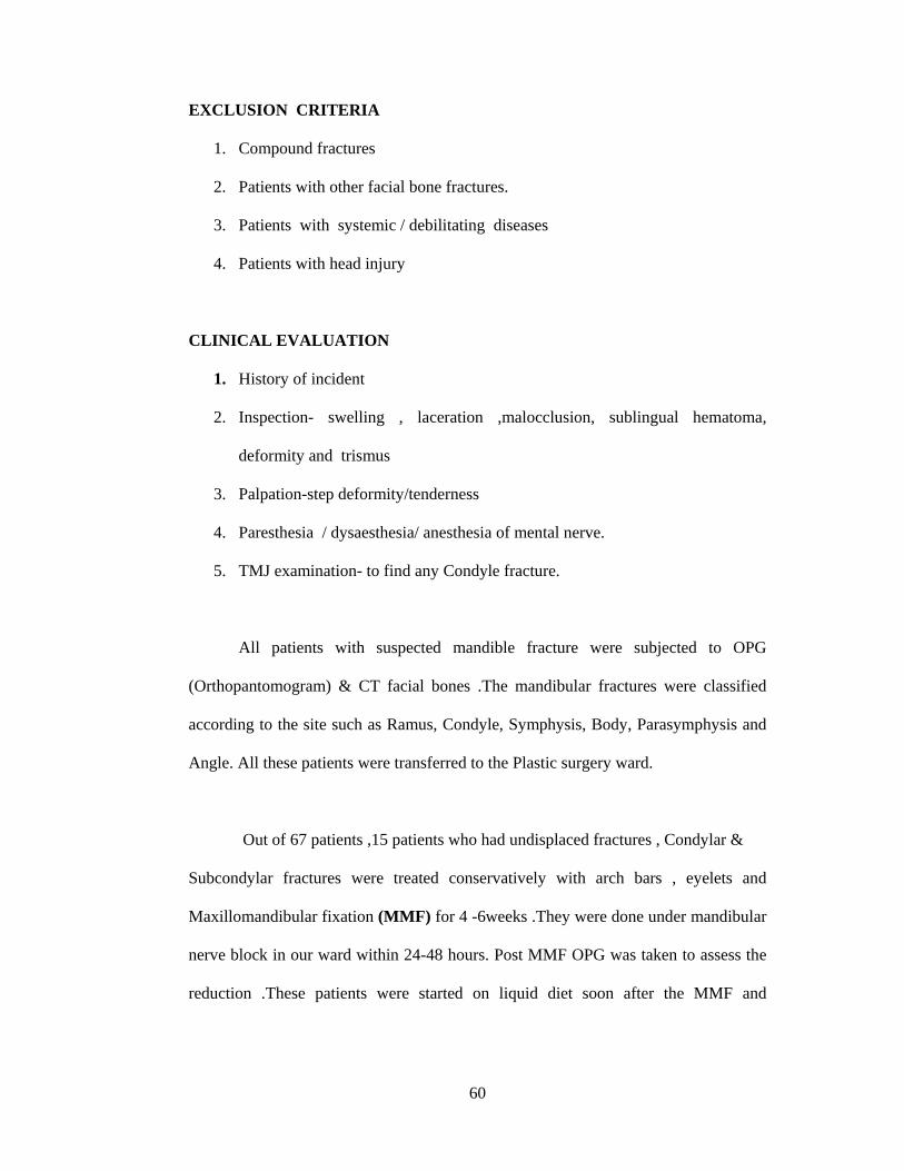
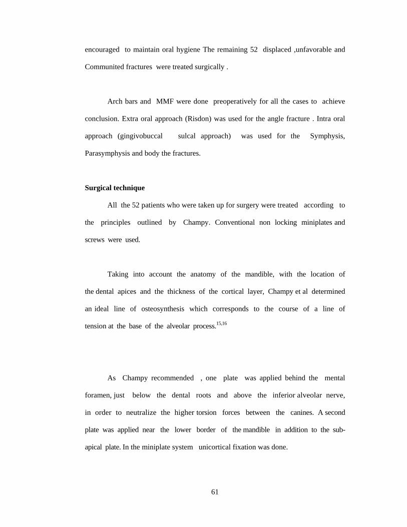
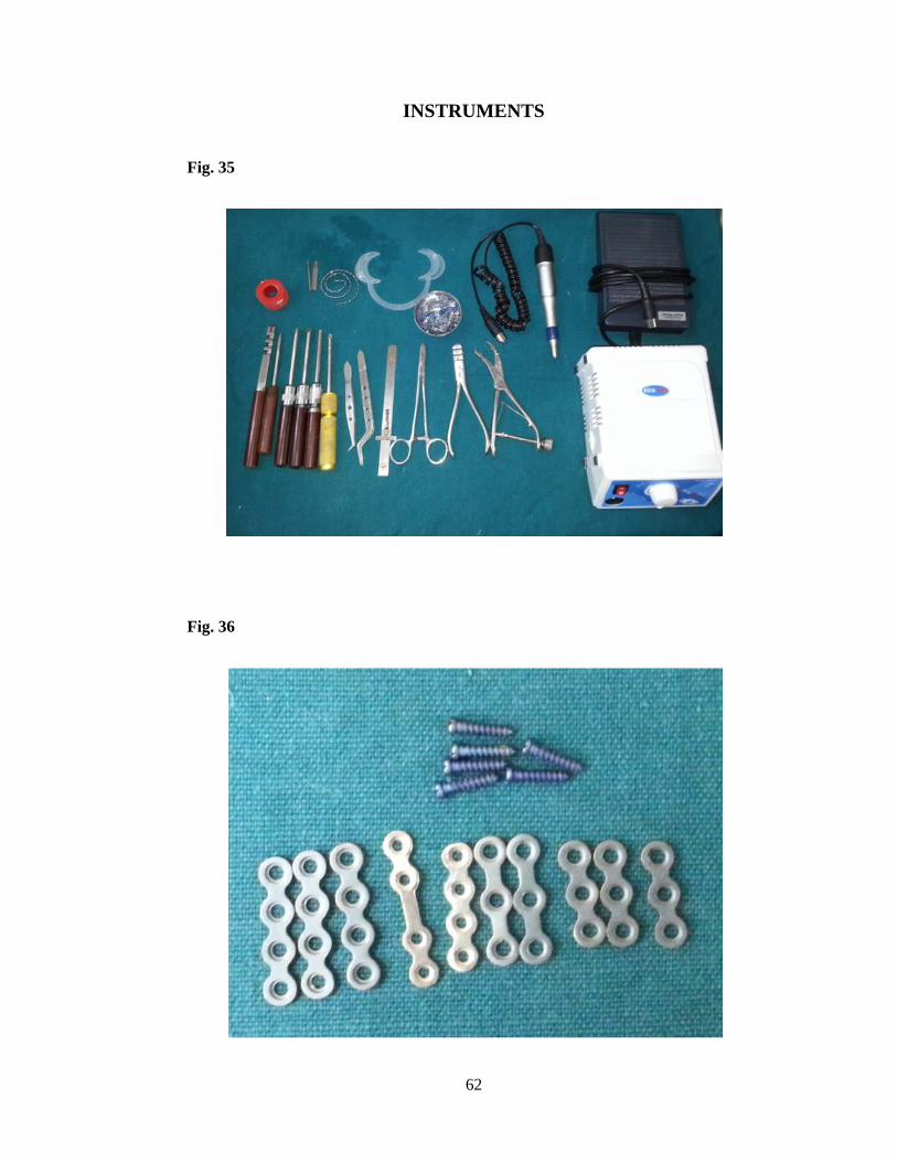
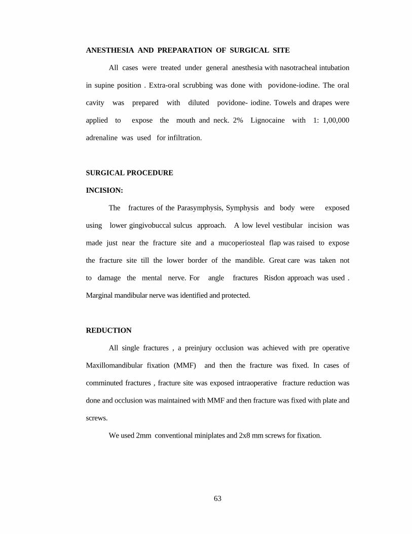
INSTRUMENTS
Fig. 35
Fig. 36
63
ANESTHESIA AND PREPARATION OF SURGICAL SITE
All cases were treated under general anesthesia with nasotracheal intubation
in supine position . Extra-oral scrubbing was done with povidone-iodine. The oral
cavity was prepared with diluted povidone- iodine. Towels and drapes were
applied to expose the mouth and neck. 2% Lignocaine with 1: 1,00,000
adrenaline was used for infiltration.
SURGICAL PROCEDURE
INCISION:
The fractures of the Parasymphysis, Symphysis and body were exposed
using lower gingivobuccal sulcus approach. A low level vestibular incision was
made just near the fracture site and a mucoperiosteal flap was raised to expose
the fracture site till the lower border of the mandible. Great care was taken not
to damage the mental nerve. For angle fractures Risdon approach was used .
Marginal mandibular nerve was identified and protected.
REDUCTION
All single fractures , a preinjury occlusion was achieved with pre operative
Maxillomandibular fixation (MMF) and then the fracture was fixed. In cases of
comminuted fractures , fracture site was exposed intraoperative fracture reduction was
done and occlusion was maintained with MMF and then fracture was fixed with plate and
screws.
We used 2mm conventional miniplates and 2x8 mm screws for fixation.
64
In placing the hole, the drill was made perpendicular to bone surface
and plate within the centre of the screw hole, with 701 or 702 bur, so that the
screw gets fitted into bone plate.
Two four hole conventional miniplates were used in the Symphysis and
Parasymphysis fractures between the mental foramina according to Champy’s line
of osteosynthesis.
These lines corresponds to the
1. Course of a line of tension at the base of alveolar process and
2. Another line near the lower border of the mandible in order to neutralize
torsion forces.
A gap of 4-5mm and parallelism were maintained between the two
plates. The upper plate was fixed first and then the lower plate with 2x8 mm
screws on either side of the fracture. Care was taken not to injure the nerve in the
mandibular canal. Marginal mandibular nerve was protected during the Risdon approach.
The occlusion was checked and the screws were tightened
finally. Maxillomandibular fixation was released depending on the stability of the
fixation.
In cases where 2 miniplates were used, MMF was removed soon after the
surgery . In cases where single plate was used, MMF retained for 2 wks. Arch bars
maintained for 4 more wks. In fractures with combinations like Parasymphysis and
Subcondyle, plating was done only for the Parasymphysis and the Subcondyle
treated conservatively with MMF for 2-3 weeks .
65
CLOSURE
The fracture site was irrigated and soft tissues closed with 2-0 vicryl in
two layers. Post operative OPG was taken to assess the stability of fixation.
POSTOPERATIVE CARE
All patients were kept under antibiotic cover for 5 days. Those for whom
MMF was removed they were advised to take liquid diet for 2days and thereafter
on a soft diet for 4to 6 weeks. Those who were advised to maintain MMF ,
continued liquid diet for 2-3 weeks .
The patients were asked to maintain oral hygiene with mouth wash. Sutures
were removed on the 5th postoperative day for patients who had underwent extra oral
approach.
At the end of second post operative week they were started on gentle
physiotherapy. Follow up was performed weekly during the first 6 weeks and
thereafter monthly for 4 to 6 months.
FOLLOW UP
During the immediate follow up the following parameters were recorded.
1. Resolution of facial edema.
2. Healing of surgical sites.
3. Sensory , motor disturbances.
4. Visual analog score for pain
5. Visual analog score for chewing ability
66
6. Angle criteria for occlusion
7. Mouth opening
8. Weight loss
Data in the form of two Visual Analogue Scales ( VAS ) related to the
degree of pain and dysfunction in terms of chewing capabilities were
collected.68
VISUAL ANALOGUE SCALES
Pain is a subjective experience reported by the patients . In clinical pain
research, pain is usually measured in rating scales . There are various rating scales
have been used like visual, verbal and numerical in clinical setting. The visual
analogue scale ( VAS ) for pain assessment was studied by Huskisson .
A commonly used visual analogue pain scale consists of a 100mm line,
anchored at each end with terms describing the amount of pain felt ( for
example: “No pain” to “worst pain possible” ). The subject makes a mark on
the line corresponding to the amount of pain felt, and the distance from the
“No pain” end of the scale to the marked point is measured in millimeters (
mm ). Thus visual analogue scale provides data on pain in the form of a
continuous variable. The other rating scale uses the verbal descriptor as ‘none’ ,
‘mild’ , ‘moderate’ and ‘severe’.
In our study we used the following:
VAS I was used to assess the level of pain ( ranging from 0 to 10 ).
VAS II was used to assess the level of disturbance in jaw function ( ranging
from 0 to 10 ).
67
Patient were given a chart with numerical marked from 0 to 10
making it more simpler for the patient to express their subjective ratings of
pain and dysfunction ( chewing capability ).
VISUAL ANALOGUE SCALE – I ( FOR PAIN )
0 – No pain
2 – Annoying pain
4 – Uncomfortable pain
6 – Dreadful pain
8 – Horrible pain
10 – Agonizing pain ( most intense pain imaginable ).
VISUAL ANALOGUE SCALE – II ( FOR CHEWING ABILITY )
0 – No impairment
2 – Mild impairment
4 – Moderate impairment
6 – Severe impairment
8 – Very severe impairment but able to chew
10 – Total inability to chew.
Patients were explained about the chart and were asked to mark the
level of their rating in both the scales.
68
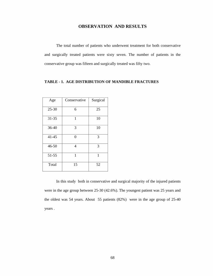
OBSERVATION AND RESULTS
The total number of patients who underwent treatment for both conservative
and surgically treated patients were sixty seven. The number of patients in the
conservative group was fifteen and surgically treated was fifty two.
TABLE - 1. AGE DISTRIBUTION OF MANDIBLE FRACTURES
In this study both in conservative and surgical majority of the injured patients
were in the age group between 25-30 (42.6%). The youngest patient was 25 years and
the oldest was 54 years. About 55 patients (82%) were in the age group of 25-40
years .
Age Conservative Surgical
25-30 6 25
31-35 1 10
36-40 3 10
41-45 0 3
46-50 4 3
51-55 1 1
Total 15 52
69
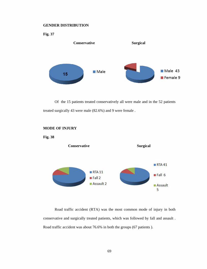
GENDER DISTRIBUTION Fig. 37
Conservative Surgical
Of the 15 patients treated conservatively all were male and in the 52 patients
treated surgically 43 were male (82.6%) and 9 were female .
MODE OF INJURY
Fig. 38
Conservative Surgical
Road traffic accident (RTA) was the most common mode of injury in both
conservative and surgically treated patients, which was followed by fall and assault .
Road traffic accident was about 76.6% in both the groups (67 patients ).
70
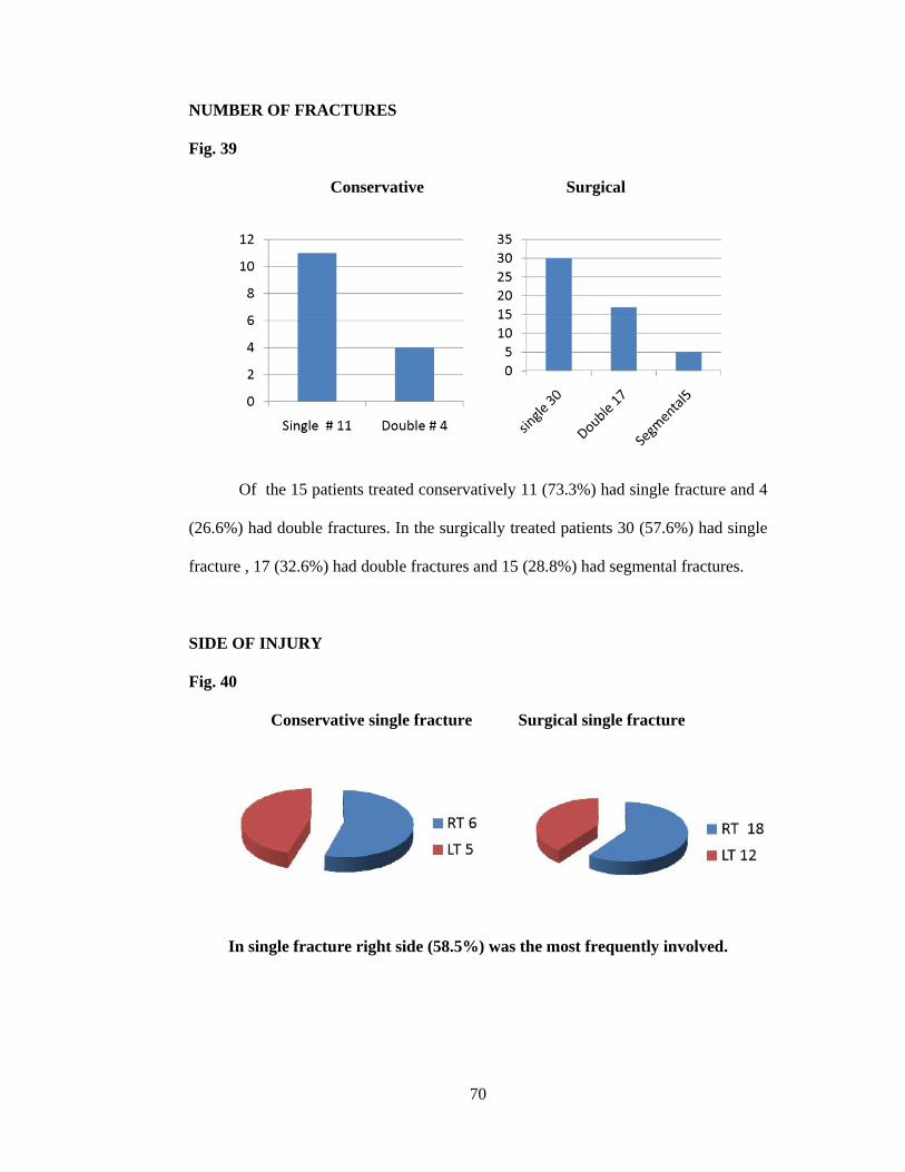
NUMBER OF FRACTURES
Fig. 39
Conservative Surgical
Of the 15 patients treated conservatively 11 (73.3%) had single fracture and 4
(26.6%) had double fractures. In the surgically treated patients 30 (57.6%) had single
fracture , 17 (32.6%) had double fractures and 15 (28.8%) had segmental fractures.
SIDE OF INJURY
Fig. 40
Conservative single fracture Surgical single fracture
In single fracture right side (58.5%) was the most frequently involved.
71
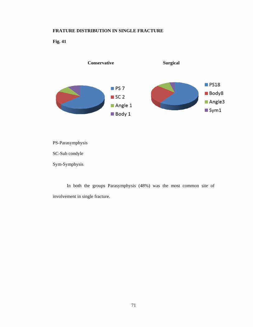
FRATURE DISTRIBUTION IN SINGLE FRACTURE
Fig. 41
Conservative Surgical
PS-Parasymphysis
SC-Sub condyle
Sym-Symphysis
In both the groups Parasymphysis (48%) was the most common site of
involvement in single fracture.
72
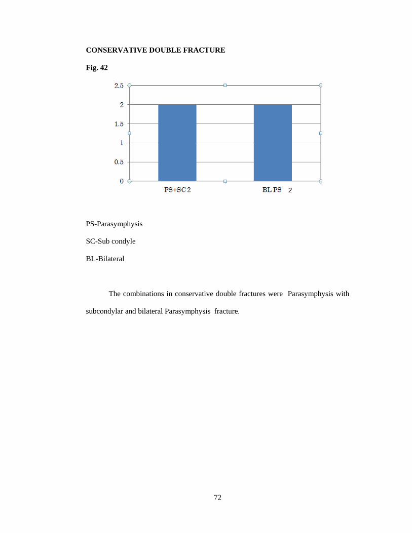
CONSERVATIVE DOUBLE FRACTURE
Fig. 42
PS-Parasymphysis
SC-Sub condyle
BL-Bilateral
The combinations in conservative double fractures were Parasymphysis with
subcondylar and bilateral Parasymphysis fracture.
73
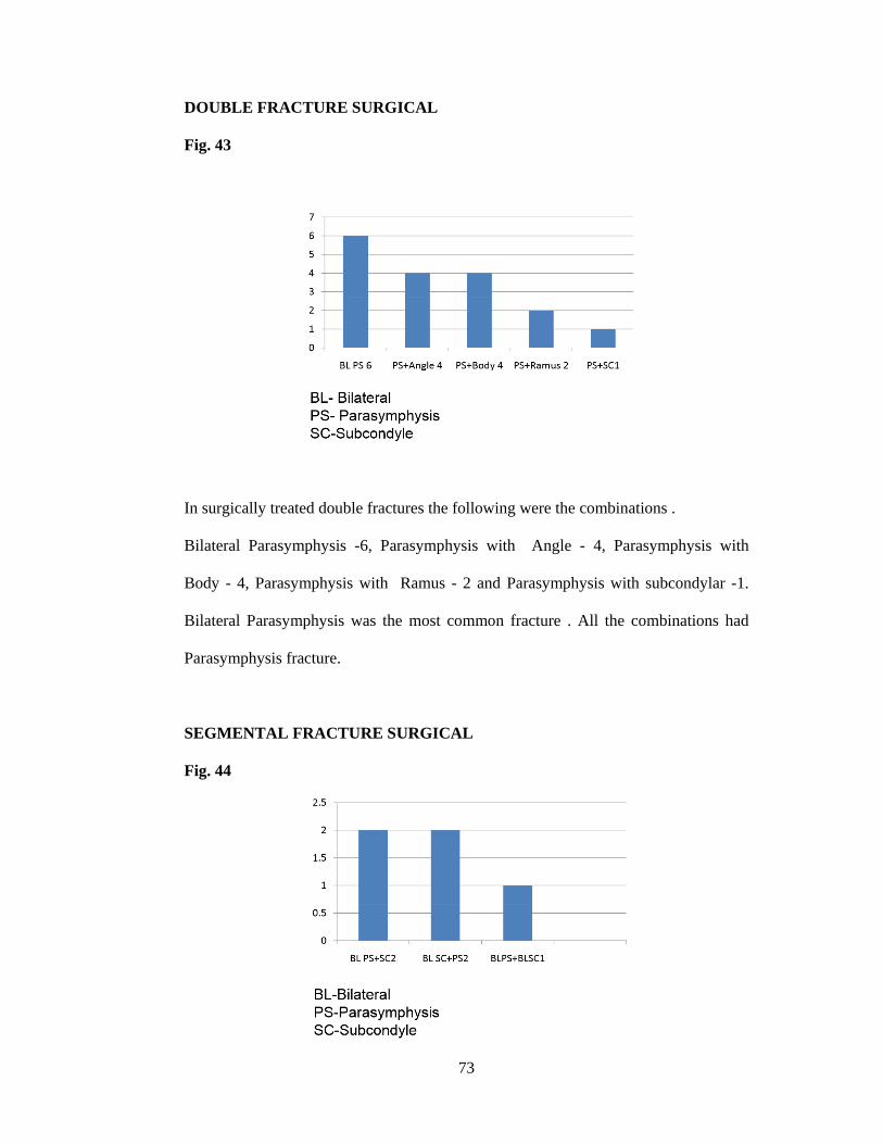
DOUBLE FRACTURE SURGICAL
Fig. 43
In surgically treated double fractures the following were the combinations .
Bilateral Parasymphysis -6, Parasymphysis with Angle - 4, Parasymphysis with
Body - 4, Parasymphysis with Ramus - 2 and Parasymphysis with subcondylar -1.
Bilateral Parasymphysis was the most common fracture . All the combinations had
Parasymphysis fracture.
SEGMENTAL FRACTURE SURGICAL
Fig. 44

74
There were five cases of segmental fracture. Of which bilateral Parasymphysis
with Subcondylar-2, bilateral subcondylar with Parasymphysis -2 and bilateral
Parasymphysis with bilateral subcondylar fracture-1.
TIME INTERVAL BETWEEN INJURY AND PROCEDURE
In the patients treated conservatively, Maxillomandibular fixation (MMF)
done within 24-48 hours. In the surgically treated patients , operated in an average
period of 7 days.
SURGICAL APPROACH
Fig. 45
Out of the 52 patients treated surgically, 43 patients underwent intraoral
approach ,3 patients underwent extra oral approach ( Risdon approach) and 6 patients
underwent both the approaches.
75
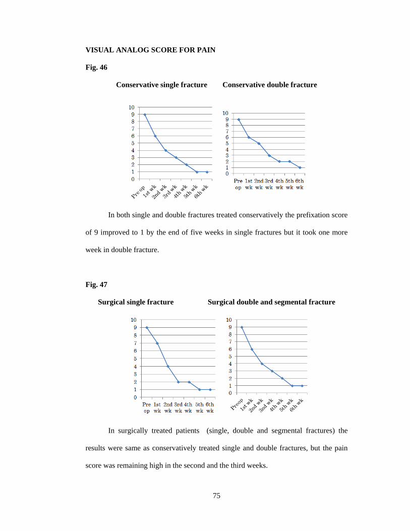
VISUAL ANALOG SCORE FOR PAIN
Fig. 46
Conservative single fracture Conservative double fracture
In both single and double fractures treated conservatively the prefixation score
of 9 improved to 1 by the end of five weeks in single fractures but it took one more
week in double fracture.
Fig. 47
Surgical single fracture Surgical double and segmental fracture
In surgically treated patients (single, double and segmental fractures) the
results were same as conservatively treated single and double fractures, but the pain
score was remaining high in the second and the third weeks.
76
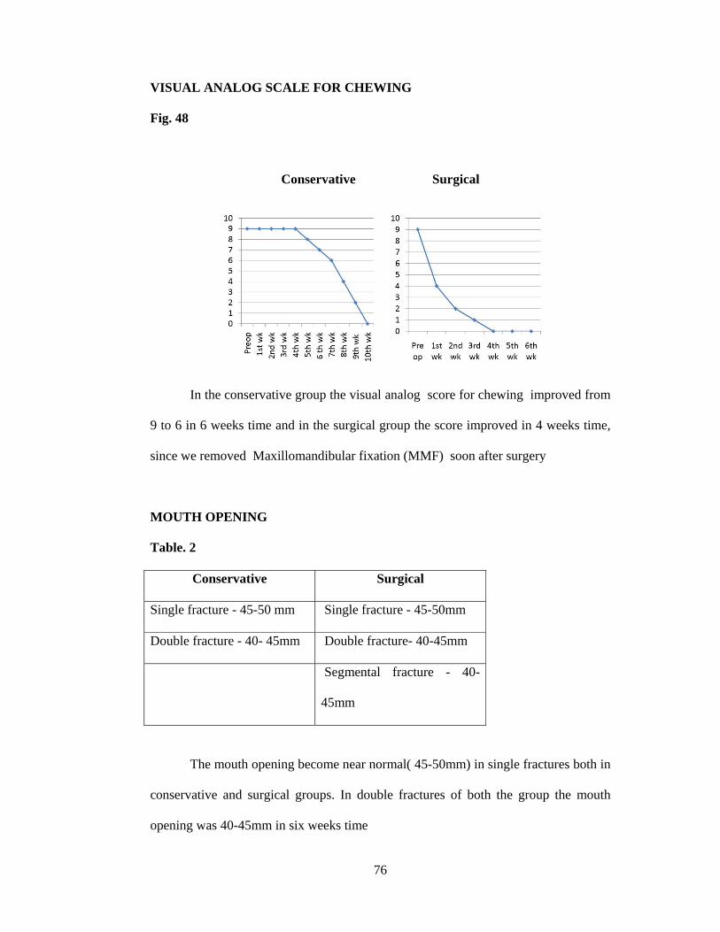
VISUAL ANALOG SCALE FOR CHEWING
Fig. 48
Conservative Surgical
In the conservative group the visual analog score for chewing improved from
9 to 6 in 6 weeks time and in the surgical group the score improved in 4 weeks time,
since we removed Maxillomandibular fixation (MMF) soon after surgery
MOUTH OPENING
Table. 2
Conservative Surgical
Single fracture - 45-50 mm Single fracture - 45-50mm
Double fracture - 40- 45mm Double fracture- 40-45mm
Segmental fracture - 40-
45mm
The mouth opening become near normal( 45-50mm) in single fractures both in
conservative and surgical groups. In double fractures of both the group the mouth
opening was 40-45mm in six weeks time
77
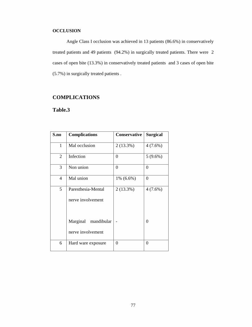
OCCLUSION
Angle Class I occlusion was achieved in 13 patients (86.6%) in conservatively
treated patients and 49 patients (94.2%) in surgically treated patients. There were 2
cases of open bite (13.3%) in conservatively treated patients and 3 cases of open bite
(5.7%) in surgically treated patients .
COMPLICATIONS
Table.3
S.no Complications Conservative Surgical
1 Mal occlusion 2 (13.3%) 4 (7.6%)
2 Infection 0 5 (9.6%)
3 Non union 0 0
4 Mal union 1% (6.6%) 0
5 Paresthesia-Mental
nerve involvement
Marginal mandibular
nerve involvement
2 (13.3%)
-
4 (7.6%)
0
6 Hard ware exposure 0 0
78
DISCUSSION
The mandible although considered the heaviest and the strongest facial bone,
is more prone for fractures because it is an open arch, located in the lower portion of
the face and atrophies with age. Facial injuries not only involves soft tissues but also
damages the bone, leading to fractures. Mandible is connected by strong muscles for
various functions .They act as a splint and give protection to the mandible, on the
other hand these powerful muscles can cause massive displacement of the fracture
fragments.68
The human face constitutes the first contact point in several human
interactions thus, injuries and mutilation of the facial structures may have a disastrous
influence on the affected person.84 Knowledge of the dentition is thus an absolute
prerequisite for the proper treatment of mandible fractures. Fractures of the mandible
invariably produce malocclusion if not treated properly.
The most common facial fractures were the mandible (61%), followed by the
maxilla (46%), the zygoma (27%) and the nasal bones (19.5%).57,58
Road traffic injury was the most common mode of injury in our study
(76.6%) followed by fall and assault. Adekeye has reported that 74% of mandibular
fractures were due to road traffic accidents.1,58 This was also reported by Subhashraj
et al in a study done in South Indian city.71 The mechanism of hyperextension and
hyper flexion of the head in traffic accidents makes it more vulnerable to fracture 34.
79
Males are predominantly involved in mandibular fractures 43,52,74. This male
predominance may be due to the greater mobility of the male and their aggressive
behavior. In our study we found that the age group between 25-30 years was the
most commonly involved . This was supported by Ajmal et al 3 and Wimon
Sirimaharaj et al 70.
There were 61.6% of single mandibular fractures and 40.6% of multiple
mandibular fractures, with an average of 1.34 fractures per person. This is similar to that
of Sirimaharaj et al.70 who reported 1.4 fractures per person . Ajmal et al 3 reported
1.5 fractures per person.
Parasymphyseal fractures were the most common fractures in our study
followed by body and angle. 52 Among double fractures the most common combination
is bilateral Parasymphysis. In segmental fractures, bilateral Parasymphysis fracture
was the most common one. Right side involvement was common. Ajmal et al 3also
reported Parasymphyseal fractures were the most frequently involved followed by
body and angle .This was also supported by Mittal et al 52 study.
Deranged occlusion followed by bony deformity was the commonest mode of
clinical presentation .This finding was supported by Laurentjoye M et al.46
All the Parasymphysis ,Symphysis and body fractures were approached intra
orally. Extra oral approach was used for angle fractures. Care was taken not to injure
the mental nerve during intraoral and marginal mandibular nerve during Risdon
approach.
80
In our study, undisplaced fractures, condylar and subcondylar fractures were
treated with Maxillomandibular fixation (MMF). with good functional results as
comparable with Ghodke et al.31
Out of 67 patients 15 ( 22.3%) underwent conservative treatment with
eyelets ,arch bars and Maxillomandibular fixation (MMF). The duration of MMF was
4-6 weeks in adults, 2-3 weeks in condylar fractures 4. Benjamin et al 10 study from
Nigeria have also reported the usage of arch bars and eyelets with same results.
The average recommended period of immobilization of fractured mandible is
4-6 weeks.27,43,57Although this is only empirical, it is usually influenced by several
factors such as age of patient, type, number and severity of fracture, presence or
otherwise of retained teeth in fracture line, and presence or absence of infection
amongst others.52
In both the conservative & surgical single fracture patients , the visual analog
score - pre operative pain score of 9 has come down to 1 during 5th week.
In surgical group the pain score was remaining high in the 1st week due to
surgical trauma ,then it has reduced to 2 during 3rd week due to stability of fixation.
In surgical double fracture the pre operative pain score of 9 has come down to 1 in 5
weeks . But it took 1 more week for the conservative double fracture to come down to
one .
81
In conservative group the pre operative chewing score improved from 9 to 0
in ten weeks. In the surgical group it improved from 9 to 0 in 4-6 weeks . After
removal of the MMF (6 weeks) in the conservative group and in the 3rd post
operative week in surgical group, patients were encouraged to do early physiotherapy.
They had impairment in speech also in conservative group. At the end of 3 months
none of the patients had mastication and speech problem , which was comparable
with Shivani et al.68
The average mouth opening was 41.5 mm in the conservatively treated group
and 47 mm in the surgically treated group. This was probably due to the TMJ
dysfunction in the conservatively treated group in whom MMF was retained for 4-5
weeks . This was comparable with studies done by Amarathunga NA4 and
Cawood et al 13 . This probably due to the muscle disuse atrophy and scarring in the
fracture site following tissue disruption and haematoma formation.69 Near normal
opening in the surgical group due to MMF removal after surgery and early
mobilization.
There was weight loss, air way related problem , difficulty in phonation and
poor oral hygiene in the conservatively treated group. Weight gain and good oral
hygiene was seen in the surgically treated patients. This study was similar to that of
Brown.J.S. et al.12 who demonstrated the advantages of miniplate
osteosynthesis over intermaxillary fixation in management of fractured
mandible. The post operative function is improved and there was weight gain .
Patient treated with intermaxillary fixation have restricted airway .
82
There was weight loss during the first postoperative week in surgically treated
patients. This was probably due to the poor intake of proper diet due to surgical
trauma.
Complications
Two patients (13.3%) had malocclusion in the conservative group ,which
was noticed in the first review and they were subjected to open reduction . There was
malocclusion in four patients (7.6%) who were treated surgically which were less
when compared with the Benjamin et al 10study. All the four patients were subjected
to redo and occlusion was achieved.
There were five cases of infection ( 9.6%) in the operated group which were
treated with higher antibiotics and the implant was retained till the fracture union .
Implant removal was done in all these five patients after the fracture union . The
infection rate was little higher when compared to Ugboko et al.57 who had 8.1%.
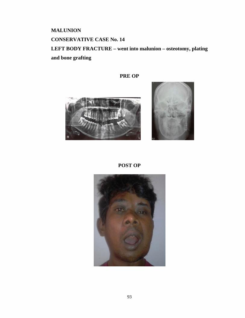
One patient who was treated conservatively developed malunion and it was
corrected with osteotomy ,bone graft and plate osteosynthesis.
The neurological deficit in the operated group was 4 (7.6%) and the
conservative group was 2 which was comparable to the study done by Okoturo and
Benjamin et al.,10 (7.1%) and Cawood 13 (8%) which improved in 6-8 weeks time .
This deficit was not due to the surgical procedure but related to the nature of injury.
83
CONCLUSION
The treatment of mandible fractures requires adequate fracture reduction and
stabilization through a closed or open technique. Success relies on the restoration of
normal dental occlusion and bony union. The treatment chosen may differ as there are
many factors like cost of treatment, affordability by the patient, feasibility in the hospital,
doctor’s decision and skill, and patient’s willingness to avail the treatment advised; all of
which may vary from one country to another.
This study is not comparing the results of closed reduction and open reduction
techniques. It is an analysis of the mandibular fracture demographic variables and
outcome of the management adopted in patients presented to our department. The
results of the patients treated both closed and open methods were same as reported in
the literature.
In single fracture, the results both in the surgical and conservative groups are
equal.
Conservative group took longer time for improvement than surgical group, since
we maintain MMF for 4-6 Weeks.
In double and segmental fracture, surgical management had good outcome with
double plate fixation.
84
Intra osseous wiring prevented distraction; however, it does not provide
sustained inter fragmentary compression.69 This has led to increased preference for
open reduction and internal fixation with miniplates. This has helped reduce
malocclusion, nonunion, improved mouth opening, speech, decreased weight loss, and
increased the ability for patients to return to work earlier.69
High levels of success can still be achieved using available materials in the form
of arch bars, eyelets and wire osteosynthesis in the treatment of mandibular fractures
using either the closed or open reduction technique in resource poor settings despite the
advent of miniplate osteosynthesis.
85
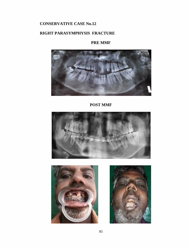
CONSERVATIVE CASE No.12
RIGHT PARASYMPHYSIS FRACTURE
PRE MMF
POST MMF
86
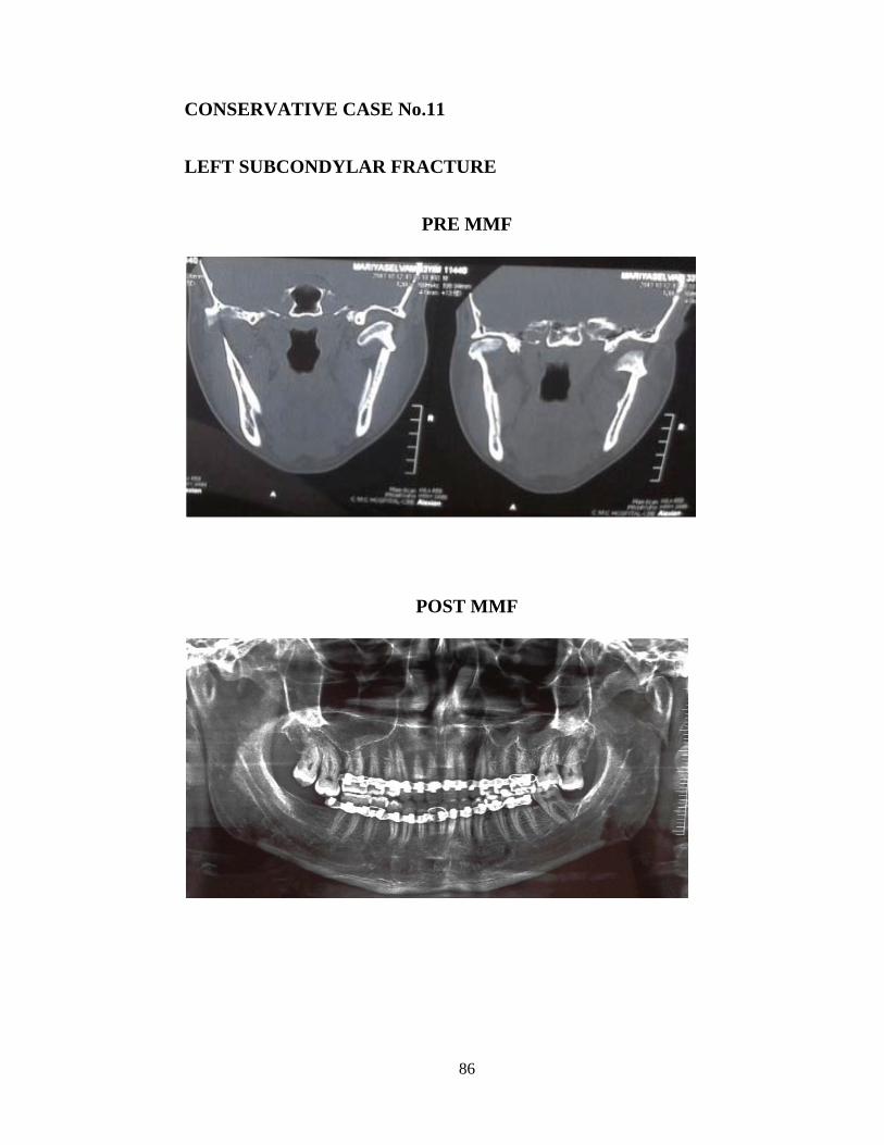
CONSERVATIVE CASE No.11
LEFT SUBCONDYLAR FRACTURE
PRE MMF
POST MMF
87
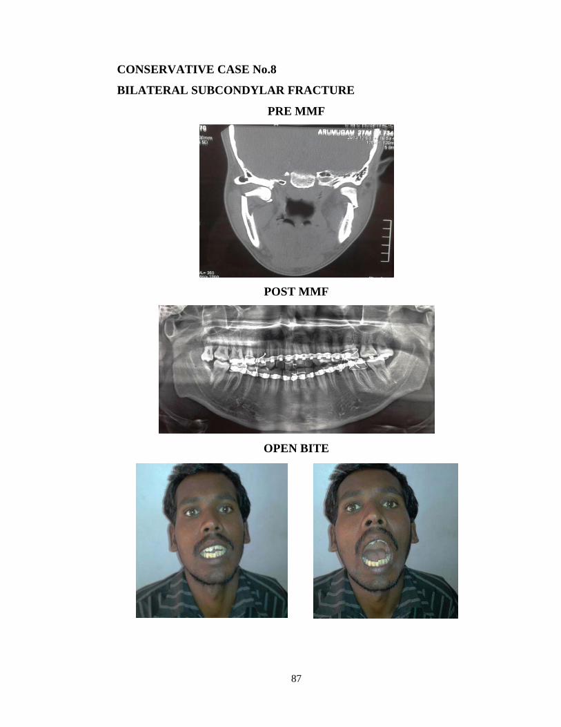
CONSERVATIVE CASE No.8
BILATERAL SUBCONDYLAR FRACTURE
PRE MMF
POST MMF
OPEN BITE
S
L
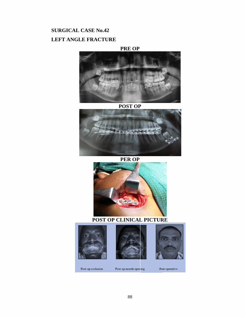
SURGICA
LEFT ANG
AL CASE N
GLE FRA
PO
No.42
ACTURE
OST OP C
88
PRE OP
POST OP
PER OP
CLINICAL
P
L PICTUR
RE
89
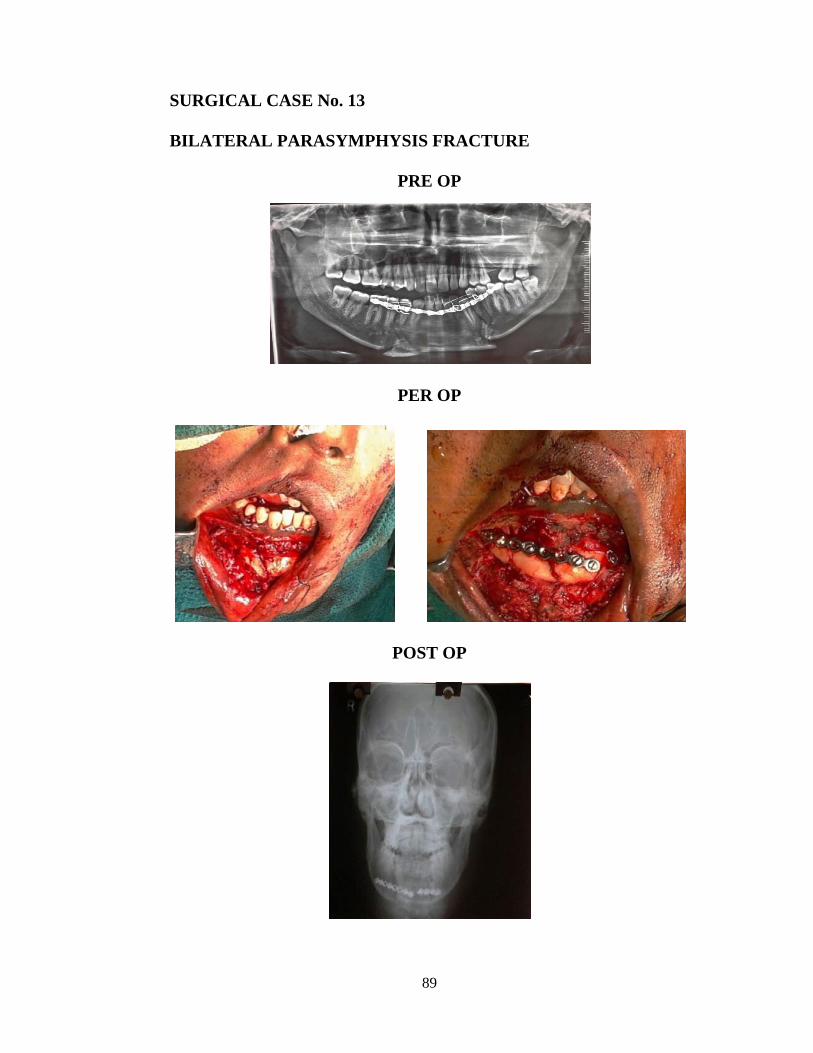
SURGICAL CASE No. 13
BILATERAL PARASYMPHYSIS FRACTURE
PRE OP
PER OP
POST OP
90
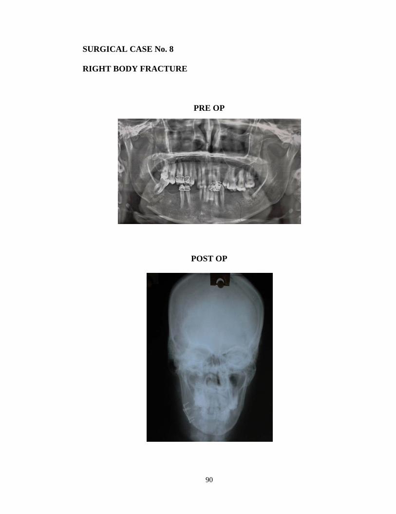
SURGICAL CASE No. 8
RIGHT BODY FRACTURE
PRE OP
POST OP
91
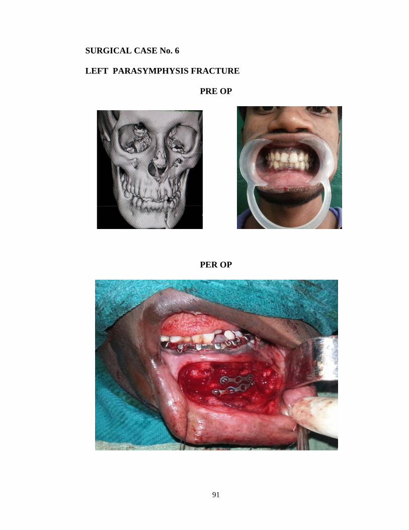
SURGICAL CASE No. 6
LEFT PARASYMPHYSIS FRACTURE
PRE OP
PER OP
C
S
IN
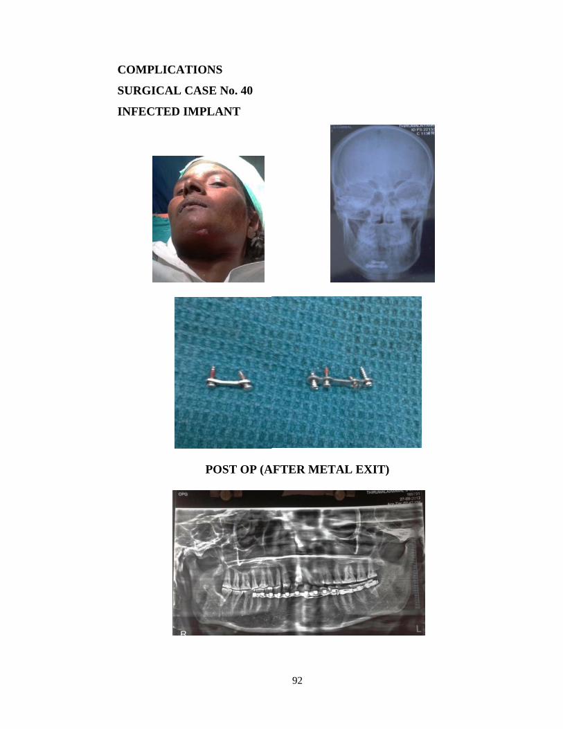
COMPLIC
SURGICA
NFECTED
CATIONS
AL CASE N
D IMPLA
PO
No. 40
ANT
ST OP (A
92
AFTER ME
ETAL EXXIT)
93
MALUNION
CONSERVATIVE CASE No. 14
LEFT BODY FRACTURE – went into malunion – osteotomy, plating
and bone grafting
PRE OP
POST OP
94
BIBLIOGRAPHY
1. Adekeye EO. The pattern of fractures of the facial skeleton in Kaduna, Nigeria. A
survey of 1,447 cases. Oral Surg Oral Med Oral Pathol 1980;49:491‑5.
2. Adewole RA. An audit of mandibular fracture treatment methods at a Military
Base Hospital Yaba, Lagos Nigeria: A 5‑year retrospective study. Nig J Clinical
Pract 2001;4:1‑4.
3. Ajmal S, Khan MA, Jadoon H, Malik SA. Management protocol of mandibular
fractures at Pakistan Institute Of Medical Sciences, Islamabad, Pakistan. J Ayub
Med Coll Abbottabad 2007; 19(3): 51-55.
4. Amartunga NA. Mouth opening after release of maxillo- mandibular
fixationinfracture patients. J Oral Maxillofac surg 1987;45:383-85
5. Andres. J.J. Gonzalez et al., “Evaluation of Trismus after treatment of mandibular
fractures”. J-Oral-Maxillofac-Surg. 1992; 50 : 223-228.
6. Ayman Chritah, Stewert .K. Lazaro, Julius. R. Berger.,“Transoral 2.0mm locking
miiniplate fixation of mandibular fractures plus I week of maxsillomandibular
fixation : a prospective study”. Journal of Oral and Maxillofacial Surgery.
2005;63: 1737-1741.
7. Bank P. Killey’s fractures of the mandible. 4th ed. Oxford: Wright
Butterworth‑Heinemann Ltd; 1991. p. 192‑216.
8. Barrera JE, Stephen GB, Hassan HR. Fractures of the mandibular angle. e‑
medicine.com, Inc. 2006;1‑12.
9. Barton JR. A systemic bandage for fractures of the lower jaw. Am Med
Recorder Phila. 1819;2:153.
95
10. Benjamin A, Sara KE, Olushola AI. Analysis of complication of mandibular
fracture. Afr J Trauma 2014;3:24-9
11. Bjorn Johnston, Leonard Krukmenov.,“Minplate osteosynthesis in infected
mandibular fractures”. Journal of Cranio Max Facial Surgery 1998; 16:22-27.
12. Brown. J.S et al., “The fate of miniplates in facial trauma and orthognathic
surgery: a restrospecitve study”. Br. J Oral Maxillofac Surg 1989. 27 (4); 306 -
315.
13. Cawood JI. Small plate osteosynthesis of mandibular fractures. Br J Oral
Maxillofac Surg 1985;23:77‑91.
14. Chu L, Gussack GS, Muller T. A treatment protocol for mandibular fractures. J
Trauma 1994;36:48‑52.
15. Champy M, Loddé JP, Schmitt Spiessl B. Rigid internal fixation of fractures of
the lower jaw. Reconstr Surg Traumatol. 1972;13:124-40. [Medline].
16. Champy.M, et al., “Mandibular osteosynthesis by miniature plates via a buccal
approach”. J Oral Maxillofac Surg. 1978; 6: 14.
17. Cienfuegos R, Cornelius CP, Ellis 3rd E, Kushner G. CMF Mandible - Diagnosis
- AO Surgery reference. AO Foundation.
18. Danis : Theory and practice of Osteosynthesis Paris, Masson, 1949. 7. 42. Kruger:
“Text book of Oral Surgery”, Mosby; 1964.
19. Dimitroulis. G., “Management of fractured mandible without the use of
intermaxillary fixation”. J Oral Maxillofac Surg. 2002;60: 1435
20. Edward Ellis et al., “Treatment of mandibular angle fractures using two 2.4 mm
dynamic compression plates”. J Oral Maxillofac Surg. 1993 ; 51: 969-973.
21. Edward Ellis et al., “Treatment of mandibular angle fractures using two
noncompression miniplates”. J Oral Maxillofac-Surg. 1994 ;52(10): 1032-6
96
22. Edward Ellis III et al., “Use of a 2 mm locking plate/screw system for mandibular
fracture surgery” .J Oral Maxillofac Surg. 2002: 60 : 642-645
23. Edwards T.J et al., “The relationship between fracture severity and complication
rate in miniplate osteosynthesis of mandibular fractures”. Br J Plast
Surg.1994;47(5):310-1.
24. Ellis E 3rd, Moos KF, el-Attar A. Ten years of mandibular fractures: an analysis
of 2,137 cases. Oral Surg Oral Med Oral Pathol. Feb 1985;59(2):120-9.
[Medline].
25. Ellis III et a!., “Treatment consideration for comminuted mandibular fractures”. J
Oral Maxillofac Surg. 2003; 61: 861-70
26. Feller. K et al., “Analysis of complications in fractures of the mandibular angle —
a study with finite element computation and evaluation of data of 277 patients”. J.
Cranio Maxillo Fac Surg 2003; Oct 31 — 290-5 .
27. Fonseca RJ, Bruce R, Walker R. Trauma. Oral and Maxillofacial Surgery.
Washington manual. Vol. 1. 2001. p. 19‑34.
28. Fordyce.A.M et al., “Intermaxillary fixation is not usually necessary to reduce
mandibular fractures”. J Oral Maxillofac Surg. 1997 : 52-57
29. Fuselier et al., “Do mandibular third molar alter the risk of Angle fracture”. J
Oral Maxillofac Surg. 2002; 60: 514-518.
30. Gerbino.G et al., “Rigid fixation with teeth in the line of mandibular fracture”.
Int J Oral Maxillofac. Surg. 1997 ; 26: 182-186.
31. Ghodke MH, Bhoyar SC, Shah SV. Prevalence of mandibular fractures reported
at C.S.M.S.S Dental College, Aurangabad from February 2008 to September
2009. J IntSoc Prevent Communit Dent 2013;3:51-8.
97
32. Gregory. S. Tate et al.,: “Bite forces in patients treated for mandibular angle
fractures : Implications for fixation- recommendations.” J Oral Maxillofac Surg.
1994; 52 : 734-736.
33. Grunst. J., “Interference with bone blood supply through plating, of intact bone.
Current concepts in the internal fixation of fractures.” 1980. .
34. Holt GR. Maxillofacial trauma. In: Cummings CW, Krause CJ, editors.
Otolaryngology‑Head and Neck Surgery. St. Louis: C.V. Mosby; 1986. p.
313‑44.
35. James RB, Fredrickson C, Kent JN. Prospective study of mandibular fractures. J
Oral Surg 1981;39:275‑81
36. J. Tams, J. P. Vanloon, F.R. Rozeno: “A three dimensional study of loads across
the fracture for different fracture sites of the mandible”. British Journal of 0ral and
Maxillofacial Surgery. 1996;34: 400-405.
37. J. Tams, J. P. Vanloon: “A three dimensional study of bending and torsion
moments for different fracture sites in the mandible: an in vitro study”.
International Journal of Maxillofacial Surgery 1997: 26: 383:388.
38. J.P. Hayter, I. Cawood., “ The functional case for miniplates in maxillofacial
surgery”. International Journal of Oral and Maxillofacial Surgery. 1993;22: 91-96.
39. J.S. Brown, N. Grew , C. Taylor: “Intermaxillary fixation compared to miniplate
osteosynthesis in the management of the fractured mandible: an audit”. British
Journal of Oral and Maxillofacial Surgery. 1991; 29:308-311
40. James W. Sikes et al, : “Comparison of fixation strength of locking head and
conventional screws, in fracture and reconstruction models”. J Oral
Maxillofac Surg. 1998 ; 56: 468- 473
98
41. Jeffrey. C. Posnick et al., “Pediatric facial fractures”. J OraI Maxillofac
Surg.1993; 51: 836- 44
42. K.E. KahnBerg, A. Ridell., “Bone plate fixation of mandibular fractures”
International Journal of Oral Surgery 1980; 9:267-273. .
43. Kamali U, Pohchi A. Mandibular fracture at HUSM: A 5‑year retrospective
study. Arch Orofacial Sci 2009;4:33‑5.
44. Kazanjian VH, Converse JM. Surgical treatment of facial injuries. 3rd ed.
Baltimore: The Williams and Wilkins Company; 1980. p. 66‑8.
45. Kawai. T et al., “Radiographic changes during bone healing after mandibular
fractures.” British Journal of oral and maxillofacial surgery. 1997 ; 35 : 312-318.
46. Laurentjoye M et al, Mandibular Fractures With Michelet Technique: Manual
Fracture Reduction Without Arch Bars. J Oral Maxillofac Surg. 2009;67:2374-2379.
47. Malik NA. Textbook of Oral and Maxillofacial Surgery. In: 2nd Ed. Applied
surgical anatomy of the mandible and classification of mandibular fractures. New
Delhi: Jaypee Brothers Medical Publishers; 2008. p. 378‑85.
48. Marisa Aparecida Cabrini Gabrielli, Mario Fransisco Real Gabrielli., “ Fixation of
mandibular fracture with 2.0mm miniplates: A review of 191 cases” Journal of
Oral and Maxillofacial Surgery. 2003; 61:430-436.
49. Mathod et al., “Non union of mandible - An analysis of contributing factors”. J
Oral Maxillofac Surg. 2000; 58 :746 -52.
50. Michelet et al., “Osteosynthesis with miniaturized screwed plates in maxillofacial
surgery”. J Oral Maxillofac Surg. 1973; 1: 79.
51. Mitchell. M. Rubin et al., : “Morbidity associated with incompletely erupted third
molar in the line of mandibular fracture. J Oral Maxillofac Surg. 1990; 1045-
1047. :
99
52. Mittal G, Mittal SR. Mandibular fractures at veer Chandra Singh Garhwali
Government Medical Science and Research Institute, Garhwal region, Uttarakhand,
India: A retrospective study. Ann Med Health Sci Res 2013;3:161-5.
53. Monty Reitzik and Willem Schrool., “Bone repair in the Mandible: a histologic
and biometric comparison between rigid and semi rigid fixation”. Journal of Oral
and Maxillofacial Surgery 1983; 215-218
54. Moon H. Mechanical appliances for treatment of fracture of the jaws. Br J Dent
Sci. 1874;17:303.
55. Nakamura - S et al., “Complications of miniplate osteosynthesis for mandibular
fractures”. J Oral Maxillofac Surg. 1994; 52(3): 233-8.
56. Nicholas Gerard et al., “Modified technique for adapting a mandibular angle
superior border plate”. J Oral Maxillofac Surg.1995; 53:220-21.
57. Oginni FO, Ugboko VI, Ogundipe O, Adegbehingbe O. Motorcycle‑related
maxillofacial injuries among Nigerian intracity road users. J Oral Maxillofac Surg
2006;64:56‑62. .
58. Oikarinen VJ, Lindquist C. The frequency of facial bone fractures in patients with
multiple injuries sustained in traffic accidents. Proc Finn Dent Soc 1975;71:53‑7.
59. R, Jaeger JH, Muster D. Mandibular osteosynthesis by miniature screwed plates
via a buccal approach. J Maxillofac Surg. Feb 1978;6(1):14-21. [Medline].
60. R.A Loukota, J.C. Shelton., “Mechanical analysis of maxillofacial miniplates”.
British Journal of Oral and Macillofacial Surgery. 1995; 33:174-179.
61. Gutwald , Brian Alpert , Rainer Schmelzeisen., “Principle and stability of
locking plates”. Keijo J Med March 2003; 52(l):21-24.
62. Raymond. J. Fonseca et al., “Oral and Maxillofacial trauma” 1997; 474, 1275 pg.
100
63. Reza Bolourian et al., “Transoral 2mm miniplate fixation of mandibular fractures
plus 2 weeks of maxillomandibular fixation: a prospective study”. J Oral
Maxillofac Surg. 2002; 60: 167- 170.
64. Richard H. Haug et al., “A microplate and screw technique for intra oral open
reduction of mandibular angle fractures”. J Oral Maxillofac Surg. 1995; 53:218-
219.
65. Richard. H. Haug., “The effects of screw number and length in two methods of
tension band plating”. J Oral Maxillofac Surg. 1993; 51: 159-62.
66. Robert .A. Rudman et al., “Photo elastic analysis of miniplate osteosynthesis for
mandibular angle fractures”. Oral Surg Oral Med Oral-Patho-Oral Radio-endo.
1997;84: 129-36.
67. Schierie. H. P et al., “One or two plate fixation of mandibular angle fractures?” J
Carniomaxillo fac Surg. 1997 25(3): 162-8.
68. Shivani Jain , Shipra Gupta. Titanium Miniplates versus Intraosseous Wires as
Methods of Fixation of Mandibular Fractures: A Clinical Study. Indian J Stomatol
2011;2(4):238-44
69. Sindet et al., “Treatment of mandibular fractures with or without intermaxillary
fixation - a comparative study.” Oral surgery Oral diagnosis. 1992; 3: 37-44.
70. Sirimaharaj W, Pyungtanasup K. The Epidemiology of mandibular fractures treated
at Chiang Mai University Hospital: A review of 198 cases. J Med Assoc Thai
2008; 91 (6): 868-74.
71. Subhashraj K, Ramkumar S, Ravindran C. Pattern of mandibular fractures in
Chennai, India. Br J Oral Maxillofac Surg 2008;46:126‑7.
72. Smith. W. P., “Delayed miniplate osteosynthesis for mandibular fractures”. Br J
Oral Maxillofac Surg. 1991; 29(2): 73-6.
101
73. Spiessl. B., “Internal fixation of the mandible : A manual of AO/ASIF principles.”
1989: Pg 5 - 37
74. Thoma. K. H., “Treatment of jaw fractures, past and present”. Oral Surgery
anaesthesia and Hosp. Dent. Ser. 1959; 17: 30.
75. Thomas KH. Historical review of methods advocated for treatment of jaw
fractures with ten commandments for modern fracture treatment. Am J Orthodont
Oral Surg. 1944;30:399.
76. Thomas. A. Chiodo et al., “Failure strength of 2.0 locking versus 2.0
conventional Synthes mandibular plates: A laboratory model”. J- Oral and
Maxillofacial Surgery. 2006 ; 64 : 1475-1479
77. Valentino et al., “Supplemental Maxillomandibular fixation with miniplate
osteosynthesis”. Otolaryngol-Head-Neck-Surg. 1995; 112(2): 215-20.
78. Vivek Shetty et al., “Fracture line stability as a function of the internal fixation
system : An in vitro comparison using a mandibular angle fracture model”. J Oral
Maxillo fac Surg. 1995; 53: 791-802.
79. Wagner. W. F et al., “Morbidity associated with extra oral open red reduction of
mandibular fractures”. Journal of Oral surgery. 1979; 37(2): 97-100.
80. Wald. R. M et al., “The trans oral treatment of mandibular fractures using non
compression miniplates : A prospective study”. Annals of plastic surgery. 1988;
20(5): 409-13.
81. Walz. C et at., “Miniplate osteosynthesis of mandibular fracture in local
anesthesia -indication and outcome in 316 patients”. Fortschr-Kiefer-
Gesichtschir. 1996; 41: 133-5.
82. Wolfgang Heidmann., “Analysis of the osseous metal interface of drill free
screws and self tapping screws”. J. Cranio maxillofacial surg. 2001; 29: 69-.
102
83. Tuovinen., “A retrospective analysis of 279 patients with isolated
mandibular fractures treated with titanium miniplates”. J Oral Maxillofac Surg.
1994; 52(9): 931-6.
84. Zargar M, Khaji A, Karbakhsh M, Zarei MR. Epidemiologystudy of facial
injuries during a 13 month of trauma registry in Tehran. Indian J Med Sci
2004;58:109‑14.
103
PROFORMA
1. NAME
2. AGE/SEX
3. PS NO
4. ADDRESS
5. MOBILE NO
6. OCCUPATION
7. SOCIOECONOMIC STATUS
8. DATE OF INJURY
9. MODE OF INJURY
10. DATE OF SURGERY/ MMF APPLICATION
11. OTHER CO MORBIDITIES
12. PREOP OPG, CT FACIAL BONES
13. SITE AND NO. OF FRACTURES
14. METHOD OF FIXATION - CLOSED/OPEN
15. TIME AT WHICH ORAL FLUIDS/DIET STARTED
16. IMMEDIATE POST OPERATIVE PERIOD
17. POST OP OPG
18. TIME AT WHICH MOBILISATION STARTED
19. REVIEW EVERY WEEK FOR THE FIRST SIX WEEK AFTER SURGERY
20. THEN MONTHLY FOR THREE MONTHS
21. END OF THIRD MONTH
22. ASSESSMENT
104
PAIN
MOUTH OPENING
CHEWING
OCCLUSION STATUS
ANY POST OP INFECTION
WEIGHT LOSS
NEUROLOGICAL DISTURBANCE

105
MASTER CHART - CONSERVATIVE
S .N0 NAME AGE SEX DIAGNOSIS TIME FROM INJURY PROCEDURE
MODE OF INJURY COMPLICATION OCCLUSION
TO TREATMENT
1 Manikandan 48 m RT Parasymphysis 2 days Upper and lower eyelet RTA CLASS I
2 Kittan 50 M RT Body ,RT Subcondylar 3 days Upper and lower arch bar RAT TRISMUS CLASS I
3 Murugesan 27 m RT Parasymphysis 2 days Upper and lower arch bar RTA CLASS I
4 Prabhu 25 m RT subcondylar 1 day Upper eyeleyt and lower arch bar RTA PARESTHESIA ,TRISMUS CLASS I
5 Kithru sirajudeen 48 m RT Parasymphysis 1 day
Upper eyeleyt and lower arch bar FALL CLASS I
6 Kannan 40 m LT Parasymphysis 1day Upper and lower arch bar FALL CLASS I
7 Chinnaswami 50 m RT Parasymphysis 1days Upper and lower arch bar ASSAULT CLASS I
8 Arumugam 28 m BL Sudcondyjar # 2 days Upper and lower arch bar RTA TRISMUS CLASS I
9 Sundaram 46 m RT Parasymphysis 1 day Upper and lower arch bar RTA CLASS I
10 Veluswami 39 m RT Parasymphysis ,RT Subcondylar 2 days Upper and lower arch bar RTA TRISMUS OPEN BITE
11 Selvam 40 m LT Subcondylar 1 day Upper and lower arch bar RTA TRISMUS CLASS I
12 Murugaraj 46 m LT Parasymphysis 1 day Upper and lower arch bar ASSAULT PARESTHESIA CLASS I
13 Balaji 31 m LT Angle 2 days Upper and lower arch bar RTA OPEN BITE
14 SENTHIL 30 M LT Body 2 days Upper and lower arch bar RTA MALOCCLUSION &MALUNION CLASS I
15 Sabarinathan 28 m RT Parasymphysis, LT Angle 2days Upper and lower arch bar RTA CLASS I
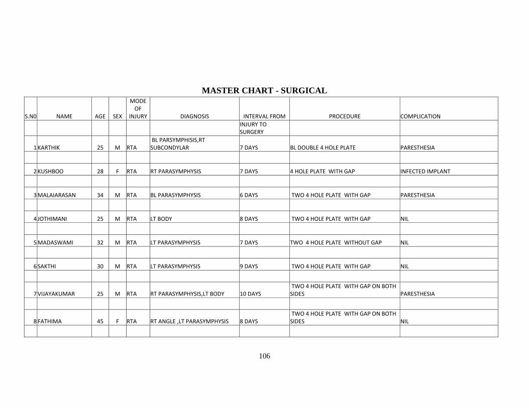
106
MASTER CHART - SURGICAL
S.N0 NAME AGE SEX
MODE OF
INJURY DIAGNOSIS INTERVAL FROM PROCEDURE COMPLICATION
INJURY TO SURGERY
1 KARTHIK 25 M RTA BL PARSYMPHISIS,RT SUBCONDYLAR 7 DAYS BL DOUBLE 4 HOLE PLATE PARESTHESIA
2 KUSHBOO 28 F RTA RT PARASYMPHYSIS 7 DAYS 4 HOLE PLATE WITH GAP INFECTED IMPLANT
3 MALAIARASAN 34 M RTA BL PARASYMPHYSIS 6 DAYS TWO 4 HOLE PLATE WITH GAP PARESTHESIA
4 JOTHIMANI 25 M RTA LT BODY 8 DAYS TWO 4 HOLE PLATE WITH GAP NIL
5 MADASWAMI 32 M RTA LT PARASYMPHYSIS 7 DAYS TWO 4 HOLE PLATE WITHOUT GAP NIL
6 SAKTHI 30 M RTA LT PARASYMPHYSIS 9 DAYS TWO 4 HOLE PLATE WITH GAP NIL
7 VIJAYAKUMAR 25 M RTA RT PARASYMPHYSIS,LT BODY 10 DAYS TWO 4 HOLE PLATE WITH GAP ON BOTH SIDES PARESTHESIA
8 FATHIMA 45 F RTA RT ANGLE ,LT PARASYMPHYSIS 8 DAYS TWO 4 HOLE PLATE WITH GAP ON BOTH SIDES NIL
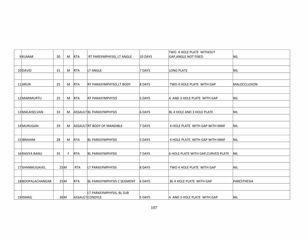
107
9 KUMAR 30 M RTA RT PARSYMPHYSIS, LT ANGLE 10 DAYS TWO 4 HOLE PLATE WITHOUT GAP,ANGLE NOT FIXED NIL
10 DAVID 31 M RTA LT ANGLE 7 DAYS LONG PLATE NIL
11 ARUN 25 M RTA RT PARASYMPHYSIS,LT BODY 8 DAYS TWO 4 HOLE PLATE WITH GAP MALOCCLUSION
12 MARIMURTU 25 M RTA RT PARASYMPHYSIS 5 DAYS 4 AND 3 HOLE PLATE WITH GAP NIL
13 MALAISELVAN 33 M ASSAULT BL PARASYMPHYSIS 6 DAYS BL 4 HOLE AND 3 HOLE PLATE NIL
14 MURUGAN 29 M ASSAULT RT BODY OF MANDIBLE 7 DAYS 4 HOLE PLATE WITH GAP WITH MMF NIL
15 IBRAHIM 28 M RTA BL PARASYMPHYSIS 5 DAYS 4 HOLE PLATE WITH GAP WITH MMF NIL
16 RASIYA BANU 35 F RTA BL PARASYMPHYSIS 7 DAYS 6 HOLE PLATE WITH GAP,CURVED PLATE NIL
17 SHANMUGAVEL 25 M RTA LT PARASYMPHYSIS 8 DAYS TWO 4 HOLE PLATE WITH GAP NIL
18 BOOPALACHANDAR 25 M RTA BL PARASYMPHYSIS C SEGMENT 6 DAYS BL 4 HOLE PLATE WITH GAP PARESTHESIA
19 ISMAIL 36 M ASSAULT LT PARASYMPHYSIS, BL SUB CONDYLE 5 DAYS 4 AND 3 HOLE PLATE WITH GAP NIL
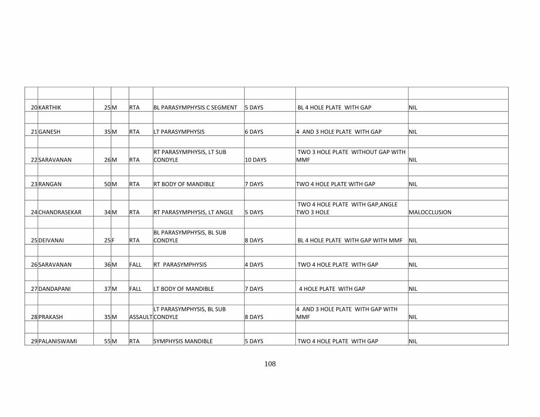
108
20 KARTHIK 25 M RTA BL PARASYMPHYSIS C SEGMENT 5 DAYS BL 4 HOLE PLATE WITH GAP NIL
21 GANESH 35 M RTA LT PARASYMPHYSIS 6 DAYS 4 AND 3 HOLE PLATE WITH GAP NIL
22 SARAVANAN 26 M RTA RT PARASYMPHYSIS, LT SUB CONDYLE 10 DAYS
TWO 3 HOLE PLATE WITHOUT GAP WITH MMF NIL
23 RANGAN 50 M RTA RT BODY OF MANDIBLE 7 DAYS TWO 4 HOLE PLATE WITH GAP NIL
24 CHANDRASEKAR 34 M RTA RT PARASYMPHYSIS, LT ANGLE 5 DAYS TWO 4 HOLE PLATE WITH GAP,ANGLE TWO 3 HOLE MALOCCLUSION
25 DEIVANAI 25 F RTA BL PARASYMPHYSIS, BL SUB CONDYLE 8 DAYS BL 4 HOLE PLATE WITH GAP WITH MMF NIL
26 SARAVANAN 36 M FALL RT PARASYMPHYSIS 4 DAYS TWO 4 HOLE PLATE WITH GAP NIL
27 DANDAPANI 37 M FALL LT BODY OF MANDIBLE 7 DAYS 4 HOLE PLATE WITH GAP NIL
28 PRAKASH 35 M ASSAULT LT PARASYMPHYSIS, BL SUB CONDYLE 8 DAYS
4 AND 3 HOLE PLATE WITH GAP WITH MMF NIL
29 PALANISWAMI 55 M RTA SYMPHYSIS MANDIBLE 5 DAYS TWO 4 HOLE PLATE WITH GAP NIL
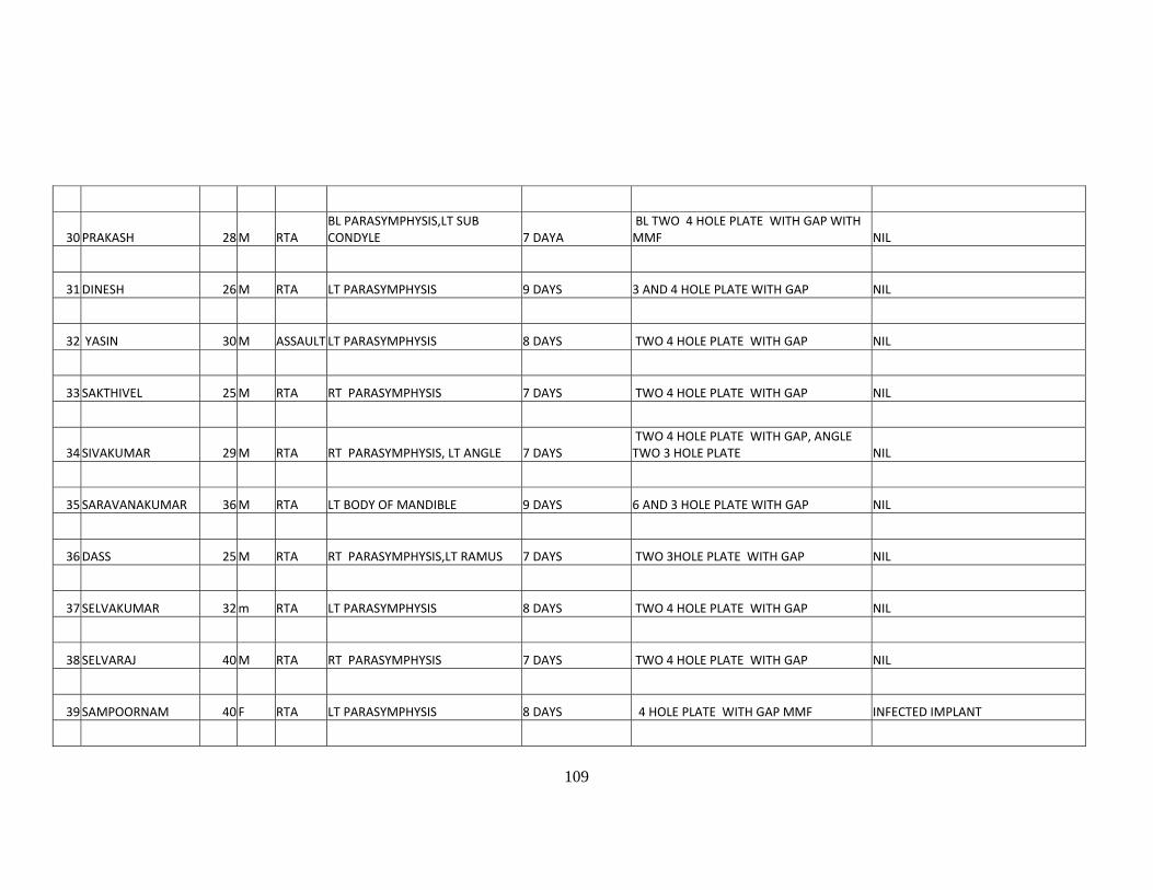
109
30 PRAKASH 28 M RTA BL PARASYMPHYSIS,LT SUB CONDYLE 7 DAYA
BL TWO 4 HOLE PLATE WITH GAP WITH MMF NIL
31 DINESH 26 M RTA LT PARASYMPHYSIS 9 DAYS 3 AND 4 HOLE PLATE WITH GAP NIL
32 YASIN 30 M ASSAULT LT PARASYMPHYSIS 8 DAYS TWO 4 HOLE PLATE WITH GAP NIL
33 SAKTHIVEL 25 M RTA RT PARASYMPHYSIS 7 DAYS TWO 4 HOLE PLATE WITH GAP NIL
34 SIVAKUMAR 29 M RTA RT PARASYMPHYSIS, LT ANGLE 7 DAYS TWO 4 HOLE PLATE WITH GAP, ANGLE TWO 3 HOLE PLATE NIL
35 SARAVANAKUMAR 36 M RTA LT BODY OF MANDIBLE 9 DAYS 6 AND 3 HOLE PLATE WITH GAP NIL
36 DASS 25 M RTA RT PARASYMPHYSIS,LT RAMUS 7 DAYS TWO 3HOLE PLATE WITH GAP NIL
37 SELVAKUMAR 32 m RTA LT PARASYMPHYSIS 8 DAYS TWO 4 HOLE PLATE WITH GAP NIL
38 SELVARAJ 40 M RTA RT PARASYMPHYSIS 7 DAYS TWO 4 HOLE PLATE WITH GAP NIL
39 SAMPOORNAM 40 F RTA LT PARASYMPHYSIS 8 DAYS 4 HOLE PLATE WITH GAP MMF INFECTED IMPLANT
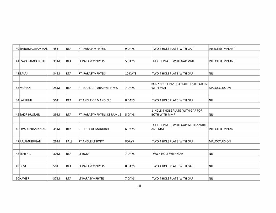
110
40 THIRUMALAIAMMAL 45 F RTA RT PARASYMPHYSIS 9 DAYS TWO 4 HOLE PLATE WITH GAP INFECTED IMPLANT
41 ESWARAMOORTHI 39 M RTA LT PARASYMPHYSIS 5 DAYS 4 HOLE PLATE WITH GAP MMF INFECTED IMPLANT
42 BALAJI 34 M RTA RT PARASYMPHYSIS 10 DAYS TWO 4 HOLE PLATE WITH GAP NIL
43 MOHAN 28 M RTA RT BODY, LT PARASYMPHYSIS 7 DAYS BODY 4HOLE PLATE,3 HOLE PLATE FOR PS WITH MMF MALOCCLUSION
44 LAKSHMI 50 F RTA RT ANGLE OF MANDIBLE 8 DAYS TWO 4 HOLE PLATE WITH GAP NIL
45 ZAKIR HUSSAIN 39 M RTA RT PARASYMPHYSIS, LT RAMUS 5 DAYS SINGLE 4 HOLE PLATE WITH GAP FOR BOTH WITH MMF NIL
46 SIVASUBRAMANIAN 45 M RTA RT BODY OF MANDIBLE 6 DAYS 4 HOLE PLATE WITH GAP WITH SS WIRE AND MMF INFECTED IMPLANT
47 RAJAMURUGAN 26 M FALL RT ANGLE LT BODY 8DAYS TWO 4 HOLE PLATE WITH GAP MALOCCLUSION
48 SENTHIL 30 M RTA LT BODY 7 DAYS TWO 4 HOLE WITH GAP NIL
49 DEVI 50 F RTA LT PARASYMPHYSIS 8 DAYS TWO 4 HOLE PLATE WITH GAP NIL
50 XAVIER 37 M RTA LT PARASYMPHYSIS 7 DAYS TWO 4 HOLE PLATE WITH GAP NIL
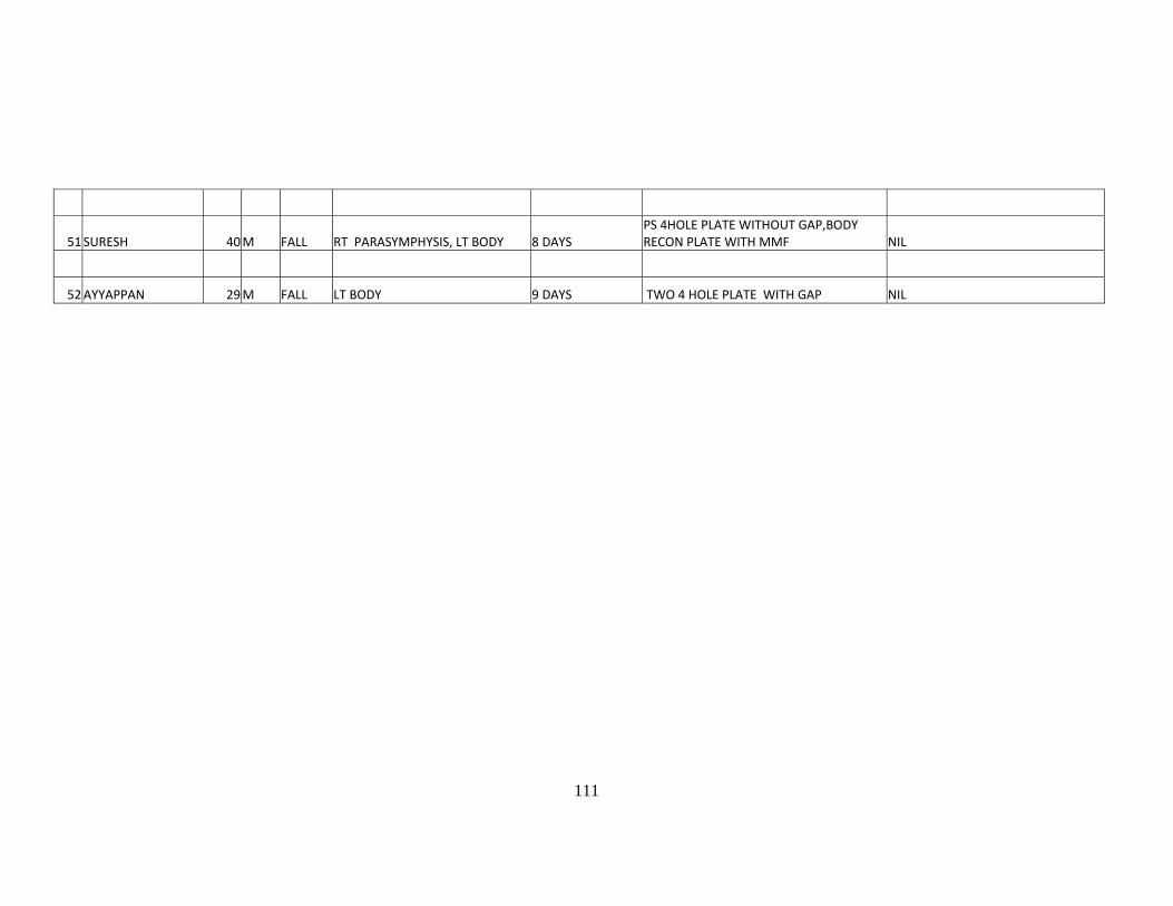
111
51 SURESH 40 M FALL RT PARASYMPHYSIS, LT BODY 8 DAYS PS 4HOLE PLATE WITHOUT GAP,BODY RECON PLATE WITH MMF NIL
52 AYYAPPAN 29 M FALL LT BODY 9 DAYS TWO 4 HOLE PLATE WITH GAP NIL