Embed Size (px)
Citation preview
CLINICAL AND VACCINE IMMUNOLOGY, July 2010, p. 1148–1154 Vol. 17, No. 71556-6811/10/$12.00 doi:10.1128/CVI.00533-09Copyright © 2010, American Society for Microbiology. All Rights Reserved.
Analytical and Biological Considerations in the Measurement ofCell-Associated CCR5 and CXCR4 mRNA and Protein�
D. E. Campbell,* J. P. Lai, N. B. Tustin, E. Riedel, R. Tustin III,J. Taylor, J. Murray, and S. D. Douglas
The Children’s Hospital of Philadelphia Research Institute, Philadelphia, Pennsylvania
Received 28 December 2009/Returned for modification 2 March 2010/Accepted 3 May 2010
The accurate measurement of T cell-associated CC chemokine receptor type 5 (CCR5) and CXC chemokinereceptor type 4 (CXCR4) expression, including expression of CCR5 and CXCR4 mRNA as an immune measureof immunologic response to highly active antiretroviral therapy (HAART) and newer agents, including entryinhibitors, is essential. Previous studies have reported alterations in lymphocyte cell membrane CCR5 expres-sion that were related to blood collection and cell separation media. Clinical trials often require the transportof specimens to central laboratories for evaluation, resulting in significant time delays between specimenprocurement and analysis. This study shows that CCR5 expression on naïve and memory T cells is influencedby blood collection media and specimen age. Peripheral blood collected in Streck Vacutainer tubes containinga cell stabilizer and fixative was found to improve detection of CCR5 expression compared to specimenscollected in K2 EDTA anticoagulant. The selection of flow cytometry gating strategies for the identification ofnaïve and memory T-helper cells can also significantly influence the sensitivity of detection of CCR5 expres-sion. Procedural methods are described that allow for the optimal measurement of naïve and memory T-helpercell CCR5 and CXCR4 expression as well as the quantitation of CCR5 and CXCR4 mRNA.
The evolution of highly active antiretroviral therapy(HAART) over the past decade has led to marked increases insurvival rates for those infected with human immunodeficiencyvirus type 1 (HIV-1) (12). Along with these successes has comethe challenge of confronting an ever-increasing occurrence ofdrug resistance (2, 4, 11). In order to meet this challenge, novelapproaches to the treatment of HIV-1 infections have emergedthat focus on the viral integrase (5), as well as viral coreceptorsessential for viral cell entry (8, 10, 16). The mechanism bywhich these coreceptors facilitate HIV-1 viral entry into hostcells involves specific binding sites within the V3 region of thegp120 envelope protein of HIV-1 (3). Changes in HIV-1 celltropism (R5 to X4) are associated with point mutations involv-ing single amino acid substitutions within the V3 region ofHIV-1 gp120 at positions 304 and 322 (14). Naïve and memoryT cells play a role in the transition from R5 to X4 tropism, withR5 viruses preferentially infecting memory T-helper cells (13).HIV CC chemokine receptor type 5 (CCR5) coreceptor an-tagonists (maraviroc and vicriviroc) in combination with otherantiretroviral agents enhance treatment outcomes in HIV-in-fected adult subjects (6, 15). The ability to accurately measureT-cell CCR5 and CXC chemokine receptor type 4 (CXCR4)expression at the protein and gene levels will provide impor-tant immune measures of patient response to CCR5 antagonisttherapy, particularly in cases of virologic failure. CCR5 andCXCR4 expression on naïve and memory T cells has beenshown to be sensitive to in vitro manipulations (1). These flowcytometry-based studies showed that simple cell isolation pro-
cedures, such as Ficoll-Hypaque density gradient sedimenta-tion, resulted in reduced expression of CCR5 on T cells com-pared to the evaluation of whole-blood samples followed byred cell lysis and fixation. These studies evaluated freshly col-lected blood samples and did not evaluate the influence ofspecimen age on CCR5 expression. This is an important con-sideration in the setting of clinical trials that often requirespecimen transport to central laboratories for evaluation, re-sulting in the study of blood samples that can often be as muchas 24 h old.
Our study evaluated the impact of blood collection media,specimen age following blood draw, and flow cytometry gatingstrategies on the measurement of CCR5 and CXCR4 cellmembrane expression on whole-blood-derived naive and mem-ory T cells. This study also evaluated individual variation inCCR5 and CXCR4 expression over time using whole-blood-derived T cells obtained from healthy adult control subjects,with samples collected at four time points over 3 weeks. HIV-1-positive subjects on maintenance antiretroviral therapy(ART) were evaluated at a single time point. Parallel studies ofvariability in CCR5 and CXCR4 mRNA expression in periph-eral blood mononuclear cells (PBMC) were performed for thesame cohorts using a previously described real-time reversetranscription (RT)-PCR assay (9).
MATERIALS AND METHODS
Study subjects, blood collection media, and specimen treatments. This studywas approved by the Institutional Review Board of The Children’s Hospital ofPhiladelphia Research Institute. Peripheral whole-blood samples collected in K2EDTA or Cyto-Chex BCT (a Vacutainer tube containing a proprietary cellstabilizer and fixative; Streck Laboratories, Omaha, NE) were obtained from anestablished donor pool of consenting healthy adult subjects and from consentingHIV-1-infected subjects on maintenance ART who are routinely followed in theUniversity of Pennsylvania Infectious Disease Clinic. Depending on experimen-tal objective, blood samples were evaluated within 6 h of blood draw or held at
* Corresponding author. Mailing address: Division of Allergy andImmunology, The Children’s Hospital of Philadelphia, 34th St. andCivic Center Blvd., Philadelphia, PA 19104. Phone: (215) 590-2353.Fax: (215) 590-3044. E-mail: [email protected].
� Published ahead of print on 12 May 2010.
1148
on May 11, 2018 by guest
http://cvi.asm.org/
Dow
nloaded from

room temperature between 20 and 24 h and between 44 and 48 h prior toevaluation.
Flow cytometry CCR5 and CXCR4 immunophenotyping. Three- and four-color flow cytometry protocols were developed for the measurement of CCR5and CXCR4 expression on CD3�/CD4� (based on CD3�/CD8� staining) Tcells, including memory (CD45RO�) and naïve (CD45RO�) subsets. The finalgating strategy (see Fig. 3) was based on preliminary studies of the impact onCCR5 and CXCR4 measurements using T-helper cell anchor gating employingeither CD3-peridinin chlorophyll protein (PerCP)/CD4-allophycocyanin (APC),CD3-PerCP/CD4-Alexa Fluor 647, or CD45-fluorescein isothiocyanate (FITC)/CD3-PerCP/CD4-Alexa Fluor 647 (gating on the CD3�/CD4� cells) or CD3-PerCP/CD8-APC, CD3-PerCP/CD8-Alexa Fluor 647, or CD45-FITC/CD3-PerCP/CD8-Alexa Fluor 647 (gating on CD3�/CD8� cells). Naïve and memoryT-helper cells were differentiated based on CD45RO-FITC-positive and -nega-tive fluorescence signals. Becton Dickinson FACS Calibur and BD LSR II flowcytometers were used for data acquisition and analysis employing Cell QuestPRO or FACS Diva analytical software, respectively (Becton Dickinson, SanDiego, CA). The fluorochrome-conjugated antibodies used were CD3-FITC,CD3-PerCP, CD4-Per-CP, CD4-Alexa Fluor 647, CD8-PerCP, CD8-APC, CD8-Alexa Fluor 647, CD45RO-FITC, CCR5-phycoerythrin (PE), and CXCR4-PE(BD PharMingen, San Diego, CA). One hundred-microliter aliquots of whole-blood samples were stained using a lyse-wash procedure (BD Biosciences, SanDiego, CA). CCR5 and CXCR4 expression on naïve and memory T-helper cellswas evaluated using PE-conjugated, mouse anti-CCR5 antibody (clone 2D7) andPE-conjugated, mouse anti-CXCR4 antibody (clone12G5; BD PharMingen, SanDiego, CA). Samples stained with PE-conjugated mouse IgG2a isotype controlantibody were used to establish cursor settings that allow for the differentiationof positive and negative fluorescence signals. The same lot of PE-conjugatedcontrol and epitope-specific antibody was used throughout the study to allowlongitudinal comparisons. After staining of samples for 30 min at 4°C, red bloodcells were lysed with the remaining white cells fixed in 2% (wt/vol) paraformal-dehyde. The samples were stored at 4°C in the dark until evaluated for fluores-cence intensity by flow cytometry. Prior to the flow cytometric evaluation ofpatient samples, the instruments were optically aligned with optimization offluorescence compensation using FACSComp software for the BD FACS Cali-bur flow cytometer and FACS Diva software for the BD LSR II instrument(Becton Dickinson, San Diego, CA). A total of 2,500 events were accumulatedfor each target cell population. Geometric mean values for the fluorescencedistribution and percent positive cells for both the isotype control and CCR5-and CXCR4-specific, PE-conjugated antibodies were recorded for total T-helpercells, as well as T-helper cell naïve and memory subsets.
Quantitation of CCR5 and CXCR4 mRNA by real-time RT-PCR. Real-timeRT-PCR was used for the quantitative measurement of CCR5 and CXCR4mRNA copy number in RNA extracts prepared from peripheral blood mono-nuclear cells as previously described (9). RNA was extracted from freshly iso-lated, whole-blood-derived PBMC by routine Ficoll-Hypaque density gradientsedimentation. The assay employed CCR5 and CXCR4 up and down primersdesigned specifically to amplify a 189-bp segment of the CCR5 gene and a 100-bpsegment of the CXCR4 gene. The molecular beacon probe was labeled at the 5�end with 6-carboxyfluorescein and at the 3� end with quencher 4-(4�-dimethyl-aminophenylaso) benzoic acid. The assay has a dynamic detection range of 102
to 106 molecules. Data were calculated as copies of CCR5 and CXCR4 mRNAper 104 copies of glyceraldehyde-3-phosphate dehydrogenase (GAPDH).
Statistical analyses. The statistical analyses of differences in the levels ofCCR5 and CXCR4 protein expression on T-cell subsets between the varioustreatment groups were performed by the Student two-tailed t test for pairedobservations using GraphPad Prism version 5.01 software (GraphPad Software,Inc., La Jolla, CA). The Student two-tailed t test for unpaired observations wasused in the analyses of CCR5 and CXCR4 expression between control andHIV-infected subjects.
RESULTS
Influence of flow cytometry T-helper cell anchor gating onCCR5 expression. Figures 1 to 3 illustrate the effects of severalthree- and four-color flow cytometry gating methods on themeasurement of whole-blood-derived T-helper cell CCR5 ex-pression using T-helper cell-positive (CD3�/CD4�) and -neg-ative (CD3�/CD8�) gating strategies. CD3�/CD8� stainingconsistently yielded higher T-helper cell CCR5 percentage andmean fluorescence intensity (MFI) values than those for
CD3�/CD4�-stained cells. The effect of staining cells withCCR5-specific antibodies before and after staining with posi-tive T-helper cell-specific antibodies (CD3�/CD4�) on CCR5measurements was also investigated. Marked increases in boththe percentage and MFI of CCR5 expression was observedwhen cells were first stained with CCR5-specific antibodiesbefore staining with T-helper cell-specific antibodies (Fig. 2).Taken together, these data suggest that T-helper cell CD4 andCCR5 are in close proximity on the cell surface and antibodiesdirected against one or the other of the molecules may inter-fere with antigen antibody binding presumably by steric hin-drance. Figure 3 shows the gating strategy selected for theoptimal flow cytometric evaluation of CCR5 and CXCR4 ex-pression on naïve and memory T-helper cells using a CD3�/CD8� gating strategy. Figure 4 summarizes the influence ofT-helper cell gating strategies on the flow cytometric detectionof positive CCR5 signals within total, naïve, and memorywhole-blood-derived T-helper cells from healthy adult controlsubjects and HIV-infected adult patients. CD3�/CD8� anchorgating consistently yielded higher levels of CCR5-positive sig-nals than a CD3�/CD4� gating strategy.
Longitudinal variation in T-helper cell CCR5 and CXCR4mRNA and protein expression in a healthy adult cohort asassessed by flow cytometry and RT-PCR. A three-color flowcytometry protocol using a CD3�/CD8� T cell anchor gatingstrategy was first applied to the longitudinal evaluation of T-helper cell CCR5 and CXCR4 expression in a healthy adultcohort. K2 EDTA anti-coagulated whole-blood samples werecollected at 4 time points over a 3-week period and wereevaluated within 6 h of collection. Parallel studies of CCR5 andCXCR4 mRNA copy number as assessed by RT-PCR werealso performed using RNA extracted from PBMC isolatedwithin 6 h of blood draw from K2 EDTA anti-coagulatedwhole blood. Figure 5 illustrates the individual subject variabil-ity in the measurement of T-helper cell CCR5 and CXCR4expression as assessed by flow cytometry (Fig. 5A) and RT-PCR (Fig. 5B). The percent coefficient of variation for the flowcytometric measurement of T-helper cell CCR5 and CXCR4expression as percent positive cells ranged from 7.7% to 24.9%and 1.0% to 5.2%, respectively. The percent coefficient ofvariation for the measurement of PBMC-derived CCR5 andCXCR4 mRNA ranged from 4.4% to 32.3% and 18.3% to43.7%, respectively. No significant differences were observedin CCR5 and CXCR4 protein levels over the four time pointsevaluated. In addition, no significant differences were observedin CCR5 and CXCR4 mRNA levels between week 0, week 1,and week 2. Minor increases in CCR5 mRNA levels wereobserved at week 3 compared to week 0, week 1, and week 2.The significance of this increase in CCR5 mRNA levels mea-sured at week 3 is unclear, as it was not reflected in increasesin CCR5 protein levels. In healthy adult controls (Fig. 5) andHIV-positive subjects (Fig. 6), the level of CXCR4 expressionat both the protein and mRNA levels was consistently higherthan the CCR5 levels.
Influence of specimen age on the flow cytometric evaluationof K2 EDTA anti-coagulated whole-blood-derived T-helper cellCCR5 and CXCR4 expression. Figure 7 illustrates the resultsof the evaluation of CCR5 and CXCR4 expression on K2EDTA anti-coagulated whole-blood-derived T-helper cellsfrom healthy control subjects collected at 4 time points over a
VOL. 17, 2010 MEASUREMENT OF CCR5 AND CXCR4 1149
on May 11, 2018 by guest
http://cvi.asm.org/
Dow
nloaded from
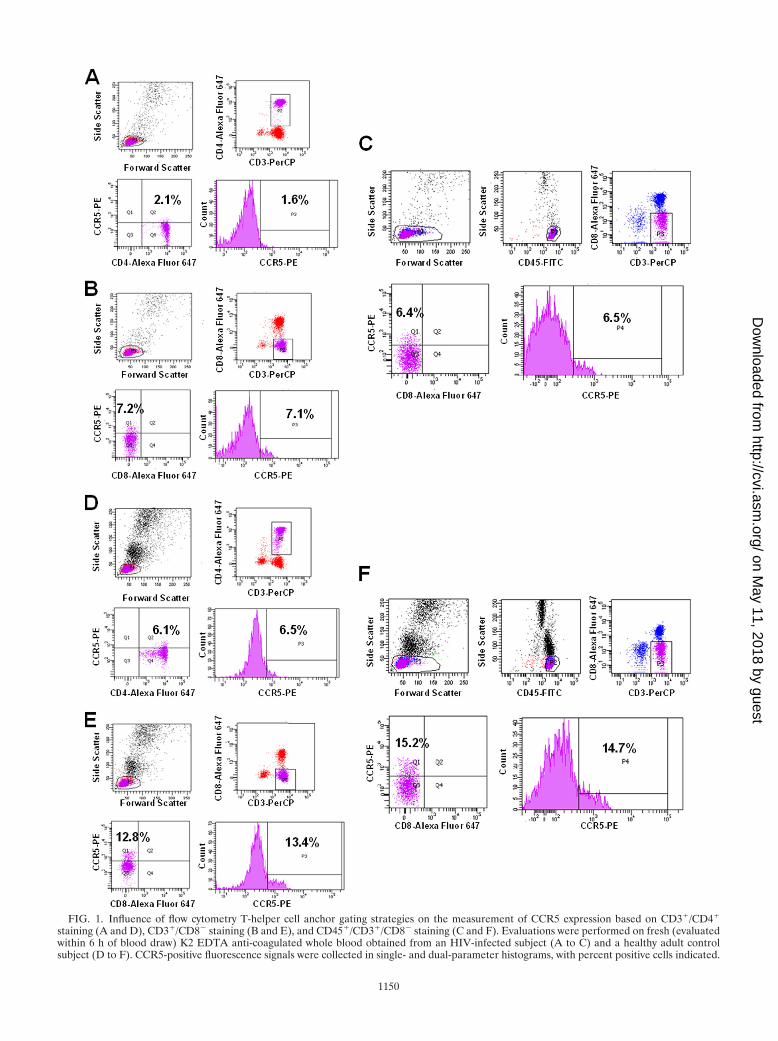
FIG. 1. Influence of flow cytometry T-helper cell anchor gating strategies on the measurement of CCR5 expression based on CD3�/CD4�
staining (A and D), CD3�/CD8� staining (B and E), and CD45�/CD3�/CD8� staining (C and F). Evaluations were performed on fresh (evaluatedwithin 6 h of blood draw) K2 EDTA anti-coagulated whole blood obtained from an HIV-infected subject (A to C) and a healthy adult controlsubject (D to F). CCR5-positive fluorescence signals were collected in single- and dual-parameter histograms, with percent positive cells indicated.
1150
on May 11, 2018 by guest
http://cvi.asm.org/
Dow
nloaded from
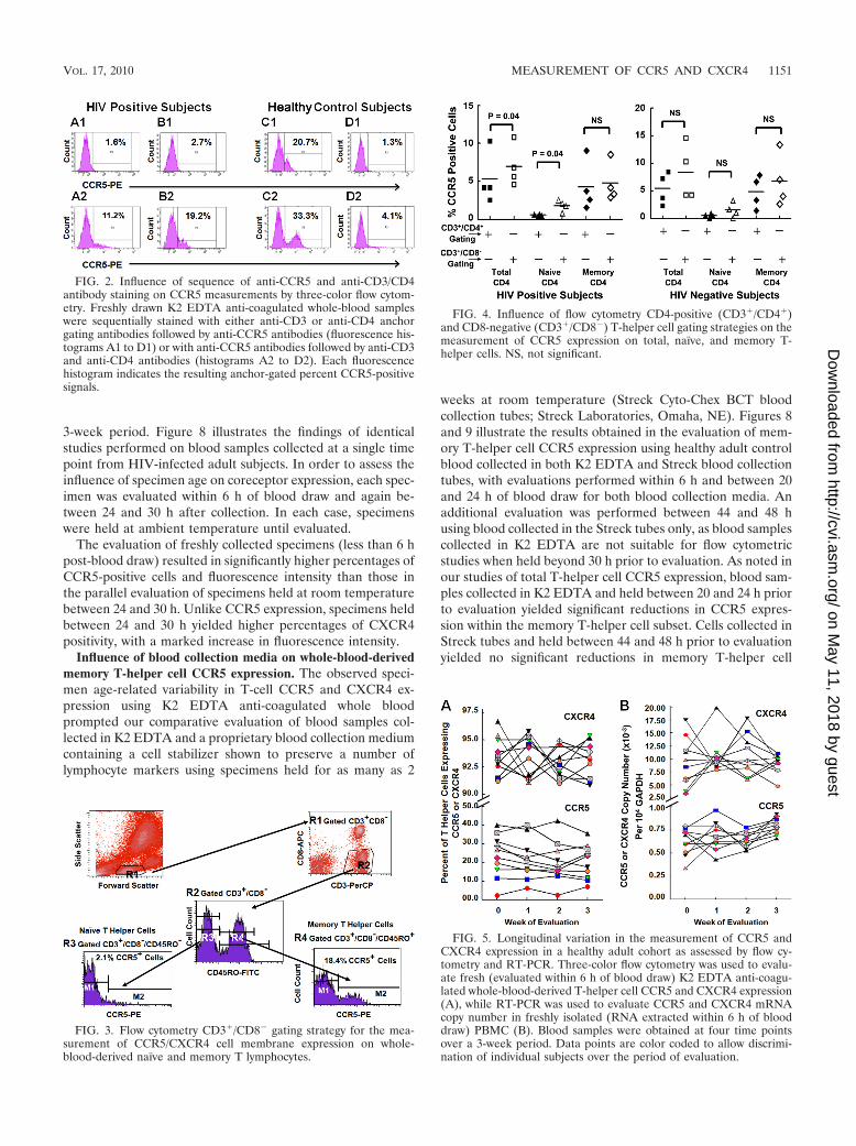
3-week period. Figure 8 illustrates the findings of identicalstudies performed on blood samples collected at a single timepoint from HIV-infected adult subjects. In order to assess theinfluence of specimen age on coreceptor expression, each spec-imen was evaluated within 6 h of blood draw and again be-tween 24 and 30 h after collection. In each case, specimenswere held at ambient temperature until evaluated.
The evaluation of freshly collected specimens (less than 6 hpost-blood draw) resulted in significantly higher percentages ofCCR5-positive cells and fluorescence intensity than those inthe parallel evaluation of specimens held at room temperaturebetween 24 and 30 h. Unlike CCR5 expression, specimens heldbetween 24 and 30 h yielded higher percentages of CXCR4positivity, with a marked increase in fluorescence intensity.
Influence of blood collection media on whole-blood-derivedmemory T-helper cell CCR5 expression. The observed speci-men age-related variability in T-cell CCR5 and CXCR4 ex-pression using K2 EDTA anti-coagulated whole bloodprompted our comparative evaluation of blood samples col-lected in K2 EDTA and a proprietary blood collection mediumcontaining a cell stabilizer shown to preserve a number oflymphocyte markers using specimens held for as many as 2
weeks at room temperature (Streck Cyto-Chex BCT bloodcollection tubes; Streck Laboratories, Omaha, NE). Figures 8and 9 illustrate the results obtained in the evaluation of mem-ory T-helper cell CCR5 expression using healthy adult controlblood collected in both K2 EDTA and Streck blood collectiontubes, with evaluations performed within 6 h and between 20and 24 h of blood draw for both blood collection media. Anadditional evaluation was performed between 44 and 48 husing blood collected in the Streck tubes only, as blood samplescollected in K2 EDTA are not suitable for flow cytometricstudies when held beyond 30 h prior to evaluation. As noted inour studies of total T-helper cell CCR5 expression, blood sam-ples collected in K2 EDTA and held between 20 and 24 h priorto evaluation yielded significant reductions in CCR5 expres-sion within the memory T-helper cell subset. Cells collected inStreck tubes and held between 44 and 48 h prior to evaluationyielded no significant reductions in memory T-helper cell
FIG. 2. Influence of sequence of anti-CCR5 and anti-CD3/CD4antibody staining on CCR5 measurements by three-color flow cytom-etry. Freshly drawn K2 EDTA anti-coagulated whole-blood sampleswere sequentially stained with either anti-CD3 or anti-CD4 anchorgating antibodies followed by anti-CCR5 antibodies (fluorescence his-tograms A1 to D1) or with anti-CCR5 antibodies followed by anti-CD3and anti-CD4 antibodies (histograms A2 to D2). Each fluorescencehistogram indicates the resulting anchor-gated percent CCR5-positivesignals.
FIG. 3. Flow cytometry CD3�/CD8� gating strategy for the mea-surement of CCR5/CXCR4 cell membrane expression on whole-blood-derived naïve and memory T lymphocytes.
FIG. 4. Influence of flow cytometry CD4-positive (CD3�/CD4�)and CD8-negative (CD3�/CD8�) T-helper cell gating strategies on themeasurement of CCR5 expression on total, naïve, and memory T-helper cells. NS, not significant.
FIG. 5. Longitudinal variation in the measurement of CCR5 andCXCR4 expression in a healthy adult cohort as assessed by flow cy-tometry and RT-PCR. Three-color flow cytometry was used to evalu-ate fresh (evaluated within 6 h of blood draw) K2 EDTA anti-coagu-lated whole-blood-derived T-helper cell CCR5 and CXCR4 expression(A), while RT-PCR was used to evaluate CCR5 and CXCR4 mRNAcopy number in freshly isolated (RNA extracted within 6 h of blooddraw) PBMC (B). Blood samples were obtained at four time pointsover a 3-week period. Data points are color coded to allow discrimi-nation of individual subjects over the period of evaluation.
VOL. 17, 2010 MEASUREMENT OF CCR5 AND CXCR4 1151
on May 11, 2018 by guest
http://cvi.asm.org/
Dow
nloaded from
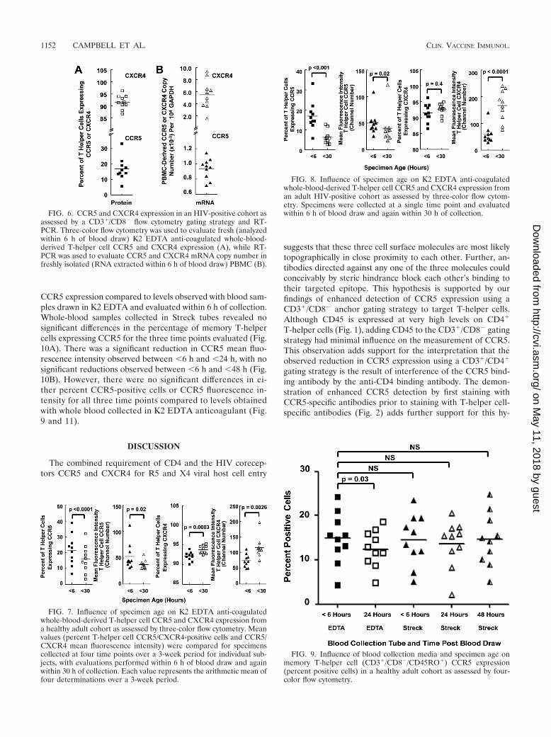
CCR5 expression compared to levels observed with blood sam-ples drawn in K2 EDTA and evaluated within 6 h of collection.Whole-blood samples collected in Streck tubes revealed nosignificant differences in the percentage of memory T-helpercells expressing CCR5 for the three time points evaluated (Fig.10A). There was a significant reduction in CCR5 mean fluo-rescence intensity observed between �6 h and �24 h, with nosignificant reductions observed between �6 h and �48 h (Fig.10B). However, there were no significant differences in ei-ther percent CCR5-positive cells or CCR5 fluorescence in-tensity for all three time points compared to levels obtainedwith whole blood collected in K2 EDTA anticoagulant (Fig.9 and 11).
DISCUSSION
The combined requirement of CD4 and the HIV corecep-tors CCR5 and CXCR4 for R5 and X4 viral host cell entry
suggests that these three cell surface molecules are most likelytopographically in close proximity to each other. Further, an-tibodies directed against any one of the three molecules couldconceivably by steric hindrance block each other’s binding totheir targeted epitope. This hypothesis is supported by ourfindings of enhanced detection of CCR5 expression using aCD3�/CD8� anchor gating strategy to target T-helper cells.Although CD45 is expressed at very high levels on CD4�
T-helper cells (Fig. 1), adding CD45 to the CD3�/CD8� gatingstrategy had minimal influence on the measurement of CCR5.This observation adds support for the interpretation that theobserved reduction in CCR5 expression using a CD3�/CD4�
gating strategy is the result of interference of the CCR5 bind-ing antibody by the anti-CD4 binding antibody. The demon-stration of enhanced CCR5 detection by first staining withCCR5-specific antibodies prior to staining with T-helper cell-specific antibodies (Fig. 2) adds further support for this hy-
FIG. 6. CCR5 and CXCR4 expression in an HIV-positive cohort asassessed by a CD3�/CD8� flow cytometry gating strategy and RT-PCR. Three-color flow cytometry was used to evaluate fresh (analyzedwithin 6 h of blood draw) K2 EDTA anti-coagulated whole-blood-derived T-helper cell CCR5 and CXCR4 expression (A), while RT-PCR was used to evaluate CCR5 and CXCR4 mRNA copy number infreshly isolated (RNA extracted within 6 h of blood draw) PBMC (B).
FIG. 7. Influence of specimen age on K2 EDTA anti-coagulatedwhole-blood-derived T-helper cell CCR5 and CXCR4 expression froma healthy adult cohort as assessed by three-color flow cytometry. Meanvalues (percent T-helper cell CCR5/CXCR4-positive cells and CCR5/CXCR4 mean fluorescence intensity) were compared for specimenscollected at four time points over a 3-week period for individual sub-jects, with evaluations performed within 6 h of blood draw and againwithin 30 h of collection. Each value represents the arithmetic mean offour determinations over a 3-week period.
FIG. 8. Influence of specimen age on K2 EDTA anti-coagulatedwhole-blood-derived T-helper cell CCR5 and CXCR4 expression froman adult HIV-positive cohort as assessed by three-color flow cytom-etry. Specimens were collected at a single time point and evaluatedwithin 6 h of blood draw and again within 30 h of collection.
FIG. 9. Influence of blood collection media and specimen age onmemory T-helper cell (CD3�/CD8�/CD45RO�) CCR5 expression(percent positive cells) in a healthy adult cohort as assessed by four-color flow cytometry.
1152 CAMPBELL ET AL. CLIN. VACCINE IMMUNOL.
on May 11, 2018 by guest
http://cvi.asm.org/
Dow
nloaded from

pothesis. In addition, the reduced level of cell surface expres-sion of CCR5 in blood samples held for 24 to 30 h prior toevaluation is accompanied by an increase in the detection ofCXCR4. This conclusion is also supported by fluorescenceresonance energy transfer imaging studies that demonstratedcolocalization of CD4 and CCR5 on the plasma membrane ofviable CD4-yellow fluorescent protein (YFP)- and CCR5-cyanfluorescent protein (CFP)-transfected HEK293T cells follow-ing treatment with a V3-containing HIV-1 gp120 core (7, 17).Using CD3�/CD8� T-helper cell gating allows for increaseddetection of CCR5 in total, naive, and memory T-helper cells,particularly in the evaluation of cells from HIV-positive sub-jects (Fig. 4). These results provided the basis for the selectionof CD3�/CD8� T-helper cell anchor gating for the measure-ment of CCR5 and CXCR4 expression on naïve and memoryT cells. The gating strategy described in our study couplesT-helper cell gating with additional gates that allow for thedifferentiation of naïve and memory phenotypes based onCD45RO-positive and -negative signals (Fig. 3).
K2 EDTA anti-coagulated whole blood has been the bloodcollection medium of choice for flow cytometry-based studiesof lymphocyte cell surface markers and has been used exten-sively in AIDS clinical trials, in which such evaluations, includ-ing T-cell CD4 percentages and absolute counts, are importantend points for the measurement of response to various thera-peutic interventions. T-cell markers, including CD4 and CD8,have been shown to be relatively stable in K2 EDTA anti-coagulated whole blood over a 24- to 30-h period, while othermarkers such as CD62L and CCR5 are sensitive to various invitro manipulations (1). Our studies have extended the findingsof Berhanu et al. (1) by showing that the level of detection ofT-helper cell CCR5 and CXCR4 expression using K2 EDTAanti-coagulated whole blood is dependent on a number offactors, including the appropriate selection of T-helper cellanchor gating and the age of the specimen prior to analysis.This study has also demonstrated the practicality of measuringCCR5 and CXCR4 expression at both the protein and mRNAlevels. The accurate measurement of whole-blood-derived T-cell CCR5 expression in transported specimens of various ageshas become an important capability in clinical studies of CCR5antagonists as adjuvants to antiretroviral therapies. Our studyhas identified a number of factors that minimize variation in
T-cell CCR5 expression that allow for specimen transportwithin at least a 48-h window. The repetitive analysis of T-helper cell CCR5 measurements using peripheral blood sam-ples collected in Streck Cyto-Chex BCT Vacutainer tubes andheld at room temperature for up to 48 h was found to yieldmore reproducible results than specimens collected in K2EDTA. These findings will allow for the transport of clinicalspecimens to central laboratories having the expertise to per-form such studies not available at many local clinical sites, thusproviding important measures of immune function in responseto therapy.
ACKNOWLEDGMENTS
Overall support for the International Maternal Pediatric AdolescentAIDS Clinical Trials Group (IMPAACT) was provided by the Na-tional Institute of Allergy and Infectious Diseases (NIAID) (U01AI068632), the Eunice Kennedy Shriver National Institute of ChildHealth and Human Development (NICHD), and the National Insti-tute of Mental Health (NIMH) (AI068632). This work was supportedby the Statistical and Data Analysis Center at Harvard School of PublicHealth, under the National Institute of Allergy and Infectious Diseasescooperative agreement number 5 U01 AI41110 with the PediatricAIDS Clinical Trials Group (PACTG) and number 1 U01 AI068616with the IMPAACT Group. Support of the sites was provided by theNational Institute of Allergy and Infectious Diseases (NIAID) and theNICHD International and Domestic Pediatric and Maternal HIVClinical Trials Network funded by the NICHD (contract number N01-DK-9-001/HHSN267200800001C). This study was performed by theChildren’s Hospital of Philadelphia IMPAACT Specialty Laboratory.
The content is solely the responsibility of the authors and does notnecessarily represent the official views of the NIH.
REFERENCES
1. Berhanu, D., F. Mortari, S. C. DeRosa, and M. Roederer. 2003. Optimizedlymphocyte isolation methods for analysis of chemokine receptor expression.J. Immunol. Methods 279:199–207.
2. Cane, P. A. 2009. New developments in HIV drug resistance. J. Antimicrob.Chemother. 64(Suppl. 1):i37–i40.
3. Cardozo, T., T. Kimura, S. Philpott, B. Weiser, H. Burger, and S. Zolla-Pazner. 2007. Structural basis for coreceptor selectivity by the HIV type 1 V3loop. AIDS Res. Hum. Retroviruses 23:415–426.
FIG. 11. Influence of blood collection media and specimen age onmemory T-helper cell (CD3�/CD8�/CD45RO�) CCR5 expression(mean fluorescence intensity) in a healthy adult cohort as assessed byfour-color flow cytometry.
FIG. 10. Influence of specimen age prior to analysis of memoryT-helper cells (CD3�/CD8�/CD45RO�) CCR5 expression (% positivecells [A] and mean fluorescence intensity [B]) in whole-blood samplescollected in Streck Vacutainer tubes. Data points are color coded toallow discrimination of individual subjects over the period ofevaluation.
VOL. 17, 2010 MEASUREMENT OF CCR5 AND CXCR4 1153
on May 11, 2018 by guest
http://cvi.asm.org/
Dow
nloaded from

4. Chen, T. K., and G. M. Aldrovandi. 2008. Review of HIV antiretroviral drugresistance. Pediatr. Infect. Dis. J. 27:749–751.
5. Croxtall, J. D., and S. J. Keam. 2009. Raltegravir: a review of its use in themanagement of HIV infection in treatment-experienced patients. Drugs69:1059–1075.
6. Gulick, R. M., J. Lalezari, J. Goodrich, N. Clumeck, E. DeJesus, A. Horban,J. Nadler, B. Clotet, A. Karlson, M. Wohlfeiler, J. B. Montana, M. McHale,J. Sullivan, C. Ridgway, S. Felstead, M. W. Dunne, E. van der Ryst, and H.Mayer, for the MOTIVATE Study Teams. 2008. Maraviroc for previouslytreated patients with R5 HIV-1 infection. N. Engl. J. Med. 359:1429–1441.
7. Huang, C.-C., M. Tang, M.-Y. Zhang, S. Majeed, E. Montabana, R. L.Stanfield, D. S. Dimitrov, B. Korber, J. Sodroski, I. A. Wilson, R. Wyatt, andP. D. Kwong. 2005. Structure of a V3-containing HIV-1 gp120 core. Science310:1025–1028.
8. Kondru, R., J. Zhang, C. Ji, T. Mirzadegan, D. Rotstein, S. Sankuratri, andM. Dioszegi. 2008. Molecular interactions of CCR5 with major classes ofsmall-molecule anti-HIV CCR5 antagonists. Mol. Pharmacol. 73:789–800.
9. Lai, J.-P., J.-H. Yang, S. D. Douglas, X. Wang, E. Riedel, and W.-Z. Ho. 2003.Quantification of CCR5 mRNA in human lymphocytes and macrophages byreal-time reverse transcriptase PCR assay. Clin. Diagn. Lab. Immunol. 10:1123–1128.
10. Lederman, M. M., A. Penn-Nicholson, M. Cho, and D. Mosfer. 2006. Biologyof CCR5 and its role in HIV infection ad treatment. JAMA 296:815–826.
11. Little, S. J., S. Holte, J.-P. Routy, E. S. Daar, M. Markowitz, A. C. Collier,R. A. Koup, J. W. Mellors, E. Connick, B. Conway, M. Kilby, L. Wang, J. M.Whitcomb, N. S. Hellmann, and D. D. Richman. 2002. Antiretroviral-drug
resistance among patients recently infected with HIV. N. Engl. J. Med.347:385–394.
12. Murphy, E. L., A. C. Collier, L. A. Kalish, S. F. Assman, M. F. Para, T. P.Flanigan, P. N. Kumar, L. Mintz, F. R. Wallach, and G. J. Nemo, for theViral Activation Transfusion Study Investigators. 2001. Highly active anti-retroviral therapy decreases mortality and morbidity in patients with ad-vanced HIV disease. Ann. Intern. Med. 135:17–26.
13. Ribeiro, R. M., M. D. Hazenberg, A. S. Perelson, and M. P. Davenport. 2006.Naïve and memory cell turnover as drivers of CCR5-to-C4CR4 tropismswitch in human immunodeficiency virus type 1: implications for therapy.J. Virol. 80:802–809.
14. Rosen, O., M. Sharon, S. R. Quadt-Akabayov, and J. Anglister. 2006. Mo-lecular switch for alternative conformations of the HIV-1 V3 region: impli-cations for phenotype conversion. Proc. Natl. Acad. Sci. U. S. A. 103:13950–13955.
15. Su, Z., R. M. Gulick, A. Krambrink, E. Coakley, M. D. Hughes, D. Han, C.Flexner, T. J. Wilkin, P. R. Skolnik, W. L. Greaves, D. R. Kuritzkes, J. D.Reeves, and AIDS Clinical Trials Group A5211 Team. 2009. Response tovicriviroc in treatment-experienced subjects, as determined by an enhanced-sensitivity coreceptor tropism assay: reanalysis of AIDS clinical trials groupA5211. J. Infect. Dis. 200:1724–1728.
16. Tsibris, A. M. N., and D. R. Kuritzres. 2007. Chemokine antagonists astherapeutics: focus on HIV-1. Annu. Rev. Med. 58:445–459.
17. Yi, L., J. Fang, N. Isik, J. Chim, and T. Jin. 2006. HIV gp120-inducedinteraction between CD4 and CCR5 requires cholesterol-rich microenviron-ments revealed by live fluorescence resonance energy transfer imaging.J. Biol. Chem. 281:35446–35453.
1154 CAMPBELL ET AL. CLIN. VACCINE IMMUNOL.
on May 11, 2018 by guest
http://cvi.asm.org/
Dow
nloaded from