Embed Size (px)
Citation preview
Analytical X-ray Analytical X-ray Safety TrainingSafety Training
ENVIRONMENTAL HEALTH AND SAFETY DEPARTMENTENVIRONMENTAL HEALTH AND SAFETY DEPARTMENTRADIATION SAFETYRADIATION SAFETY
486-3613486-3613
No individual shall be permitted to operate analytical x-ray diffraction or spectrographic equipment until such individual has:
1. completed this on-line training module;
2. completed the required laboratory-based training conducted by the equipment supervisor;
3. viewed an x-ray safety video; and
4. successfully completed the x-ray Radiation Safety exam.
Upon completion of this on-line training module, contact Radiation Safety at 486-3613 to schedule an appointment to view the x-ray safety video and take an exam.
The Laboratory-Based Training form can be downloaded at: Analytical X-Ray Lab Based Training Form
RADIATION SAFETY TRAINING REQUIREMENTS
• Identification of radiation hazards associated with the use of the equipment;
• Significance of the various radiation warning, safety devices and interlocks incorporated into the equipment, or the reasons they have not been installed on certain pieces of equipment and the extra precautions required in such cases;
• Proper operating procedures for the equipment;
• Recognition of the symptoms of an acute localized exposure; and
• Proper procedures for reporting an actual or suspected exposure.
Competence as to the following must be demonstrated prior to operating analytical x-ray equipment:
Radiation Safety training and/or experience completed at another university and documented with the University of Connecticut’s Radiation Safety Officer may be
sufficient to fulfill a portion(s) of the initial training requirements.
X-RAY REGISTRATION
Analytical x-ray producing equipment in the possession of the University of Connecticut is subject to registration with the State of Connecticut DEP Radiation Control Division and applicable state and federal regulations. The University of Connecticut’s Radiation Safety Office must be notified at 486-3613 of the possession of such equipment prior to utilization.
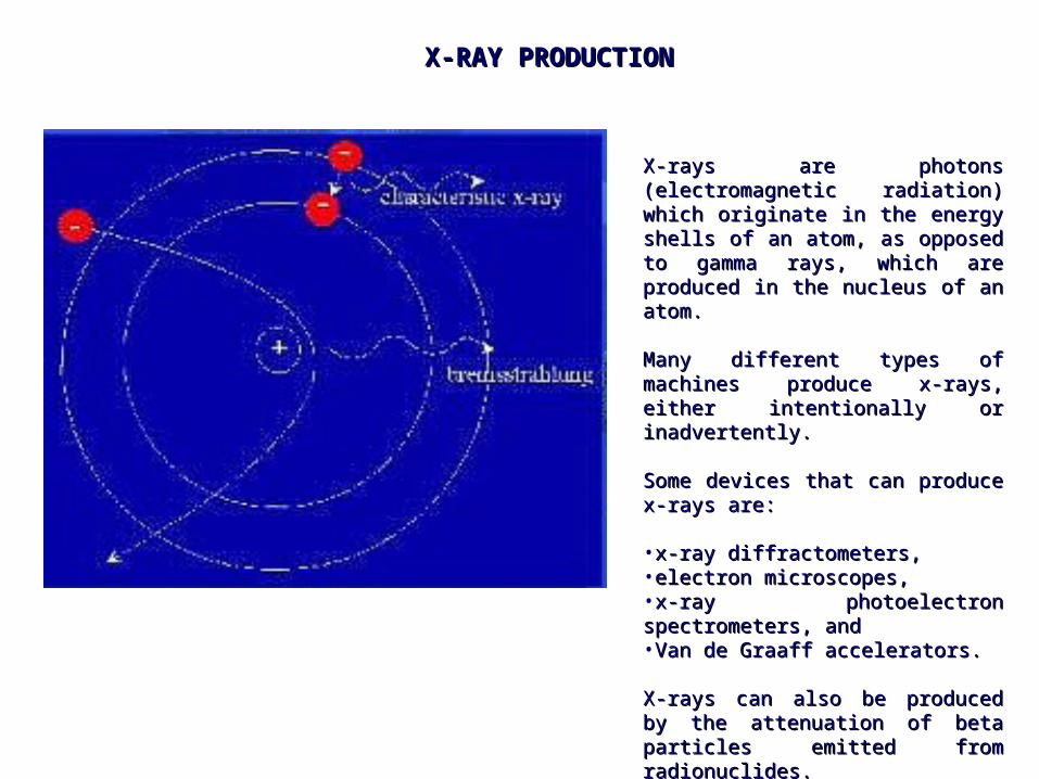
X-rays are photons (electromagnetic X-rays are photons (electromagnetic radiation) which originate in the energy radiation) which originate in the energy shells of an atom, as opposed to shells of an atom, as opposed to gamma rays, which are produced in the gamma rays, which are produced in the nucleus of an atom. nucleus of an atom.
Many different types of machines Many different types of machines produce x-rays, either intentionally or produce x-rays, either intentionally or inadvertently. inadvertently.
Some devices that can produce x-rays Some devices that can produce x-rays are: are:
•x-ray diffractometers, x-ray diffractometers, •electron microscopes, electron microscopes, •x-ray photoelectron spectrometers, andx-ray photoelectron spectrometers, and•Van de Graaff accelerators. Van de Graaff accelerators.
X-rays can also be produced by the X-rays can also be produced by the attenuation of beta particles emitted attenuation of beta particles emitted from radionuclides. from radionuclides.
X-RAY PRODUCTIONX-RAY PRODUCTION
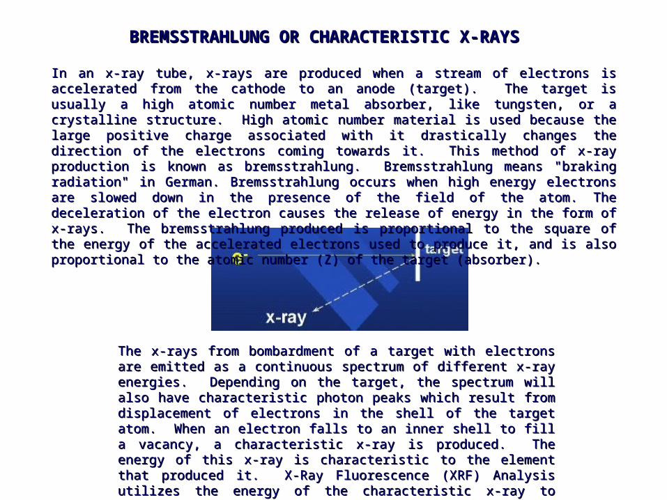
The x-rays from bombardment of a target with electrons are emitted as a The x-rays from bombardment of a target with electrons are emitted as a continuous spectrum of different x-ray energies. Depending on the target, continuous spectrum of different x-ray energies. Depending on the target, the spectrum will also have characteristic photon peaks which result from the spectrum will also have characteristic photon peaks which result from displacement of electrons in the shell of the target atom. When an electron displacement of electrons in the shell of the target atom. When an electron falls to an inner shell to fill a vacancy, a characteristic x-ray is produced. falls to an inner shell to fill a vacancy, a characteristic x-ray is produced. The energy of this x-ray is characteristic to the element that produced it. X-The energy of this x-ray is characteristic to the element that produced it. X-Ray Fluorescence (XRF) Analysis utilizes the energy of the characteristic x-Ray Fluorescence (XRF) Analysis utilizes the energy of the characteristic x-ray to identify the particular elements in a sample.ray to identify the particular elements in a sample.
In an x-ray tube, x-rays are produced when a stream of electrons is accelerated from the cathode In an x-ray tube, x-rays are produced when a stream of electrons is accelerated from the cathode to an anode (target). The target is usually a high atomic number metal absorber, like tungsten, or to an anode (target). The target is usually a high atomic number metal absorber, like tungsten, or a crystalline structure. High atomic number material is used because the large positive charge a crystalline structure. High atomic number material is used because the large positive charge associated with it drastically changes the direction of the electrons coming towards it. This associated with it drastically changes the direction of the electrons coming towards it. This method of x-ray production is known as bremsstrahlung. Bremsstrahlung means "braking method of x-ray production is known as bremsstrahlung. Bremsstrahlung means "braking radiation" in German. Bremsstrahlung occurs when high energy electrons are slowed down in the radiation" in German. Bremsstrahlung occurs when high energy electrons are slowed down in the presence of the field of the atom. The deceleration of the electron causes the release of energy in presence of the field of the atom. The deceleration of the electron causes the release of energy in the form of x-rays. The bremsstrahlung produced is proportional to the square of the energy of the form of x-rays. The bremsstrahlung produced is proportional to the square of the energy of the accelerated electrons used to produce it, and is also proportional to the atomic number (Z) of the accelerated electrons used to produce it, and is also proportional to the atomic number (Z) of the target (absorber)the target (absorber)..
BREMSSTRAHLUNG OR CHARACTERISTIC X-RAYSBREMSSTRAHLUNG OR CHARACTERISTIC X-RAYS
X-RAY ENERGIES
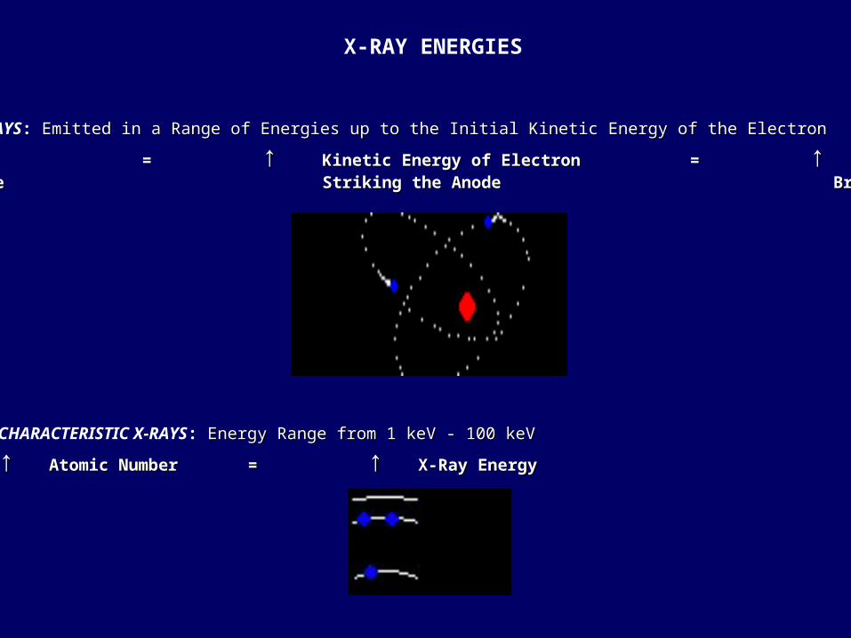
BREMSSTRAHLUNG X-RAYS: Emitted in a Range of Energies up to the Initial Kinetic Energy of the Electron Emitted in a Range of Energies up to the Initial Kinetic Energy of the Electron
↑ ↑ AppliedApplied = = ↑ ↑ Kinetic Energy of Electron = Kinetic Energy of Electron = ↑ ↑ Energy ofEnergy of High Voltage Striking the AnodeHigh Voltage Striking the Anode Bremsstrahlung X-Rays Bremsstrahlung X-Rays
CHARACTERISTIC X-RAYS: Energy Range from 1 keV - 100 keV Energy Range from 1 keV - 100 keV
↑ ↑ Atomic Number = Atomic Number = ↑ ↑ X-Ray EnergyX-Ray Energy
RADIATION TERMS AND UNITS
DOSE EQUIVALENT:
The quantity used by regulatory agencies related to risk of biological damage to radiation workers from ionizing radiation. This quantity generally pertains to external exposure. It is the product of the absorbed dose in tissue and all other necessary factors at the location of interest.
The unit of dose equivalent is the rem.
1000 mrem = one rem
DOSE
The energy imparted to matter by ionizing radiation per unit mass of irradiated material.
The unit of absorbed dose is the rad.
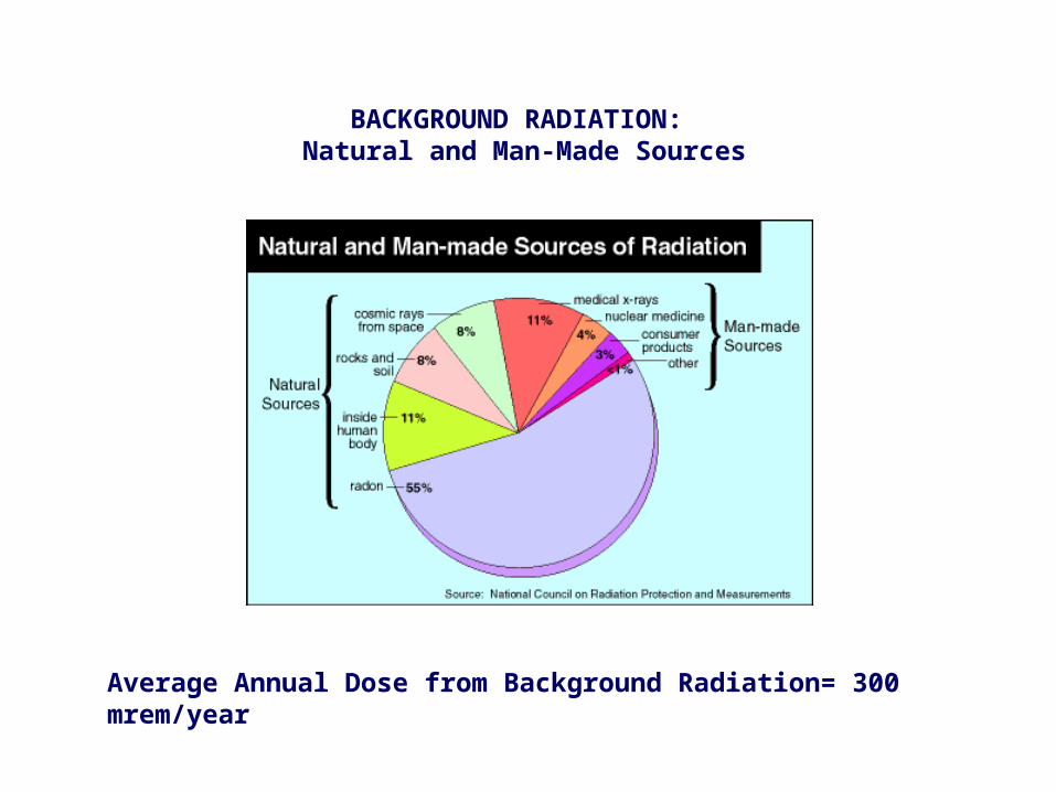
Average Annual Dose from Background Radiation= 300 mrem/year
BACKGROUND RADIATION: Natural and Man-Made Sources
NRC: Regulatory Guide 8.13 - Instruction Concerning Prenatal Radiation Exposure
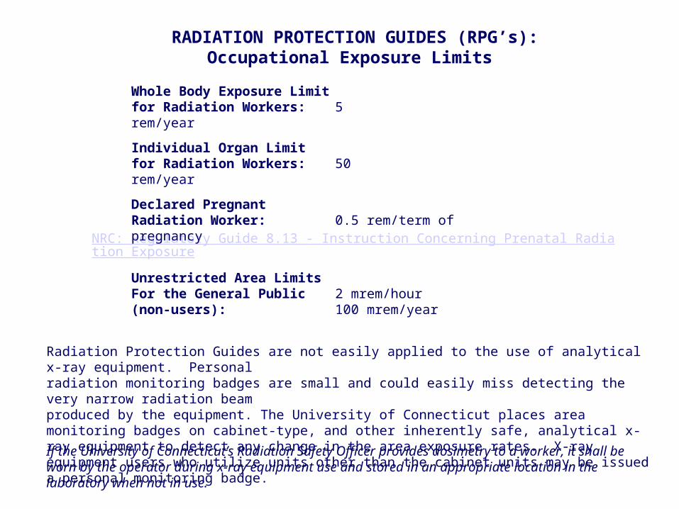
RADIATION PROTECTION GUIDES (RPG’s):Occupational Exposure Limits
Unrestricted Area LimitsFor the General Public 2 mrem/hour(non-users): 100 mrem/year
Whole Body Exposure Limit for Radiation Workers: 5 rem/year
Individual Organ Limit for Radiation Workers: 50 rem/year
Declared Pregnant Radiation Worker: 0.5 rem/term of pregnancy
Radiation Protection Guides are not easily applied to the use of analytical x-ray equipment. Personal radiation monitoring badges are small and could easily miss detecting the very narrow radiation beam produced by the equipment. The University of Connecticut places area monitoring badges on cabinet-type, and other inherently safe, analytical x-ray equipment to detect any change in the area exposure rates. X-ray equipment users who utilize units other than the cabinet units may be issued a personal monitoring badge.
If the University of Connecticut’s Radiation Safety Officer provides dosimetry to a worker, it shall be worn by the operator during x-ray equipment use and stored in an appropriate location in the laboratory when not in use.

UConn ALARA Goals: Ten percent of RPG’s
chievable
Whole body: 0.5 rem/year or 500 mrem/year
Individual Organs: 5 rem/year
s ow s is easonably
The University of Connecticut is committed to keeping exposures to radiation ALARA (As Low As is Reasonably Achievable). This means that every reasonable effort shall be made to maintain radiation exposures as far below the dose limits as practical, taking into account the state of the technology, the economics of the improvements in relation to the benefits, and other socioeconomic considerations.
Each worker shall keep radiation exposures to themselves and others ALARA
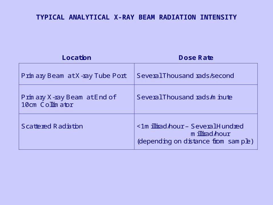
Location
Dose Rate
Primary Beam at X-ray Tube Port
Several Thousand rads/second
Primary X-ray Beam at End of 10cm Collimator
Several Thousand rads/minute
Scattered Radiation
<1millirad/hour – Several Hundred
millirad/hour (depending on distance from sample)
TYPICAL ANALYTICAL X-RAY BEAM RADIATION INTENSITY
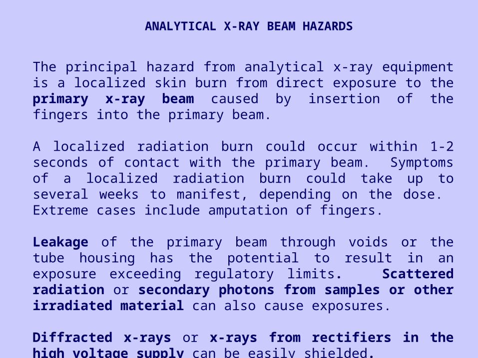
ANALYTICAL X-RAY BEAM HAZARDS
The principal hazard from analytical x-ray equipment is a localized skin burn from direct exposure to the primary x-ray beam caused by insertion of the fingers into the primary beam.
A localized radiation burn could occur within 1-2 seconds of contact with the primary beam. Symptoms of a localized radiation burn could take up to several weeks to manifest, depending on the dose. Extreme cases include amputation of fingers.
Leakage of the primary beam through voids or the tube housing has the potential to result in an exposure exceeding regulatory limits. Scattered radiation or secondary photons from samples or other irradiated material can also cause exposures.
Diffracted x-rays or x-rays from rectifiers in the high voltage supply can be easily shielded.
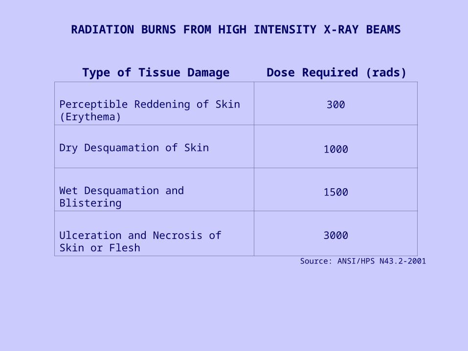
RADIATION BURNS FROM HIGH INTENSITY X-RAY BEAMS
Perceptible Reddening of Skin (Erythema)
300
Dry Desquamation of Skin 1000
Wet Desquamation and Blistering 1500
Ulceration and Necrosis of Skin or Flesh 3000
Type of Tissue Damage Dose Required (rads)
Source: ANSI/HPS N43.2-2001
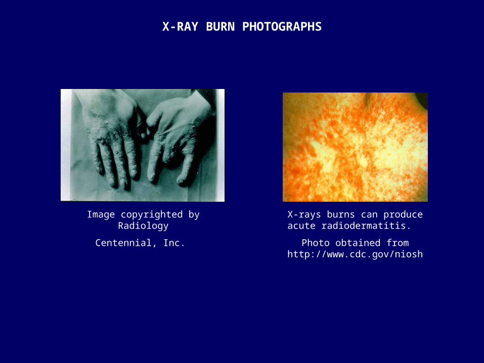
Image copyrighted by Radiology
Centennial, Inc.
X-rays burns can produce acute radiodermatitis.
Photo obtained from http://www.cdc.gov/niosh
X-RAY BURN PHOTOGRAPHS
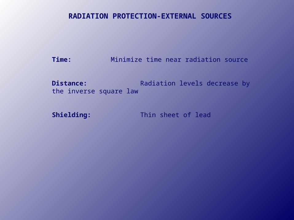
Time: Minimize time near radiation source
Distance: Radiation levels decrease by the inverse square law
Shielding: Thin sheet of lead
RADIATION PROTECTION-EXTERNAL SOURCES
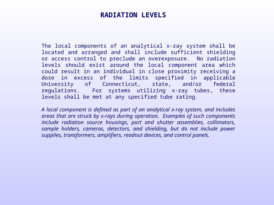
RADIATION LEVELS
The local components of an analytical x-ray system shall be located and arranged and shall include sufficient shielding or access control to preclude an overexposure. No radiation levels should exist around the local component area which could result in an individual in close proximity receiving a dose in excess of the limits specified in applicable University of Connecticut, state, and/or federal regulations. For systems utilizing x-ray tubes, these levels shall be met at any specified tube rating. A local component is defined as part of an analytical x-ray system, and includes areas that are struck by x-rays during operation. Examples of such components include radiation source housings, port and shutter assemblies, collimators, sample holders, cameras, detectors, and shielding, but do not include power supplies, transformers, amplifiers, readout devices, and control panels.
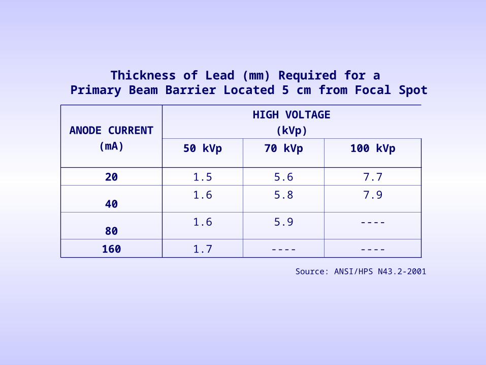
ANODE CURRENT
(mA)
HIGH VOLTAGE
(kVp)
50 kVp 70 kVp 100 kVp
20 1.5 5.6 7.7
401.6 5.8 7.9
801.6 5.9 ----
160 1.7 ---- ----
Thickness of Lead (mm) Required for a Primary Beam Barrier Located 5 cm from Focal Spot
Source: ANSI/HPS N43.2-2001
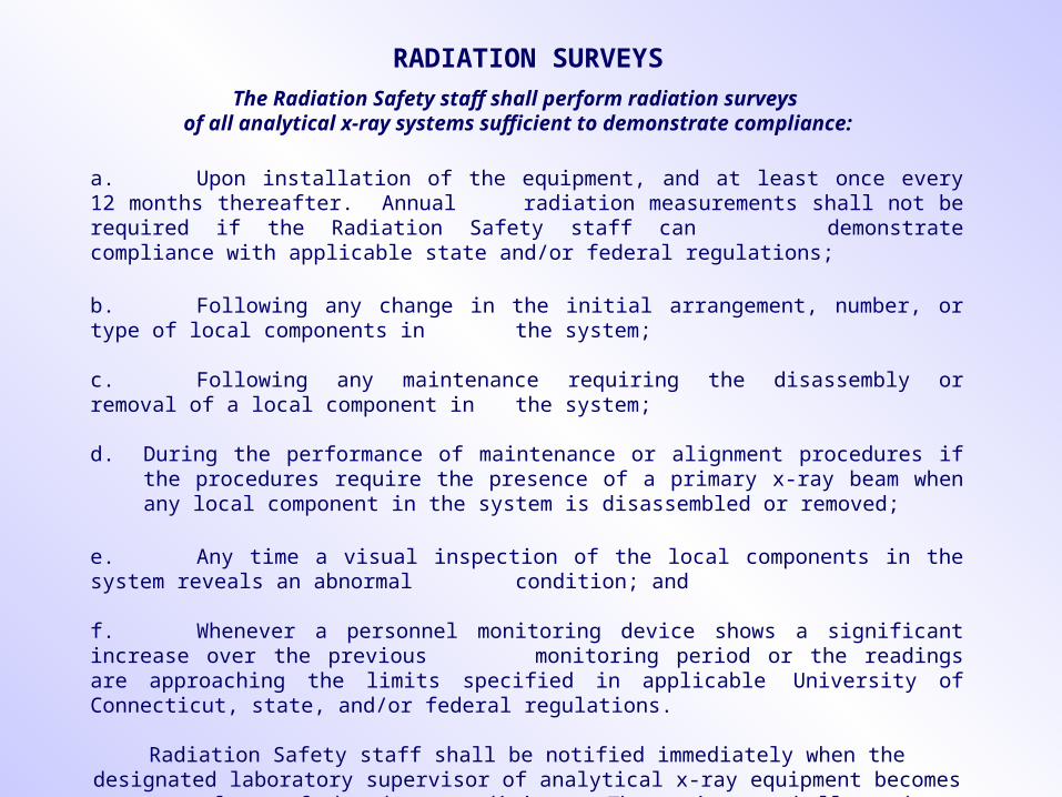
RADIATION SURVEYS
a. Upon installation of the equipment, and at least once every 12 months thereafter. Annual radiation measurements shall not be required if the Radiation Safety staff can demonstrate compliance with applicable state and/or federal regulations;
b. Following any change in the initial arrangement, number, or type of local components in the system;
c. Following any maintenance requiring the disassembly or removal of a local component in the system;
d. During the performance of maintenance or alignment procedures if the procedures require the presence of a primary x-ray beam when any local component in the system is disassembled or removed;
e. Any time a visual inspection of the local components in the system reveals an abnormal condition; and
f. Whenever a personnel monitoring device shows a significant increase over the previous monitoring period or the readings are approaching the limits specified in applicable
University of Connecticut, state, and/or federal regulations.
Radiation Safety staff shall be notified immediately when the designated laboratory supervisor of analytical x-ray equipment becomes aware of any of the above conditions. The equipment
shall not be utilized until Radiation Safety demonstrates compliance with University regulations.
The Radiation Safety staff shall perform radiation surveys of all analytical x-ray systems sufficient to demonstrate compliance:
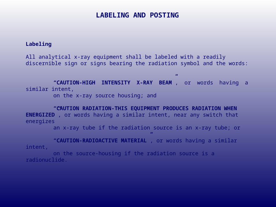
LABELING AND POSTING
Labeling All analytical x-ray equipment shall be labeled with a readily discernible sign or signs bearing the radiation symbol and the words:
“CAUTION-HIGH INTENSITY X-RAY BEAM”, or words having a similar intent, on the x-ray source housing; and
“CAUTION RADIATION-THIS EQUIPMENT PRODUCES RADIATION WHEN ENERGIZED”, or words having a similar intent, near any switch that energizes
an x-ray tube if the radiation source is an x-ray tube; or
“CAUTION-RADIOACTIVE MATERIAL”, or words having a similar intent, on the source-housing if the radiation source is a radionuclide.
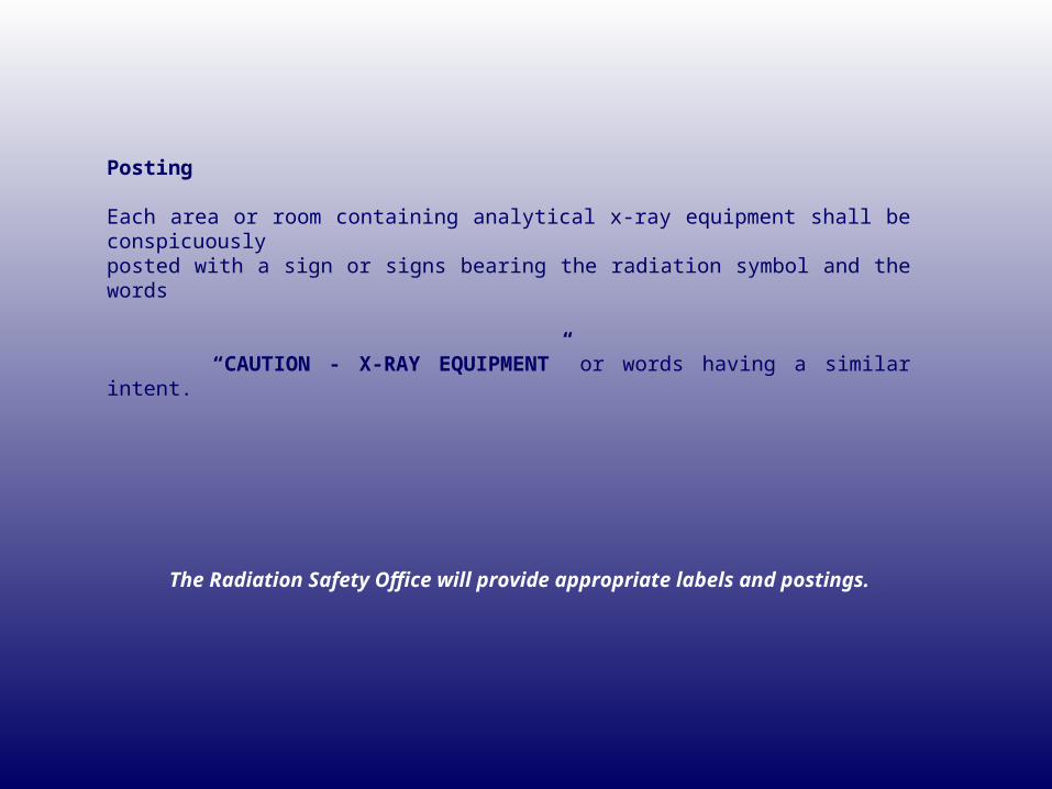
Posting Each area or room containing analytical x-ray equipment shall be conspicuously posted with a sign or signs bearing the radiation symbol and the words
“CAUTION - X-RAY EQUIPMENT” or words having a similar intent.
The Radiation Safety Office will provide appropriate labels and postings.
UNIVERSITY OF CONNETICUT’S RADIATION SAFETY REQUIREMENTS
FOR OPERATORS OF ANALYTICAL X-RAY EQUIPMENT
The following requirements are applicable to equipment utilized for x-ray diffraction or fluorescence analysis. Individuals in charge of the installation and all equipment operators shall be familiar with applicable operating procedures and regulations governing the x-ray installation. The Radiation Safety Officer (RSO) is empowered by the Radiation Safety Committee to immediately terminate the operation of analytical x-ray equipment found to be a threat to health, safety, or property until the violation is corrected.

EQUIPMENT REQUIREMENTS FOR AN ENCLOSED BEAM X-RAY SYSTEM
1 .The radiation source, beam paths, sample, detector and/or other devices e.g. analyzing crystal, filters, etc. shall be enclosed in a chamber, coupled chambers, beam pipes, whole system enclosure, etc. that cannot be entered by any part of the body during normal operation. 2. The inherent shielding of the chamber/enclosure walls shall be sufficient to limit the dose rate in all regions 5 cm from its outer surface to 0.25 mrem/hr during normal operations. 3. The system enclosure, sample chamber, etc. closure shall be interlocked with the x-ray tube high voltage supply and/or a shutter in the primary beam, such that no x-ray beam can enter the sample chamber while it is open unless the interlock has been consciously and deliberately defeated. The interlock shall be of fail-safe design.* * A fail-safe characteristic means a design feature that causes beam port shutters to close upon failure of a safety or warning device.
If there is more than one port in the radiation source housing or more than one radiation source, all requirements must be satisfied for each port in every source housing associated with the system.
If the entire system, including the x-ray tube, is under one contiguous vacuum, and radiation leakage is less than 0.25 mrem/hr at 5cm, and a change in any part of the system will not increase the radiation level, then the entire system shall be considered to be an enclosed beam system.
Ports Unused ports on radiation source housings shall be secured in the closed position in a manner that will prevent casual opening.
A safety device is that which prevents entry of any portion of the individual’s body into the primary x-ray beam path or which causes the beam to be shut off upon entry into its path shall be provided on all open-beam configurations. An open beam system is defined as an analytical x-ray system in which an individual could accidentally place some part of his/her body into the primary beam path during normal operation. An analytical x-ray system is considered to be an open-beam system unless the requirements for an enclosed beam system are met. The primary beam is the radiation that passes through an aperture of the source housing by a direct path from the x-ray tube or a radioactive source located in the source housing.
SAFETY DEVICE FOR AN OPEN BEAM SYSTEM
Definitions
Shutters
On open-beam configurations, each port on the radiation source housing shall be equipped with a shutter that cannot be opened unless a collimator or coupling has been connected to the port.
a. A description of the various safety devices that have been evaluated;
b. The reason each of these devices cannot be used; and
c. A description of the alternative methods that will be employed to minimize the possibility of an accidental exposure, including procedures to assure thatoperators and others in the area will be informed of the absence of safety devices.
An analytical x-ray equipment supervisor may apply for an exemption from the requirement of a safety device if compliance with the requirement is not feasible. The application shall be submitted to the University of Connecticut’s Radiation Safety Officer for review and shall include the following:
An exemption is dependant upon review and formal approval from the University of Connecticut’s Radiation Safety Officer. The x-ray equipment shall not be utilized until approval of the exemption.
An open-beam configuration shall be provided with a readily discernible indication of:
a. X-ray tube “on-off” status located near the radiation source housing, if the primary beam is controlled in this manner; and/or
b. Shutter “open-closed” status located near each on the radiation source housing, if the primary beam is controlled in this manner.
An easily visible warning light labeled with the words “X-RAY ON”, or words having similar intent shall be located near any switch that energizes an x-ray tube and shall be illuminated only when the tube is energized. Warning devices shall be labeled so that their purpose is easily identified. The warning devices shall have fail-safe characteristics.
WARNING DEVICES FOR AN OPEN BEAM SYSTEM
Radiation Source Housing Each radiation source housing shall be subject to the following requirements:
a. Each x-ray tube housing shall be equipped with an interlock that shuts off the tube if it is removed from the radiation source housing or if the housing is
disassembled.
b. Each radioactive source housing or port cover or each x-ray tube housing shall be so constructed that, when all shutters are closed, the radiation measured ata distance of 5 centimeters from its surface is not capable of producing a dose in excess of 2.5 mrem (0.025 mSv) in any one hour. For systems utilizing x-ray tubes, this limit shall be met at any specified tube rating.
Generator Cabinet
Each x-ray generator shall be supplied with a protective cabinet that limits leakage radiation measured at a distance of 5 centimeters from its surface, such that it is not capable of producing a dose in excess of 0.25 mrem (2.5 µSv) in any one hour.
No individual shall bypass a safety device or interlock unless such individual has obtained prior approval from the University of Connecticut’s Radiation Safety Officer.
Such approval, if granted, shall only be made for a specified period of time. During the approved time that the safety device or interlocks are bypassed, a readily discernible sign bearing the words “SAFETY DEVICE NOT WORKING”, or words having a similar intent, shall be placed on the radiation source housing.
BYPASSING
REPAIR OR MODIFICATION OF X-RAY TUBE SYSTEMS
Most severe injuries have occurred during non-routine operations such as repair and alignment. Alignment procedures may only be performed by the approved equipment supervisor utilizing procedures recommended by the manufacturer of the x-ray system if available. Except as specified in the section pertaining to bypassing, no operation involving the removal of covers, shielding materials, or tube housings or modifications to shutters, collimators, or beam stops shall be performed without ascertaining that the tube is off and will remain off until safe conditions have been restored. The main switch, rather than the interlocks, shall be utilized for routine shutdown in preparation for repairs.
If a survey meter is available, it should be kept near the equipment at all times so that the operator may perform casual surveys frequently. It should never be assumed that
another operator or a service person left the equipment in a safe condition.
a. The operator shall, upon the instruction of Radiation Safety staff and/or responsible supervisor, follow the recommendations and instructions that have been developed in the interest of radiation protection. b. Each worker shall bring to the attention of the laboratory supervisor in charge any defect or deficiency in radiation protection devices, procedures or x-ray equipment function. c. Each worker shall inform his/her supervisor and the Radiation Safety Officer of known or suspected abnormal radiation exposures to themselves or others. d. Each operator shall be familiar with all radiation safety requirements for x-ray producing equipment and be familiar with the safety procedures as they apply to the machine he/she operates.
RADIATION SAFETY REQUIREMENTS FOR OPERATORS
Each operator of x-ray diffraction or spectrographic equipment shall be responsible for all operations associated with that equipment, including radiation safety. In particular:
OPERATING PROCEDURES
Normal operating procedures shall be written by the laboratory supervisor for the analytical x-ray equipment and be made available to all analytical x-ray equipment workers. No individual shall be permitted to operate analytical x-ray equipment in any manner other than that specified in the procedures unless such individual has received written approval directly from the University of Connecticut’s Radiation Safety Officer. Normal operating procedures means step-by-step instructions necessary to accomplish the analysis. These procedures shall include sample insertion and manipulation, equipment alignment, routine maintenance, and data recording procedures, which are all related to radiation safety.
The radiation producing equipment supervisor shall ensure that a usage log is maintained for each x-ray producing machine. An entry shall be recorded on the usage log for each time the equipment is utilized. A period of use is defined as a consecutive period of time when x-rays are being generated. At a minimum, the following information shall be recorded for each period of use: a. Operator Nameb. Datec. Start Timed. End Timee. X-Ray Unit Make, Model, and Serial Numberf. Equipment Supervisorg. Operating Parameters (voltage and current) The equipment supervisor shall utilize either key control or other administrative controls in order to prevent unauthorized use of the x-ray equipment. Additionally, when the x-ray unit is unattended, steps shall be taken to prevent entry to the x-ray beam.
Usage Log
REQUIREMENTS FOR EQUIPMENT SUPERVISORS
The supervisor or person in charge of a controlled area shall be responsible for the development of standard operating procedures, the evaluation of needs, and adherence to policies with respect to radiation protection. They shall be responsible for the working conditions and for the instruction of all persons working in the area regardless of radiation hazards and methods of control. They shall also be responsible for carrying out all specified instructions and maintaining prescribed operating conditions. All shields, interlocks, and other safety devices shall be inspected periodically and appropriately serviced. Defective shielding shall be promptly repaired and the inspection shall be repeated to determine the original degree of protection has been restored. If there is doubt about the adequacy of the repair, the Radiation Safety Officer shall be consulted. Additionally, the laboratory supervisor shall ensure that adequate medical surveillance and radiation monitoring of personnel are carried out as necessary. If performed, a medical examination should pay particular attention to the eyes and to the skin of the hands and face.
EMERGENCY PROCEDURES
If a local radiation exposure is suspected, equipment operations shall be immediately terminated, the laboratory supervisor shall be notified and the Radiation Safety Officer shall be notified at 486-1108. If a radiation emergency occurs after regular working hours, the Radiation Safety Officer may be contacted by contacting the University of Connecticut Campus Police at 911.
Arrangements may be made to receive a medical examination by Student Health Services at 486-2719. The examining physician must be informed that exposure to low-energy x-rays may have occurred.
Emergency contact information shall be posted in each analytical x-ray laboratory.


![ACC 3613 Grp Audit Presentation Edited]](https://img.pdfslide.net/doc/110x75/577d252f1a28ab4e1e9e39ee/acc-3613-grp-audit-presentation-edited.jpg)