Embed Size (px)
Citation preview
1
Anatomic Anomalies
Steven R. Singer, DDS212.305.5674
Anomalies ! Anomalies are
variations in the:! Size! Morphology! Number ! Eruption
of the teeth
Anomalies There are two
categories:! Developmental! Acquired
Anomalies ! Developmental
anomalies occur during the formation of the tooth or teeth.
! Acquired anomalies are changes to the teeth after their formation.
Supernumerary Teeth
! Teeth that form in addition to the normal complement of 20 Primary or 32 Permanent teeth.
! May have morphology similar to other nearby teeth. (Supplemental)
! Tend to be familial, polygenic, initial spontaneous gene mutations
Supernumerary Teeth
! Occur twice as often in males! When erupted, tends to be positioned
outside of the arches, either buccally or lingually.

2
Supernumerary Teeth
Mesiodens is a single supernumerary tooth found in the maxilla between the two central incisors. Mandibular mesiodens is rare.! It may erupted or unerupted. Unerupted
mesiodens may interfere with normal eruption of the central incisors.
Mesiodens
Mesiodens Mesiodens
Mesiodens Supernumerary Teeth
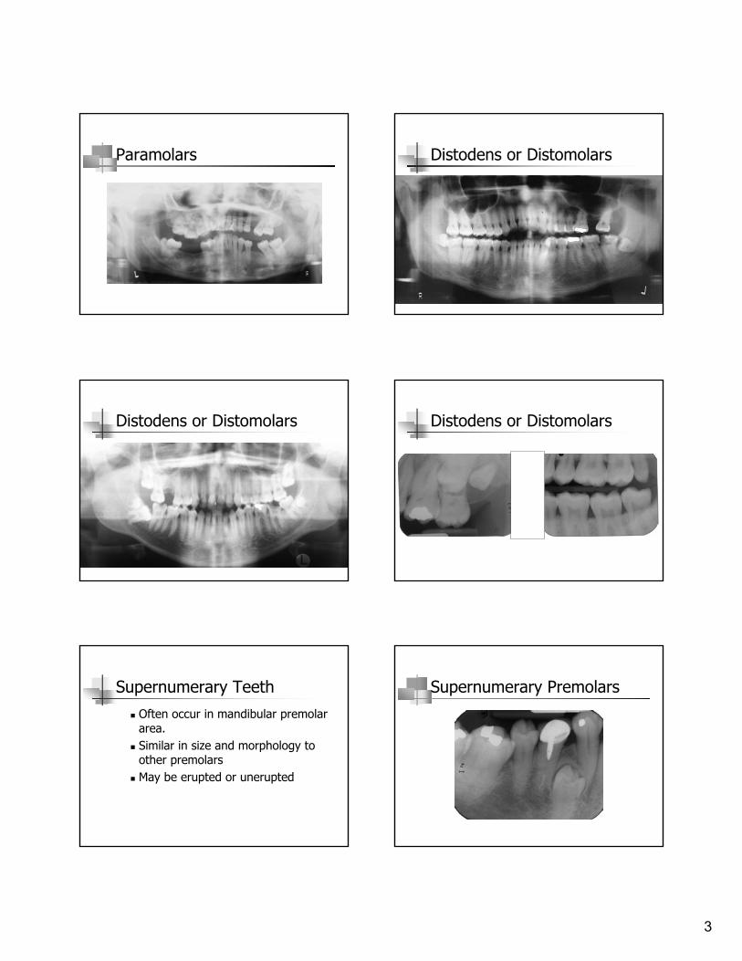
! Paramolars are additional molar teeth.! When they are positioned distal to the third
molar, they are called distodens or distomolars
Supernumerary Teeth
3
Paramolars Distodens or Distomolars
Distodens or Distomolars Distodens or Distomolars
Supernumerary Teeth
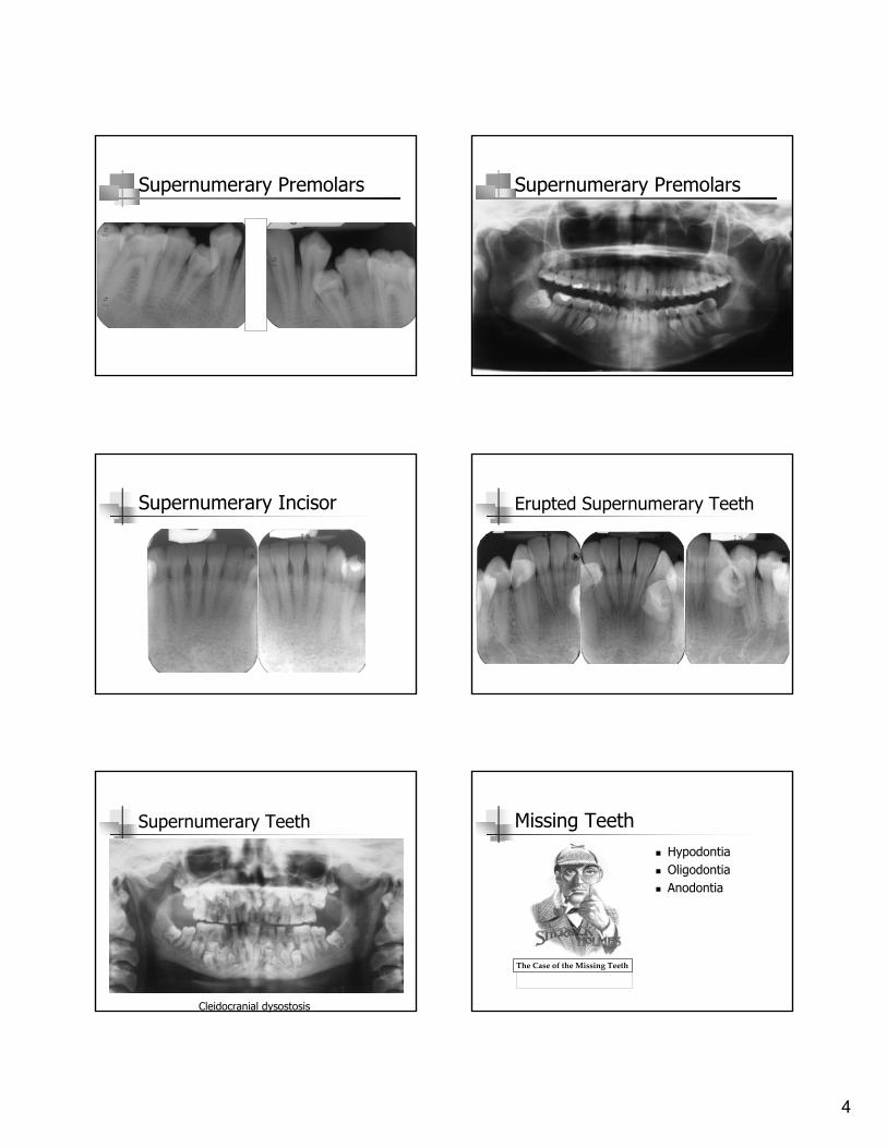
! Often occur in mandibular premolar area.
! Similar in size and morphology to other premolars
! May be erupted or unerupted
Supernumerary Premolars
4
Supernumerary Premolars Supernumerary Premolars
Supernumerary Incisor Erupted Supernumerary Teeth
Supernumerary Teeth
Cleidocranial dysostosis
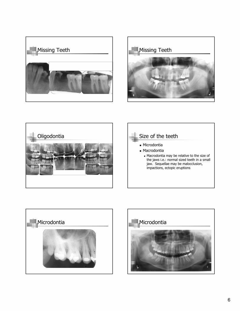
Missing Teeth! Hypodontia! Oligodontia! Anodontia
The!Case!of!the!Missing!Teeth
5
Missing Teeth
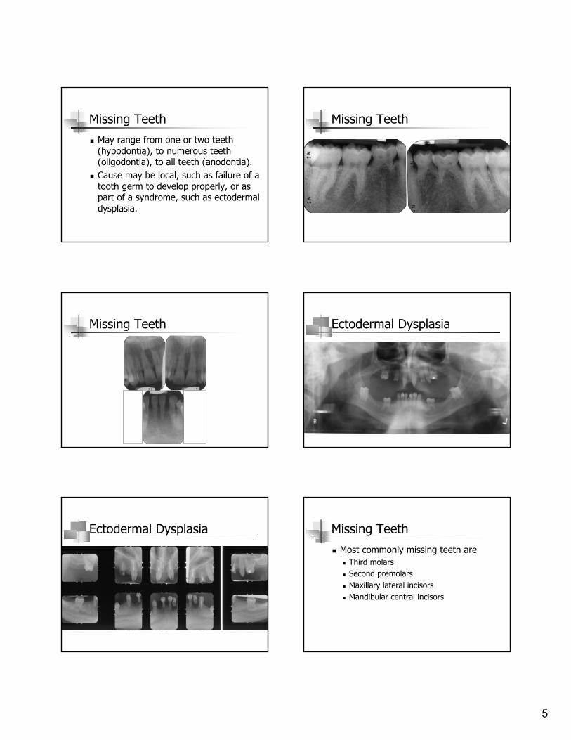
! May range from one or two teeth (hypodontia), to numerous teeth (oligodontia), to all teeth (anodontia).
! Cause may be local, such as failure of a tooth germ to develop properly, or as part of a syndrome, such as ectodermal dysplasia.
Missing Teeth
Missing Teeth Ectodermal Dysplasia
Ectodermal Dysplasia Missing Teeth
! Most commonly missing teeth are! Third molars! Second premolars! Maxillary lateral incisors! Mandibular central incisors
6
Missing Teeth Missing Teeth
Oligodontia Size of the teeth
! Microdontia ! Macrodontia
! Macrodontia may be relative to the size of the jaws i.e.: normal sized teeth in a small jaw. Sequellae may be malocclusion, impactions, ectopic eruptions
Microdontia Microdontia

7
Macrodontia Macrodontia
Morphology Morphology
Multiple canals in lower anterior teeth
Eruption of the teeth
! Transposition! Exchange of position of two teeth! Usually canine and premolar! Not reported in the primary dentition
Transposition

8
Impaction Impaction
Impaction Impaction
Altered Morphology - Fusion! Fusion is the union
of two developing teeth
! Results in fewer teeth in the arch
Cold Fusion
Altered Morphology - Fusion! Occurs in both
primary and permanent dentitions
! Morphology and mesiodistal width of the clinical crown varies
Cold Fusion

9
Fusion of the central and lateral incisors
Fusion of the central and lateral incisors
Fusion of the Central and Lateral Incisors
Fusion of the Central and Lateral Incisors (Maxillary)
Fusion of the Central and Lateral Incisors (Mandibular) Altered Morphology
! Concrescence is the union of the roots two teeth by cementum only
! May be developmental or acquired

10
Concrescence Concrescence
Hypercementosis
! Excessive deposition of cementum at apex of root
! May be:! Idiopathic! Response to inflammation! Responses to hyperocclusion! Seen with Paget’s Disease of Bone
Hypercementosis
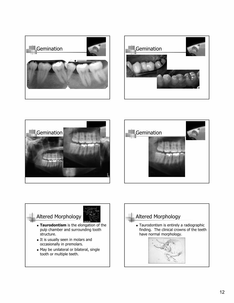
Altered Morphology! Gemination happens
when a single tooth bud attempts to divide
! Morphology varies from partial division to complete replication of all dental structures and may even result in a supernumerary tooth
Jean Arp (Hans Arp) (French, born Alsace. 1886–1966. Lived in Switzerland 1959–66.)Two Heads [Deux têtes]. (1927
Gemination

11
Gemination Gemination
Gemination Gemination
Gemination Gemination
12
Gemination Gemination
Gemination Gemination
Altered Morphology
! Taurodontism is the elongation of the pulp chamber and surrounding tooth structure.
! It is usually seen in molars and occasionally in premolars.
! May be unilateral or bilateral, single tooth or multiple teeth.
Altered Morphology
! Taurodontism is entirely a radiographic finding. The clinical crowns of the teeth have normal morphology.

13
Taurodontism
Images courtesy of SUNY Buffalo School of Dental Medicine
Taurodontism
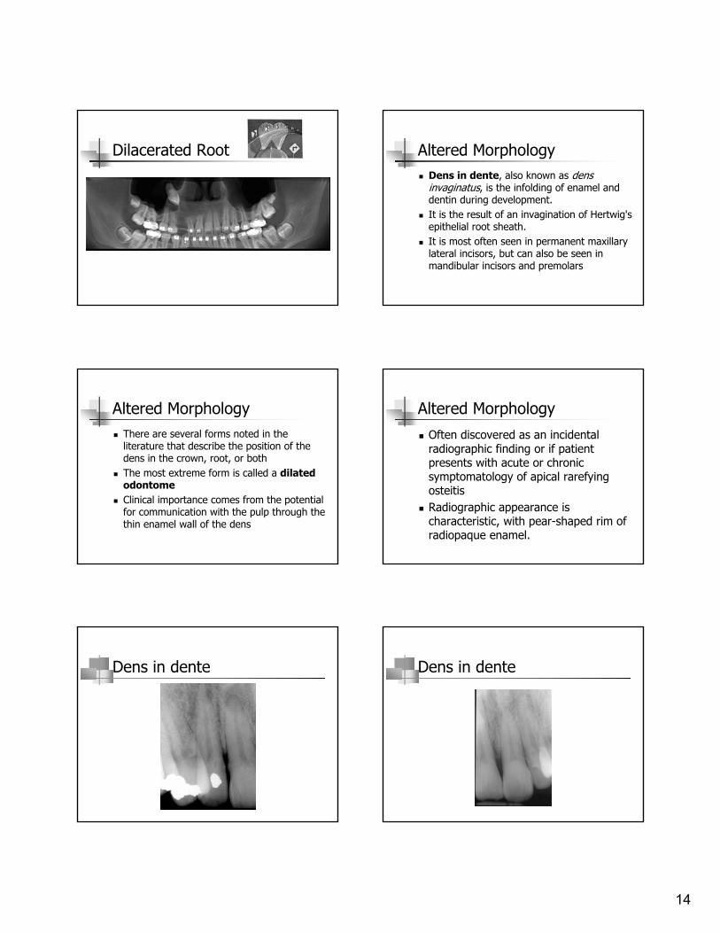
Altered Morphology-Dilaceration! A sharp bend or
angulations seen in a portion of the root
Image courtesy of www.dental.mu.edu
Dilacerated Root
Image courtesy of www.dental.mu.edu
Dilacerated Root Dilacerated Root
14
Dilacerated Root Altered Morphology! Dens in dente, also known as dens
invaginatus, is the infolding of enamel and dentin during development.
! It is the result of an invagination of Hertwig's epithelial root sheath.
! It is most often seen in permanent maxillary lateral incisors, but can also be seen in mandibular incisors and premolars
Altered Morphology! There are several forms noted in the
literature that describe the position of the dens in the crown, root, or both
! The most extreme form is called a dilated odontome
! Clinical importance comes from the potential for communication with the pulp through the thin enamel wall of the dens
Altered Morphology
! Often discovered as an incidental radiographic finding or if patient presents with acute or chronic symptomatology of apical rarefying osteitis
! Radiographic appearance is characteristic, with pear-shaped rim of radiopaque enamel.
Dens in dente Dens in dente

15
Dens in dente Dens in dente in a peg lateral
Dialated Odontome Altered Morphology! Dens evaginatus, also known as
Leong’s premolar or talon cusp, is an outpocketing of the enamel.
! It also occurs occasionally in a molar or canine.
! Often includes dentin and pulp, which may become exposed as the tubercle wears
Dens evaginatus Dens evaginatus

16
Dens Evaginatus Altered Morphology
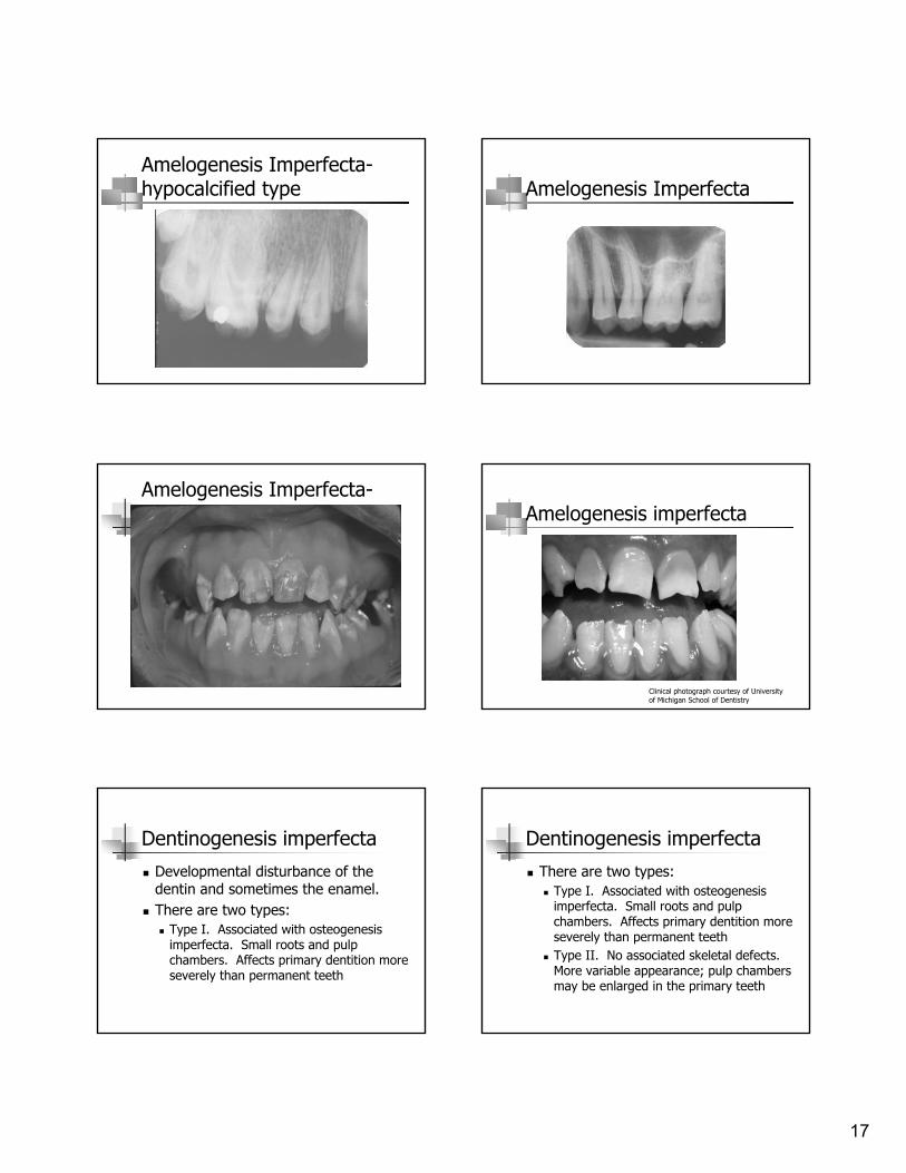
! Amelogenesis imperfect is due to a developmental disturbance and results in altered enamel formation
! 1 in 14,000 people are affected
Altered Morphology! Three varieties:
! Hypoplastic. Enamel is thin and discolored from the underlying dentin. Surface may be pitted or smooth. Teeth generally have open contacts and altered shape of crowns. There may be an anterior open bite
! Hypomaturation. Enamel has normal thickness, but is softer and may separate from dentin. The enamel is also discolored
Altered Morphology! Hypocalcification. Teeth have normal
morphology and thickness of enamel on eruption. Soft enamel fractures away easily in function. Teeth can wear to the level of gingiva in extreme cases. Caries is rare in these teeth, but they do tend to stain
Amelogenesis imperfecta (hypoplastic form) Amelogenesis Imperfecta
! Smooth hypoplastic type
17
Amelogenesis Imperfecta-hypocalcified type Amelogenesis Imperfecta
Amelogenesis Imperfecta-hypomaturation type Amelogenesis imperfecta
Clinical photograph courtesy of University of Michigan School of Dentistry
Dentinogenesis imperfecta
! Developmental disturbance of the dentin and sometimes the enamel.
! There are two types:! Type I. Associated with osteogenesis
imperfecta. Small roots and pulp chambers. Affects primary dentition more severely than permanent teeth
Dentinogenesis imperfecta
! There are two types:! Type I. Associated with osteogenesis
imperfecta. Small roots and pulp chambers. Affects primary dentition more severely than permanent teeth
! Type II. No associated skeletal defects. More variable appearance; pulp chambers may be enlarged in the primary teeth
18
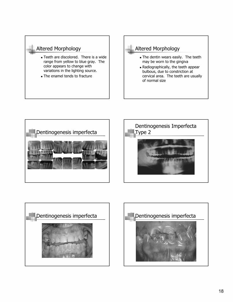
Altered Morphology
! Teeth are discolored. There is a wide range from yellow to blue gray. The color appears to change with variations in the lighting source.
! The enamel tends to fracture
Altered Morphology
! The dentin wears easily. The teeth may be worn to the gingiva
! Radiographically, the teeth appear bulbous, due to constriction at cervical area. The teeth are usually of normal size
Dentinogenesis imperfectaDentinogenesis Imperfecta Type 2
Dentinogenesis imperfecta Dentinogenesis imperfecta

19
Dentinogenesis imperfecta
Image courtesy of Marquette University School of Dentistry
Dentin dysplasia
! Resembles dentinogenesis imperfecta, but is more rare.
! Two types:! Type I Radicular. Short and malformed
roots are radiographically apparent! Type II Coronal.
Dentin Dysplasia – Type I Radicular
Image courtesy of Marquette University School of Dentistry
Odontodysplasia
PhalangeomaThank you!
![[MESIODENS OG BEHANDLINGSOVERVEJELSER] - ato.dk · Definition af Mesiodens Ifølge lærebogen: Oral og Maxillofacial Pathology, Nerville mfl (13) benævnes en overtallig tand centralt](https://img.pdfslide.net/doc/110x75/5e0b32e62d6b2c3c236771e7/mesiodens-og-behandlingsovervejelser-atodk-definition-af-mesiodens-iflge.jpg)