Embed Size (px)
Citation preview

Anatomical Modelling of the MusculoskeletalSystem from MRI
Benjamin Gilles, Laurent Moccozet, and Nadia Magnenat-Thalmann
MIRALab, University of Geneva, CH-1211 Geneva, Switzerland,{gilles, moccozet, thalmann}@miralab.unige.ch
Abstract. This paper presents a novel approach for multi-organ (mus-culoskeletal system) automatic registration and segmentation from clini-cal MRI datasets, based on discrete deformable models (simplex meshes).We reduce the computational complexity using multi-resolution forces,multi-resolution hierarchical collision handling and large simulation timesteps (implicit integration scheme), allowing real-time user control andcost-efficient segmentation. Radial forces and topological constraints (at-tachments) are applied to regularize the segmentation process. Basedon a medial axis constrained approximation, we efficiently characterizeshapes and deformations. We validate our methods for the hip joint andthe thigh (20 muscles, 4 bones) on 4 datasets: average error=1.5mm,computation time=15min.
1 Introduction
For the diagnosis, the surgical planning and the post-operative assessment ofmusculoskeletal disorders, the automatic segmentation of the patient muscu-loskeletal system is important for orthopaedists, biomechanicians and kinesiol-ogists that would like to simulate, visualize and navigate through articulationswith a minimum amount of manual tasks. As stressed by [1], [2] and [3], usualsimplified models (stick-figures, muscle action lines) are not able to take intoaccount large attachment areas, as well as global constraints such as volumepreservation and non-penetration; although they are important biomechanicalparameters. Therefore, the relationship between musculoskeletal dynamics andorgan shapes need to be better studied through image segmentation. MagneticResonance Imaging (MRI) is a flexible modality for imaging both soft and bonytissues non-invasively. However, due to the large amount of textural information,noise, low-resolution, organ imbrication, and large spatial variability; automatic,fast and robust musculoskeletal segmentation is a difficult task. As a conse-quence, existing methods for musculoskeletal modelling are interactive [2] [4] [1][3] and therefore time-consuming. A common approach to constrain a segmen-tation process is to use prior information: shape constraints rely on assumptionsabout surface regularity (smoothness, curvature) and variability across the pop-ulation, while topological constraints exploit prior knowledge about organ inter-relationships. Contrary to traditional pixel-based segmentation (e.g. level-sets)and registration (e.g. FFD) methods, these constraints can be efficiently applied
R. Larsen, M. Nielsen, and J. Sporring (Eds.): MICCAI 2006, LNCS 4190, pp. 289–296, 2006.c© Springer-Verlag Berlin Heidelberg 2006

290 B. Gilles, L. Moccozet, and N. Magnenat-Thalmann
on deformable models [5]. Physical-based simulation approaches (volumetric),including finite element or finite volume are appropriate for enforcing mechan-ical constraints, but their computational cost would exclude time-efficient anduser-controlled segmentation. In this context, we propose to use scalable discretedeformable surfaces (simplex meshes), that can benefit from efficient geometricmethods, popular in computer graphics applications.
Simplex meshes were first described by H. Delingette [6] for constrained 3Dshape reconstruction and segmentation, and extended in 4D by J. Montagnat [7],with application to the heart (single model and resolution). A k-simplex meshis defined by a set of vertices and a connectivity function (each vertex is con-nected to exactly k+1 neighbors). In this paper, we use 2-simplex meshes (dualto triangle meshes). Mesh geometric quality (uniformity of vertices repartition)and topological quality (uniformity of edge number among faces) are improvedusing simple topological operators. The most interesting property is its simplegeometric description: three parameters (two barycentric coordinates plus thecurvature) uniquely define vertex positions from their three neighbors. Based,on these parameters, smoothing and shape constraint forces are computed to reg-ularize the segmentation. External forces are obtained through 1D registrationof intensity profiles (at vertex positions and in normal direction) with genericprofiles from a reference segmentation. The external force field is regularizedusing a local smoothing and global regularization based on the closest affinetransformation.
Discrete models are commonly considered as punctual masses evolving underthe Newtonian law of motion. The Newton equation leads to a first-order differ-ential equation system relating forces to particle state (velocity and position).After forces evaluation and time discretisation, particle state can be explicitlyresolved (forward Euler, Runge-Kutta) with tight time step restrictions for en-suring stability. We prefer the more stable implicit (or backward) scheme [8][9], that however requires the resolution of a large sparse linear system. To sim-plify force derivatives evaluation, we consider that forces have a independentanisotropic action on each particle. Non-penetration constraints are based oncollision handling techniques [10]. In this paper, we apply a hierarchical collisiondetection scheme based on 18-discrete oriented polytope(DOP) quadtrees [10].We perform collision correction and response on particle positions, speeds andaccelerations, such as in [9].
From these background studies, we propose improvements in terms of compu-tational speed and robustness by extending the simplex mesh framework with amulti-resolution scheme, topological constraints and medial surfaces. We showthat the medial axis is suited for muscle shape analysis.
2 Methods
2.1 Multi-resolution Scheme
The use of levels of details (LODs) aims at reducing system complexity andsensitiveness to local solutions. As shape constraints spatial influence depends

Anatomical Modelling of the Musculoskeletal System from MRI 291
on the resolution level, the idea is to quickly propagate forces from lower reso-lutions to a current simulation LOD in order to get multiscale regularization. Inaddition, collision detection (often considered as the bottle-neck for simulation)is more efficiently performed on coarse LODs, collision response being passed tofine LODs. Indeed, exact contact computation is most of the time not relevant asfat separates organs. The tessellation of dual triangle meshes leads to a system-atic and computationally efficient LOD generation scheme for simplex meshes(linear combination of vertex positions). Shape features are preserved as lowlevel vertices are contained in higher levels. The number of vertices is quadru-pled when increasing the resolution. During the simulation, forces from lowerresolutions are linearly combined like vertex positions and added to current res-olution forces. Even if this is not exact (vertices having moved relatively to theirneighbors, since resolution increase), the estimation is still relevant, assumingthat shape constraints have enforced mesh local regularity.
2.2 Topological Constraints
The human musculoskeletal anatomy exhibits various organs interrelationships:muscles are attached to bones, they can merge into common tendon units, fasciabinds muscles and enforces frictionless contact between them. We have developeda spline-based method for generating attachment areas, so that the placementand adjustment of areas have a reduced number of degrees of freedom. Splinecontrol points are projected onto bone surfaces, while soft-tissues vertices areattached to the spline through curvilinear coordinates. These vertices are con-strained using mass modification [8]: M−1 = 0. For individualization, we wrapsplines from a generic model using spline control point barycentric coordinateson bone surface. As shown in [11], this approach is valid for most attachmentsas they rely on bone geometrical features. For merging deformable models, wesum forces and masses of the attached vertices. This is interesting for mod-elling common tendons (e.g. quadriceps femoris tendon), but also specific partswith high curvature, where smoothing forces are not appropriate (e.g. attach-ment between the adductor magnus and its inferior tendon). Fascia is modelledthrough collision handling: once generic collisions and proximities have beendetected, they are subsequently used as a reference (springs) to smoothly en-force the relative position between models. In other words, we deform genericcontacts.
2.3 Radial Constraints
Muscles generally have a smooth and tubular shape. Hence, they can be effi-ciently represented by an underlying piece-wise action line [1] [4], where iso-tonic contractions are modelled through action line shrinking/stretching, andisometric contractions through radial constraints applied to a wrapped surface.This has been applied in biomechanics for calculating joint moment arms [3].However, muscles with large attachment areas and/or several origins/insertionsrequire many action lines. We propose a continuous representation using medialsurfaces, leading to a continuous radial regularization of the overlying surface
292 B. Gilles, L. Moccozet, and N. Magnenat-Thalmann
and enhanced mechanisms for detecting collisions and characterizing shapes.Anchored medial surfaces (M-reps) have been successfully used in [5] [12] forconstraining deformable model-based segmentation. The medial axis transform(computation of maximal ball centers and radii inside an object [13]) is an in-vertible transform that allows an efficient reduction of parameters dimension.Several methods have been presented for approximating the medial axis (MA),based on Voronoi diagrams, on distance maps, or on thinning.
We propose an iterative method based on forces applied to constrained de-formable surfaces. Hook’s spring interaction forces f∗
j are applied to medialsurface vertices for MA approximation (j indexes medial vertices). Reciprocally,forces fi are applied to overlying surface vertices for MA-based shape regu-larization (i indexes model vertices). Each model vertex Pi is associated to amedial surface point Pi⊥ with a certain radius ri. Radii are linearly interpo-lated between medial surface vertices Qj of radius Rj to allow continuous forcedefinition (leading to a smooth reconstructed surface from the MA). Given theweights wij , we have Pi⊥ =
∑j wij .Qj and ri =
∑j wij .Rj . From a reference
state (Fig. 1a), weights are obtained by projecting orthogonally Pi onto the MA,and radii by the weighted mean Rj =
∑i wij .PiPi⊥/
∑i wij . Weights wij and
radii Rj are subsequently memorized to compute radial forces for deformed states(Fig. 1b). Model forces fi are simply defined by fi = k.(1 − ri/PiPi⊥)PiPi⊥where k is the spring stiffness (or force weight). By applying the momentumconservation law [9], we calculate the force contribution fij for one spring iat Qj such as fij = −wij .fi/
∑j w2
ij . We average all spring forces associatedto a MA vertex: f∗
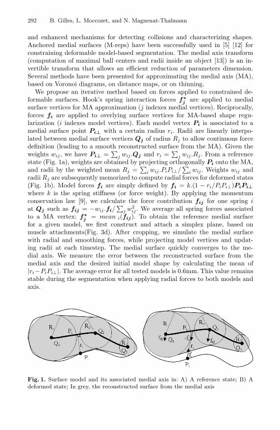
j = mean i(fij). To obtain the reference medial surfacefor a given model, we first construct and attach a simplex plane, based onmuscle attachments(Fig. 3d). After cropping, we simulate the medial surfacewith radial and smoothing forces, while projecting model vertices and updat-ing radii at each timestep. The medial surface quickly converges to the me-dial axis. We measure the error between the reconstructed surface from themedial axis and the desired initial model shape by calculating the mean of|ri−PiPi⊥|. The average error for all tested models is 0.6mm. This value remainsstable during the segmentation when applying radial forces to both models andaxis.
Fig. 1. Surface model and its associated medial axis in: A) A reference state; B) Adeformed state; In grey, the reconstructed surface from the medial axis

Anatomical Modelling of the Musculoskeletal System from MRI 293
In addition, we use medial axis for collision detection: 18-DOP quadtrees hier-archies, inflated according to radii, are generated/updated for medial axis cells.Muscles vertices at a certain resolution are subsequently tested towards DOPsand projected to medial axis for the inclusion test (comparison with interpolatedradii ri). Independently of the model resolution, medial axis based-collision de-tection is around 3 times faster than surface-based collision detection. On topof MA based-collision detection, multi-resolution collision handling (section 2.1)is used to improve computation time (by a factor of 7 per LOD). Given a cur-rent resolution, we use its first coarser LOD for collision handling in order tominimize errors.
3 Results
3.1 Automatic Segmentation of the Musculoskeletal System
MRI protocol definition has been done in close collaboration with physicians fromradiology and orthopaedics. The goal was to obtain images carrying sufficientinformation with clinically achievable protocols (fast). The final protocol wasT1-weighted spin echo with TR/TE=578/18ms, Matrix/ FOV=512x512/40cmand slice thickness=2mm to 10mm between the foot the the iliac crests (fiveseries in total, ∼150 slices, ∼30 minutes).
Based on interactive segmentation, we have reconstructed a generic modelof the hip and the thigh, composed of a skin, 20 muscles, 4 bones and thecorresponding attachment splines. After topological optimization, the differentLODs (3 for soft-tissues, 4 for bones) and medial surfaces were generated. Thefinal surface model (highest resolution) is composed of 71328 vertices for mus-cles and 85100 vertices for bones. Alternatively, muscles can be representedby their medial surfaces (1 resolution, 3821 vertices) with an average error of0.6mm. The compression factor (parameter dimension reduction) is equal to(3x71328)/(4x3821)=14 (3 dimensions for model points, 4 for axis points, in-cluding radii).
Before automatic individualization, the generic model is coarsely initialized us-ing ten manually placed landmarks corresponding to anatomical landmarks, andusing thin-plate-spline (TPS) interpolation. After this step, bones are deformed(Euler implicit integration) from the coarse level to the fine level using multi-resolution internal forces (shape and smoothing constraints), non-penetrationconstraints (multi-resolution collision handling) and intensity profile-based ex-ternal forces. Muscles are subsequently individualized (Fig. 2) as follows: A)attachment splines are initialized on bone surfaces from their generic barycen-tric coordinates; B) a skeleton-driven deformation algorithm [14] (skinning) isapplied to generic muscles and medial surfaces according to joint angles; C)soft-tissues/medial surfaces are deformed using internal forces (radial/shape andsmoothing constraints), proximity constraints (deforming contacts), and skinsurface matching (gradient-based external forces); D) soft-tissues are deformedfrom the coarse level to the fine level using internal forces, proximity/non-penetration constraints and intensity profile-based external forces. The best

294 B. Gilles, L. Moccozet, and N. Magnenat-Thalmann
intensity profile size and resolution have been experimentally defined from in-teractively segmented models by minimizing excursions. Being more variable,the external part of the profile is less relevant (thus shorter). The intensity pro-file search depth decreases from 20mm to 5mm during segmentation (Fig. 2).We found that normalized cross-correlation is the most robust metric for inten-sity profile similarity measure. The affine regularization contribution decreasesfrom 100% to 20%. For fine resolutions, internal forces from coarse levels arepropagated (global/local constraints) as presented in 2.1. Muscle shape con-straints are derived from their medial surfaces with generic radii and weights.However, radii are updated for fine levels to give more freedom to surfaces.Radial forces are applied simultaneously to surfaces and to medial axis withan equal contribution. For coarse levels, generic proximity constraints are ap-plied (deforming contacts). These constraints are released for the finest level,where multi-resolution medial axis-based collision detection is applied to allowsurface sliding.
Fig. 2. Automatic muscle segmentation process and result on a sample slice: A) Genericmodel with wrapped attachments; B) Skeleton-driven model initialization; C) Segmen-tation using internal forces and skin matching (min. resolution); D) Final segmentationusing external forces (max. resolution)
During the segmentation, it is possible to interactively place constraint pointson the images, to get a faster matching and a more accurate segmentation.Using our collision detection scheme, deformable models are forced to include orexclude these constraint points. In addition to the generic model, this methodhas been applied by a medical student to segment accurately 4 datasets from 4different healthy subjects (2 females and 2 males). We compared automaticallysegmented models to these reference models (Fig. 2). The average distance (std.dev.) was 1.25mm (1mm) for bones and 1.7mm (1.8mm) for muscles. The overallcomputation time for the automatic method is around 15min on a standard PC,for which 3/4 of the time is spent on external forces computation. Computationtime for each timestep, including visualization, is around 0.5sec.

Anatomical Modelling of the Musculoskeletal System from MRI 295
3.2 Shape Analysis
Musculoskeletal shape characterization is important for anthropometric compar-ison between individuals, and deformation analysis (temporal and longitudinalstudies). For this purpose, our method registers anatomical features throughshape and topological constraints. High-level descriptors such as the medial axisconvey more information than local descriptors (curvature). Muscle thickness canbe simply analyzed through medial axis radii comparison, as shown in Fig. 3b.Using geodesic distances to attachments, we compute normalized coordinates Xand Y along medial surfaces, from which a thickness profile can be extracted(maximum radius in Y direction). For some muscles showing thickness steepchanges, tendons lengths (which is an important biomechanical parameter) canbe automatically extracted (Fig. 3f).
Fig. 3. A) Reconstructed generic model; B) Muscle thickness comparison with an in-dividualized model (blue:≥5mm; red:≤5mm); C) Biceps femoris generic model; D) Ini-tialized medial axis (blue: max radius, red: min radius); E) Medial axis after croppingand fitting; F) Tendon selection (in red) after thickness profile analysis along Y
4 Discussion and Future Work
Prior low-level (e.g. curvature) and high-level (e.g. medial axis) shape informa-tion and topological relationships (e.g. proximities, attachments) are relevantfor musculoskeletal modelling, and complexity can be efficiently decreased usinga multi-resolution approach for force and contact computation. By constrainingthe problem, our goal is to get a fast and accurate segmentation from a minimumamount of information: we want to extend our previous work on bone motion ex-traction from real-time dynamic MRI [15] (6 low resolution slices) by extractingsoft tissue deformation. We believe that we can get a higher accuracy through adeeper study of intensity profile forces (combination of several metrics, weightingaccording to profile relevance, etc.) and by adding statistical constraints appliedto high-level descriptors (e.g. medial axis radii) according to joint angles. The

296 B. Gilles, L. Moccozet, and N. Magnenat-Thalmann
next step will be to relate these descriptors with dynamic (e.g. moment arms)and physiological parameters (e.g. muscle activation from EMG). This will pro-vide useful information for validating functional biomechanical models.
Acknowledgments. This work is supported by CO-ME (Computer Aided andImage Guided Medical Interventions) project funded by Swiss National ResearchFoundation. We would like to thank Dr. Kolo-Christophe, Dr. N’Guyen and Dr.Sadri from the Geneva University Hospital for their collaboration.
References
1. Teran, J., Sifakis, E., Blemker, S., Ng-Thow-Hing, V., Lau, C., Fedkiw, R.: Creatingand simulating skeletal muscle from the visible human data set. IEEE TVCG 11(2005) 317–328
2. Ng-Thow-Hing, V.: Anatomically-based models for physical and geometric recon-struction of humans and other animals. Ph.D. Thesis, Department of ComputerScience, University of Toronto (2000)
3. Blemker, S.S., Delp, S.L.: Three-dimensional representation of complex musclearchitectures and geometries. Annals of Biomedical Eng. 33 (2005) 661–673
4. Aubel, A., Thalmann, D.: Interactive modeling of the human musculature. Proc.of Computer Animation (2001)
5. Terzopoulos, D., Witkin, A., Kass, M.: Symmetry-seeking models and 3d objectreconstruction. International Journal of Computer Vision 1 (1987) 211–221
6. Delingette, H.: General object reconstruction based on simplex meshes. Interna-tional Journal of Computer Vision 32 (1999) 111–146
7. Montagnat, J., Delingette, H.: 4d deformable models with temporal constraints:application to 4d cardiac image segmentation. MIA 9 (2005) 87–100
8. Baraff, D., Witkin, A.: Large steps in cloth simulation. Proc. of SIGGRAPH98,Computer Graphics 32 (1998) 106–117
9. Volino, P., Magnenat-Thalmann, N.: Implementing fast cloth simulation with col-lision response. Proc. of the Int. Conference on Computer Graphics (2000) 257–268
10. Teschner, M., Kimmerle, S., Zachmann, G., Heidelberger, B., Raghupathi, L.,Fuhrmann, A., Cani, M.P., Faure, F., Magnenat-Thalmann, N., Strasser, W.: Col-lision detection for deformable objects. Proc. of Eurographics State-of-the-ArtReport (2004) 119–135
11. Kaptein, B.L., VanDerHelm, F.C.T.: Estimating muscle attachment contours bytransforming geometrical bone models. Journal of Biomechanics 37 (2004) 263–273
12. Pizer, S.M., Fletcher, P.T., Joshi, S., Thall, A., Chen, J.Z., Fridman, Y., Fritsch,D.S., Gash, A.G., Glotzer, J.M., Jiroutek, M.R., Lu, C., Muller, K.E., Tracton, G.,Yushkevich, P., Chaney, E.L.: Deformable m-reps for 3d medical image segmenta-tion. International Journal of Computer Vision 55 (2003) 85–106
13. Blum, H.: A transformation for extracting new descriptors of shape. Models forthe Perception of Speech and Visual Form (1967)
14. Kalra, P., Magnenat-Thalmann, N., Moccozet, L., Sannier, G., Aubel, A., Thal-mann, D.: Real-time animation of realistic virtual humans. Computer Graphicsand Applications 18 (1998) 42–56
15. Gilles, B., Perrin, R., Magnenat-Thalmann, N., Vallee, J.P.: Bones motion analysisfrom dynamic mri: acquisition and tracking. Proc. of MICCAI’04 2 (2004) 942–949