Embed Size (px)
Citation preview
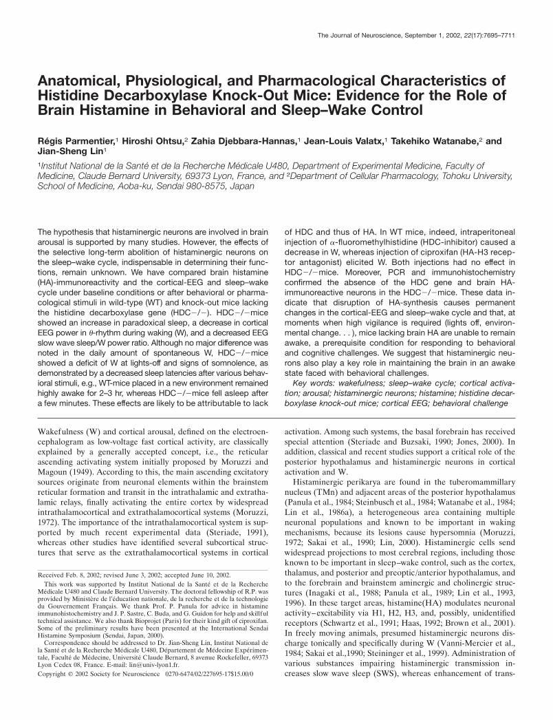
Anatomical, Physiological, and Pharmacological Characteristics ofHistidine Decarboxylase Knock-Out Mice: Evidence for the Role ofBrain Histamine in Behavioral and Sleep–Wake Control
Regis Parmentier,1 Hiroshi Ohtsu,2 Zahia Djebbara-Hannas,1 Jean-Louis Valatx,1 Takehiko Watanabe,2 andJian-Sheng Lin1
1Institut National de la Sante et de la Recherche Medicale U480, Department of Experimental Medicine, Faculty ofMedicine, Claude Bernard University, 69373 Lyon, France, and 2Department of Cellular Pharmacology, Tohoku University,School of Medicine, Aoba-ku, Sendai 980-8575, Japan
The hypothesis that histaminergic neurons are involved in brainarousal is supported by many studies. However, the effects ofthe selective long-term abolition of histaminergic neurons onthe sleep–wake cycle, indispensable in determining their func-tions, remain unknown. We have compared brain histamine(HA)-immunoreactivity and the cortical-EEG and sleep–wakecycle under baseline conditions or after behavioral or pharma-cological stimuli in wild-type (WT) and knock-out mice lackingthe histidine decarboxylase gene (HDC�/�). HDC�/�miceshowed an increase in paradoxical sleep, a decrease in corticalEEG power in �-rhythm during waking (W), and a decreased EEGslow wave sleep/W power ratio. Although no major difference wasnoted in the daily amount of spontaneous W, HDC�/�miceshowed a deficit of W at lights-off and signs of somnolence, asdemonstrated by a decreased sleep latencies after various behav-ioral stimuli, e.g., WT-mice placed in a new environment remainedhighly awake for 2–3 hr, whereas HDC�/�mice fell asleep aftera few minutes. These effects are likely to be attributable to lack
of HDC and thus of HA. In WT mice, indeed, intraperitonealinjection of �-fluoromethylhistidine (HDC-inhibitor) caused adecrease in W, whereas injection of ciproxifan (HA-H3 recep-tor antagonist) elicited W. Both injections had no effect inHDC�/�mice. Moreover, PCR and immunohistochemistryconfirmed the absence of the HDC gene and brain HA-immunoreactive neurons in the HDC�/�mice. These data in-dicate that disruption of HA-synthesis causes permanentchanges in the cortical-EEG and sleep–wake cycle and that, atmoments when high vigilance is required (lights off, environ-mental change. . . ), mice lacking brain HA are unable to remainawake, a prerequisite condition for responding to behavioraland cognitive challenges. We suggest that histaminergic neu-rons also play a key role in maintaining the brain in an awakestate faced with behavioral challenges.
Key words: wakefulness; sleep–wake cycle; cortical activa-tion; arousal; histaminergic neurons; histamine; histidine decar-boxylase knock-out mice; cortical EEG; behavioral challenge
Wakefulness (W) and cortical arousal, defined on the electroen-cephalogram as low-voltage fast cortical activity, are classicallyexplained by a generally accepted concept, i.e., the reticularascending activating system initially proposed by Moruzzi andMagoun (1949). According to this, the main ascending excitatorysources originate from neuronal elements within the brainstemreticular formation and transit in the intrathalamic and extratha-lamic relays, finally activating the entire cortex by widespreadintrathalamocortical and extrathalamocortical systems (Moruzzi,1972). The importance of the intrathalamocortical system is sup-ported by much recent experimental data (Steriade, 1991),whereas other studies have identified several subcortical struc-tures that serve as the extrathalamocortical systems in cortical
activation. Among such systems, the basal forebrain has receivedspecial attention (Steriade and Buzsaki, 1990; Jones, 2000). Inaddition, classical and recent studies support a critical role of theposterior hypothalamus and histaminergic neurons in corticalactivation and W.
Histaminergic perikarya are found in the tuberomammillarynucleus (TMn) and adjacent areas of the posterior hypothalamus(Panula et al., 1984; Steinbusch et al., 1984; Watanabe et al., 1984;Lin et al., 1986a), a heterogeneous area containing multipleneuronal populations and known to be important in wakingmechanisms, because its lesions cause hypersomnia (Moruzzi,1972; Sakai et al., 1990; Lin, 2000). Histaminergic cells sendwidespread projections to most cerebral regions, including thoseknown to be important in sleep–wake control, such as the cortex,thalamus, and posterior and preoptic/anterior hypothalamus, andto the forebrain and brainstem aminergic and cholinergic struc-tures (Inagaki et al., 1988; Panula et al., 1989; Lin et al., 1993,1996). In these target areas, histamine(HA) modulates neuronalactivity–excitability via H1, H2, H3, and, possibly, unidentifiedreceptors (Schwartz et al., 1991; Haas, 1992; Brown et al., 2001).In freely moving animals, presumed histaminergic neurons dis-charge tonically and specifically during W (Vanni-Mercier et al.,1984; Sakai et al.,1990; Steininger et al., 1999). Administration ofvarious substances impairing histaminergic transmission in-creases slow wave sleep (SWS), whereas enhancement of trans-
Received Feb. 8, 2002; revised June 3, 2002; accepted June 10, 2002.This work was supported by Institut National de la Sante et de la Recherche
Medicale U480 and Claude Bernard University. The doctoral fellowship of R.P. wasprovided by Ministere de l’education nationale, de la recherche et de la technologiedu Gouvernement Francais. We thank Prof. P. Panula for advice in histamineimmunohistochemistry and J. P. Sastre, C. Buda, and G. Guidon for help and skillfultechnical assistance. We also thank Bioprojet (Paris) for their kind gift of ciproxifan.Some of the preliminary results have been presented at the International SendaiHistamine Symposium (Sendai, Japan, 2000).
Correspondence should be addressed to Dr. Jian-Sheng Lin, Institut National dela Sante et de la Recherche Medicale U480, Departement de Medecine Experimen-tale, Faculte de Medecine, Universite Claude Bernard, 8 avenue Rockefeller, 69373Lyon Cedex 08, France. E-mail: [email protected] © 2002 Society for Neuroscience 0270-6474/02/227695-17$15.00/0
The Journal of Neuroscience, September 1, 2002, 22(17):7695–7711
mission promotes W (Lin et al., 1988; Monti et al., 1991).Muscimol-induced inactivation of the posterior hypothalamuscontaining HA cells results in hypersomnia in both normal orexperimentally induced insomniac cats (Lin, 2000). Finally, inhi-bition of HA synthesis in the same area increases SWS, whereasinhibition of HA degradation elicits long-lasting arousal (Lin etal., 1986b, 1988).
These results support the potential importance of histaminer-gic neurons in arousal, but the effects of selective long-termabolition of HA neurons on the sleep–wake cycle, indispensablein determining their role, remain unknown. Although attemptshave been made in cats to abolish neuronal activity in theposterior-hypothalamus either temporarily (Lin et al., 1989) orpermanently (Sallanon et al., 1988), these experimental ap-proaches affect not only histaminergic cells, but also nonhista-minergic neurons codistributed in the same or adjacent regions,and so the observed hypersomnia cannot be definitively attrib-uted to the loss of HA cells. Among the nonhistaminergic cellspresent in the posterior-hypothalamus, a subpopulation, locatedin the perifornical area, just rostrodorsal to the histaminergicTMn and containing orexin–hypocretin neuropeptides has re-cently been identified in the mammalian CNS (de Lecea et al.,1998; Sakurai et al., 1998). There is evidence that orexin defi-ciency might be responsible for the pathogenesis of human andanimal narcolepsy (Lin et al., 1999) and that, like HA neurons,orexin-containing cells may be involved in arousal by their wide-spread projections. It is therefore necessary to clarify the respec-tive role of histaminergic and orexin neurons in sleep–wakecontrol using animal models with selective abolition of each ofthese systems. In terms of the orexin system, the sleep–wake cyclehas been studied in knock-out (KO) (Chemelli et al., 1999) andgenetic abolition (Hara et al., 2001) models. As to the HA system,Ohtsu et al. (2001) have recently developed KO mice lackinghistidine decarboxylase (HDC), the sole enzyme responsible forHA synthesis, and have demonstrated the absence of HA synthe-sis in these HDC�/�mice, thus providing a unique experimentalmodel for determining the impact of long-term selective abolitionof the HA system on the sleep–wake cycle. The present study wastherefore designed to compare the brain HA system, corticalEEG, and sleep–wake cycle under spontaneous conditions orafter behavioral or pharmacological stimuli in wild-type andHDC�/� mice.
MATERIALS AND METHODSAnimals and surgeryFifteen pairs of male inbred (129/Sv strain) wild-type (HDC�/� or WT)and histidine decarboxylase gene knock-out (HDC�/� or KO) micewere used. The HDC�/� mice were generated according to previouslydescribed procedures (Ohtsu et al., 2001). Briefly, linearized targetingconstruct was electroporated into R1 embryonic stem cells, derived from129/Sv embryo (Nagy et al., 1993). The chimeric mice, generated with theconfirmed embryonic stem cells, were crossed with 129/Sv mice to obtainthe inbred �/� mice. The animals were kept on 129/Sv genomic back-ground. All experiments followed EEC Directive (86/609/EEC), andevery effort was made to minimize the number of animals used and anypain and discomfort.
At the age of 12 weeks and with a body weight of 27–33 gm, the animalswere chronically implanted, under deep sodium pentobarbital anesthesia(60–70 mg/kg, i.p.), with six cortical electrodes (gold-plated tinnedcopper wire, � � 0.4 mm; Filotex, Draveil, France) and three muscleelectrodes (fluorocarbon-coated gold-plated stainless steel wire, � � 0.03mm; Cooner Wire, Chatworth, CA) to record the electroencephalogram(EEG) and electromyogram (EMG) and to monitor the sleep–wakecycle. All electrodes were previously soldered to a multichannel electricalconnector, and each was separately insulated with a covering of heat-
shrinkable polyolefin–polyester tubing. The cortical electrodes were in-serted into the dura through three pairs of holes (� � 0.5 mm) made inthe skull, located, respectively, in the frontal (1 mm lateral and anteriorto the bregma), parietal (1 mm lateral to the midline at the midpointbetween the bregma and lambda), and occipital (2 mm lateral to themidline and 1 mm anterior to the lambda) cortices. The muscle elec-trodes were inserted into the neck muscles. Finally, the electrode assem-bly was anchored and fixed to the skull with Super-Bond (Sun MedicalCo., Shiga, Japan) and dental cement. This implantation allowed stablepolygraphic recordings to be made for �4 months.
Polygraphic recording and data acquisition and analysisAfter surgery, the animals were housed individually in transparent bar-rels (� 20 cm, height 30 cm) in an insulated sound-proofed recordingroom maintained at an ambient temperature of 22 � 1°C and on a 12 hrlight /dark cycle (lights on at 7:00 A.M.), food and water being availablead libitum. In some animals and for some experiments, a video camerawith infrared and digital time recording capabilities was set up in therecording room to observe and score the animal’s behavior during boththe light and dark phases. After a 3–4 d recovery period, the mice werehabituated to the recording cable for 10 d before starting polygraphicrecordings.
Cortical EEG (ipsilateral and contralateral frontoparietal and fronto-occipital leads) and EMG signals were amplified, digitized with a reso-lution of 200 and 100 Hz, respectively, and computed on a CED 1401 Plus(Cambridge, UK). Using a Spike2 script and with the assistance ofspectral analysis using the fast Fourier transform, polygraphic recordswere visually scored by 30 sec epochs for wakefulness (W), slow wavesleep (SWS), and paradoxical sleep (PS) according to previously de-scribed criteria validated for mice (Valatx, 1971; Valatx and Bugat,1974).
To avoid any variation caused by the positioning of cortical electrodes,the cortical EEG used for power spectral density analysis was capturedfrom frontoparietal leads, set with reference to the bregma, lambda, andmidline in all mice. EEG power spectra were computed for consecutive30 sec epochs within the frequency range of 0.8–60 Hz using a fastFourier transform routine. The data were collapsed in 0.4 Hz bins. Onthe basis of visual and spectral analysis, epochs containing artifactsoccurring during active waking (with large movements) were visuallyidentified and omitted from the spectral analysis when the thresholdvalue in the 0–1 Hz band was exceeded; this represented 1.96 � 0.94%of the total recording time. The power densities obtained for each statewere summed over the frequency band of 0.8–60 Hz (total power). Tostandardize the data, all power spectral densities at the different fre-quency ranges, i.e., �, 0.8–2.4 Hz; �, 3–9 Hz; �, (spindle) 9–19 Hz; �,20–30 Hz; �, 30–60 Hz; and ���, 20–60 Hz, were expressed as apercentage relative to the total power (e.g., power in the � range/powerin the 0.8–60 Hz range) of the same epochs. To evaluate contrast in thecortical EEG between SWS and W or PS, we used an EEG power ratiodetermined by the averaged cortical EEG total power density duringSWS divided by that during either W or PS.
Experimental proceduresIn each experiment, recordings were simultaneously made from an equalnumber (usually in batches of three) of HDC�/� and HDC�/� mice.The mice were submitted to the following experimental procedures.
Spontaneous cortical EEG and sleep–wake cycle. During the period ofdays 15–45 after surgery, drug-naive mice (11 pairs) were subjected tothree separate 24 hr recording sessions, beginning at 7:00 P.M. Duringeach recording session, the animals were left undisturbed. The data fromthe two sets of mice were then compared.
Cortical EEG and sleep–wake cycle af ter behavioral stimuli. Recordingswere made from HDC�/� and HDC�/� mice for 24 hr after each of thethree tests described below, which were performed in a random se-quence. As a criterion of sedation and drowsiness, the latencies to SWSand PS, defined as the time between the end of the stimuli and the onsetof the first SWS or PS episode lasting �30 sec, were also measured. Thethree tests consisted of: (1) a simulation of injection (at either 10:00 A.M.or 8:00 P.M.; n � 26 from 13 pairs of animals), consisting of the handlingof the animal and sham intraperitoneal injection without needle inser-tion; (2) a change of litter (at 2:00 P.M.; n � 36 from 15 pairs of mice),which was a routine care performed at light phase every 4–6 d to cleanthe cage and which usually causes a period of waking and behavioralexcitation in rodents; in this test, we compared the excitability of the twogroups of mice following this routine care; (3) a new environment, the
7696 J. Neurosci., September 1, 2002, 22(17):7695–7711 Parmentier et al. • Cortical EEG and Sleep–Wake Cycle in Mice Lacking Histamine Synthesis
mice being transferred for 4 hr from their habitual transparent barrel toan opaque rectangular box (21 � 30 cm, height 20 cm, with open field);in this test, the ability of the two genotypes to remain awake after thisenvironmental change was tested. Each mouse was subjected to this testfour times separated by an interval of 10–14 d, twice at 2:00 P.M. whenthe animals normally being sleepy for �80% of the time (defined as“sleepy period”; n � 22 from 11 pairs of mice) (Fig. 1 A), and twice at6:00 P.M. when they would normally be awake a majority of the time(defined as “awake period”; n � 18 from nine pairs of mice) (Fig. 1 A).Sleep–wake stages during their stay in the new environment were com-pared between the two groups and with the baseline recordings for thesame group.
Cortical EEG and the sleep–wake cycle af ter drug administration. Tocompare the effects of drugs acting on the histaminergic system, the twogenotypes of mice were injected intraperitoneally with the followingagents with an interval of at least 7 d between injections, subsequentrecordings being made for 24 hr: (1) saline alone (0.1 ml) and salinecontaining ( S)-�-fluoromethylhistidine (�-FMH; 50 mg/kg; Merck,Sharp, & Dohme; n � 14 for each genotype; seven pairs of mice), aspecific HDC inhibitor (Kollonitsch et al., 1978; Garbarg et al., 1980;Maeyama et al., 1982); injections were given at 4:00 P.M. (2) Saline alone(0.1 ml) and saline containing cyclopropyl-(4-(3-(1H-imidazol-4-yl) pro-pyloxy) phenyl) ketone (ciproxifan, 1 mg/kg; Bioprojet, Paris, France)(n � 9 for each genotype; nine pairs of mice), a potent and specific
antagonist of histamine-H3 receptor (Ligneau et al., 1998) that controlshistamine release and synthesis by autoinhibition; injections were givenat 10:00 A.M. (sleepy period). In addition, some animals (nine pairs)were injected subcutaneously with either saline, alone or containinghistamine dihydrochloride (HA; Sigma, St. Louis, MO; 1 mg/kg, dis-solved in saline and adjusted to pH 7) during either the light (10:00 A.M.;n � 9) or dark phase (8:00 P.M.; n � 9) to assess the effect of peripheralHA on the cortical EEG and the sleep–waking cycle. All drugs, ex-pressed as salt weight, were dissolved immediately before use. Results inthe saline- and drug-injected animals were compared.
Detection of the HDC gene using the PCR. To confirm the genotypes ofthe mice used with respect to the HDC gene, at the end of the experi-ments, tail biopsies were taken from all mice and analyzed by PCR. TheWT allele was amplified using primers located within the HDC genefragment that was replaced by the neomycin resistant gene in the KOmice. These primers were 5�-AGT GAG GGA CTG TGG CTC CACGTC GAT GCT-3� (complementary to HDC gene 833–862) and 5�-TACAGT CAA AGT GTA CCA TCA TCC ACT TGG-3� (HDC gene980–951), the expected product size being 147 base pairs. The mutantallele was amplified using primers located within the neo r gene, thesebeing 5�-AAA CAT CGC ATC GAG CGA GCA CGT ACT CGG-3�and 5�-ATG TCC TGA TAG CGG TCC GCC ACA CCC AGC-3�, withan expected product size of 244 base pairs. These two sets of primerswere included concomitantly and PCR was performed using 40 cycles of
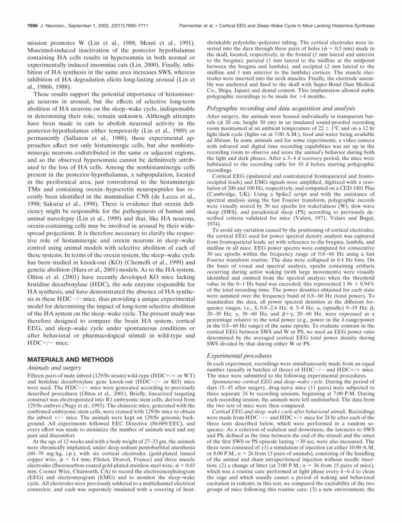
Figure 1. Quantitative comparison of spontaneous sleep–wake parameters in inbred HDC�/� and HDC�/� mice. HDC�/� mice, filled symbols andcolumns; HDC�/� mice, unfilled symbols and columns. A, Mean hourly values (� SE) of the sleep–wake states. The gray area corresponds to the periodbetween 6:00 and 10:00 P.M., and the total sleep–wake values for each state during this period for both genotypes are indicated in the histogram. B,Means (� SE) of sleep–wake stages for the 12 hr light (Day) and dark (Night) periods and the 24 hr period. C, Mean values (� SE) of episode durationand number of episodes of each sleep–wake stage for all 24 hr recordings. Note that, compared with HDC �/� mice, HDC �/� mice exhibit thefollowing: (1) a deficit of W immediately before and after lights-off (A), without major change in the daily amount of W or SWS (B); (2) an increasein PS, mainly in the light phase, because of an increase in the number of episodes (A–C); and (3) a fragmented sleep–wake architecture, with shortenedepisode duration and increased number of episodes in W and SWS (C). Note also the small interindividual SDs for the sleep–wake stages within eachgenotype group, indicating that each group was genetically homogenous (n � 33, corresponding to 3 � 24 hr recordings for 11 animals of each genotype).*p 0.05; **p 0.01; ***p 0.001; ****p 0.0001, using a two-tailed t test after significance in a two-way ANOVA for repeated measures).
Parmentier et al. • Cortical EEG and Sleep–Wake Cycle in Mice Lacking Histamine Synthesis J. Neurosci., September 1, 2002, 22(17):7695–7711 7697
30 sec at 94°C, 1 min at 64°C, and 1 min at 72°C, followed by one cycleat 72°C for 10 min. The whole reaction mix was then fractionated on a 2%agarose gel, and the PCR product was visualized by ethidium bromidestaining.
Histamine immunohistochemistry. To examine the fate of brain hista-minergic neurons after HDC gene disruption, at the end of the otherexperiments, HA immunohistochemistry was performed as described byPanula et al. (1988), Lin et al. (1993, 1996), and Eriksson et al. (2001).Briefly, the WT and KO animals were anesthetized with an overdose ofsodium pentobarbital (�100 mg/kg) and perfused transcardially with 50ml of Ringer’s lactate solution containing 0.1% heparin, followed by 60ml of ice-cold 0.1 M phosphate buffer (PB), pH 7.4, containing 4%1-ethyl-3(3-dimethyl-aminopropyl)-carbodiimide and 0.1% N-hydroxy-succinimide (both from Sigma). After 48 hr postfixation in the samesolution and 48 hr rinsing in PB containing 30% sucrose and 0.1%sodium azide, the brains were coronally sectioned (25 �m thickness) ona freezing cryostat. Free-floating sections were then incubated at 4°C for84–96 hr on an agitator with rabbit anti-HA antibody (Delichon, Masala,Finland) diluted 1:20,000–80,000 in PB saline containing 0.3% TritonX-100 (PBS-T) and 0.1% sodium azide. The specificity of the anti-HAantibody has been demonstrated in several species in previous studies
(Panula et al., 1988; Lin et al., 1993, 1996), including mice (Airaksinen etal., 1992). After rinses, the sections were submitted to one of thefollowing procedures:
(1) Some were incubated at 4°C overnight on an agitator with CyTM3-conjugated anti-rabbit IgG (1: 800; Jackson ImmunoResearch, WestGrove, PA) in PBS-T, then immediately mounted on glass slides, andcoverslipped with glycerin and examined on a light microscope equippedwith epifluorescent illumination with a filter to view CyTM3. A positivereaction was seen as gold–orange fluorescent staining of the labeledstructures (see Figs. 11,12).
(2) Others were incubated with either anti-rabbit IgG antibody (1:1000–2000; Jackson ImmunoResearch, West Grove, PA) or biotinylatedanti-rabbit IgG antibody (1: 1000–2000; Vector Laboratories, Burlin-game, CA), then, after several rinses, were incubated with rabbitperoxidase-anti-peroxidase (PAP; 1:2000–40,000; Jackson ImmunoRe-search) or horseradish peroxidase-conjugated streptavidin (1:40,000;Jackson ImmunoResearch), or a Vectastain ABC kit (1:2000; VectorLaboratories). Both incubations were in PBS-T at 4°C overnight onan agitator. The sections were then immersed for 5–10 min at roomtemperature in 0.05 M Tris-HCl buffer, pH 7.6, containing 0.02% 3,3�dia-minobenzidine-4HCl, 0.6% nickel ammonium sulfate, and 0.003% H2O2.
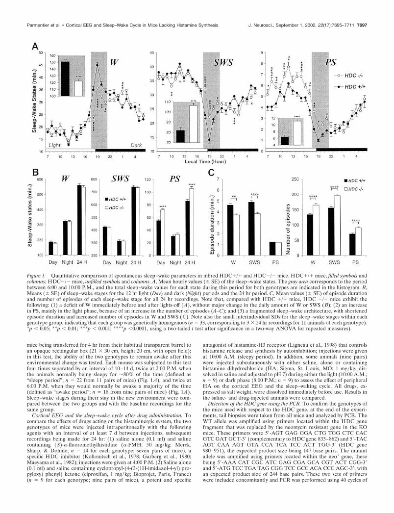
Figure 2. Typical examples of polygraphic recordings and corresponding hypnograms illustrating the spontaneous sleep–wake cycle before and afterlights-off and sleep–wake state transitions in HDC�/� and HDC�/� mice. Samples on the hour range (A, B) or second range (a, b) scale from anHDC�/� (A, a) or HDC�/� (B, b) mouse, showing the following: (1) the cortical EEG signs of both genotypes (a, b); and (2) the decreased waking(A, B) around the lights-off and reduced cortical electroencephalogram (EEG) SWS/W amplitude ratio (A, B, a, b) in the HDC�/� mouse. Calibration:200 �V, 1 sec. EMG, Electromyogram.
7698 J. Neurosci., September 1, 2002, 22(17):7695–7711 Parmentier et al. • Cortical EEG and Sleep–Wake Cycle in Mice Lacking Histamine Synthesis
A positive reaction resulted in blue–black staining of the labeled struc-tures (somata, dendrites, axons, and varicosities). Some sections werecounterstained with neutral red to identify topographic and cellularstructures. Finally, all sections were immediately mounted on gelatin-coated glass slides, dried, and coverslipped with Depex for lightmicroscopy.
Whereas the biotin-conjugated reagents proved to be incompatiblewith the carbodiimide-perfused mouse tissue, specific clear labeling wasseen using fluorescent staining or nonconjugated IgG/PAP, and the datapresented are those using these methods. The atlas of Franklin andPaxinos (1996) was used for the anatomical nomenclature of cerebralregions and for their abbreviations.
Statistical analysisANOVA and the post hoc Student’s t test (two-tailed) were used toevaluate differences between HDC�/� and HDC�/�mice in the corti-cal EEG and sleep–wake parameters under normal conditions or aftertreatment and differences in these parameters between control data(baseline recordings or saline injection) and data after treatment in thesame group of animals; in the latter case, individual animal served as itsown control.
RESULTSGeneral observationsAs wild-type (WT or HDC�/�) mice, HDC gene disrupted(HDC�/� or KO) mice appeared to develop normally. No ap-parent troubles were noted in terms of fertility, general morphol-ogy, movement, or other behaviors under basal conditions. Nev-ertheless, compared with HDC�/� animals, HDC�/� miceseemed to be less reactive when handled, and, at the age of �12weeks, their body weight was greater (31.6 � 0.9 gm at the age of97 � 7 d; n � 15 vs 27.8 � 0.5 gm at the age of 93 � 3 d; n � 15;p 0.001; Student’s t test). This difference in body weightincreased with age (e.g., the respective weights at 42 weeks were40.5 � 2.6 gm; n � 11 vs 33.0 � 1.1 gm; n � 11; p 0.02;Student’s t test).
Spontaneous sleep–wake cycle in HDC�/� miceUnder basal conditions in which the animals were left undis-turbed, both sets of mice exhibited a circadian sleep–waking
rhythm characteristic of 129/Sv (Huber et al., 2000) and othermice (Franken et al., 1999), i.e., with larger amounts of W duringthe dark period than during the light one (Fig. 1A,B). Quantita-tive analysis of the time spent in each sleep–wake stage duringthe diurnal and nocturnal periods or over 24 hr showed a signif-icantly greater amount of PS in HDC�/�mice. This increaseoccurred mainly during the light phase because of an increasednumber of PS episodes and also led to a PS augmentation of�23% over 24 hr (Fig. 1B), PS episode duration remainingunchanged during both light/dark phases (Fig. 1C). No directonset of PS phases from W was seen in either the lights-on orlights-off period. With respect to W and SWS, HDC�/� micedisplayed a significant decrease in episode duration and an in-crease in episode number for both states (Fig. 1C) and duringboth light/dark phases, e.g., the mean W episode duration over 24hr was 3.7 min � 0.1 instead of 4.6 min � 0.2 seen in HDC�/�mice. However, these changes did not result in a significantchange in the total amount of W and SWS during either the lightor dark phase or over 24 hr (Fig. 1B).
Despite this lack of a major change in the daily amount ofspontaneous W, hourly analysis of sleep–wake states (Fig. 1A)revealed a significantly smaller amount of W in HDC�/� miceduring the period of 6:00–10:00 P.M, corresponding to the peri-ods before and after lights-off at 7:00 P.M. Thus, like mostrodents, the HDC�/� mice anticipated and responded to lights-off with a significant increase in W (as a result of increased Wepisode duration: 6.5 min � 0.3 vs 4.6 min � 0.2; the mean valueover 24 hr; p 0.001), accompanied by a high level of behavioralactivity, whereas this characteristic phenomenon was markedlyreduced or almost absent in the HDC�/� mice (4.1 � 0.1 vs 3.7min � 0.1, the mean value over 24 hr; p � 0.05) (see Fig. 1A andexamples of polygraphic recordings in Fig. 2A,B). Concomitant tothe deficit in W in HDC�/� mice (122.6 � 2.8 vs 151.2 � 3.3 minin HDC�/� mice; p 0.001) there was an increase in both SWSand PS (Fig. 1A, boxed areas). The W deficit during this periodwas compensated for over 24 hr, because there was no majorchange in the daily total W (Fig. 1B).
Because an age-related increase in body weight was found inHDC�/� mice and to determine the possible effect this mighthave on the sleep–wake cycle or vice versa, we also examined therelationship between body weight and sleep–wake parameters inall mice. No correlation was found between body weight (at age of12 weeks) and the daily amount of sleep–wake stages in eithergenotype, except for a negative correlation with PS (linear re-gression, p � 0.034) in the HDC�/�group. At the age of �40weeks, the body weight of HDC�/� mice increased significantly,whereas their daily amount of PS remained unchanged (data notshown). These data indicate that there is no simple causal linkbetween body weight and the sleep–wake change seen inHDC�/� mice. Further studies, such as controlling the animal’sfood intake, activity, and metabolism, are required to determinethe mechanisms by which HDC�/� mice regulate their bodyweight.
Characteristics of cortical EEG in HDC�/� miceFrom the frontoparietal leads (Fig. 2; see Fig. 4) as the fronto-occipital ones (data not shown), the cortical EEG of both sets ofanimals manifested marked and specific changes across the be-havioral states and signs characteristic of mice, i.e., with domi-nant presence of � frequencies, notably during PS and W. Nev-ertheless, compared with HDC�/� ones, HDC�/�mice showedchanges in the following aspects:
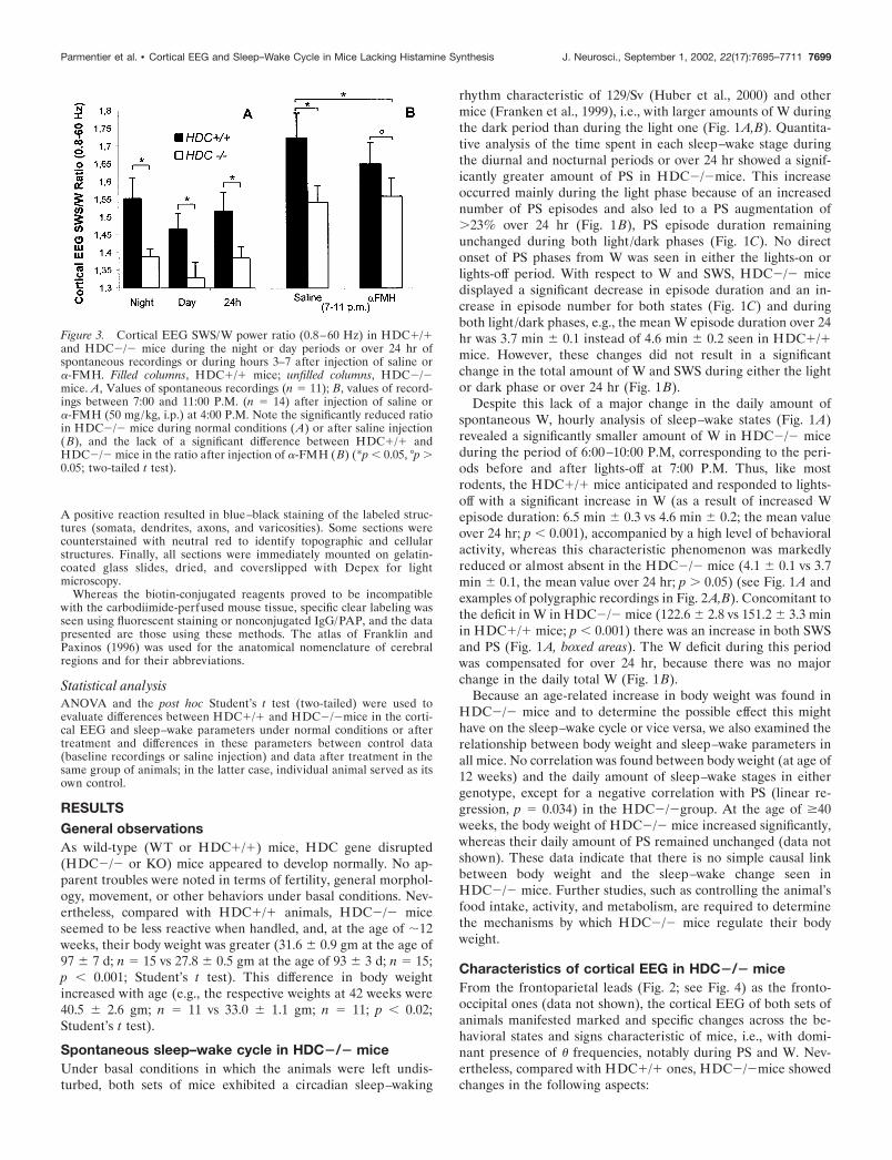
Figure 3. Cortical EEG SWS/W power ratio (0.8–60 Hz) in HDC�/�and HDC�/� mice during the night or day periods or over 24 hr ofspontaneous recordings or during hours 3–7 after injection of saline or�-FMH. Filled columns, HDC�/� mice; unfilled columns, HDC�/�mice. A, Values of spontaneous recordings (n � 11); B, values of record-ings between 7:00 and 11:00 P.M. (n � 14) after injection of saline or�-FMH (50 mg/kg, i.p.) at 4:00 P.M. Note the significantly reduced ratioin HDC�/� mice during normal conditions (A) or after saline injection(B), and the lack of a significant difference between HDC�/� andHDC�/� mice in the ratio after injection of �-FMH (B) (*p 0.05, °p �0.05; two-tailed t test).
Parmentier et al. • Cortical EEG and Sleep–Wake Cycle in Mice Lacking Histamine Synthesis J. Neurosci., September 1, 2002, 22(17):7695–7711 7699

A reduced cortical EEG SWS/W power ratioAs shown in Figure 2 (see also Figs. 5, 8), one remarkable andvisually detectable change in the cortical EEG in HDC�/� micewas a reduction in the EEG SWS/W amplitude ratio. This obser-vation is confirmed by an analysis of the averaged cortical EEGSWS/W power (0.8–60 Hz) (Fig. 3A) showing that, in HDC�/�mice, the power ratio was higher during the darkness than duringthe light phase, with the maximal ratio found around lights-off(4:00–10:00 P.M.) (Fig. 3B), and that the ratio in HDC�/� micewas significantly lower than that in HDC�/� mice, during eitherphase or over 24 hr (Fig. 3A). This decreased ratio was seenduring all recorded baseline periods (i.e., days 15–45 after sur-
gery). The reduced SWS/W power ratio was mainly attributableto a reduced peak power and amplitude of the cortical EEG ofSWS. Although these parameters also decreased during W, thatcould not contribute to such a reduced ratio (Fig. 4). In addition,the cortical EEG SWS/PS power ratio was also lower inHDC�/� mice (data not shown). Because of these changes in thecortical EEG desynchronization/synchronization power ratios inHDC�/� mice, we then analyzed each cortical EEG frequencyduring all sleep–wake states in both HDC�/� and HDC�/�mice. As shown in Figure 4, A1 and A2, both genotypes displayedstate-dependent cortical EEG spectral profiles, with peak powersof W, SWS, and PS similar to those reported for the 129/Sv
Figure 4. Mean spectral distribution of cortical EEG power density in spontaneous sleep–wake states in inbred HDC�/� and HDC�/� mice. The datawere obtained from 14 pairs of animals by pooling consecutive 30 sec epochs during the period of 7:00–10:00 P.M. using the fast Fourier transformroutine within the frequency range of 0.8–60 Hz. A1, A2, Mean absolute power values (in square microvolts) in each 0.4 Hz frequency bin. Note thestate-dependent profiles of cortical EEG spectra across wakefulness (W ), slow wave sleep (SWS), and paradoxical sleep (PS) in HDC�/� (A1) andHDC�/� (A2) mice. B1–B3, Mean percentage power density calculated as the mean power (in square microvolts) in each 0.4 Hz frequency bin dividedby the total power (0.8–60 Hz) in the same epoch. The spectra from HDC�/� mice were set to the same apparent sizes to those of the same animalsin A1 to facilitate comparison. The inset on B1 is enlarged view for 0.8–2.4 Hz. B4, EEG power spectra in HDC�/� mice (columns, n � 14) expressedas a mean percentage change (� SE) relative to those (� SE) in HDC�/� mice (baseline 0; n � 14). Note that the HDC�/� mice show an increasein power density of cortical � frequency (0.8–2.4 Hz) during W, a deficit of power density of cortical slow � rhythm (3–9 Hz) during W and SWS, andan increase in power density of cortical fast rhythms (���, 20–60 Hz) during SWS (*p 0.05; **p 0.01; ***p 0.001; two-tailed t test).
7700 J. Neurosci., September 1, 2002, 22(17):7695–7711 Parmentier et al. • Cortical EEG and Sleep–Wake Cycle in Mice Lacking Histamine Synthesis
(Huber et al., 2000), 129/Ola, or DBA/2J (Franken et al., 1998)strains. When the power spectral density in each 0.4 Hz bin/totalpower (0.8–60 Hz) of each 30 sec epoch was compared betweenthe genotypes (Fig. 4B1–B4), we found further changes in theHDC�/� mice:
An increased power density of cortical activity in � rangeduring WThis increase, limited to the slow � range (0.8–2.4 Hz), was seenonly during W (Fig. 4B1, B4).
A decrease in the power density of cortical � rhythm (3–9 Hz)This was most marked during W, less prominent during SWS, andnot significant during PS (Fig. 4B1–B4). This deficit of � rhythmappears to contribute largely to the decrease in the peak powerand amplitude of the cortical EEG of W and SWS (Fig. 4).
An increase in the power density of cortical fast rhythm (� and� ranges, 20–60 Hz) during SWS (Fig. 4B2,B4)A smaller increase was also seen during PS and W, but did notreach statistical significance. No major change was seen in powerdensity in the spindle or � frequencies (including the 9–19 Hzrange) during any state (Fig. 4B1–B4).
Effects of behavioral stimuli on sleep latencies and thesleep–wake cycleConsistent with the observation that HDC�/� mice were lessreactive when handled, three behavioral tests confirmed an ob-jective sedative behavior in these mice.
Recordings of sleep–wake parameters after a routine litterchange during the light phase or after a behavioral challenge,such as a simulation of injection, during either the light or darkphase showed significant shorter latencies to SWS and PS inHDC�/� mice than in HDC�/� mice (Table 1). There werehowever, no major differences between the two genotypes in thesleep–wake cycle after sleep onset (data not shown).
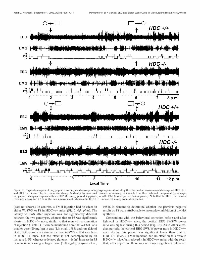
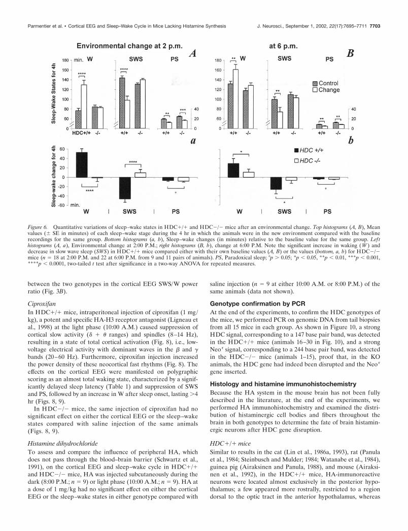
A greater difference in the sleep–wake cycle was seen betweenthe two genotypes in the new environment test, which consistedof transferring the mice from their habitual home cage (a trans-parent barrel) to a new cage (an opaque rectangular box withopen field). Thus, as shown in Figures 5 and 6 and Table 1, thetransfer of the HDC�/� mice to the new cage elicited an increasein W over the whole 4 hr period that the mice were in the newcage, including a significant increase in the latencies to SWS and
PS (Table 1) and an increase in W and decrease in SWS aftersleep onset during their stay in the new cage (Figs. 5, 6). Theyappeared to be interested by the new environment, because videorecording showed several exploratory behaviors, such as ambula-tion around the new cage and rearing. In contrast, HDC�/� miceseemed indifferent and unresponsive, because they fell asleepsoon after the environmental change (Fig. 5, Table 1), and therewas no change in W and SWS during their 4 hr stay in the newcage, compared with their own control data during basal condi-tions (Fig. 6). This difference in responsiveness to the new envi-ronment was seen both at 2:00 P.M. (sleepy period; Fig. 5, toppanels, Table 1), and at 6:00 P.M. (awake period; Fig. 5, bottompanels, Table 1). Both sets of mice showed a decrease of �4–8min in PS in the new environment compared with their baselinerecordings (Fig. 6) in which they were undisturbed, presumably asa result of handling. Interestingly, when the animals were placedback to their home cages after the 4 hr stay, no any significantdifference in term of sleep latencies was noted between the twogenotypes (Table 1), suggesting that novelty plays an importantrole in the new environment-elicited awakening in the HDC�/�mice. It should be mentioned that the object recognition testshowed that the vision of both phenotypes was intact and that themice appeared not to be stressed during their stay in the newcage, because a similar degree of water and food intake andgrooming occurred as in the home cage. No immobility or hyper-activity or other apparently unusual behavioral signs were seen,suggesting that the animals did not suffer stress or anxiety in thenew environment.
Effects of drug administration on the cortical EEG andsleep–wake cycle�-FMHBefore and after lights-off, W and locomotion increased in theHDC�/� mice, whereas these effects were markedly less evidentin HDC�/� mice. When the animals were injected intraperito-neally with �-FMH (specific HDC inhibitor, 50 mg/kg,) at 4:00P.M. (3 hr before lights-off, in view of the latency of drug actionshown in Garbarg et al., 1980; Maeyama et al., 1982), a progres-sively developing significant decrease in W and increase in bothSWS and PS were seen in HDC�/� mice (Fig. 7, lef t plots),results similar to the reduction in W seen in untreated HDC�/�mice during this period under basal conditions. The increase inSWS and PS was caused by a prolongation of episode duration
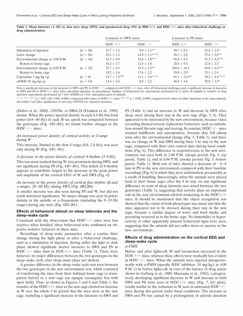
Table 1. Mean latencies (� SE) to slow wave sleep (SWS) and paradoxical sleep (PS) in HDC�/� and HDC�/� mice after behavioral challenge ordrug administration
Latencies to SWS (min) Latencies to PS (min)
HDC �/� HDC �/� HDC �/� HDC �/�
Simulation of injection (n � 26) 15.7 � 1.3 9.9 � 1.1** 48.7 � 8.5 23.8 � 2.5*Litter change (n � 36) 25.1 � 1.2 14.9 � 1.1**** 50.1 � 2.8 37.8 � 2.8**Environmental change at 2:00 P.M. (n � 18) 42.5 � 8.9 18.4 � 1.8*** 91.0 � 9.3 51.3 � 4.2***
Return to home cage 16.2 � 3.7 12.4 � 1.8 28.5 � 9.3 21.0 � 2.3Environmental change at 6:00 P.M. (n � 22) 35.7 � 6.5 15.3 � 1.5** 109.6 � 14.3 49.0 � 4.6***
Return to home cage 19.2 � 1.4 17.6 � 2.2 39.9 � 2.9 32.1 � 2.4Ciproxifan 1 mg/kg i.p. (n � 9) 53.7 � 7.2°°°° 11.1 � 3.4**** 91.1 � 13.5°°° 30.3 � 4.6****�FMH 50 mg/kg i.p. (n � 14) 12.4 � 2.6 8.8 � 2.2 46.8 � 4.6 35.0 � 3.9*
Note a significant decrease in the latencies to SWS and PS in HDC�/� compared with HDC�/� mice after all behavioral challenges and a significant increase in latenciesto SWS and PS in HDC�/� mice after ciproxifan injection. In parentheses, Number of behavioral test experiments performed on 11 pairs of animals or number of druginjection experiments performed on 7 (for �FMH) or 9 (for ciproxifan) pairs of animals.*, **, ***, **** p 0.05, 0.01, 0.001, 0.0001, compared with values of HDC�/� mice; °°°, °°°° p 0.001, 0.0001 compared with values of saline injections in the same animals,two-tailed t test after significance in two-way ANOVA for repeated measures.
Parmentier et al. • Cortical EEG and Sleep–Wake Cycle in Mice Lacking Histamine Synthesis J. Neurosci., September 1, 2002, 22(17):7695–7711 7701
(data not shown). In contrast, �-FMH injection had no effect oneither W, SWS, or PS in HDC�/� mice, (Fig. 7, right plots). Thelatency to SWS after injection was not significantly differentbetween the two genotypes, whereas that to PS was significantlyshorter in HDC�/� mice, similar to that seen with a simulationof injection (Table 1). It can be mentioned here that �-FMH at asmaller dose (20 mg/kg) in cats (Lin et al., 1988) and rats (Montiet al., 1988) results in a similar increase in SWS to that seen herein HDC�/� mice, but the effect is not accompanied by anincrease in PS; whereas a delayed (latency �16 hr) increase in PSis seen in rats using a larger dose (100 mg/kg; Kiyono et al.,
1984). It remains to determine whether the previous negativeresults on PS were attributable to incomplete inhibition of the HAsynthesis.
Concomitant with the behavioral activation before and afterlights-off in HDC�/� mice, the cortical EEG SWS/W powerratio was highest during this period (Fig. 3B). As in other circa-dian periods, the cortical-EEG SWS/W power ratio in HDC�/�mice during this period was significant lower than that inHDC�/� mice. �-FMH injection had no effect on this ratio inHDC�/� mice, but reduced it in HDC�/� mice, with the resultthat, after injection, there was no longer significant difference
Figure 5. Typical examples of polygraphic recordings and corresponding hypnograms illustrating the effects of an environmental change on HDC�/�and HDC�/� mice. The environmental change (indicated by an arrow) consisted of moving the animals from their habitual transparent barrel cagesto opaque rectangular cages at either 2:00 P.M. (sleepy period; top panels) or 6:00 P.M. (awake period; bottom panels). Note that the HDC �/� mouseremained awake for �2 hr in the new environment, whereas the HDC�/� mouse fell asleep soon after the test.
7702 J. Neurosci., September 1, 2002, 22(17):7695–7711 Parmentier et al. • Cortical EEG and Sleep–Wake Cycle in Mice Lacking Histamine Synthesis
between the two genotypes in the cortical EEG SWS/W powerratio (Fig. 3B).
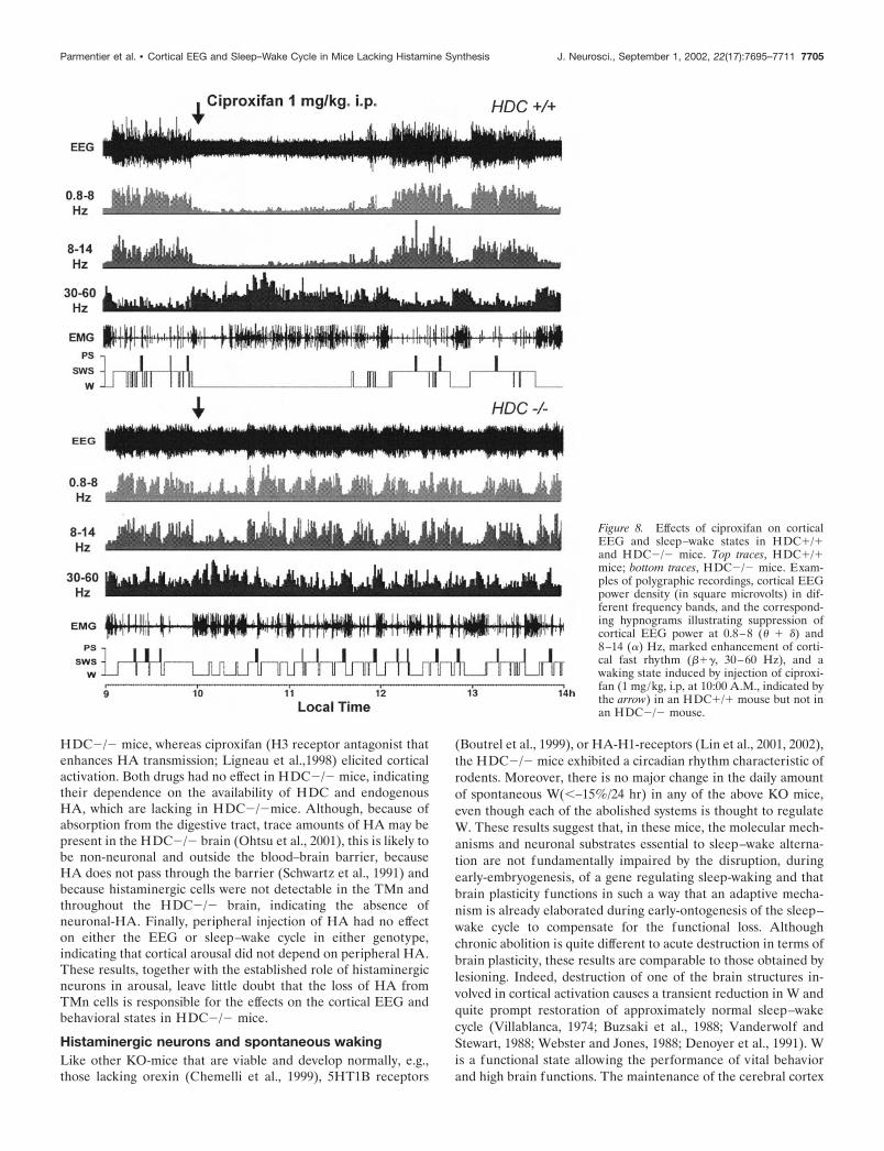
CiproxifanIn HDC�/� mice, intraperitoneal injection of ciproxifan (1 mg/kg), a potent and specific HA-H3 receptor antagonist (Ligneau etal., 1998) at the light phase (10:00 A.M.) caused suppression ofcortical slow activity (� � � ranges) and spindles (8–14 Hz),resulting in a state of total cortical activation (Fig. 8), i.e., low-voltage electrical activity with dominant waves in the � and �bands (20–60 Hz). Furthermore, ciproxifan injection increasedthe power density of these neocortical fast rhythms (Fig. 8). Theeffects on the cortical EEG were manifested on polygraphicscoring as an almost total waking state, characterized by a signif-icantly delayed sleep latency (Table 1) and suppression of SWSand PS, followed by an increase in W after sleep onset, lasting �4hr (Figs. 8, 9).
In HDC�/� mice, the same injection of ciproxifan had nosignificant effect on either the cortical EEG or the sleep–wakestates compared with saline injection of the same animals(Figs. 8, 9).
Histamine dihydrochlorideTo assess and compare the influence of peripheral HA, whichdoes not pass through the blood–brain barrier (Schwartz et al.,1991), on the cortical EEG and sleep–wake cycle in HDC�/�and HDC�/� mice, HA was injected subcutaneously during thedark (8:00 P.M.; n � 9) or light phase (10:00 A.M.; n � 9). HA ata dose of 1 mg/kg had no significant effect on either the corticalEEG or the sleep–wake states in either genotype compared with
saline injection (n � 9 at either 10:00 A.M. or 8:00 P.M.) of thesame animals (data not shown).
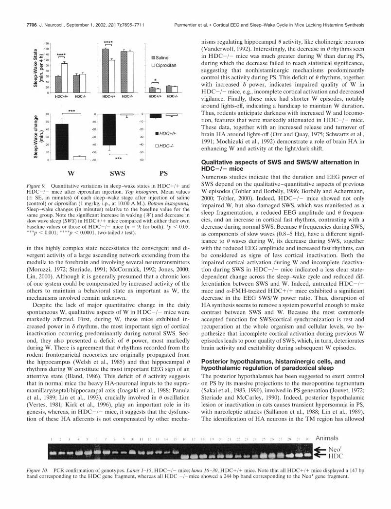
Genotype confirmation by PCRAt the end of the experiments, to confirm the HDC genotypes ofthe mice, we performed PCR on genomic DNA from tail biopsiesfrom all 15 mice in each group. As shown in Figure 10, a strongHDC signal, corresponding to a 147 base pair band, was detectedin the HDC�/� mice (animals 16–30 in Fig. 10), and a strongNeor signal, corresponding to a 244 base pair band, was detectedin the HDC�/� mice (animals 1–15), proof that, in the KOanimals, the HDC gene had indeed been disrupted and the Neor
gene inserted.
Histology and histamine immunohistochemistryBecause the HA system in the mouse brain has not been fullydescribed in the literature, at the end of the experiments, weperformed HA immunohistochemistry and examined the distri-bution of histaminergic cell bodies and fibers throughout thebrain in both genotypes to determine the fate of brain histamin-ergic neurons after HDC gene disruption.
HDC�/� miceSimilar to results in the cat (Lin et al., 1986a, 1993), rat (Panulaet al., 1984; Steinbusch and Mulder; 1984; Watanabe et al., 1984),guinea pig (Airaksinen and Panula, 1988), and mouse (Airaksi-nen et al., 1992), in the HDC�/� mice, HA-immunoreactiveneurons were located almost exclusively in the posterior hypo-thalamus; a few appeared more rostrally, restricted to a regiondorsal to the optic tract in the anterior hypothalamus, whereas
Figure 6. Quantitative variations of sleep–wake states in HDC�/� and HDC�/� mice after an environmental change. Top histograms (A, B), Meanvalues (� SE in minutes) of each sleep–wake stage during the 4 hr in which the animals were in the new environment compared with the baselinerecordings for the same group. Bottom histograms (a, b), Sleep–wake changes (in minutes) relative to the baseline value for the same group. Lefthistograms (A, a), Environmental change at 2:00 P.M.; right histograms (B, b), change at 6:00 P.M. Note the significant increase in waking (W ) anddecrease in slow wave sleep (SWS) in HDC�/� mice compared either with their own baseline values (A, B) or the values (bottom, a, b) for HDC�/�mice (n � 18 at 2:00 P.M. and 22 at 6:00 P.M. from 9 and 11 pairs of animals). PS, Paradoxical sleep; °p � 0.05; *p 0.05, **p 0.01, ***p 0.001,****p 0.0001, two-tailed t test after significance in a two-way ANOVA for repeated measures.
Parmentier et al. • Cortical EEG and Sleep–Wake Cycle in Mice Lacking Histamine Synthesis J. Neurosci., September 1, 2002, 22(17):7695–7711 7703
most HA-immunoreactive cell bodies aggregated in both theventral and dorsal divisions of the tuberomammillary nucleus(TMn), in the adjacent lateral hypothalamic area, and in theperimammillary and supramammillary areas (Fig. 11). The ven-tral division of the histaminergic TMn was more compactly or-ganized than the dorsal division. The HA-immunoreactive neu-rons, estimated to number 2500–3500 in the whole brain, weremedium to large in size (15 � 30 �m), mostly ovoid or polygonalin shape, and possessed two to four prominent, long, thick pro-cesses. The majority of the HA-immunoreactive perikaryashowed strong immunoreactivity, although a few were moderatelystained (Fig. 11).
HA-immunoreactive fibers and terminal-like dots were de-tected in virtually all brain regions. For example, numerous finevaricose fibers were present in the various neocortical areas,hippocampal formation, basal forebrain, thalamus, preoptic/ante-rior and posterior hypothalamus including the perifornical area,and the forebrain and brainstem aminergic and cholinergic struc-
tures, such as the substantia innominata, ventral tegmental area ofTsai, mesopontine tegmentum, raphe nuclei, and locus coeruleus(Figs. 11, 12). Omission of anti-HA antibody or pre-incubation ofthe sections with excess exogenous free or ovalbumin-conjugatedHA resulted in no immunolabeling of any part of the mouse brain(data not shown), demonstrating the specificity of labeling. Ex-amples of the presence of HA-immunoreactive fibers in some ofthese structures are shown in Figure 12.
HDC�/� miceIn contrast to the dense HA immunoreactivity present in theHDC�/� brains, no HA immunostaining was found throughoutthe HDC�/� brains using either immunofluorescent or PAPtechniques, and no HA-immunoreactive perikarya, dendrites,fibers, or terminal-like dots were identified in the TMn and theadjacent posterior hypothalamus or elsewhere in the brain (Figs.11, 12).
Because the lack of the neurotransmitter, HA, TMn neurons inHDC�/� mice can no longer be qualified as histaminergic neu-rons. However, as demonstrated by neutral red counterstaining(Fig. 12) or immunohistochemistry of type B monoamine oxidase(data not shown), a marker for TMn neurons (Lin et al., 1993),both dorsal and ventral divisions of the TMn, although nonim-munoreactive with anti-HA antibody, seemed to be intact in thesemice, without obvious visual difference in either the number ofneurons or their morphology (see example from the ventraldivision in Fig. 12). Finally, no visually apparent structuralchanges were seen in the brain sections examined. Because thenumber and morphology of TMn neurons under the light micros-copy appeared to be unchanged in HDC�/� mice, the nature ofthe functional change after HDC disruption and the neurotrans-mitter(s), if any, which replaces HA remains to be determined.
Summary of the principal findingsThe present study reveals the absence of detectable brain HA-immunoreactive neurons and the absence of response to admin-istration of HA-related agents in the PCR-confirmed HDC�/�mice. Moreover, we have shown that the sleep–wake cycle ofthese mice was affected both quantitatively and qualitatively. Onthe one hand, these mice exhibited an increase in the dailyamount of PS (�23%) and a deficit in W just before and afterlights-off. On the other hand, their cortical-EEG showed a re-duced SWS/W power ratio and a significant increase in � frequen-cies (0.8–2.5Hz) and a deficit of �-rhythms (3–9Hz) during W.These changes are likely to have an effect on the animal’s behav-ior, because the HDC�/� mice presented clear signs of sedation,manifested as a significant decrease in sleep latencies after severalbehavioral stimuli, and more importantly, unlike normal mice, inbeing unable to remain awake in a new environment.
DISCUSSIONSeveral lines of evidence from our study indicated that the corti-cal EEG and behavioral signs seen in HDC�/� mice are causedby the lack of HA synthesis. First, the mouse strain, sex, and ageand the experimental conditions were identical for the wild-typeand KO mice, and the only correlation with the observed effectswas with the mouse genotype. Because our study used inbredmice, the genomic background of the two genotypes was identical,except for the HDC gene. The small interindividual SDs for thesleep–wake stages within genotype also indicated that each groupwas homogenous (Fig. 1). Second, in HDC�/� mice, �-FMH(HDC inhibitor) produced the same changes seen in untreated
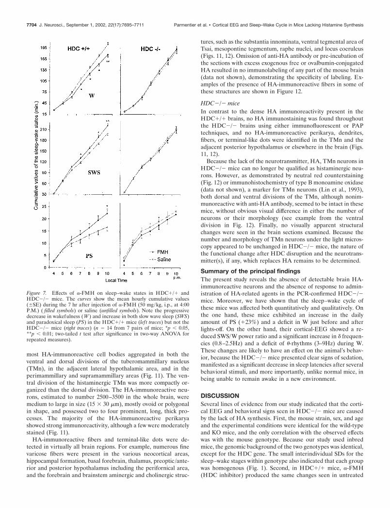
Figure 7. Effects of �-FMH on sleep–wake states in HDC�/� andHDC�/� mice. The curves show the mean hourly cumulative values(�SE) during the 7 hr after injection of �-FMH (50 mg/kg, i.p., at 4:00P.M.) ( filled symbols) or saline (unfilled symbols). Note the progressivedecrease in wakefulness (W ) and increase in both slow wave sleep (SWS)and paradoxical sleep (PS) in the HDC�/� mice (lef t traces) but not theHDC�/� mice (right traces) (n � 14 from 7 pairs of mice; *p 0.05,**p 0.01; two-tailed t test after significance in two-way ANOVA forrepeated measures).
7704 J. Neurosci., September 1, 2002, 22(17):7695–7711 Parmentier et al. • Cortical EEG and Sleep–Wake Cycle in Mice Lacking Histamine Synthesis
HDC�/� mice, whereas ciproxifan (H3 receptor antagonist thatenhances HA transmission; Ligneau et al.,1998) elicited corticalactivation. Both drugs had no effect in HDC�/� mice, indicatingtheir dependence on the availability of HDC and endogenousHA, which are lacking in HDC�/�mice. Although, because ofabsorption from the digestive tract, trace amounts of HA may bepresent in the HDC�/� brain (Ohtsu et al., 2001), this is likely tobe non-neuronal and outside the blood–brain barrier, becauseHA does not pass through the barrier (Schwartz et al., 1991) andbecause histaminergic cells were not detectable in the TMn andthroughout the HDC�/� brain, indicating the absence ofneuronal-HA. Finally, peripheral injection of HA had no effecton either the EEG or sleep–wake cycle in either genotype,indicating that cortical arousal did not depend on peripheral HA.These results, together with the established role of histaminergicneurons in arousal, leave little doubt that the loss of HA fromTMn cells is responsible for the effects on the cortical EEG andbehavioral states in HDC�/� mice.
Histaminergic neurons and spontaneous wakingLike other KO-mice that are viable and develop normally, e.g.,those lacking orexin (Chemelli et al., 1999), 5HT1B receptors
(Boutrel et al., 1999), or HA-H1-receptors (Lin et al., 2001, 2002),the HDC�/� mice exhibited a circadian rhythm characteristic ofrodents. Moreover, there is no major change in the daily amountof spontaneous W(–15%/24 hr) in any of the above KO mice,even though each of the abolished systems is thought to regulateW. These results suggest that, in these mice, the molecular mech-anisms and neuronal substrates essential to sleep–wake alterna-tion are not fundamentally impaired by the disruption, duringearly-embryogenesis, of a gene regulating sleep-waking and thatbrain plasticity functions in such a way that an adaptive mecha-nism is already elaborated during early-ontogenesis of the sleep–wake cycle to compensate for the functional loss. Althoughchronic abolition is quite different to acute destruction in terms ofbrain plasticity, these results are comparable to those obtained bylesioning. Indeed, destruction of one of the brain structures in-volved in cortical activation causes a transient reduction in W andquite prompt restoration of approximately normal sleep–wakecycle (Villablanca, 1974; Buzsaki et al., 1988; Vanderwolf andStewart, 1988; Webster and Jones, 1988; Denoyer et al., 1991). Wis a functional state allowing the performance of vital behaviorand high brain functions. The maintenance of the cerebral cortex
Figure 8. Effects of ciproxifan on corticalEEG and sleep–wake states in HDC�/�and HDC�/� mice. Top traces, HDC�/�mice; bottom traces, HDC�/� mice. Exam-ples of polygraphic recordings, cortical EEGpower density (in square microvolts) in dif-ferent frequency bands, and the correspond-ing hypnograms illustrating suppression ofcortical EEG power at 0.8–8 (� � �) and8–14 (�) Hz, marked enhancement of corti-cal fast rhythm (���, 30–60 Hz), and awaking state induced by injection of ciproxi-fan (1 mg/kg, i.p, at 10:00 A.M., indicated bythe arrow) in an HDC�/� mouse but not inan HDC�/� mouse.
Parmentier et al. • Cortical EEG and Sleep–Wake Cycle in Mice Lacking Histamine Synthesis J. Neurosci., September 1, 2002, 22(17):7695–7711 7705
in this highly complex state necessitates the convergent and di-vergent activity of a large ascending network extending from themedulla to the forebrain and involving several neurotransmitters(Moruzzi, 1972; Steriade, 1991; McCormick, 1992; Jones, 2000;Lin, 2000). Although it is generally presumed that a chronic lossof one system could be compensated by increased activity of theothers to maintain a behavioral state as important as W, themechanisms involved remain unknown.
Despite the lack of major quantitative change in the dailyspontaneous W, qualitative aspects of W in HDC�/� mice weremarkedly affected. First, during W, these mice exhibited in-creased power in � rhythms, the most important sign of corticalinactivation occurring predominantly during natural SWS. Sec-ond, they also presented a deficit of � power, most markedlyduring W. There is agreement that � rhythms recorded from therodent frontoparietal neocortex are originally propagated fromthe hippocampus (Welsh et al., 1985) and that hippocampal �rhythms during W constitute the most important EEG sign of anattentive state (Bland, 1986). This deficit of � activity suggeststhat in normal mice the heavy HA-neuronal inputs to the supra-mamillary/septal /hippocampal axis (Inagaki et al., 1988; Panulaet al., 1989; Lin et al., 1993), crucially involved in � oscillation(Vertes, 1981; Kirk et al., 1996), play an important role in itsgenesis, whereas, in HDC�/� mice, it suggests that the dysfunc-tion of these HA afferents is not compensated by other mecha-
nisms regulating hippocampal � activity, like cholinergic neurons(Vanderwolf, 1992). Interestingly, the decrease in � rhythms seenin HDC�/� mice was much greater during W than during PS,during which the decrease failed to reach statistical significance,suggesting that nonhistaminergic mechanisms predominantlycontrol this activity during PS. This deficit of � rhythms, togetherwith increased � power, indicates impaired quality of W inHDC�/� mice, e.g., incomplete cortical activation and decreasedvigilance. Finally, these mice had shorter W episodes, notablyaround lights-off, indicating a handicap to maintain W duration.Thus, rodents anticipate darkness with increased W and locomo-tion, features that were markedly attenuated in HDC�/� mice.These data, together with an increased release and turnover ofbrain HA around lights-off (Orr and Quay, 1975; Schwartz et al.,1991; Mochizuki et al., 1992) demonstrate a role of brain HA inenhancing W and activity at the light/dark shift.
Qualitative aspects of SWS and SWS/W alternation inHDC�/� miceNumerous studies indicate that the duration and EEG power ofSWS depend on the qualitative–quantitative aspects of previousW episodes (Tobler and Borbely, 1986; Borbely and Achermann,2000; Tobler, 2000). Indeed, HDC�/� mice showed not onlyimpaired W, but also damaged SWS, which was manifested as asleep fragmentation, a reduced EEG amplitude and � frequen-cies, and an increase in cortical fast rhythms, contrasting with adecrease during normal SWS. Because � frequencies during SWS,as components of slow waves (0.8–5 Hz), have a different signif-icance to � waves during W, its decrease during SWS, togetherwith the reduced EEG amplitude and increased fast rhythms, canbe considered as signs of less cortical inactivation. Both theimpaired cortical activation during W and incomplete deactiva-tion during SWS in HDC�/� mice indicated a less clear state-dependent change across the sleep–wake cycle and reduced dif-ferentiation between SWS and W. Indeed, untreated HDC�/�mice and �-FMH-treated HDC�/� mice exhibited a significantdecrease in the EEG SWS/W power ratio. Thus, disruption ofHA synthesis seems to remove a system powerful enough to makecontrast between SWS and W. Because the most commonlyaccepted function for SWS/cortical synchronization is rest andrecuperation at the whole organism and cellular levels, we hy-pothesize that incomplete cortical activation during previous Wepisodes leads to poor quality of SWS, which, in turn, deterioratesbrain activity and excitability during subsequent W episodes.
Posterior hypothalamus, histaminergic cells, andhypothalamic regulation of paradoxical sleepThe posterior hypothalamus has been suggested to exert controlon PS by its massive projections to the mesopontine tegmentum(Sakai et al., 1983, 1990), involved in PS generation (Jouvet, 1972;Steriade and McCarley, 1990). Indeed, posterior hypothalamiclesion or inactivation in cats causes transient hypersomnia in PS,with narcoleptic attacks (Sallanon et al., 1988; Lin et al., 1989).The identification of HA neurons in the TM region has allowed
Figure 10. PCR confirmation of genotypes. Lanes 1–15, HDC�/� mice; lanes 16–30, HDC�/� mice. Note that all HDC�/� mice displayed a 147 bpband corresponding to the HDC gene fragment, whereas all HDC �/�mice showed a 244 bp band corresponding to the Neo r gene fragment.
Figure 9. Quantitative variations in sleep–wake states in HDC�/� andHDC�/� mice after ciproxifan injection. Top histogram, Mean values(� SE, in minutes) of each sleep–wake stage after injection of saline(control) or ciproxifan (1 mg/kg, i.p., at 10:00 A.M.). Bottom histograms,Sleep–wake changes (in minutes) relative to the baseline value for thesame group. Note the significant increase in waking (W ) and decrease inslow wave sleep (SWS) in HDC�/� mice compared with either their ownbaseline values or those of HDC�/� mice (n � 9; for both). *p 0.05;***p 0.001; ****p 0.0001, two-tailed t test).
7706 J. Neurosci., September 1, 2002, 22(17):7695–7711 Parmentier et al. • Cortical EEG and Sleep–Wake Cycle in Mice Lacking Histamine Synthesis
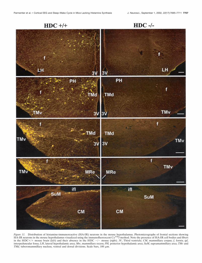
Figure 11. Distribution of histamine-immunoreactive (HA-IR) neurons in the mouse hypothalamus. Photomicrographs of frontal sections showingHA-IR neurons in the mouse hypothalamus visualized using the immunofluorescent CyTM3 method. Note the presence of HA-IR cell bodies and fibersin the HDC�/� mouse brain (lef t) and their absence in the HDC �/� mouse (right). 3V, Third ventricle; CM, mammillary corpus; f, fornix; ipf,interpeduncular fossa; LH, lateral hypothalamic area; Mre, mammillary recess; PH, posterior hypothalamic area; SuM, supramammillary area; TMv andTMd, tuberomammillary nucleus, ventral and dorsal divisions. Scale bars, 100 �m.
Parmentier et al. • Cortical EEG and Sleep–Wake Cycle in Mice Lacking Histamine Synthesis J. Neurosci., September 1, 2002, 22(17):7695–7711 7707
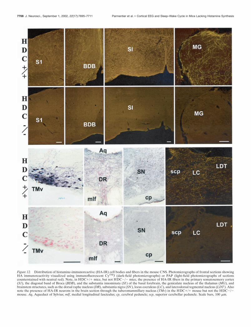
Figure 12. Distribution of histamine-immunoreactive (HA-IR) cell bodies and fibers in the mouse CNS. Photomicrographs of frontal sections showingHA immunoreactivity visualized using immunofluorescent CyTM3 (dark-field photomicrographs) or PAP (light-field photomicrographs of sectionscounterstained with neutral red). Note, in HDC�/� mice, but not HDC�/� mice, the presence of HA-IR fibers in the primary somatosensory cortex(S1), the diagonal band of Broca (BDB), and the substantia innominata (SI ) of the basal forebrain, the geniculate nucleus of the thalamus (MG), andbrainstem structures, such as the dorsal raphe nucleus (DR), substantia nigra (SN ), locus coeruleus (LC), and laterodorsal tegmental nucleus (LDT ). Alsonote the presence of HA-IR neurons in the brain section through the tuberomammillary nucleus (TMv) in the HDC�/� mouse but not the HDC�/�mouse. Aq, Aqueduct of Sylvius; mlf, medial longitudinal fasciculus; cp, cerebral peduncle; scp, superior cerebellar peduncle. Scale bars, 100 �m.
7708 J. Neurosci., September 1, 2002, 22(17):7695–7711 Parmentier et al. • Cortical EEG and Sleep–Wake Cycle in Mice Lacking Histamine Synthesis
the assumption that this control could be, in part, histaminergic.This hypothesis has not yet been proven, because posterior hy-pothalamic lesion or inactivation causes larger increase in SWSthan in PS and because the use of various pharmacological agentsimpairing HA transmission produces an increase in SWS, but notin PS (Lin et al., 1988; Monti et al., 1988; Lin, 2000). In this study,the most marked quantitative change seen with HDC gene dis-ruption was an increase in PS. Inhibition of HA synthesis by�-FMH in HDC�/� mice also enhanced PS. Whereas it remainsto determine whether the previous negative results on PS werecaused by incomplete inactivation of or limited selectivity of thedrugs used for the HA system or whether histaminergic regula-tion of PS is more pronounced in mice than in other species, theenhanced SP seen here is consistent with the PS-off dischargepattern of presumed HA neurons, which, like other aminergiccells, cease firing during PS (Sakai et al., 1990). Our results thuspoint out that HA cells are involved in PS-permissive mecha-nisms and, together with other data (Sakai et al., 1990; Lin, 2000),suggest that HA-cells exert a control, via their descending inputs,over the mesopontine PS-generating mechanisms.
In view of the presence of orexin neurons adjacent to the TMnand given the role of orexin deficiency in narcolepsy, the hypo-thalamic mechanisms controlling PS should be multiple and in-clude both HA and orexin neurons, which might act in a syner-gistic complementary manner and which might explain, in part,the importance of the posterior hypothalamus in sleep–wakecontrol. Indeed, HDC and orexin KO mice exhibit a similarincrease in PS. However, the increase in HDC�/� mice is seenduring light phase, whereas that in orexin KO mice occurs duringdarkness, accompanied by narcoleptic phases (Chemelli et al.,1999). Narcolepsy was not seen either in HDC �/�mice or innormal animals treated with compounds impairing HA transmis-sion. Thus, the narcoleptic phases seen after posterior hypotha-lamic lesion or inactivation (Sallanon et al., 1988; Lin et al., 1989),which also involved the perifornical area containing orexin cells,should result from a loss of orexin cells rather than HA neurons,whereas the decreased HA transmission seen in narcoleptic dogs(Nishino et al., 2001) would account for their excessive sleep,rather than for narcolepsy. Some interactions between HA andorexin neurons have been identified. Orexin neurons constituteimportant excitatory afferents to HA neurons (Peyron et al., 1998;Eriksson et al., 2001; Marcus et al., 2001). The arousing effect oforexin seems to depend on H1 receptors (Huang et al., 2001).However, the role of histaminergic inputs to orexin neuronsremains unknown. In view of dense histaminergic fibers andterminal dots (Inagaki et al., 1988; Panula et al., 1989; Lin et al.,1993) and H1 receptors (Bouthenet et al., 1988) in the periforni-cal area, and considering the excitatory action of H1 receptors(McCormick, 1992; Brown et al., 2001), we hypothesize that HAneurons also excite orexin cells during W and that the reciprocalexcitatory interactions between HA and orexin neurons consti-tute important hypothalamic arousal mechanisms. Their interac-tion during PS, however, remains to be determined, because HAcells are silent during this sleep stage.
Histaminergic neurons and maintenance of wakingfaced with behavioral challengesThe impaired cortical EEG in HDC�/� mice might be expectedto have behavioral consequence. Indeed, they presented signs ofsomnolence (reduced sleep latencies, e.g.) after routine change oflitter or simulation of injection. This decreased arousal reactionin response to external stimuli is consistent with their deficit of W
at lights-off under unstimulated conditions. Moreover, HDC�/�mice placed in a new environment failed to remain awake, dem-onstrated by a significant decrease in sleep latencies and in Wduration. Obviously, in the new environment, exploration andother behaviors (curiosity, e.g.) of HDC�/� mice should also beaffected, and this seems to be attributable to their inability toremain vigilant, rather than to a direct effect of loss of HA onspecific behaviors, because this sedation was seen in several tests.These results are consistent with the well known drowsiness andimpaired performance caused by H1 receptor antagonists (Doug-las, 1985; Nicholson and Stone, 1986; Schwartz et al., 1991; Yanaiet al., 1999). Although, by as yet unknown compensatory mech-anisms, HDC�/� mice can reach, under normal conditions, asimilar daily amount of W to normal mice, thus allowing theperformance of behaviors indispensable for survival, this is by nomeans apparent under other circumstances. Thus, when a highlevel of vigilance is required, e.g., lights-off or environmentalchange, they are unable to maintain awake. Because W is thebasis for all other high brain functions, like attention, perfor-mance, and learning, and because an alert waking state is aprerequisite condition for responding to behavioral–cognitivechallenges, we suggest that the high brain functions of HDC�/�mice should also be secondarily affected.
Our findings thus extend the current understanding of the roleof HA neurons, which cannot simply be regarded as a system inwhich neuronal activity is positively linked with instantaneouscortical activation of W. Long-term abolition of HA synthesisimpairs cortical EEG, affects all sleep–wake states, and causesbehavioral deficits. We suggest that, in addition to their impor-tance in arousal under normal conditions (see introductory re-marks), histaminergic neurons also play a key role in maintainingthe brain in an awake state in the presence of behavioralchallenges.
REFERENCESAiraksinen MS, Panula P (1988) The histaminergic system in the guinea
pig central nervous system: an immunocytochemical mapping studyusing an antiserum against histamine. J Comp Neurol 273:163–186.
Airaksinen MS, Alanen S, Szabat E, Visser TJ, Panula P (1992) Multipleneurotransmitters in the tuberomammillary nucleus: comparison of rat,mouse, and guinea pig. J Comp Neurol 323:103–116.
Bland BH (1986) The physiology and pharmacology of hippocampalformation theta rhythms. Prog Neurobiol 26:1–54.
Borbely AA, Achermann P (2000) Sleep homeostasis and models ofsleep regulation. In: Principles and practice of sleep medicine (KrygerMH, Roth T, Dement WC, eds), pp 377–390. Philadelphia: Saunders.
Bouthenet ML, Ruat M, Sales N, Garbarg M, Schwartz JC (1988) Adetailed mapping of histamine H1-receptors in guinea-pig central ner-vous system established by autoradiography with [125I]iodobolpyra-mine. Neuroscience 26:553–600.
Boutrel B, Franc B, Hen R, Hamon M, Adrien J (1999) Key role of5-HT1B receptors in the regulation of paradoxical sleep as evidenced in5-HT1B knock-out mice. J Neurosci 19:3204–3212.
Brown RE, Stevens DR, Haas HL (2001) The physiology of brain his-tamine. Prog Neurobiol 63:637–672.
Buzsaki G, Bickford RG, Ponomareff G, Thal LJ, Mandel R, Gage FH(1988) Nucleus basalis and thalamic control of neocortical activity inthe freely moving rat. J Neurosci 8:4007–4026.
Chemelli RM, Willie JT, Sinton CM, Elmquist JK, Scammell T, Lee C,Richardson JA, Williams SC, Xiong Y, Kisanuki Y, Fitch TE, Naka-zato M, Hammer RE, Saper CB, Yanagisawa M (1999) Narcolepsy inorexin knock-out mice: molecular genetics of sleep regulation. Cell98:437–451.
de Lecea L, Kilduff TS, Peyron C, Gao X, Foye PE, Danielson PE,Fukuhara C, Battenberg EL, Gautvik VT, Bartlett FS, Frankel WN,van den Pol AN, Bloom FE, Gautvik KM, Sutcliffe JG (1998) Thehypocretins: hypothalamus-specific peptides with neuroexcitatory activ-ity. Proc Natl Acad Sci USA 95:322–327.
Denoyer M, Sallanon M, Buda C, Kitahama K, Jouvet M (1991) Neu-rotoxic lesion of the mesencephalic reticular formation and/or theposterior hypothalamus does not alter waking in the rat. Brain Res539:287–303.
Parmentier et al. • Cortical EEG and Sleep–Wake Cycle in Mice Lacking Histamine Synthesis J. Neurosci., September 1, 2002, 22(17):7695–7711 7709
Douglas WW (1985) Histamine and 5-hydroxytryptamine (serotonin)and their antagonists. In: The pharmacological basis of therapeutics(Gilman AG, Goodman LS, Rall TW, Murad F, eds), pp 605–638. NewYork: Macmillan.
Eriksson KS, Sergeeva O, Brown RE, Haas HL (2001) Orexin/hypocre-tin excites the histaminergic neurons of the tuberomammillary nucleus.J Neurosci 21:9273–9279.
Franken P, Malafosse A, Tafti M (1998) Genetic variation in EEGactivity during sleep in inbred mice. Am J Physiol 275:1127–1137.
Franken P, Malafosse A, Tafti M (1999) Genetic determinants of sleepregulation in inbred mice. Sleep 22:155–169.
Franklin KB, Paxinos G (1996) The mouse brain in stereotaxic coordi-nates. San Diego: Academic.
Garbarg M, Barbin G, Rodergas E, Schwartz JC (1980) Inhibition ofhistamine synthesis in brain by alpha-fluoromethylhistidine, a newirreversible inhibitor: in vitro and in vivo studies. J Neurochem35:1045–1052.
Haas HL (1992) Electrophysiology of histamine-receptors. In: The his-tamine receptors (Schwartz JC, Haas HL, eds), pp 161–178. New York:Wiley-Liss.
Hara J, Beuckmann CT, Nambu T, Willie JT, Chemelli RM, Sinton CM,Sugiyama F, Yagami K, Goto K, Yanagisawa M, Sakurai T (2001)Genetic ablation of orexin neurons in mice results in narcolepsy,hypophagia, and obesity. Neuron 30:345–354.
Huang ZL, Qu WM, Li WD, Mochizuki T, Eguchi N, Watanabe T,Urade Y, Hayaishi O (2001) Arousal effect of orexin A depends onactivation of the histaminergic system. Proc Natl Acad Sci USA98:9965–9970.
Huber R, Deboer T, Tobler I (2000) Effects of sleep deprivation on sleepand sleep EEG in three mouse strains: empirical data and simulations.Brain Res 857:8–19.
Inagaki N, Yamatodani A, Ando-Yamamoto M, Tohyama M, WatanabeT, Wada H (1988) Organization of histaminergic fibers in rat brain.J Comp Neurol 273:283–300.
Jones BE (2000) Basic mechanisms of sleep–wake states. In: Principlesand practice of sleep medicine (Kryger MH, Roth T, Dement WC,eds), pp 134–154. Philadelphia: Saunders.
Jouvet M (1972) The role of monoamines and acetylcholine containingneurons in the regulation of the sleep-waking cycle. Ergebn Physiol64:166–307.
Kirk IJ, Oddie SD, Konopacki J, Bland BH (1996) Evidence for differ-ential control of posterior hypothalamic, supramammillary, and medialmammillary theta-related cellular discharge by ascending and descend-ing pathways. J Neurosci 16:5547–5554.
Kiyono S, Seo M, Shibagaki M, Watanabe T, Maeyama K, Wada H(1984) Effects of �-fluoromethylhistidine on sleep-waking parametersin rats. Physiol Behav 34:615–617.
Kollonitsch J, Perkins LM, Patchett AA, Doldouras GA, Marburg S,Duggan DE, Maycock AL, Aster SD (1978) Selective inhibitors ofbiosynthesis of aminergic neurotransmitters. Nature 274:906–908.
Ligneau X, Lin JS, Vanni-Mercier G, Jouvet M, Muir JL, Ganellin CR,Stark H, Elz S, Schunack W, Schwartz J (1998) Neurochemical andbehavioral effects of ciproxifan, a potent histamine H3-receptor antag-onist. J Pharmacol Exp Ther 287:658–666.
Lin JS (2000) Brain structures and mechanisms involved in the control ofcortical activation and wakefulness, with emphasis on the posteriorhypothalamus and histaminergic neurons. Sleep Med Rev 4:471–503.
Lin JS, Luppi PH, Salvert D, Sakai K, Jouvet M (1986a) Histamine-containing neurons in the cat hypothalamus. C R Acad Sci (III)303:371–376.
Lin JS, Sakai K, Jouvet M (1986b) Role of hypothalamic histaminergicsystems in the regulation of the states of vigilance in the cat. C R AcadSci 303:469–474.
Lin JS, Sakai K, Jouvet M (1988) Evidence for histaminergic arousalmechanisms in the hypothalamus of cat. Neuropharmacology 27:111–122.
Lin JS, Sakai K, Vanni-Mercier G, Jouvet M (1989) A critical role of theposterior hypothalamus in the mechanisms of wakefulness determinedby microinjection of muscimol in freely moving cats. Brain Res479:225–240.
Lin JS, Kitahama K, Fort P, Panula P, Denney RM, Jouvet M (1993)Histaminergic system in the posterior hypothalamus in the cat withreference to type B monoamine oxidase. J Comp Neurol 330:405–420.
Lin JS, Hou Y, Sakai K, Jouvet M (1996) Histaminergic descendinginputs to the mesopontine tegmentum and their role in the control ofcortical activation and wakefulness in the cat. J Neurosci 16:1523–1537.
Lin JS, Vanni-Mercier G, Parmentier R (2001) Histaminergic neuronsand sleep–wake regulation. In: Histamine research in the new millen-nium (Watanabe T, Timmerman H, Yanai K, eds), pp 125–135. Am-sterdam: Elsevier.
Lin JS, Parmentier R, Valatx JL, Watanabe T (2002) Cortical EEG andsleep–wake cycle in histamine H1-receptor knock-out mice. Soc Neu-rosci Abstr 28:XXX.
Lin L, Faraco J, Li R, Kadotani H, Rogers W, Lin X, Qiu X, de Jong PJ,Nishino S, Mignot E (1999) The sleep disorder canine narcolepsy is
caused by a mutation in the hypocretin (orexin) receptor 2 gene. Cell98:365–376.
Maeyama K, Watanabe T, Taguchi Y, Yamatodani A, Wada H (1982)Effect of alpha-fluoromethylhistidine, a suicide inhibitor of histidinedecarboxylase, on histamine levels in mouse tissues. Biochem Pharma-col 31:2367–2370.
Marcus JN, Aschkenasi CJ, Lee CE, Chemelli RM, Saper CB, Yanagi-sawa M, Elmquist JK (2001) Differential expression of orexin recep-tors 1 and 2 in the rat brain. J Comp Neurol 435:6–25.
McCormick DA (1992) Neurotransmitter actions in the thalamus andcerebral cortex and their role in neuromodulation of thalamocorticalactivity. Prog Neurobiol 39:337–388.
Mochizuki T, Yamatodani A, Okakura K, Horii A, Inagaki N, Wada H(1992) Circadian rhythm of histamine release from the hypothalamusof freely moving rats. Physiol Behav 51:391–394.
Monti JM, D’Angeto L, Jantos H, Pazos S (1988) Effects of�-fluoromethylhistidine on sleep and wakefulness in the rat. J NeuralTransm 72:141–145.
Monti JM, Jantos H, Boussard M, Altier H, Orellana C, Olivera S (1991)Effects of selective activation or blockade of the histamine H3 receptoron sleep and wakefulness. Eur J Pharmacol 205:283–287.
Moruzzi G (1972) The sleep-waking cycle. Ergeb Physiol 64:1–165.Moruzzi G, Magoun HW (1949) Brainstem reticular formation and ac-
tivation of the EEG. Electroencephalogr Clin Neurophysiol 1:455–473.Nagy A, Rossant J, Nagy R, Abramow-Newerly W, Roder JC (1993)
Derivation of completely cell culture-derived mice from early-passageembryonic stem cells. Proc Natl Acad Sci USA 90:8424–8428.
Nicholson AN, Stone BM (1986) Antihistamines: impaired performanceand the tendency to sleep. Eur J Clin Pharmacol 30:27–32.
Nishino S, Fujiki N, Ripley B, Sakurai E, Kato M, Watanabe T, MignotE, Yanai K (2001) Decreased brain histamine content in hypocretin/orexin receptor-2 mutated narcoleptic dogs. Neurosci Lett 313:125–128.
Ohtsu H, Tanaka S, Terui T, Hori Y, Makabe-Kobayashi Y, Pejler G,Tchougounova E, Hellman L, Gertsenstein M, Hirasawa N, Sakurai E,Buzas E, Kovacs P, Csaba G, Kittel A, Okada M, Hara M, Mar L,Numayama-Tsuruta K, Ishigaki-Suzuki S, Ohuchi K, Ichikawa A, FalusA, Watanabe T, Nagy A (2001) Mice lacking histidine decarboxylaseexhibit abnormal mast cells. FEBS Lett 502:53–56.
Orr E, Quay WB (1975) Hypothalamic 24-hour rhythms in histamine,histidine decarboxylase and histamine-N-methyltransferase. Endocri-nology 96:941–945.
Panula P, Yang HY, Costa E (1984) Histamine-containing neurons inthe rat hypothalamus. Proc Natl Acad Sci USA 81:2572–2576.
Panula P, Happola O, Airaksinen S, Auvinen S, Virkamaki A (1988)Carbodiimide as a tissue fixative in histamine immunohistochemistryand its application in developmental neurobiology. J Histochem Cyto-chem 36:259–269.
Panula P, Pirvola U, Auvinen S, Airaksinen MS (1989) Histamine-immunoreactive nerve fibers in the rat brain. Neuroscience 28:585–610.
Peyron C, Tighe DK, van den Pol AN, de Lecea L, Heller HC, SutcliffeJG, Kilduff TS (1998) Neurons containing hypocretin (orexin) projectto multiple neuronal systems. J Neurosci 18:9996–10015.
Sakai K, Salvert D, Kimura H, Maeda T, Jouvet M (1983) Ascendingand descending projections of caudal hypothalamic neurons stained byserotoine immunohistochemistry after administration of 5-hydroxy-triptophan in the cat. C R Acad Sci 296:1013–1018.
Sakai K, El Mansari M, Lin JS, Zhang JG, Vanni-Mercier G (1990) Theposterior hypothalamus in the regulation of wakefulness and paradox-ical sleep. In: The diphencephalon and sleep (Mancia M, Marini G,eds), pp 171–198. New York: Raven.
Sakurai T, Amemiya A, Ishii M, Matsuzaki I, Chemelli RM, Tanaka H,Williams SC, Richardson JA, Kozlowski GP, Wilson S, Arch JR,Buckingham RE, Haynes AC, Carr SA, Annan RS, McNulty DE, LiuWS, Terrett JA, Elshourbagy NA, Bergsma DJ, Yanagisawa M (1998)Orexins and orexin receptors: a family of hypothalamic neuropeptidesand G protein-coupled receptors that regulate feeding behavior. Cell92:573–585.
Sallanon M, Sakai K, Buda C, Puymartin M, Jouvet M (1988) Increase ofparadoxical sleep induced by microinjections of ibotenic acid into theventrolateral part of the posterior hypothalamus in the cat. Arch ItalBiol 126:87–97.
Schwartz JC, Arrang JM, Garbarg M, Pollard H, Ruat M (1991) Hista-minergic transmission in the mammalian brain. Physiol Rev 71:1–51.
Steinbusch HWM, Mulder AH (1984) Immunohistochemical localiza-tion of histamine in neurons and mast cells in the rat brain. In:Handbook of chemical neuroanatomy (Bjorklund A, Hokfelt T, andKuhar MJ, eds), pp 126–140. Amsterdam: Elsevier.
Steininger TL, Alam MN, Gong H, Szymusiak R, McGinty D (1999)Sleep-waking discharge of neurons in the posterior lateral hypothala-mus of the albino rat. Brain Res 840:138–147.
Steriade M (1991) Alertness, quiet sleep, dreaming. In: Cerebral cortex,Vol 9 (Peters A, ed), pp 279–357, New York: Plenum.
Steriade M, Buzsaki G (1990) Parallel activation of thalamic and cortical
7710 J. Neurosci., September 1, 2002, 22(17):7695–7711 Parmentier et al. • Cortical EEG and Sleep–Wake Cycle in Mice Lacking Histamine Synthesis
neurons by brainstem and basal forebrain cholinergic system. In: Braincholinergic systems (Steriade M, Biesold D, eds), pp 3–64, Oxford:Oxford UP.
Steriade M, McCarley RW (1990) Brainstem control of wakefulness andsleep. New York: Plenum.
Tobler I (2000) Phylogeny of sleep regulation. In: Principles and practiceof sleep medicine (Kryger MH, Roth T, Dement WC, eds), pp 72–81.Philadelphia: Saunders.
Tobler I, Borbely AA (1986) Sleep EEG in the rat as a function of priorwaking. Electroencephalogr Clin Neurophysiol 64:74–76.
Valatx JL (1971) Enregistrement chronique des activites electriques ce-rebrales, musculaires et oculaires chez la souris. C R Seances Soc BiolFil 165:112–115.
Valatx JL, Bugat R (1974) Facteurs genetiques dans le determinisme ducycle veille-sommeil chez la souris. Brain Res 69:315–330.
Vanderwolf CH (1992) The electrocorticogram in relation to physiologyand behavior: a new analysis. Electroencephalogr Clin Neurophysiol82:165–175.
Vanderwolf CH, Stewart DJ (1988) Thalamic control of neocortical ac-tivation: a critical reevaluation. Brain Res Bull 20:529–538.
Vanni-Mercier G, Sakai K, Jouvet M (1984) Specific neurons for wake-
fulness in the posterior hypothalamus in the cat. C R Acad Sci III298:195–200.
Vertes RP (1981) An analysis of ascending brain stem systems involvedin hippocampal synchronization and desynchronization. J Neuro-physiol 46:1140–1159.
Villablanca J (1974) Role of the thalamus in sleep control: Sleep–wake-fulness in chronic diencephalic and athalamic cats. In: Basic sleepmechanisms (Petre-Quadens O, Schlag JD, eds), pp. 51–78. New York:Academic.
Watanabe T, Taguchi Y, Shiosaka S, Tanaka J, Kubota H, Terano Y,Tohyama M, Wada H (1984) Distribution of the histaminergic neuronsystem in the central nervous system of rats; a fluorescent immunohis-tochemical analysis with histidine decarboxylase as a marker. Brain Res295:13–25.
Webster HH, Jones BE (1988) Neurotoxic lesions of the dorsolateralpontomesencephalic tegmentum-cholinergic cell area in the cat. II.Effects upon sleep-waking states. Brain Res 458:285–302.
Welsh DK, Richardson GS, Dement WC (1985) A circadian rhythm ofhippocampal theta activity in the mouse. Physiol Behav 35:533–538.
Yanai K, Okamura N, Tagawa M, Itoh M, Watanabe T (1999) Newfindings in pharmacological effects induced by antihistamines: fromPET studies to knock-out mice. Clin Exp Allergy [Suppl 3] 29:29–36.
Parmentier et al. • Cortical EEG and Sleep–Wake Cycle in Mice Lacking Histamine Synthesis J. Neurosci., September 1, 2002, 22(17):7695–7711 7711