Embed Size (px)
Citation preview

New Phytol. (1981) 88, 595-605
ANATOMICAL VARIATION ALONG THE LENGTHOF THE ZEA MA YS LEAF IN RELATION TO
PHOTOSYNTHESIS
BY V A L E N T I N E MIRANDA, NEIL R. BAKERAND STEPHEN P. LONG
Department of Biology, University of Essex, Wivenhoe Park, ColchesterCO4 3SQ, U.K.
{Accepted 24 December 1980)
SUMMARY
Anatomical features of the leaf, related to C4 photosynthetic activity, were examined along thelength of the second leaf of 7-day-old plants grown under a diurnal light regime. Changes ofsignificance to photosynthetic activity were observed in cuticle thickness, stomatal size andfrequency, mesophyll anatomy and interveinal distance. Anatomical changes within the basal3 cm of the leaf were attributable to development. Above this region, variation in someanatomical characteristics, notably stomatal frequency and interveinal distance, could only beattributed to ontogenetic differences between parts of the leaf.
INTRODUCTION
The leaves of Graminae exhibit a progressive gradient of cell age from the baseto the tip and have been used to provide a model for chloroplast development undernatural diurnal light regimes (Leech, Rumsby and Thomson, 1973; Hawke,Rumsby and Leech, 1974). Ultrastructural (Leech et al., 1973) and biochemicalstudies (Leeched a/., 1973; Hawke ef a/., 1974; Leese and Leech, 1976) have shownthat a sequential development of chloroplasts occurs from the base to the tip ofthe second leaf in maize. To achieve a complete understanding of the developmentof photosynthesis and its control in this system a knowledge of the basic anatomicalchanges that occur along the maize leaf is needed; changes in the structure ofphotosynthetic tissue could be reflected in changes in photosynthetic activity. Inmaize and many other tropical plants ' Kranz' leaf anatomy is highly correlated,if not essential, to C4 photosynthesis and there is a compartmentation of metabolicpathways between the two chlorenchyrhatous tissues of the C4 leaf (Ray and Black,1979). The synthesis of phosphoenolpyruvate (PEP), the primary acceptor of OCjin C4 photosynthesis, the carboxylation of PEP and the reduction of oxaloacetateoccurs in the mesophyll, whilst the decarboxylation of the C4 dicarboxylate andthe reactions of photosynthetic carbon reduction cycle are limited to the bundlesheath cells (Gutierrez, Gracen and Edwards, 1974; Edwards and Huber, 1979).Thus differential rates of development of these two interdependent tissues mightexplain, or be indicative of, changes in the capacity for CO2 assimilation along theleaf. Similarly, a knowledge of stomatal development and function is importantto the interpretation of the observations made in studies on photosynthesis. Eora critical understanding of the development of in vivo photosynthetic activityassociated anatomical changes must be known.
This paper examines anatomical changes in the cuticle, stomatal apparatus (and
O028-646X/81/080595 + 11 S02.00/0 © '98! The New Phytologist
20 ANP88

596 V. MIRANDA, N . R. BAKER AND S. P. LONG
its resistance to COj diffusion), mesophyll, bundle sheath and vascular tissues alongthe length of the second leaf of 7-day-old maize plants grown under a diurnal lightregime. The significance of these structural features in the regulation of carbonassimilation by the leaf are considered. Also, this anatomical study is used to assessthe validity of using regions along the length of a single Z. mays leaf as a progressivedevelopmental gradient of the C4 photosynthetic apparatus.
MATERIALS AND M E T H O D S
Plant materialCaryopses of Zea mays (var. L.G. 11, Nickersons Seed Specialists Ltd.,
Grimsby, U.K.) were washed in running water for 17 h and sown in John InnesNo. 2 potting compost. Plants were grown at 25 °C and in an 18 h photoperiod witha photon flux density of 390/^molm"2 s~i produced by a bank of 10, 40 Wfluorescent tubes (warm white BIP IN, Osram-GEC Ltd.). After 7 days plantshaving a second leaf of 17 cm in length were selected; 17 cm was the mean lengthof normally developing second leaves. Leaves were measured from the base of thesheath at the mesocotyl insertion to the tip of the lamina.
Leaf anatomyThe second leaf was divided into 1 cm segments for anatomical studies.
Sections for light microscopy were cut by hand, stained with either Sudan IV,haematoxylin or phloroglucinol and cone. HCl. From these stained transactions,the degrees of cutinization and lignification were determined. Epidermal cell lengthand breadth and stomatal length were measured from epidermal strips. Interveinaldistance and the number of vascular bundles per unit leaf breadth were determinedfrom cleared whole mounts according to the method of Crookston and Moss (1974).Structural details of vascular tissue were examined from microtome sections.
Stomatal resistanceBoth maximum and minimum values of r̂ . were estimated for each 1 cm
segment of the leaf by measuring stomatal apertures in a high COj concentration(2600 mg m -̂'') to induce partial stomatal closure and a low COj concentration(Omgm"^) to induce maximal stomatal opening, respectively. The mean numberof stomata per unit area in each segment was determined from epidermal stripsfrom both the upper and lower surfaces of five leaves. Mean depth of stomatal porewas determined from transverse sections taken along the length of five leaves. Theaperture length and width of 30 stomata in each 1 cm segment were measuredon attached leaves enclosed in a chamber mounted on the stage of an invertedmicroscope (Nikon Model MS, Nippon Kogaku K.K.). A Dyson's reflectiveparabolic objective (N.A. 0-57 x 40, Vickers-AEI. Ltd.) allowed sufficient workingdistance between the objective and the specimen to observe the stoma on the leafenclosed in the chamber. The chamber was maintained at 25 °C, and the leaf wasirradiated with a photon flux density of 1500/tmolm"^s"^, and was aspirated withair containing either 2600 mgm"^ or Omgm^^ of COj. The equation of Parlangeand Waggoner (1970), for eliptical stoma, was used to estimate r,.

Photosynthetic apparatus of maize leaf 597
RESULTS
At the leaf base the cuticles of both surfaces were c. 0 3/^m thick and stained paleorange with Sudan IV, which is a specific stain for fatty acids (Fahn, 1974). Thecuticle thickness increased from c. 0-3 to \-0 fim at 3 cm from the leaf base on bothsurfaces. The staining of the cuticle changed with the increases in thickness; thecuticle stained deep red with Sudan IV at 3 cm from leaf base suggesting that thefatty acid content of the cuticle increased with maturity. No further changes inthe cuticle were observed above 3 cm.
In surface view the epidermal cells were four or five sided on both the upperand lower leaf surfaces and the cells arranged in parallel rows along the long axisof the leaf. The majority of the epidermal cells on the lower surface were similarto those found on the upper surface although one or two rows of smaller cells knownas 'short-cells' (Metcalf, 1960) were found above the regions of the vascularbundles. In the ligule region the upper and lower epidermal cells were muchsmaller and hexagonal in shape. The pattern of change of epidermal cell lengthand breadth was clearly different for the two surfaces (Fig. 1). The marked decrease
300 •
2 0 0 •
100
3 0 0 •
2 0 0
100
5 10
Distance from leaf base (cm)
Fig. 1. Changes in epidermal cell length ( • ) and breadth (D) along the upper surface (a) and thelower surface (b) of the leaf. Vertical bars indicate twice the standard error of the mean, n = 20.
in upper epidermal cell length between 3 and 5 cm and the decrease in both theupper and lower epidermal cell length in the ligule region and along the leaf blademust be attributed to ontogenetic differences between the different regions of theleaf, rather than to developmental change.

598 V. MIRANDA, N . R. BAKER AND S. P. LONG
Cytological aspects of stomatal development in Z. mays have been reportedpreviously (Stebbins and Shah, 1960; Srivastava and Singh, 1972; Ziegler,Shmueli and Lange, 1974). The stomata of both surfaces showed similar structuraldevelopment and first exhibited pores at c. 0-35 cm, although they were neithermature nor functional. On the lower epidermis the stomatal pores were open underall conditions up to 1-75 cm from the leaf base whereas in the upper epidermis thestomatal pores remained open until the ligule region and mature, fully functionalstomata were found only in the blade. Although the pattern of change of stomatallength and frequency was similar for both surfaces, their magnitude was significantlydifferent (Figs 2 and 3). In the first 1 cm of the leaf sheath stomatal length was
125
100 •
75 •
£ 50oo
CO
2 5 •
0
0
155 10
Distance from leaf base (cm)
Fig. 2. Changes in stomatal frequency along the upper surface (O) and the lower surface ( # ) ofthe leaf. Vertical bars indicate twice the standard error of the mean, n = 5.
E
JZ
CPcOJ
D
Eo
60
50
40
30
20
10
1/
if
/
_i 1
5 10
Disfonce from leaf base (cm)
15
Fig. 3. Changes in stomatal length along the upper surface (A) and the lower surface (A) of theleaf. Vertical hars indicate twice the standard error of the mean, n = 30.
similar for both surfaces, however above 1 cm the stomatal length was significantlygreater on the lower epidermis. On both surfaces stomatal length followed anexpected developmental pattern until a maximum was reached at the ligule region.However, above the ligule a decrease in the stomatal length was observed. Such

Photosynthetic apparatus of maize leaf 599
5 10
Distance from leaf base (cm)
15
Fig. 4. Estimated changes in stomatal resistance to COj assimilation, r,, along the length of theleaf in COj free air (O) and air containing 2600mgm"-' COj ( • ) .
ontogenetic variation was also shown by changes in stomatal frequency. Frequencydecreased over the first 2 cm of the sheath, presumably due to expansion of thetissue. Above this region there was a progressive increase in frequency. Similarchanges in stomatal frequency have also been observed in the mature maize leaf(Heichel, 1971).
The changes in the resistance of the stomata to COg diffusion, r^, estimated atthe two different atmospheric COg concentrations, C^, are shown in Figure 4. Thefunctional stoma of Z. mays showed a decreased aperture in response to increasedC(j (Raschke, 1970), thus r^ estimated in C^ = 0 will be the minimum whilst r^estimated in C^ = 2600 mgm~^ will approach the maximum. The r^ of the basal2 cm was insensitive to COj concentration, suggesting that the stomata werenon-functional. The open pores of these non-functional stomata and the highstomatal frequency would account for the low r^ of these two segments. Above 2 cmthe stomata showed an increased capacity to limit COj diffusion by an increasedr^ at high C^. The maximal response of r^ to variations in C^ was obtained at theligule region. The increase in r^ from leaf base to the ligule at the two C^ levelscould be attributed to decreases in stomatal frequency.
At the leaf base the mesophyll is c. seven cells thick and composed of six or eightsided cells, which were compactly arranged without intercellular spaces. At c. 1 cmfrom the leaf base the mesophyll cells on the lower side of the leaf were irregularin shape and intercellular spaces were found [Fig. 5(b)]. Within the ligule regionthe mesophyll cells revert to a compact arrangement with no intercellular spaces.Above the ligule region a well developed intercellular space system was againpresent. Mature mesophyll cells in the sheath were six or eight sided and 40 to115/*m across [Fig. 5(a), (b)], whereas in the blade the mesophyll cells were only20 to 30//m across [Fig. 6(b),(d)].
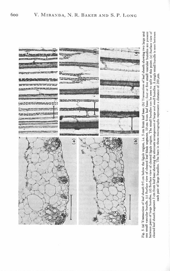
The bundle sheath cells were arranged tightly around the vascular bundles inboth the sheath and blade [Fig. 6(a)-(d)]. The bundle sheath cells were smallerthan the mesophyll cells in the leaf sheath [Fig. 6(a),(c)], but as large or largerin the leaf blade [Fig. 6(b), (d)]. The centrifugal arrangement of the bundle sheathchloroplasts was found 3 cm from the leaf base [Fig. 6(a)]. Within the leaf sheath.
6oo V. MIRANDA, N . R. BAKER AND S. P. LONG

Photosynthetic apparatus of maize leaf 601

6O2 V. MIRANDA, N . R. BAKER AND S. P. LONG
chloroplasts were found only in the mesophyll cells situated next to the bundlesheath.
Two types of vascular bundles, large and small, have been found in the Z. maysleaf (Sharman, 1942). Within the basal regions of the leaf sheath large bundlesalternate with small bundles [Fig. 5(b),(e)], however within the leaf blade threeto eight small bundles were found between each pair of large bundles [Fig. 5(c)].The large vascular bundles matured at a lower position in the leaf than the smallbundles. The vascular elements of the large bundles appeared fully developed at0-5 cm from base, whereas the small bundles did not show clear differentiationof the vascular tissue until c. 3 cm [Fig. 6(a)]. The size, structure and distributionof the vascular bundles in the leaf sheath showed marked variations compared tothose of the leaf blade. Structural details of the vascular bundles from the sheathand blade are shown in Figure 6. Large vascular bundles of the sheath generallyhad incomplete bundle sheaths which were often confluent with the hypodermalschlerenchyma. The bundles were near the lower surface in the sheath [Fig.5(a),(b)], but midway between the surfaces in the blade.
E ^2 •
500
400
300
200
100
Ea.
sta
nce
1In
ter
0 5 10 15
Distance from leaf base (cm)
Fig. 1. Changes in the number of vascular bundles per unit leaf breadth ( • ) and in the interveinaldistance (O) along the length of the leaf. Standard errors were less than 1 % of mean values.
The changes in the number of vascular bundles per unit leaf breadth and theinterveinal distance along the length of the leaf are shown in Fig. 7. The numberof bundles per unit leaf breadth in the leaf sheath is c. 2-5 mm^^ and this increasedto c. 7-5 mm~i in the leaf blade. This threefold increase in the number of vascularbundles per unit leaf breadth occurred at the ligule region where the small vascularbundles split [Fig. 5(d)]. At the leaf base adjacent bundles were separated by fourto seven mesophyll cells [Fig. 5(b)] and had an interveinal distance of c. 295//m.This distance increased to 433/<m at c. 3 cm from leaf base due to expansion ofthe mesophyll cells. Above 4 cm the interveinal distance showed a progressivedecrease, reaching 125 to 140/<m in the leaf blade. The decreases in the interveinaldistances were consistent with the number of mesophyll cells separating theadjacent vascular bundles, which was two or three at the ligule region [Fig. 5(a)]and was always found to be two within the leaf blade [Fig. 6(b)].

Photosynthetic apparatus of maize leaf 603
DISCUSSION
The first objective of this study was to describe anatomical changes, which couldaffect photosynthetic activity, along the length of the second leaf in the 7-day-oldZ. mays plant. Cuticle thickness, stomatal size and frequency and the presence ofintercellular spaces within the mesophyll tissue are all features which will infiuencethe gas exchange characteristics of the leaf. The cuticular thickening observedwithin the basal 3 cm of the leaf would be expected to increase the cuticularresistance to COj diffusion. The cuticle at 3 cm appeared to be fully developed andwould thus provide a large resistance to COj diffusion. Above 3 cm the majorpathway for COj diffusion would be through the functional stomata.
The absence of intercellular spaces in the basal cm would seriously limit ratesof CO2 diffusion to the sites of COj assimilation because rates of diffusion are lowerin liquid rather than gaseous phase. In addition, the path length of liquid phasediffusion for CO2 would be greater in the sheath than in the blade because of thepresence of large non-photosynthetic parenchyma cells around the chlorenchyma.It is possible that the high respiratory activity of the developing tissue in this region(Miranda, Baker and Long, unpublished data) will maintain a high internal CO,concentration and obviate the need for COj diffusion from the atmosphere. Abovethe basal 1 cm, intercellular spaces were found in the mesophyll and there wascontinuum of air spaces from this point to the ligule. Lack of intercellular spaceswithin the ligule region prohibits rapid gaseous exchange between blade andsheath.
Variations in stomatal size and frequency along the length of the leaf accountedfor changes in r,, which could modify the rate of COj diffusion into the leaf. Thevery low r^ values, observed in the basal 2 cm of the sheath, were unlikely to limitCO2 diffusion. However, as r^ increases with leaf development stomata are likelyto become increasingly important in regulating the capacity for photosynthetic CO2assimilation. In mature leaves of C4 plants r., has been shown to be a major factorin limiting COg assimilation (Ludlow and Wilson, 1971; Akita and Moss, 1972;Gifford, 1974).
The morphological and anatomical transition from the sheath to the blade occursat the ligule. The sheath and blade exhibited large differences in the sizes ofchlorenchymatous cells, the number of mesophyll layers, interveinal distance andin the size, structure and distribution of the vascular bundles. The leaves of C4plants have a low interveinal distance, usually below 160/<m (Takeda andFukuyama, 1971; Crookston and Moss, 1974; Hattersley and Watson, 1975) dueto the presence of only two mesophyll cells between adjacent vascular bundles(Crookston and Moss, 1974; Laetsch, 1974). The large interveinal distancerecorded for the sheath segments of the second leaf of Z. mays shows deviationsfrom this typical C4 character. The leaf blade exhibits typical 'Kranz' anatomy.In transections the sheath consisted of about seven layers of mesophyll cells,however chloroplasts were observed only in the mesophyll cells adjacent to bundlesheath cells, thus it would appear that only a fraction of the mesophyll cells maybe capable of photosynthesis. The vascular bundles within the leaf sheath aresituated close to the lower epidermis. Such structural organization would facilitatelight and COj availability to the chlorenchymatous tissue since only the lowersurface of the leaf sheath is exposed to the external atmosphere. The upper surfaceof the leaf sheath was always rolled over the inner leaves.
Although the sheath lacks typical 'Kranz' anatomy it did possess two types of

6o4 V. MIRANDA, N . R. BAKER AND S. P. LONG
chlorenchymatous cells, mesophyll and bundle sheath, which were in intimateassociation, a feature considered essential for C4 metabolism (Ray and Black, 1979).Leaf tissue of other species has been shown to exhibit non-classical 'Kranz'anatomy whilst employing C4 metabolism for COj assimilation (Hattersley,Watson and Osmond, 1977). On the basis of the C4 acid decarboxylatingmechanism found in the bundle sheath cells C4 plants have been divided into threegroups: NADP-malic enzyme, NAD-malic enzyme and PEP-carboxykinase species(Gutierrezes a/., 1974; Hatch, Kagawaand Craig, 1975). These three biochemicallydistinct C4 metabolic groups have been associated with certain structuralcharacteristics, such as bundle sheath chloroplast position and the degree of granalstacking within these chloroplasts (Hatch et al., 1975). Z. mays is known to be aNADP-malic enzyme species, in which the bundle sheath chloroplasts tend to beagranal and are situated in a centrifugal position (Hatch et al., 1975). Hattersleyand Watson (1976) have shown that the condition where there are no cells betweenthe metaxylem vessel elements and the laterally adjacent sheath cells of largebundles in grass leaf blades, i.e. the 'XyMS-condition', is a characteristic ofNADP-malic enzyme species. This 'XyMS-condition' was present in the largebundles of leaf sheath and blade of the second leaf of Z. mays [Fig. 6(c), (d)]. Thisanatomical evidence suggests that C4 metabolism should be present within the leafsheath and blade of Z. mays leaf.
The second objective of this study was to utilize anatomical information to assessthe validity of using regions along the length of a single Z. mays leaf as a progressivedevelopmental gradient of the C4 photosynthetic apparatus. Changes in anatomicalcharacteristics expected during the normal course of leaf development are limitedto the basal 3 cm of the leaf. Above this region many anatomical changes wereclearly ontogenetic rather than developmental. The most striking example of thiswas found at the ligule, where features such as interveinal distance, cell size andmesophyll anatomy show marked discontinuities. In conclusion, the use of thesingle Z. mays leaf in studies of development of aspects of the photosyntheticprocesses, which are dependent on anatomical features, may be complicated byontogeny.
ACKNOWLEDGEMENTS
The authors are grateful to Dr K. Parkinson for the loan of the microscopemounted leaf chamber and to Mrs S. Corbett for skilled technical assistance ingrowing the plants.
REFERENCES
AKITA, S . & Moss, D. N. (1972). Differential stomatal response between C., and C, species to atmosphericCOj concentration and light. Crop Science, 12, 789-793.
CROOKSTON, R. K . & Mess, D. N. (1974). Interveinal distance for carbohydrate transport in leaves of C^and C,] grasses. Crop Science, 14, 123^125.
EDWARDS, G . E . & HIIBER, S . C . (1979). C4 metabolism in isolated cells and protoplasts. In: Encyclopediaof Plant Physiology, vol. 6 (ed. by M. Gibbs and E. Latzko), pp. 102-112. Springer-Verlag, Berlin.
FAHN, A . (1974). Plant Anatomy 2nd edn, Pergamon Press, Oxford.GiFFORD, R. M. (1974). A comparison of potential photosynthesis, productivity and yield of plant species
with differing photosynthetic nnetaboUsm. Australian Journal of Plant Physiology, 1, 107 117.GuTiFKHF.z, M., GRACEN, V. E. & EDWARDS, G . E . (1974). Biochemical and cytological relationship in C^
plants. P/anta 119, 279-300.HATCH, M . D . , KAGAWA, T . & CRAIG, S . (1975). Sub-division of C,,-pathway species based on differing C^

Photosynthetic apparatus of maize leaf 605
acid decarboxylating systems and ultrastructural features. Australian Journal of Plant Pliysiology, 2,111-128.
HATTERSI.EV, P . W . & WATSON, L . (1975). Anatomical parameters for predicting photosynthetic pathwaysof grass leaves: the 'maximum lateral count' and the 'maximum cells distant count'. Phytonwrphology,25, 325-333.
HATTERSI.I-V, P . W . & WATSON, L . (1976). C, Grasses: an anatomical criterion for distinguishing betweenNADP-malic enzyme species and PCK or NAD-malic enzyme species. Australian Journal of Botany,24, 297-308.
HATTERSI.EY, P . W . , WATSON, L . & OSMOND, C . B . (1977). In situ immunoHuorescent labelling ofribulose-l,5-bisphosphate carboxylase in leaves of C;, and C,, plants. Australian Journal of PlantPhysiology, 4, 523-539.
HAWKE, J . C , RUMSDY, M . G . & LEECH, R. M . (1974). Lipid biosynthesis in green leaves of developingmaize. Plant Physiology, 53, 555-561.
HEICHEL, G . H . (1971). Genetic control of epidermal cell and stomatal frequency in maize. Crop Science,11, 830-832.
LAET.SCH, W . M . (1974). The C, syndrome: a structural analysis. Annual Review of Plant Physiology, 25,27-52.
LEECH, R. M . , RUMSBY, M . G . & THOMSON, W . W . (1973). Plastid differentiation, acyl lipid, and fatty acidchanges in developing green maize leaves. Plant Physiology, 52, 240-245.
LEESE, B . M . & LEECH, R . M . (1976). Sequential changes in the lipids of developing proplastids isolatedfrom green maize leaves. Plant Physiology, 57, 789-794.
LuDLow, M. & WH.SON, G. L. (1971). Photosynthesis of tropical pasture plants IH. Leaf age. AustralianJournal of Biological Sciences, 24, 1077-1(387.
METCALEE, C . R. (1960). Anatomy of the monocotyledons. \. Gramineae. Clarendon Press, Oxford.PARLANGE, J . Y . & WAGGONER, P. E. (1970). Stomatal dimensions and resistance to diffusion. Plant
Physiology, 46, 337-342.RASCHKE, K . (1970). The temperature dependence of COj assimilation and stomatal aperture in leaf sections
of Zea mays. Planta, 91, 336-363.RASCHKE, K . (1975). Stomatal action. Annual Reviejv of Plant Physiology, 26, 309-340.RAY, T . B . & BLACK, C . C . (1979). The C,, pathway and its regulation. In: Encyclopedia of Plant Physiology,
vol. 6 (ed. by M. Gibbs & E. Latzko), pp. 77-101. Springer-Verlag, Berlin.SHARMAN, B . C . (1942). Developmental anatomy of the shoot of Zea mays. Annals of Botany, 6, 245-282.SRIVASTAVA, L . M . & SINGH, S. S . (1972). Stomatal structure in theccorn leaves. Jo»/Hfl/o/ Ultrastructural
Research, 39, 345 363.STEIIHINS, G . L . & SHAH, S . S. (1960). Developmental studies of cell differentiation in the epidermis of
monocots. U. Cytological features of stomatal development in the Graminae. Developmental Biology,2, 477-500.
TAKEDA, T . & FuKUYAMA, M. (1971). Studies on the photosynthesis of the Gramineae. 1. Difference inphotosynthesis among subfamilies and their relations with the systematics of the Gramineae. (InJapanese with English summary.) Crop Science Society of Japan Proceedings, 40, 12-19.
ZlEGLER, H., SHMUELI, E . & LANGE, G . (1974). The structure and function of the stomata of Zea mays.I. The development. Cytobiologie, 9, 162-168.


![Biodiversity and conservation Genetic diversity: within species variation (e.g corn [Zea mays] in North vs. Central America) Species diversity: species](https://img.pdfslide.net/doc/110x75/56649db45503460f94aa4ef5/biodiversity-and-conservation-genetic-diversity-within-species-variation.jpg)