Embed Size (px)
Citation preview
Anatomie, histologie a embryologie
Témata:
- Vývin pohlavních buněk, samčí gametofyt
- Mikrosporogeneze
- Mikrogametogeneze
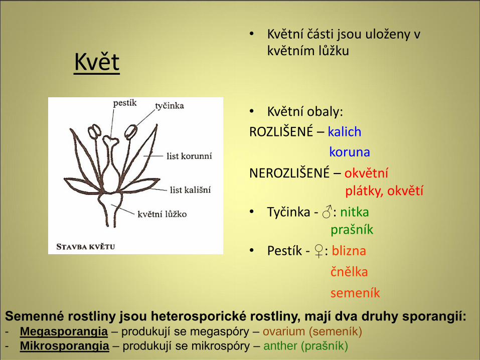
• Květní části jsou uloženy v květním lůžku
• Květní obaly:
ROZLIŠENÉ – kalich
koruna
NEROZLIŠENÉ – okvětní plátky, okvětí
• Tyčinka - ♂: nitka prašník
• Pestík - ♀: blizna
čnělka
semeník
Květ
Semenné rostliny jsou heterosporické rostliny, mají dva druhy sporangií: - Megasporangia – produkují se megaspóry – ovarium (semeník)
- Mikrosporangia – produkují se mikrospóry – anther (prašník)
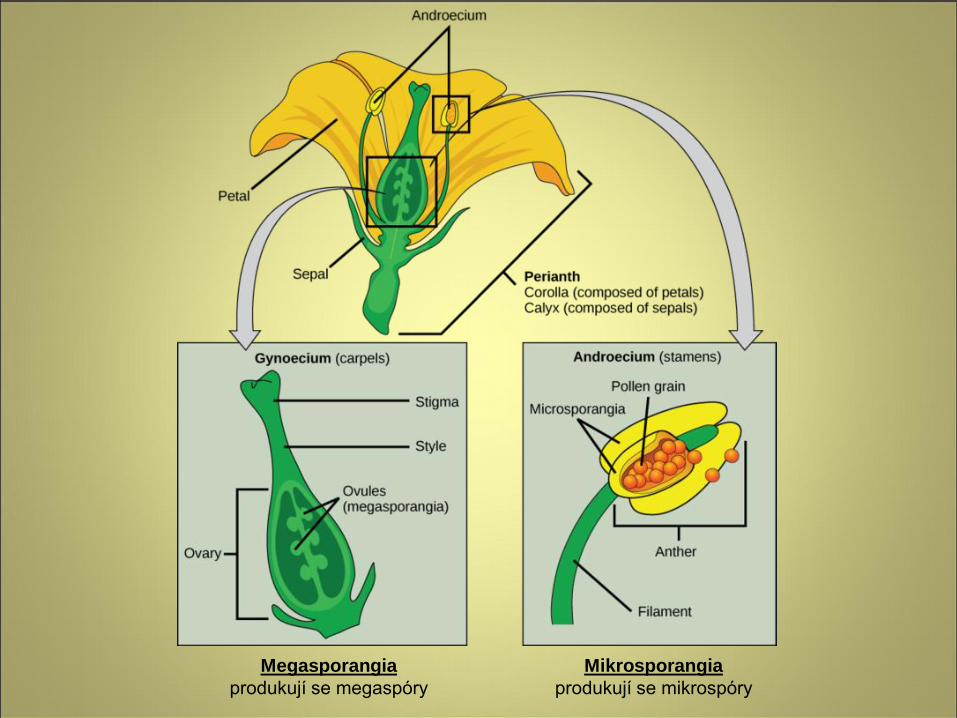
Megasporangia
produkují se megaspóry
Mikrosporangia
produkují se mikrospóry
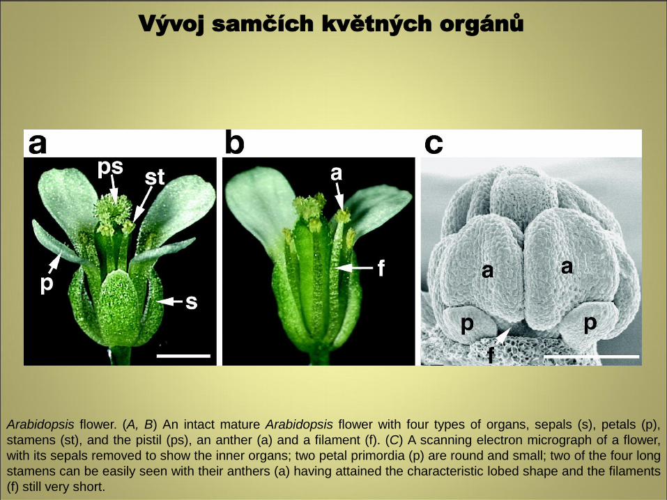
Arabidopsis flower. (A, B) An intact mature Arabidopsis flower with four types of organs, sepals (s), petals (p),
stamens (st), and the pistil (ps), an anther (a) and a filament (f). (C) A scanning electron micrograph of a flower,
with its sepals removed to show the inner organs; two petal primordia (p) are round and small; two of the four long
stamens can be easily seen with their anthers (a) having attained the characteristic lobed shape and the filaments
(f) still very short.
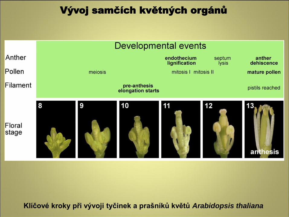
Vývoj samčích květných orgánů
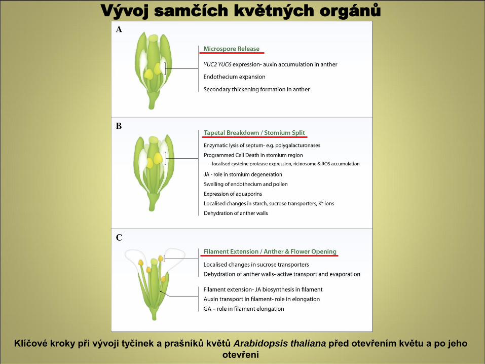
Klíčové kroky při vývoji tyčinek a prašníků květů Arabidopsis thaliana před otevřením květu a po jeho
otevření
Vývoj samčích květných orgánů
Vývoj samčích květných orgánů
Klíčové kroky při vývoji tyčinek a prašníků květů Arabidopsis thaliana
Vývoj samčích květných orgánů
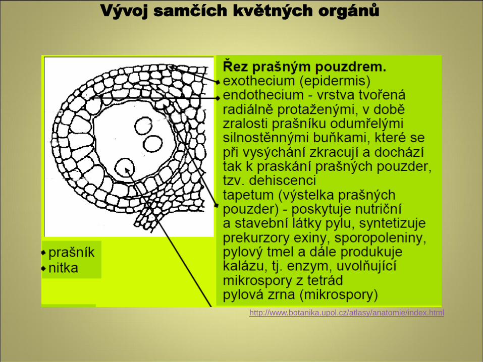
Vývoj samčích květných orgánů
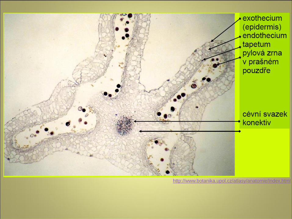
http://www.botanika.upol.cz/atlasy/anatomie/index.html
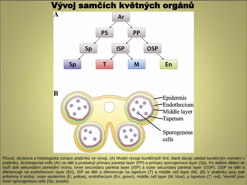
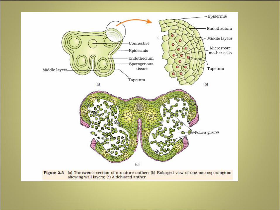
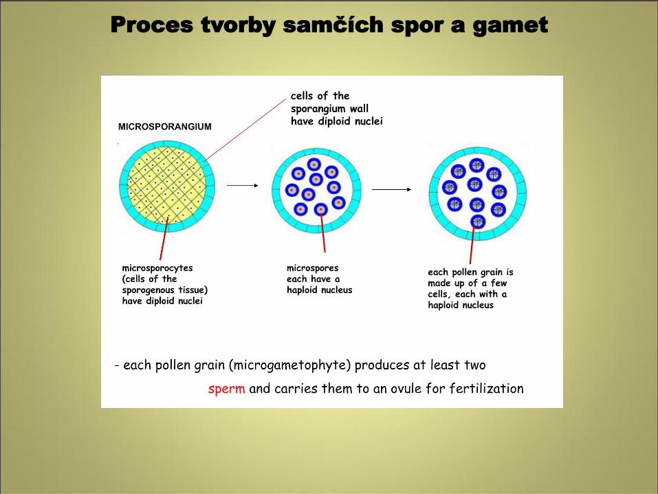
Původ, struktura a histologická zonace prašníků ve vývoji. (A) Model vývoje buněčných linií, které dávají základ buněčným vrstvám v prašníku. Archesporial cells (Ar) se dělí a produkují primary parietal layer (PP) a primary sporogenous layer (Sp). Po dalším dělení se tvoří dvě sekundární parietální vrstvy, inner secondary parietal layer (ISP) a outer secondary parietal layer (OSP). OSP se dělí a diferencuje na endothecium layer (En), ISP se dělí a diferencuje na tapetum (T) a middle cell layer (M). (B) V prašníku jsou pak prítomny 4 vrstvy: outer epidermis (E; yellow), endothecium (En; green), middle cell layer (M; blue), a tapetum (T; red). Vevnitř jsou inner sporogenous cells (Sp; purple).
Vývoj samčích květných orgánů
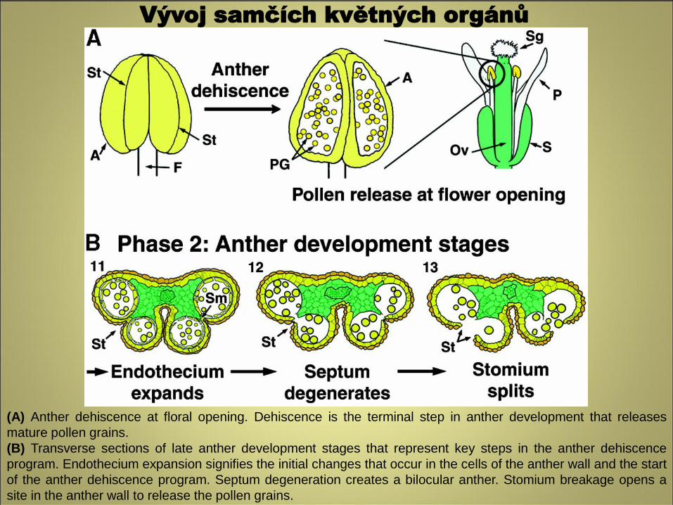
(A) Anther dehiscence at floral opening. Dehiscence is the terminal step in anther development that releases
mature pollen grains.
(B) Transverse sections of late anther development stages that represent key steps in the anther dehiscence
program. Endothecium expansion signifies the initial changes that occur in the cells of the anther wall and the start
of the anther dehiscence program. Septum degeneration creates a bilocular anther. Stomium breakage opens a
site in the anther wall to release the pollen grains.
Vývoj samčích květných orgánů
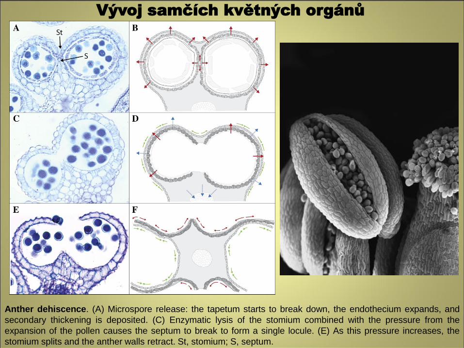
Anther dehiscence. (A) Microspore release: the tapetum starts to break down, the endothecium expands, and
secondary thickening is deposited. (C) Enzymatic lysis of the stomium combined with the pressure from the
expansion of the pollen causes the septum to break to form a single locule. (E) As this pressure increases, the
stomium splits and the anther walls retract. St, stomium; S, septum.
Vývoj samčích květných orgánů
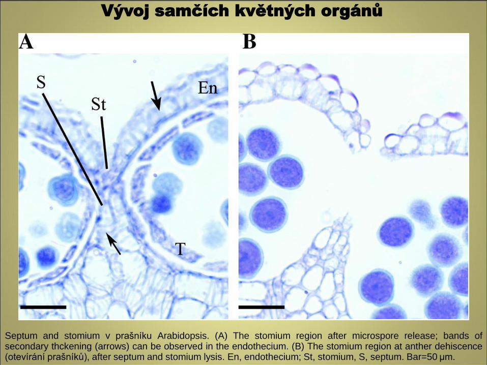
Septum and stomium v prašníku Arabidopsis. (A) The stomium region after microspore release; bands of secondary thckening (arrows) can be observed in the endothecium. (B) The stomium region at anther dehiscence (otevírání prašníků), after septum and stomium lysis. En, endothecium; St, stomium, S, septum. Bar=50 μm.
Vývoj samčích květných orgánů
http://www.botanika.upol.cz/atlasy/anatomie/index.html
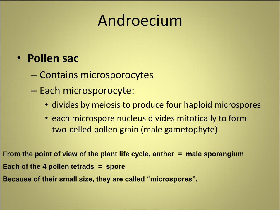
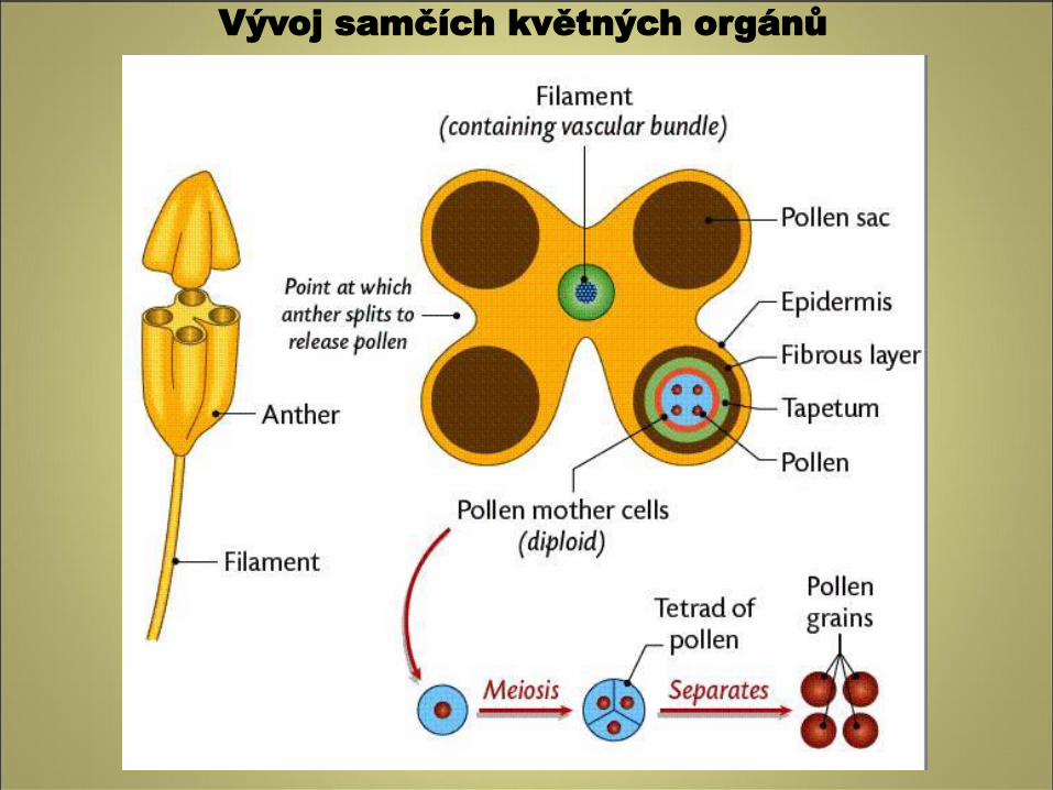
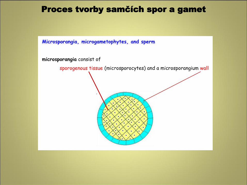
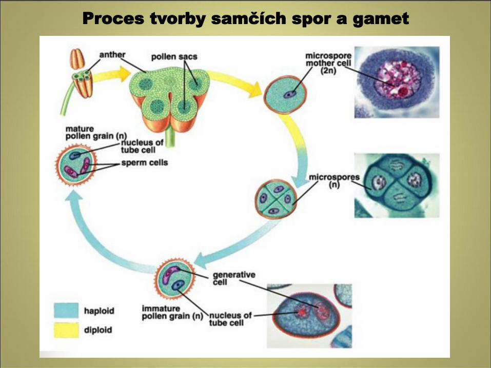
Androecium
• Pollen sac
– Contains microsporocytes
– Each microsporocyte:
• divides by meiosis to produce four haploid microspores
• each microspore nucleus divides mitotically to form two-celled pollen grain (male gametophyte)
From the point of view of the plant life cycle, anther = male sporangium
Each of the 4 pollen tetrads = spore
Because of their small size, they are called “microspores”.
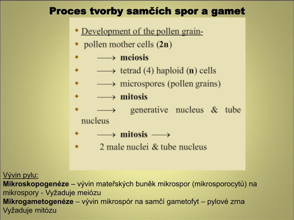
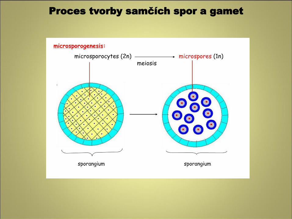
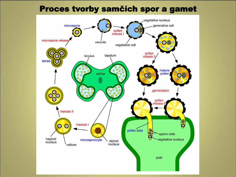
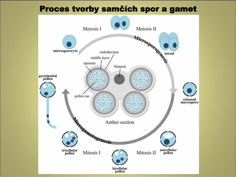
Vývin pylu:
Mikroskopogenéze – vývin mateřských buněk mikrospor (mikrosporocytů) na
mikrospory - Vyžaduje meiózu
Mikrogametogenéze – vývin mikrospór na samčí gametofyt – pylové zrna
Vyžaduje mitózu
Proces tvorby samčích spor a gamet
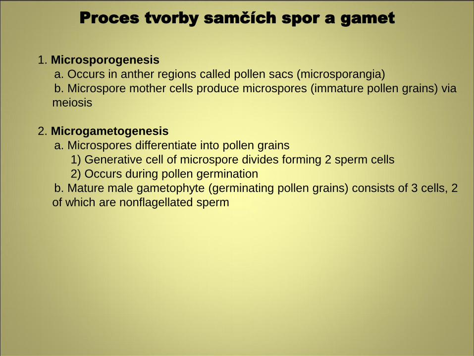
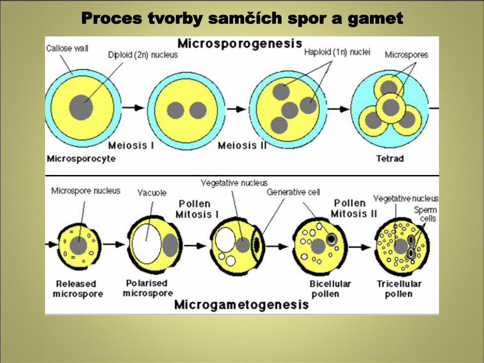
1. Microsporogenesis
a. Occurs in anther regions called pollen sacs (microsporangia)
b. Microspore mother cells produce microspores (immature pollen grains) via
meiosis
2. Microgametogenesis
a. Microspores differentiate into pollen grains
1) Generative cell of microspore divides forming 2 sperm cells
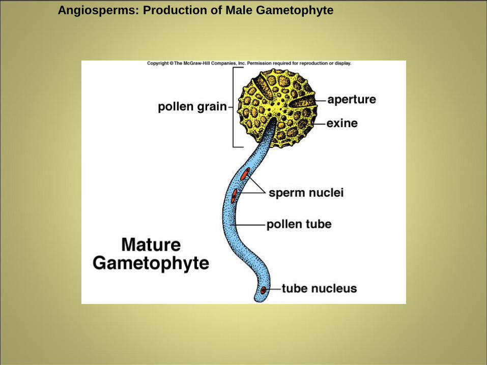
2) Occurs during pollen germination
b. Mature male gametophyte (germinating pollen grains) consists of 3 cells, 2
of which are nonflagellated sperm
Proces tvorby samčích spor a gamet
Proces tvorby samčích spor a gamet
Proces tvorby samčích spor a gamet
Proces tvorby samčích spor a gamet
Proces tvorby samčích spor a gamet
Proces tvorby samčích spor a gamet
Proces tvorby samčích spor a gamet
Proces tvorby samčích spor a gamet
Proces tvorby samčích spor a gamet
Proces tvorby samčích spor a gamet
Proces tvorby samčích spor a gamet
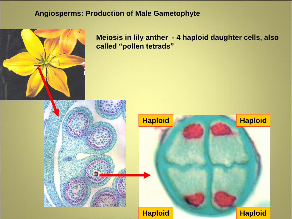
Angiosperms: Production of Male Gametophyte
Haploid Haploid
Haploid Haploid
Meiosis in lily anther - 4 haploid daughter cells, also
called “pollen tetrads”
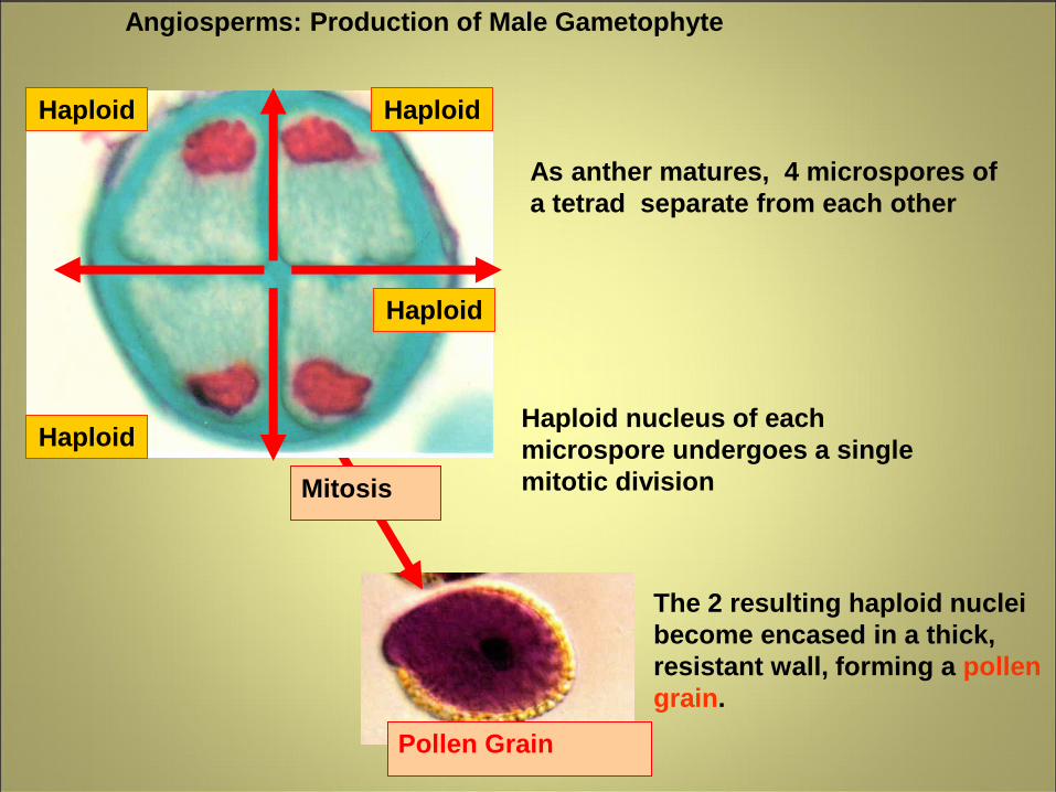
As anther matures, 4 microspores of
a tetrad separate from each other
Angiosperms: Production of Male Gametophyte
Haploid nucleus of each
microspore undergoes a single
mitotic division
Pollen Grain
Mitosis
Haploid
Haploid
Haploid Haploid
The 2 resulting haploid nuclei
become encased in a thick,
resistant wall, forming a pollen
grain.
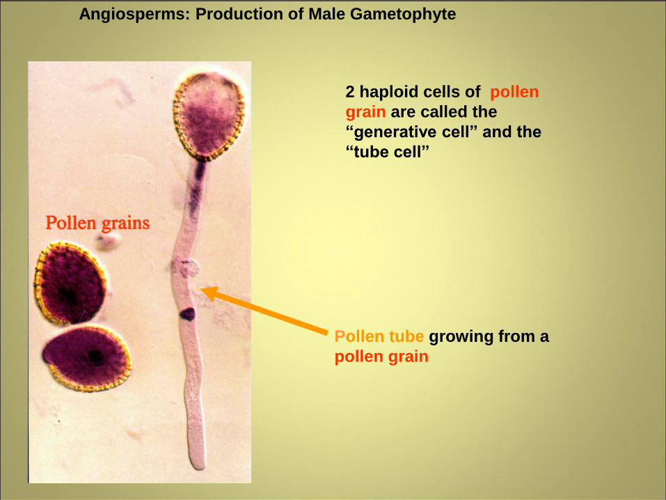
2 haploid cells of pollen
grain are called the
“generative cell” and the
“tube cell”
Pollen tube growing from a
pollen grain
Angiosperms: Production of Male Gametophyte
Angiosperms: Production of Male Gametophyte
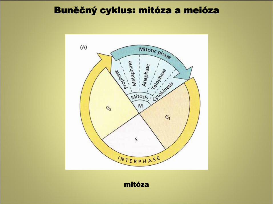
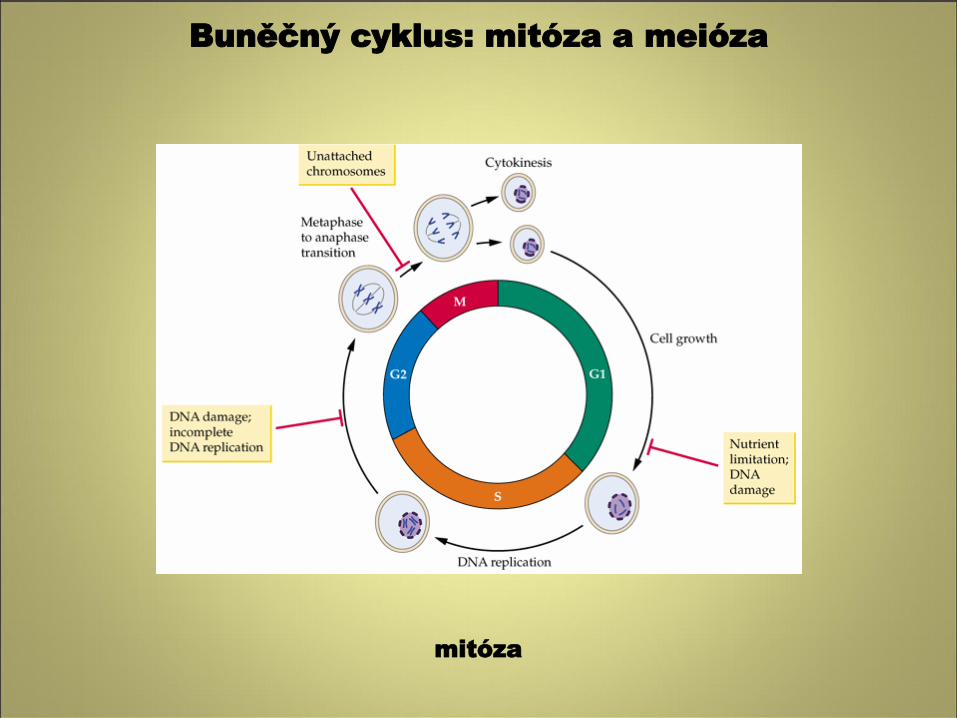
Buněčný cyklus: mitóza a meióza
mitóza
Buněčný cyklus: mitóza a meióza
mitóza
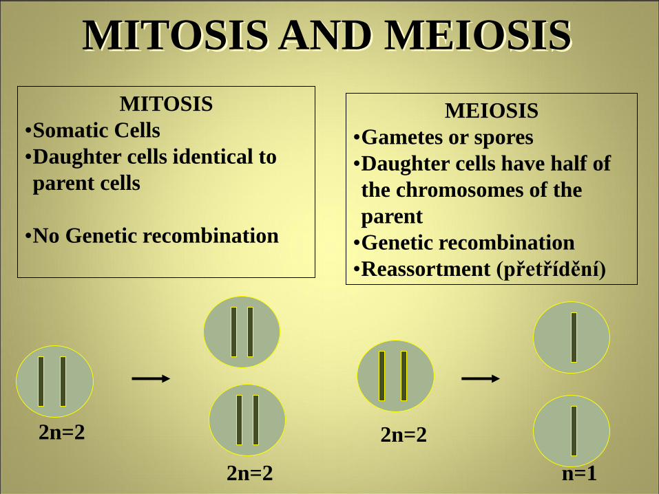
MITOSIS AND MEIOSIS
MITOSIS
•Somatic Cells
•Daughter cells identical to
parent cells
•No Genetic recombination
MEIOSIS
•Gametes or spores
•Daughter cells have half of
the chromosomes of the
parent
•Genetic recombination
•Reassortment (přetřídění)
2n=2
2n=2
2n=2
n=1
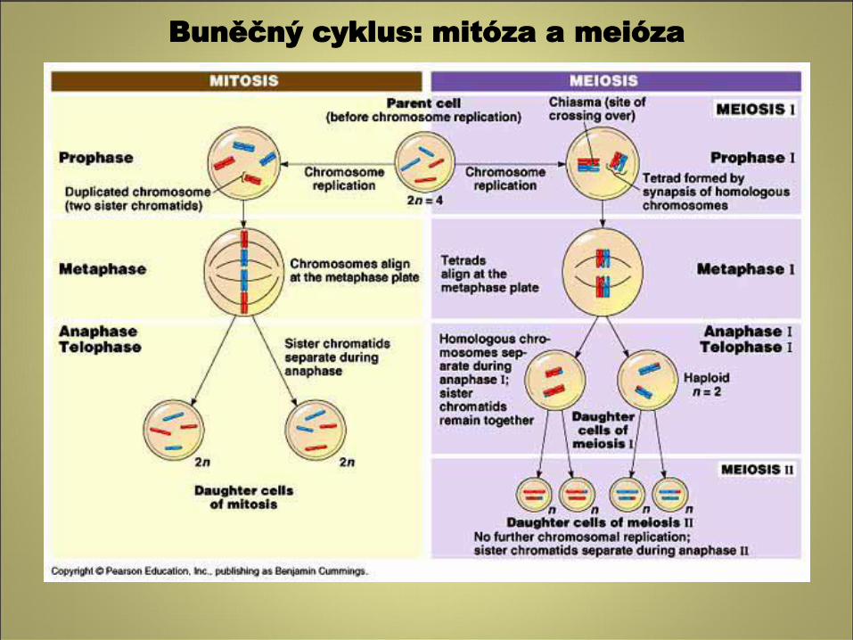
Buněčný cyklus: mitóza a meióza
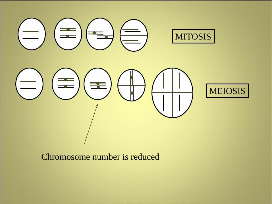
MITOSIS
MEIOSIS
Chromosome number is reduced
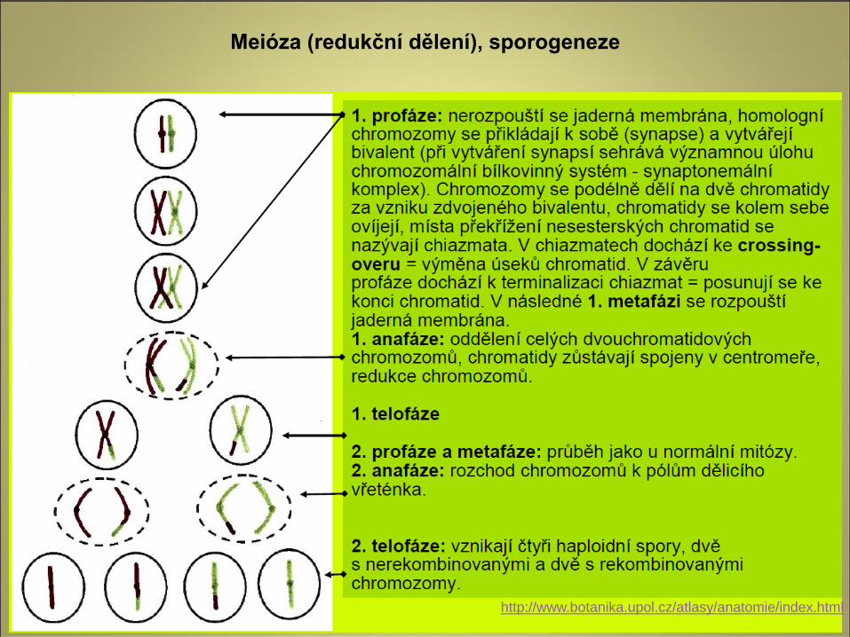
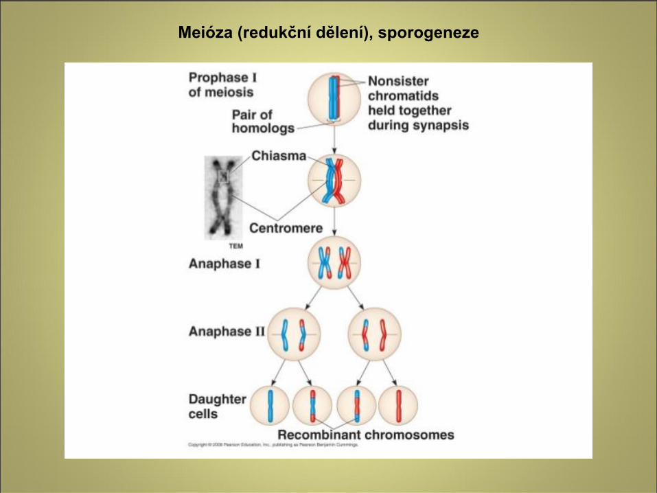
Meióza (redukční dělení), sporogeneze
http://www.botanika.upol.cz/atlasy/anatomie/index.html
Meióza (redukční dělení), sporogeneze
Meióza (redukční dělení), sporogeneze
Meiózou vznikají u rostlin haploidní spory z diploidních buněk sporogenního pletiva
Meióza probíhá ve dvou po sobě následujících mitotických děleních:
1. meióza (heterotypické dělení):
- V profázi dochází k podélnému přikládání a spojování homologických chromozomů,
vzniká tzv. bivalent.
- Chromozomy se podélně rozdělí na dvě chromatidy a vzniká čtyřchromatidový
bivalent.
- Nesesterské chromatidy homologických chromozomů se mohou překřížit a vyměnit si
úseky chromatid (crossing – over). Z těchto chromatid vznikají po rozdělení centromery
(v anafázi homeotypického dělení) rekombinované chromozomy.
- Teprve v metafázi se rozpouští jaderná membrána, mizí jadérko a diferencuje se
dělicí vřeténko. Zdvojené bivalenty se posunují do ekvatoriální roviny.
- V anafázi se homologické chromozomy od sebe oddělují a dvouchromatidové
chromozomy se po mikrotubulech dělicího vřeténka posunují k opačným pólům
buňky, kde se nakonec shromáždí úplná haploidní sada chromozomů (= redukce
počtu chromozomů).
- V telofázi vznikají dvě haploidní buňky.
2. meióza (homeotypické dělení): je v podstatě normální mitóza. Výsledkem je tetráda
haploidních jednobuněčných (později většinou dvou- nebo někalikabuněčných) spor.
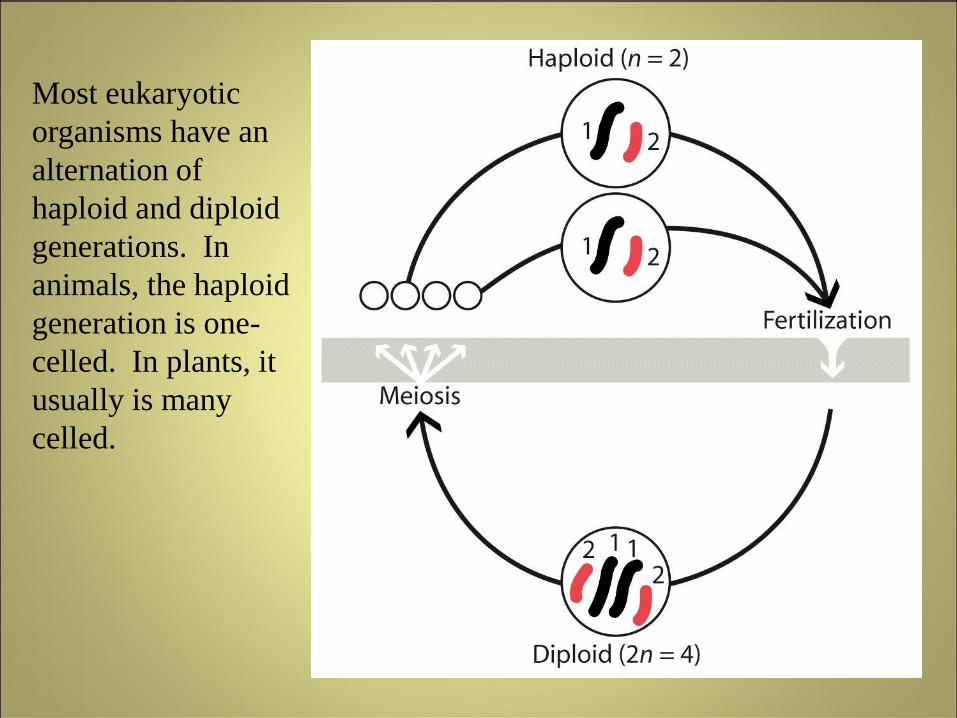
Most eukaryotic
organisms have an
alternation of
haploid and diploid
generations. In
animals, the haploid
generation is one-
celled. In plants, it
usually is many
celled.
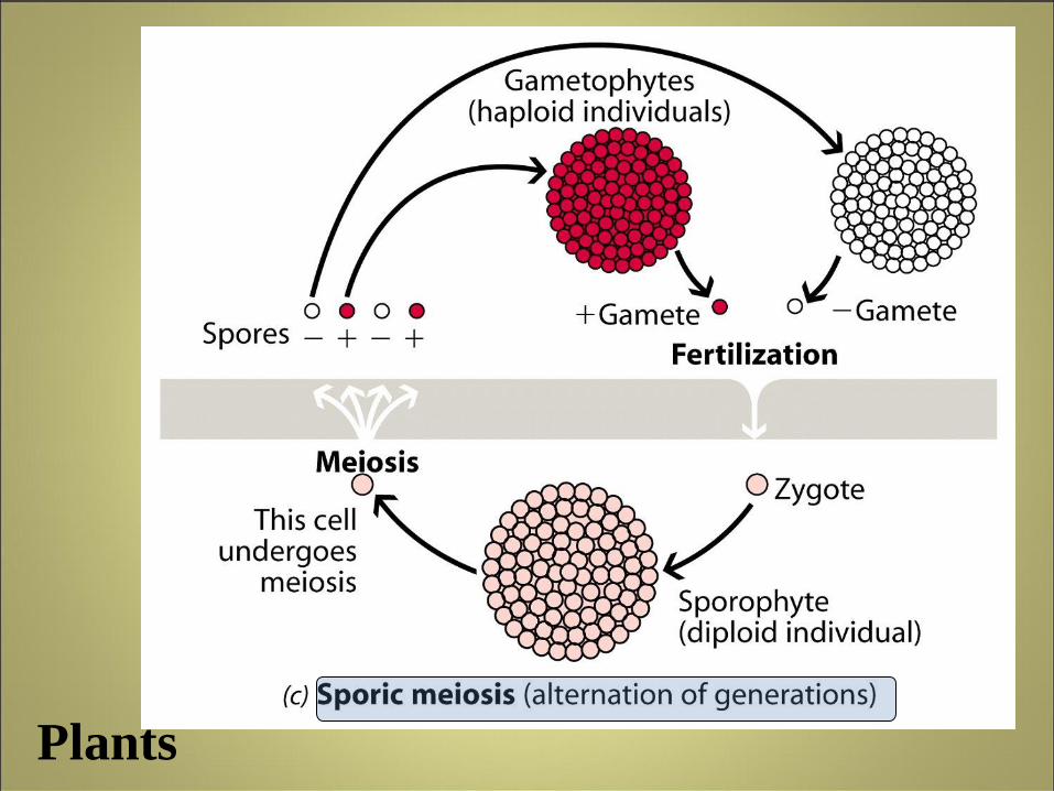
Plants
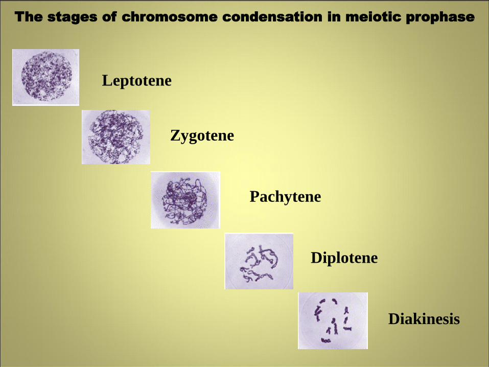
The stages of chromosome condensation in meiotic prophase
Leptotene
Zygotene
Diplotene
Pachytene
Diakinesis
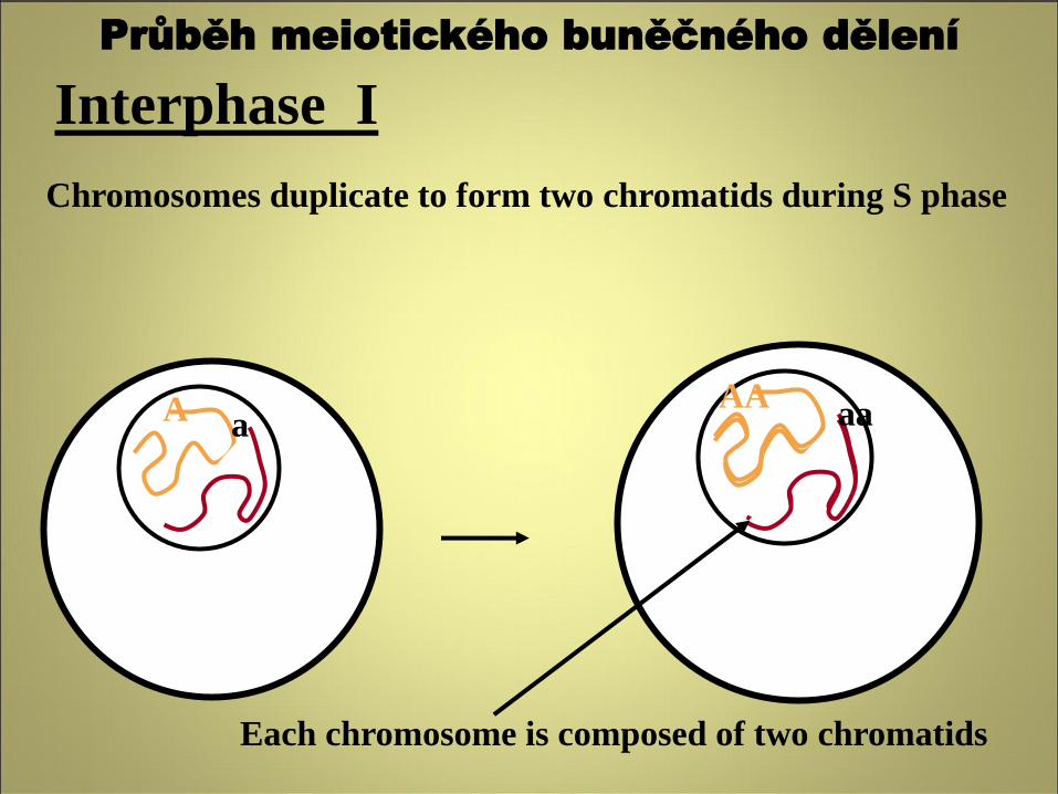
Interphase I
Chromosomes duplicate to form two chromatids during S phase
Each chromosome is composed of two chromatids
A a AA
aa
Průběh meiotického buněčného dělení
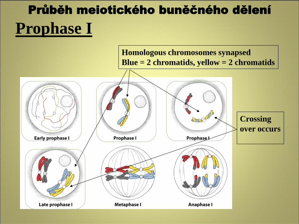
Prophase I
Crossing
over occurs
Homologous chromosomes synapsed
Blue = 2 chromatids, yellow = 2 chromatids
Průběh meiotického buněčného dělení
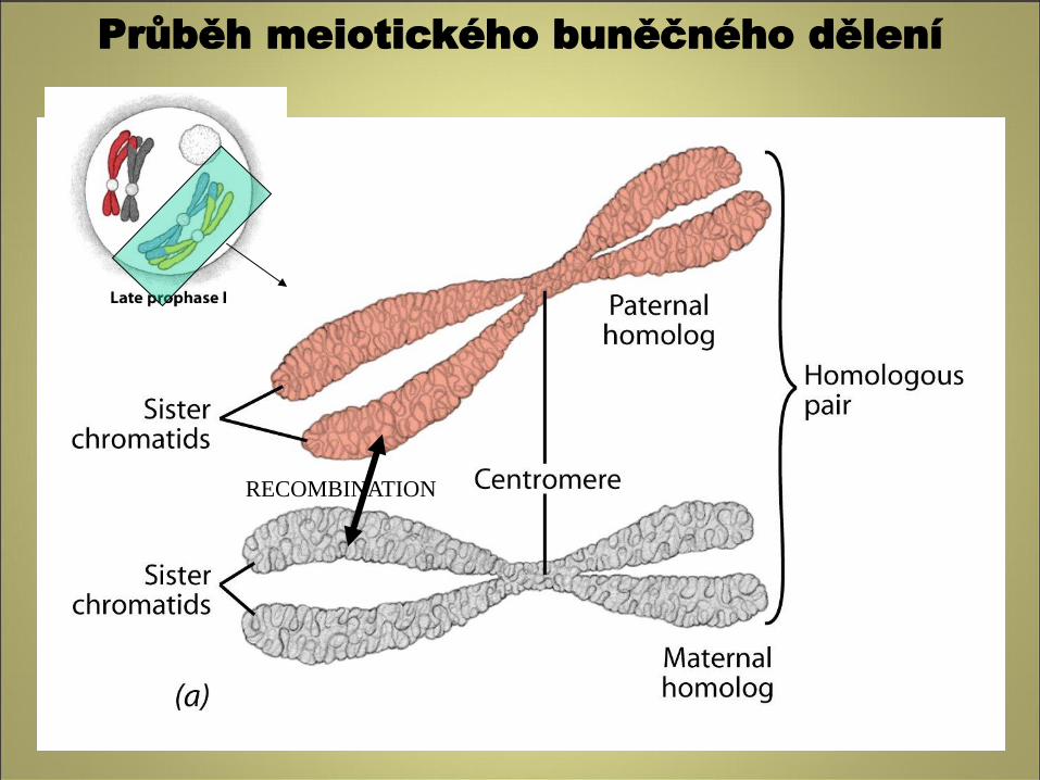
RECOMBINATION
Průběh meiotického buněčného dělení
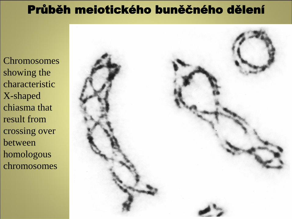
Chromosomes
showing the
characteristic
X-shaped
chiasma that
result from
crossing over
between
homologous
chromosomes
Průběh meiotického buněčného dělení
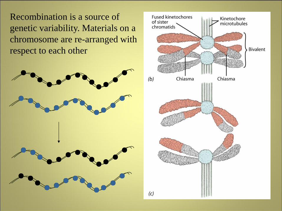
Recombination is a source of
genetic variability. Materials on a
chromosome are re-arranged with
respect to each other
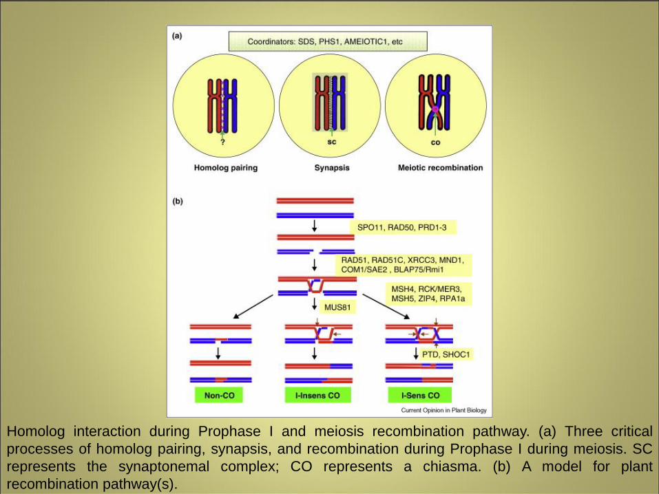
Homolog interaction during Prophase I and meiosis recombination pathway. (a) Three critical
processes of homolog pairing, synapsis, and recombination during Prophase I during meiosis. SC
represents the synaptonemal complex; CO represents a chiasma. (b) A model for plant
recombination pathway(s).
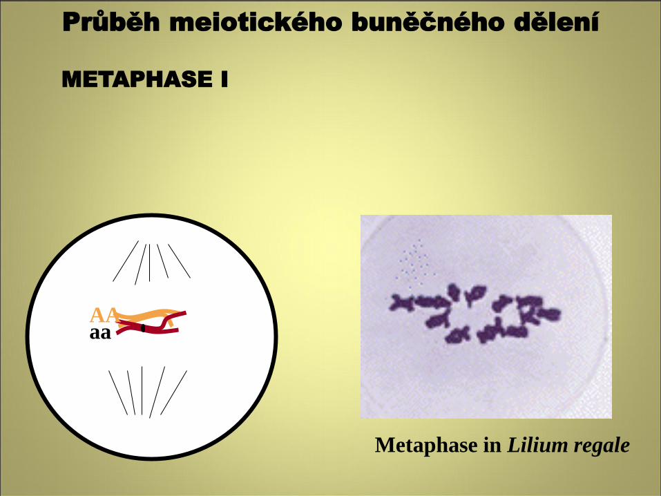
METAPHASE I
Metaphase in Lilium regale
AA aa
Průběh meiotického buněčného dělení
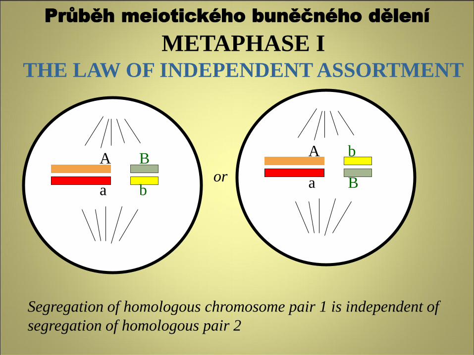
METAPHASE I THE LAW OF INDEPENDENT ASSORTMENT
Segregation of homologous chromosome pair 1 is independent of
segregation of homologous pair 2
or A
a
B
b
A
a
b
B
Průběh meiotického buněčného dělení
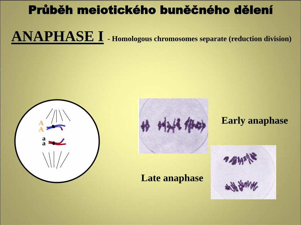
ANAPHASE I - Homologous chromosomes separate (reduction division)
Early anaphase
Late anaphase
A A
a a
Průběh meiotického buněčného dělení
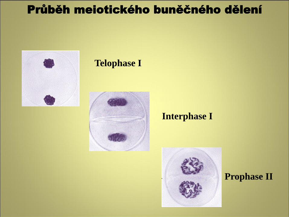
Telophase I
Interphase I
Prophase II
Průběh meiotického buněčného dělení
METAPHASE II
A A
aa
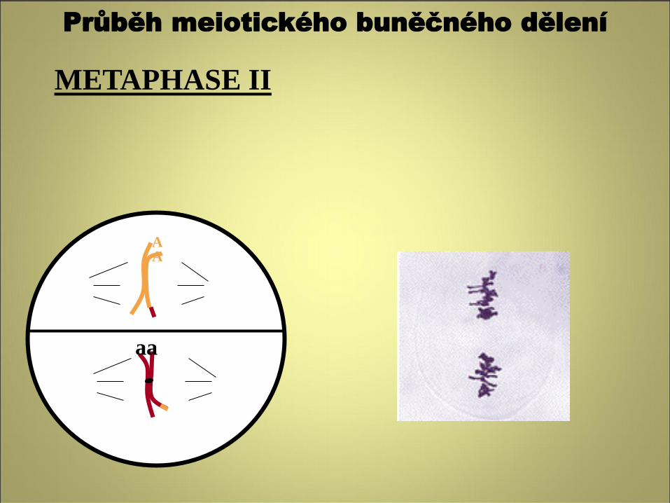
Průběh meiotického buněčného dělení
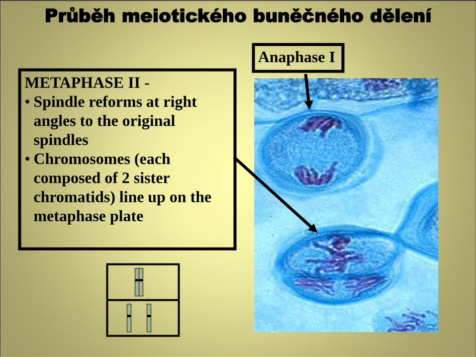
METAPHASE II -
• Spindle reforms at right
angles to the original
spindles
• Chromosomes (each
composed of 2 sister
chromatids) line up on the
metaphase plate
Anaphase I
Průběh meiotického buněčného dělení
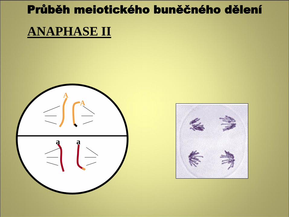
ANAPHASE II
A
a
A
a
Průběh meiotického buněčného dělení
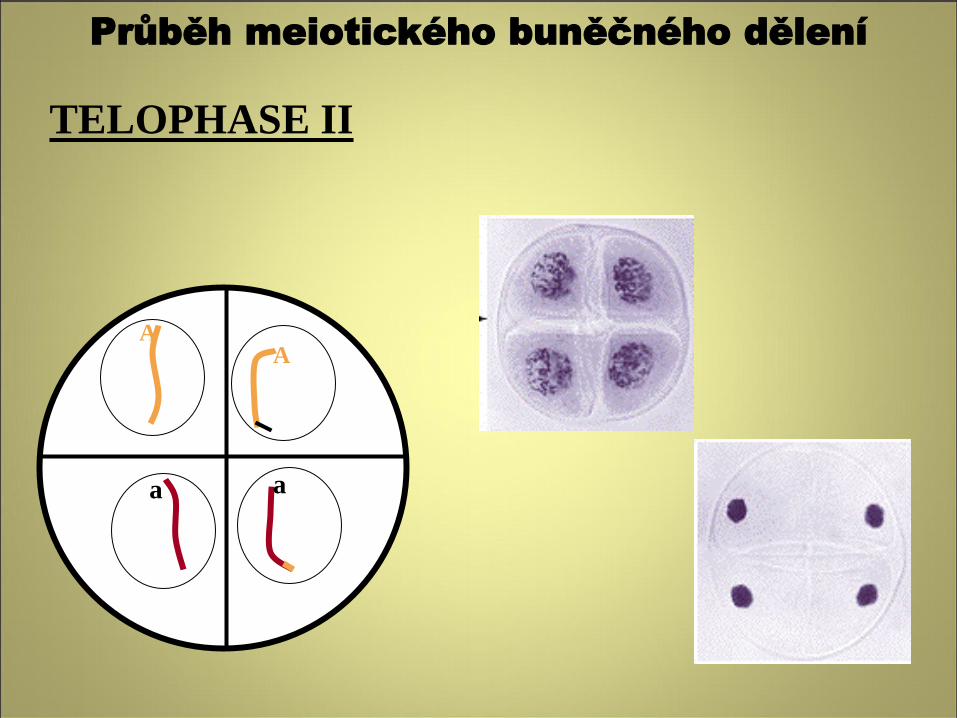
TELOPHASE II
A
a
A
a
Průběh meiotického buněčného dělení
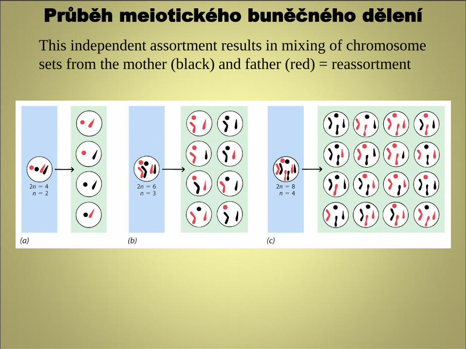
This independent assortment results in mixing of chromosome
sets from the mother (black) and father (red) = reassortment
Průběh meiotického buněčného dělení
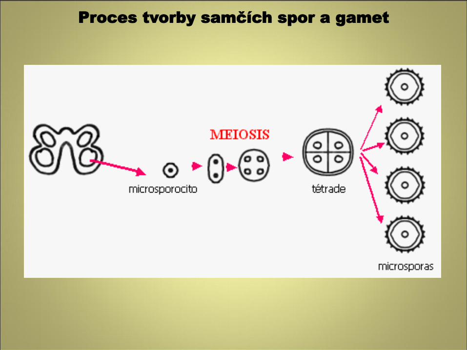
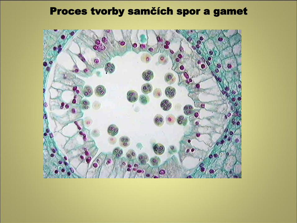
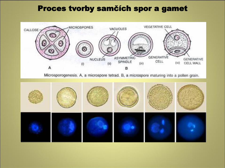
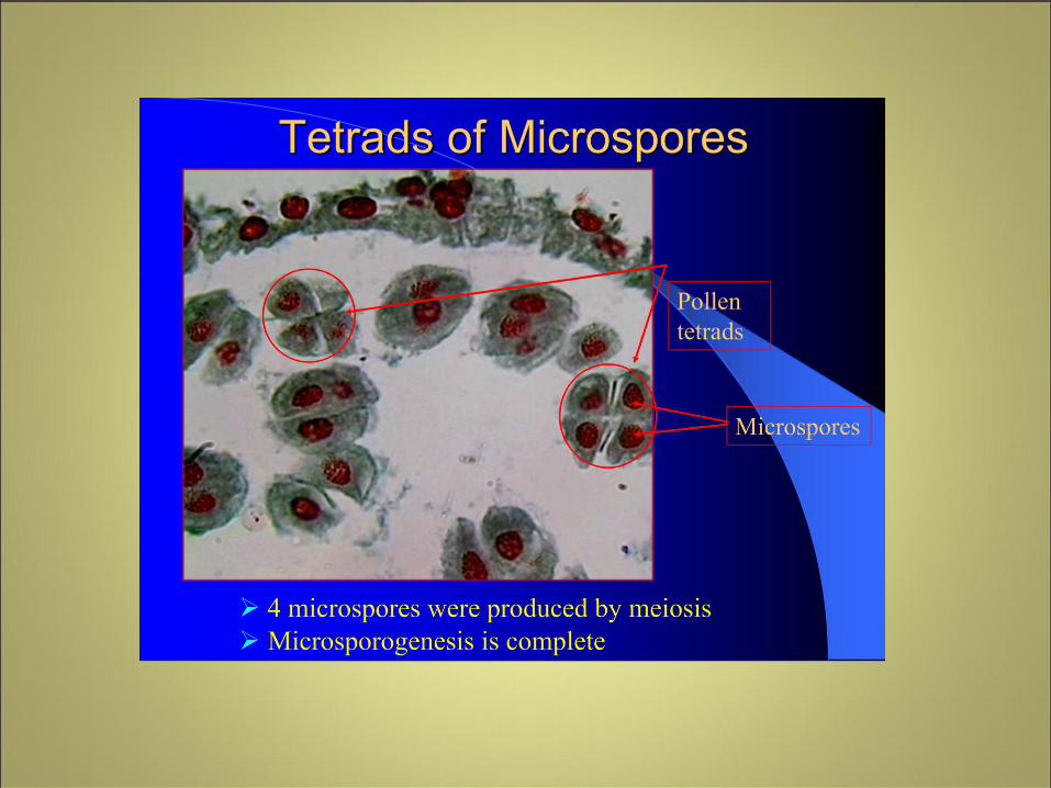
Mikrosporogeneze (vývoj mikrospory = pylového zrna)
• Ze sporogenních buněk vznikají po několika mitózách mateřské buňky mikrospor, tzv.
mikrosporocyty
• Z mikrosporocytů meiotickým redukčním dělením vznikají čtyři haploidní jádra
• V tetrádách mohou být mikrospory uspořádány různým způsobem (nejčastěji
tetraedricky, izobilaterálně, příčně, lineárně).
• Pylová zrna jsou v tetrádách spojena kalózou, u většiny rostlin však dochází k jejímu
rozrušení enzymem kalázou a k rozpadu tetrád. U některých rostlin zůstávají tetrády
pylových zrn zachovány
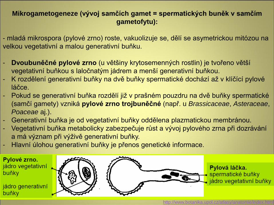
Mikrogametogeneze (vývoj samčích gamet = spermatických buněk v samčím
gametofytu):
- mladá mikrospora (pylové zrno) roste, vakuolizuje se, dělí se asymetrickou mitózou na
velkou vegetativní a malou generativní buňku.
- Dvoubuněčné pylové zrno (u většiny krytosemenných rostlin) je tvořeno větší
vegetativní buňkou s laločnatým jádrem a menší generativní buňkou.
- K rozdělení generativní buňky na dvě buňky spermatické dochází až v klíčící pylové
láčce.
- Pokud se generativní buňka rozdělí již v prašném pouzdru na dvě buňky spermatické
(samčí gamety) vzniká pylové zrno trojbuněčné (např. u Brassicaceae, Asteraceae,
Poaceae aj.).
- Generativní buňka je od vegetativní buňky oddělena plazmatickou membránou.
- Vegetativní buňka metabolicky zabezpečuje růst a vývoj pylového zrna při dozrávání
a má význam při výživě generativní buňky.
- Hlavní úlohou generativní buňky je přenos genetické informace.
http://www.botanika.upol.cz/atlasy/anatomie/index.html
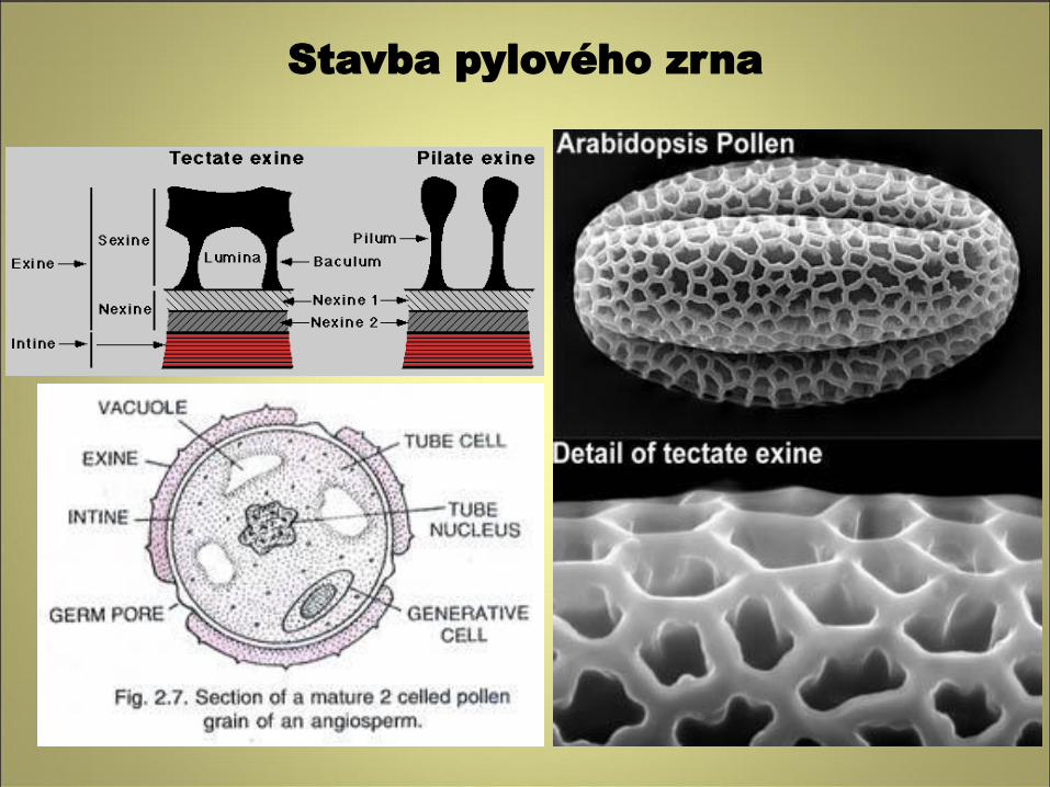
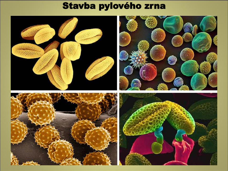
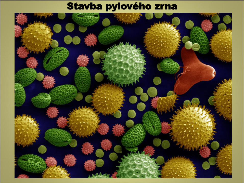
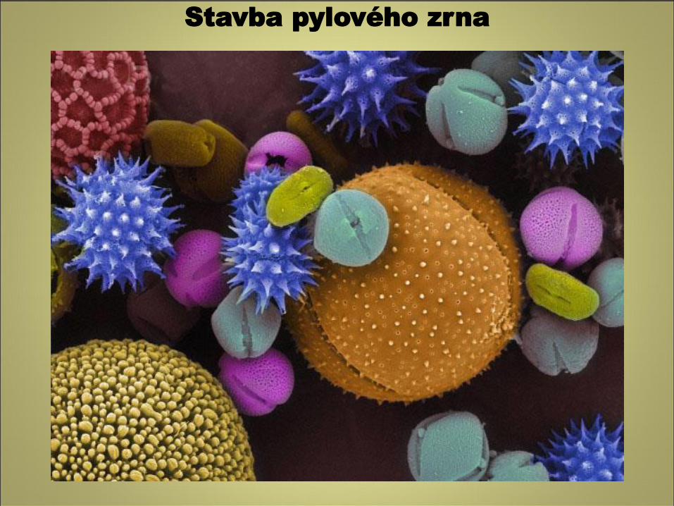
Stavba pylového zrna
Sporoderma (stěna spory):
- sestává z vnitřní tenké pektocelulózové intiny a vnější silné exiny tvořené celulózou,
pektiny, sporopoleniny, karoteny.
- Exina je rozlišena na vnitřní nexinu a vnější sexinu.
- Pokud se v exině nacházejí dutiny oddělené sloupky (columela) jedná se o exinu
tektátní.
- Povrch exiny je u entomogamních rostlin rozmanitě skulpturovaný a lepkavý, u
anemogamních rostlin bývá hladký a nelepkavý.
Pollen grains
- Outer wall, exine, contains chemicals that interact with stigma of flower
- Aperture(s) in wall involved in pollen tube formation
Stavba pylového zrna
Stavba pylového zrna
Stavba pylového zrna
Stavba pylového zrna