Embed Size (px)
Citation preview
Anatomy and Development of the Nervous Systemof Nematostella vectensis, an Anthozoan Cnidarian
Heather Q. Marlow,1 Mansi Srivastava,2 David Q. Matus,3 Daniel Rokhsar,2
Mark Q. Martindale1
1 Kewalo Marine Laboratory, Pacific Biomedical Research Center, University of Hawaii, Honolulu,Hawaii 96813
2 Center for Integrative Genomics and Department of Molecular and Cell Biology,University of California, Berkeley, California 94720
3 Department of Biology, Duke University, Durham, North Carolina 27708
Received 28 August 2008; revised 27 October 2008; accepted 20 November 2008
ABSTRACT: Nematostella vectensis, an anthozoan
cnidarian, whose genome has been sequenced and is
suitable for developmental and ecological studies, has a
complex neural morphology that is modified during de-
velopment from the larval to adult form. N. vectensis’
nervous system is a diffuse nerve net with both ectoder-
mal sensory and effector cells and endodermal multipo-
lar ganglion cells. This nerve net consists of several dis-
tinct neural territories along the oral–aboral axis
including the pharyngeal and oral nerve rings, and the
larval apical tuft. These neuralized regions correspond
to expression of conserved bilaterian neural developmen-
tal regulatory genes including homeodomain transcrip-
tion factors and NCAMs. Early neurons and stem cell
populations identified with NvMsi, NvELAV, and
NvGCM, indicate that neural differentiation occurs
throughout the animal and initiates prior to the conclu-
sion of gastrulation. Neural specification in N. vectensisappears to occur through an independent mechanism
from that in the classical cnidarian model Hydra. ' 2009
Wiley Periodicals, Inc. Develop Neurobiol 69: 235–254, 2009
Keywords: cnidarian; evolution; nerve net
INTRODUCTION
An ability to sense variation in the environment and
coordinate appropriate behavioral response is a hall-
mark of morphologically diverse extant eumetazoan
nervous systems. Many questions surrounding early
neural evolution remain unanswered. To what extent
were neural axes patterned along the major body
axes, as in flies, nematodes, and vertebrates, or in the
dispersed nerve nets of earlier branching taxa? How
many neural cell types existed, how were they speci-
fied, and what was the mode of communication
between them? Characteristics of the ancestral eume-
tazoan nervous system can only be inferred by studies
of extant species (structural, developmental, and mo-
lecular traits), including lineages, which predate the
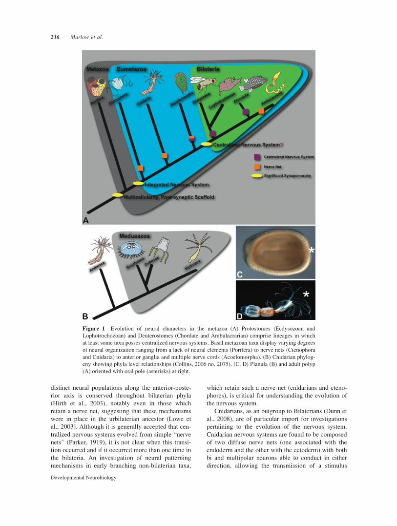
origin of central nervous systems [Fig. 1(A)]. Investi-
gations into early branching metazoans are particu-
larly useful in this regard. Nematostella vectensis is
an anthozoan cnidarian and thus, a member of the sis-
ter clade to all Bilateria.
Although the molecular basis of the formation and
function of bilaterian nervous systems is understood
in some detail, comparatively little is known about
these processes in the basal metazoans. Patterning of
Additional Supporting Information may be found in the onlineversion of this article.
Correspondence to: M.Q. Martindale ([email protected]).
' 2009 Wiley Periodicals, Inc.Published online 23 January 2009 in Wiley InterScience (www.interscience.wiley.com).DOI 10.1002/dneu.20698
235
distinct neural populations along the anterior-poste-
rior axis is conserved throughout bilaterian phyla
(Hirth et al., 2003), notably even in those which
retain a nerve net, suggesting that these mechanisms
were in place in the urbilaterian ancestor (Lowe et
al., 2003). Although it is generally accepted that cen-
tralized nervous systems evolved from simple \nerve
nets" (Parker, 1919), it is not clear when this transi-
tion occurred and if it occurred more than one time in
the bilateria. An investigation of neural patterning
mechanisms in early branching non-bilaterian taxa,
which retain such a nerve net (cnidarians and cteno-
phores), is critical for understanding the evolution of
the nervous system.
Cnidarians, as an outgroup to Bilaterians (Dunn et
al., 2008), are of particular import for investigations
pertaining to the evolution of the nervous system.
Cnidarian nervous systems are found to be composed
of two diffuse nerve nets (one associated with the
endoderm and the other with the ectoderm) with both
bi and multipolar neurons able to conduct in either
direction, allowing the transmission of a stimulus
Figure 1 Evolution of neural characters in the metazoa (A) Protostomes (Ecdysozoan and
Lophotrochozoan) and Deuterostomes (Chordate and Ambulacrarian) comprise lineages in which
at least some taxa posses centralized nervous systems. Basal metazoan taxa display varying degrees
of neural organization ranging from a lack of neural elements (Porifera) to nerve nets (Ctenophora
and Cnidaria) to anterior ganglia and multiple nerve cords (Acoelomorpha). (B) Cnidarian phylog-
eny showing phyla level relationships (Collins, 2006 no. 2075). (C, D) Planula (B) and adult polyp
(A) oriented with oral pole (asterisks) at right.
236 Marlow et al.
Developmental Neurobiology
from any one part of the nerve net to the other (West-
fall, 1971; Anderson, 1985, 1988; Satterlie, 2002;
Brusca, 2003). These neurons serve to coordinate the
input from sensory cells and structures with the out-
put of the epitheliomuscular cells (cnidarians do not
have definitive muscle cells). Sensory structures such
as eyes and statocysts are likely to have been inde-
pendently evolved in the medusozoan lineages to
which they are confined (scyphozoa and cubozoa)
and were not present in the cnidarian-bilaterian
ancestor. Single epithelial sensory cells and unique
stinging cells called cnidae are found throughout the
cnidaria. Cnidae also act as sensory cells by respond-
ing to stimuli (Pantin, 1942) and are integrated with
the nervous system through synapses (Westfall,
2004), and like neurons, originate from multipotent
\i" cells in Hydra (Bode, 1996).
Nervous system organization differs significantly
between cnidarian and bilaterian taxa, but much is
conserved at the individual neuronal unit. Interneuro-
nal communication in cnidarians employs tranmis-
sion through both chemical synapses and electrical
synapses mediated by gap junctions (Satterlie and
Spencer, 1987), which is further supported by the dis-
covery of a pannexin-like gene in the N. vectensis ge-
nome (Putnam et al., 2007) and innexins in hydrozo-
ans (Alexopoulos et al., 2004). Both excitatory and
inhibitory postsynaptic potentials have been reported
(Anderson and Spencer, 1989). Moreover, there is
also evidence for epithelial conduction through junc-
tions between epithelial cells in some cnidarians
(Mackie, 1965, 1976; Mackie and Passano, 1968).
Studies on swimming behavior in jellyfish such as
Aglantha digitale have revealed the use of calcium
and sodium action potentials as well as their regula-
tion by potassium channels (Mackie, 1980, 2004;
Anderson and Spencer, 1989; Meech and Mackie,
1993a,b; Mackie and Meech, 2000; Greenspan,
2007). Cnidarians utilize both classical fast (acetyl-
choline, glutamate, GABA, and glycine) and slow
(catecholamines and serotonin) transmitters and neu-
ropeptides (especially of the RFamide and RWamide
families) for synaptic neurotransmission (Satterlie,
2002; Kass-Simon and Pierobon, 2007). Recently, it
has been hypothesized, with evidence from newly
available basal metazoan genomes, that the postsy-
naptic scaffold common to all metazoans was present
prior to the divergence of the cnidarian-bilaterian
ancestor (Sakarya et al., 2007). Although previous
work illustrates that morphology and transmission
mechanisms of neural cells appear quite similar in
bilaterians and cnidarians, it is unclear how the over-
all neural architecture and patterning of cnidarian
nervous systems relates to the main body axis or if
patterning occurs by way of a bilaterian-like mecha-
nism. Much of cnidarian neurobiology has been con-
ducted in adult medusozoans, including the model
Hydra. Questions regarding the development of neu-
ral organization during embryogenesis remain to be
answered.
Nematostella vectensis is an emerging cnidarian
model for investigations of the nervous system during
embryonic development (Darling et al., 2005) and is
therefore particularly suited for studies regarding the
evolution of the nervous system. Like all other antho-
zoans, N. vectensis has both a larval and polyp stage
[Fig. 1(B,C)], and lacks the derived pelagic medusa
(jellyfish) stage found in hydrozoans, scyphozoans,
and cubozoans (Collins et al., 2006). Development in
N. vectensis is well characterized to include an early
chaotic cleavage stage, a hollow blastula which gas-
trulates by unipolar invagination to a swimming pla-
nula, which settles and grows tentacles surrounding
its oral opening to the gut, finally forming a juvenile
polyp (Hand and Uhlinger, 1992; Lee et al., 2007;
Magie et al., 2007). Nematostella vectensis has many
practical advantages such as ease of culture, access to
developmental material (Fritzenwanker and Technau,
2002), and a sequenced and annotated genome
(Putnam et al., 2007). These technical advances in
addition to a growing interest in basal metzoan taxa
have fueled a rapidly accumulating body of work cen-
tering on the molecular and cellular basis of develop-
ment in N. vectensis.Cnidarians have traditionally been viewed as sim-
ple diploblasts with a loosely organized nervous
system and little axial organization. Recent work
emerged to show that cnidarians possess a great deal
of genomic and molecular complexity (Putnam et al.,
2007) much of which is found in discrete cell popula-
tions and indicates a greater number of cell types
than previously described. The N. vectensis genome
contains nearly 18,000 protein coding genes and
encompasses all of the major signaling pathways
(notch, wnt, TGFbeta, FGF, and Hedgehog), as well
as components of virtually all transcription family
members used in bilaterian patterning (Putnam et al.,
2007). This molecular and genomic complexity is
used for cryptic patterning of the N. vectensis body
plan during development by way of complex expres-
sion patterns for these transcription factors and sig-
naling pathways (Kusserow et al., 2005; Ryan et al.,
2007). Functional studies have begun to confirm con-
served roles for these genes in germ layer segregation
(Wikramanayake et al., 2003) and in formation of
larval structures (Rentzsch et al., 2008), and although
a subset of these infer a role for these pathways in
neurogenesis, virtually nothing is known about the
Neural Development in N. vectensis 237
Developmental Neurobiology
development or complexity of the nervous system of
N. vectensis.Here, we determine the neural architecture of N.
vectensis at various developmental stages, present
evidence for potentially neurogenic regions and inter-
pret these data with respect to the biphasic life history
(swimming planula versus benthic polyp). Using anti-
bodies directed against GABA, FMRFamide, and se-
rotonin and in situ hybridization for antho-Rfamideand dopamine beta hydroxylase, we have identified
populations of neurons with distinct morphology and
localization within gastrula, planula, and polyp
stages. We have also identified regions of both the
endoderm and ectoderm that express transcripts of
putative markers of stem cells and early neurons
(ELAV and Musashi) as well as those specific for sub-
sets of developing neurons (GCM, repo, and NCAM).
These results suggest that although N. vectensis does
not posses a centralized nervous system, its neural
morphology is patterned along the oral–aboral axis
and correlates with previously identified molecular
domains of gene expression, which are temporally
associated with the planula and polyp stage.
METHODS
Identification and Amplification ofTarget Genes
Orthologous genes were identified by performing tblastx,
tblastn, and blastp searches (NCBI) of the assembled N.vectensis genome and predicted gene models (JGI) using
publicly available protein and DNA sequences from Gen-
Bank. Prior to the completion of the N. vectensis genome
assembly, searches of sequenced scaffolds and ESTs
(NCBI) were conducted using tblastn searches. Nested
RACE PCR (Smart Race cDNA amplification kit, BD Bio-
sciences Clontech) was used to amplify the 50 and 30
regions of target genes from mixed stage pools of N. vecten-sis cDNA using sequence specific primers. Sequencing of
the PCR products cloned into the p-GEM T Easy vector
(Promega) was conducted by Macrogen (South Korea).
Open reading frames for submission to GenBank were
determined by comparing RACE PCR products, EST data,
and JGI gene predictions (Supp. Info. Fig. 1).
Alignment and Tree Construction
Protein sequences were aligned in MacVector using the
default settings of the ClustalW alignment program and
were trimmed by eye (Supp. Info. Fig. 2). Protein domains
were identified using the SMART database (Ponting et al.,
1999). Transmembrane, intracellular, and extracellular
domains of the NvNCAM predicted proteins were predicted
with Phobius prediction program (Kall et al., 2004). Orthol-
ogy of Nematostella proteins with bilaterian proteins was
assessed with phylogenetic trees made with MrBayes 3.1.2
(Huelsenbeck and Ronquist, 2001; Ronquist and Huelsen-
beck, 2003). Additional information regarding the MrBayes
analysis, relevant outgroups, and protein models employed
in the analysis are available in supporting Information
Figure 3.
In Situ Hybridization
In situ hybridization of probes to mRNA was performed, as
previously described (Martindale et al., 2004). DIG labeled
RNA probes of 750–2000 nucleotides were constructed
from RACE products or expressed sequence tags from a
previously constructed N. vectensis EST library using
Ambion MEGAscript reverse transcriptase kit (AM1330
and AM1334).
Antibody Staining
Early embryos (0–48 hpf) were dejellied in 4% cysteine
(pH 7.4), and polyp stages were relaxed in 7% MgCl2 prior
to fixation. Samples for antibody staining were fixed for 90
s in 4% paraformaldehyde and 0.2% gluteraldehyde in 1/
33 filtered seawater (FSW). Samples were then fixed for 1
h at 48C in 4% paraformaldehyde in FSW. Embryos were
washed three times quickly and three times for 30 min in
phosphate buffered saline (PBS) and 0.1% Tween-20
(PTw). Some samples were washed in PBS and 0.1%
Triton X-100 (PBT). Samples were stored in either PBS or
methanol.
Nematostella vectensis embryos were blocked in 5%
normal goat serum, NGS (Sigma, catalog no. G9023) in
PBT, or PTw (blocking buffer) for 1 h prior to antibody
staining. Samples were incubated overnight at 48C in pri-
mary antibody: a commercially available rabbit anti-GABA
antibody at 1:250–1:500, mouse anti-5HT at 1:200 (Dia
Sorin catalog no. 20080), or rabbit anti-FMRFamide at
1:100 (Peninsula Laboratories, catalog no. 8755). Three
quick washes (allowing the embryos to settle briefly) fol-
lowed by three 30-min washes were done at room tempera-
ture with PBT or PTw. Embryos were reblocked for 30 min
at room temperature and then incubated in secondary anti-
body in blocking buffer overnight at 48C. Three quick
washes followed by two long washes were performed prior
to counterstaining in 2 U/mL (66 nM) 488 or 647 alexa-
conjugated phalloidin (Molecular Probes, Eugene, Oregon,
USA) and propidium iodide (1 lg/mL) in the presence of
RNase (50 lg/mL). Following staining, embryos were
dehydrated through an isopropanol:PBS series (50:50,
70:30, 90:10, and 100:0) then mounted in Murray clear (2:1
benzyl benzoate to benzyl alcohol).
DAPI Staining of Cnidocytes
Cnidocytes were stained according to Szczepanek et al.
(2002). Embryos were fixed as for antibody staining with
the modification that 10 mM EDTA was added to all fixa-
238 Marlow et al.
Developmental Neurobiology
tives. Nematostella vectensis samples were stained with
140 mM DAPI for 30 min at room temperature and then
washed three times for 30 min in PBS. Counterstaining
with propidium iodide and phalloidin and mounting were
carried out, as described above.
Imaging
In situ stained embryos were visualized under 203 differ-
ential interference contrast (DIC) microscopy on a Zeiss Z-
1 imager microscope (Carl Zeiss, Jena, Germany) or Zeiss
Axioscop2 and photographed with a Zeiss AxioCam HRc
or Coolpix camera (Nikon, Tokyo, Japan). DAPI stained
embryos were imaged on an Zeiss Z-1 imager microscope
and photographed using an Orca-ER digital camera (Hama-
matsu TOA Electronics, Shizuoka, Japan). Images were
collected, processed, and false colored (DAPI cnidocytes)
using Zeiss AxioVision or Volocity Acquisition software
(Improvision, Coventry, UK). Neural antibody stained
embryos were visualized using a Zeiss LSM510 confocal
microscope using the LSM software. 3D projections were
rendered using LSM or Volocity software and edited in
Adobe Photoshop.
RESULTS
We have used crossreactive antibodies to neuropepti-
des and other neurotransmitters, staining of cnido-
cytes, and in situ hybridization of genes for neuropep-
tides and for neurotransmitter biosynthesis to reveal
several subsets of neurons distributed in distinct
regions of the planula and polyp. Neural organization
of the animal changes with developmental time as the
prominent neural region of the swimming planula,
the apical tuft, is replaced upon settlement and polyp
formation with the adult neural morphology consist-
ing of the oral nerve ring, the pharyngeal nerve ring,
continued elaboration of the innervation in newly
forming mesenteries, and highly neuralized tentacle
tips. Molecular markers of differentiating neurons
indicate that neurogenesis occurs in both the ectoder-
mal and endodermal tissue layers and is not limited
to the ectoderm as is the case in many metazoans.
Anti-GABA Immunoreactivity
In the early planula, cells stained with anti-GABA
antibody are first detected throughout the ectoderm
along the oral–aboral axis and at high densities in the
apical tuft at the aboral pole [Fig. 2(A–C)]. These
early neurons are columnar in shape and span the
entire width of the ectodermal epithelium, which like
the endoderm, are one cell layer thick. As the planula
develops, ciliary membranes of the apical tuft, cili-
ated sensory cells along the body column ectoderm
[Fig. 2(C,D)], and a ring of large neurons at the base
of the pharyngeal tissue, the pharyngeal nerve ring,
become anti-GABA reactive [Fig. 2(H,I)]. At this
stage, projections extend from the base of the cell
bodies between the ectodermal and endodermal tissue
layers and run parallel to the surface of the planula.
As the tentacle buds form during polyp stages, new
neurons form at the highest numbers in tentacle tips
[Fig. 2(F,G)]. In the polyp stage, several distinct
GABA-positive cell types are distinguishable that
include, at a minimum, sensory neurons [Fig. 2(C,D)]
pyramidal shaped neurons with long processes, which
become bifurcated in some regions [Fig. 2(E)], and
bipolar neurons in the tentacles [Fig. 2(G)]. Rather
than a strictly columnar morphology, many neurons
located at the base of the ectoderm adopt a flattened
appearance, maintaining no contact with the external
environment [Fig. 2(E,G)], whereas many presumed
sensory cells remain positioned so that contact with
the outer surface of the animal is maintained [Fig.
2(E,F)].
Neuropeptides of the RFamide Class
FMRFamide immunoreactivity, as with GABA
immunoreactivity, is present in individual ectodermal
cell bodies spread throughout the body wall by the
early planula stage [Fig. 2(J)]. At later stages, strong
staining is seen in both the body wall and in the pha-
ryngeal region at the oral pole of the planula [Fig.
2(J,K)]. By the late planula stage, cell bodies are
found throughout the ectoderm and endoderm and
many cells, particularly those in the endoderm,
extend long processes that run parallel to the surface
of the animal in the mesoglea [Fig. 2(L,M)]. Polyp
stage animals have an abundance of anti-FMRFamide
staining neural processes along the oral–aboral axis
at the interface between the ectoderm and endoderm
[Fig. 2(L,M)] and many cell bodies are found in the
oral region at the oral nerve ring and base of the ten-
tacles [Fig. 2(O)]. In comparison to GABA-labeled
polyps [Fig. 2(H)], there is a notably smaller number
of cells in the tentacles and the pharyngeal nerve ring
region of the FMRFamide-labeled polyps [Fig. 2(N)].
Many of the ectodermal neurons send projections lat-
erally toward the mesenteries and project loosely in
the oral–aboral orientation along the mesentery [Fig.
2(N–P)]. The aggregation of neural projections may
allow the animal to quickly contract along the length
of the body column by innervating the epitheliomus-
cular cells along the mesenteries (Ramsay, 1952).
Neural Development in N. vectensis 239
Developmental Neurobiology
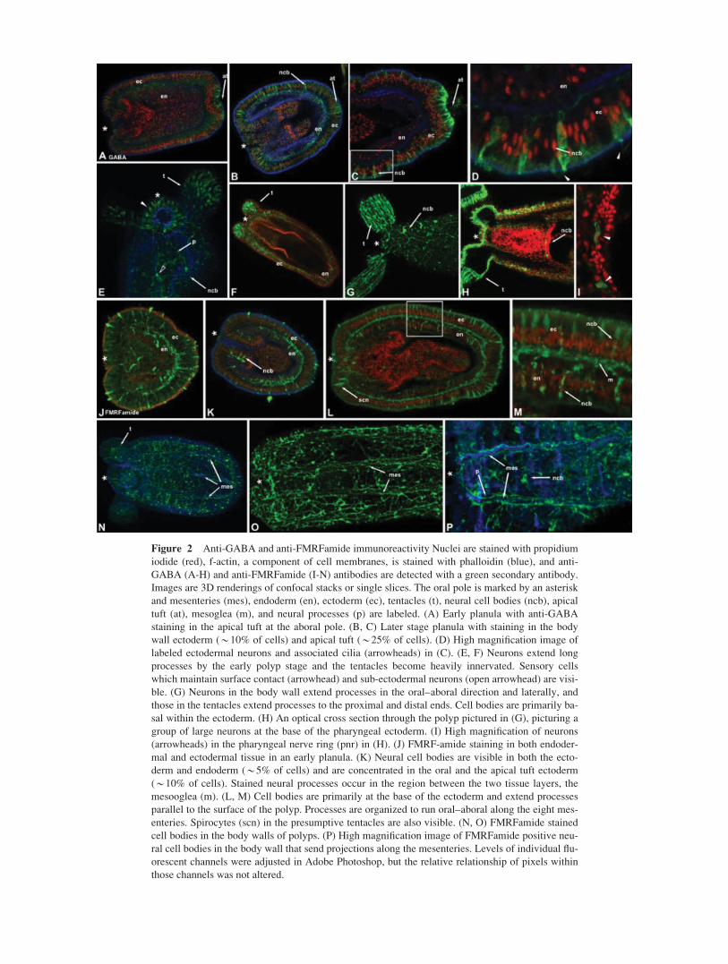
Figure 2 Anti-GABA and anti-FMRFamide immunoreactivity Nuclei are stained with propidium
iodide (red), f-actin, a component of cell membranes, is stained with phalloidin (blue), and anti-
GABA (A-H) and anti-FMRFamide (I-N) antibodies are detected with a green secondary antibody.
Images are 3D renderings of confocal stacks or single slices. The oral pole is marked by an asterisk
and mesenteries (mes), endoderm (en), ectoderm (ec), tentacles (t), neural cell bodies (ncb), apical
tuft (at), mesoglea (m), and neural processes (p) are labeled. (A) Early planula with anti-GABA
staining in the apical tuft at the aboral pole. (B, C) Later stage planula with staining in the body
wall ectoderm (*10% of cells) and apical tuft (*25% of cells). (D) High magnification image of
labeled ectodermal neurons and associated cilia (arrowheads) in (C). (E, F) Neurons extend long
processes by the early polyp stage and the tentacles become heavily innervated. Sensory cells
which maintain surface contact (arrowhead) and sub-ectodermal neurons (open arrowhead) are visi-
ble. (G) Neurons in the body wall extend processes in the oral–aboral direction and laterally, and
those in the tentacles extend processes to the proximal and distal ends. Cell bodies are primarily ba-
sal within the ectoderm. (H) An optical cross section through the polyp pictured in (G), picturing a
group of large neurons at the base of the pharyngeal ectoderm. (I) High magnification of neurons
(arrowheads) in the pharyngeal nerve ring (pnr) in (H). (J) FMRF-amide staining in both endoder-
mal and ectodermal tissue in an early planula. (K) Neural cell bodies are visible in both the ecto-
derm and endoderm (*5% of cells) and are concentrated in the oral and the apical tuft ectoderm
(*10% of cells). Stained neural processes occur in the region between the two tissue layers, the
mesooglea (m). (L, M) Cell bodies are primarily at the base of the ectoderm and extend processes
parallel to the surface of the polyp. Processes are organized to run oral–aboral along the eight mes-
enteries. Spirocytes (scn) in the presumptive tentacles are also visible. (N, O) FMRFamide stained
cell bodies in the body walls of polyps. (P) High magnification image of FMRFamide positive neu-
ral cell bodies in the body wall that send projections along the mesenteries. Levels of individual flu-
orescent channels were adjusted in Adobe Photoshop, but the relative relationship of pixels within
those channels was not altered.

Antho-RFamide is a Distinct Member ofthe RFamide Family of Neuromodulators
N. vectensis antho-RFamide mRNA expression was
assayed by in situ hybridization in early gastrula (24–
36 hpf) through polyp stages. Presumptive neurons
began expressing the antho-RFamide mRNA at the
gastrula stage by which time N. vectensis embryos
usually have begun swimming [Fig. 3(A)]. Although
early expression appears relatively uniform in the
ectoderm [Fig. 3(A)], by the planula (older larvae
with pharynx and developing mesenteries) and polyp
stages, expression was restricted to a large number of
cells in the oral nerve ring and a few scattered cells in
the body wall ectoderm, tentacles [Fig. 3(B–D)], and
an aggregation of cells at the aboral tip of the polyp,
the physa [Fig. 3(E)]. Antho-RFamide positive cells
are among the first neural cell types we detect and
their distribution in late development and in adult
polyps mark a significant morphological feature of
the adult nervous system, the oral nerve ring [Fig.
3(C,D)].
FMRFamide immunoreactivity and antho-RFamidemRNA expression occur in markedly different
expression domains. FMRFamide immunoreactivity
labels a larger number of total neurons but does not
label the antho-RFamide positive cells in the ten-
tacles [Fig. 3(C)]. Thus, it is likely that the anti-
FMRFamide antibody is crossreactive with a wide
variety of neuropeptides, which may not include the
antho-RFamide gene identified here [Fig. 3(K)].
Additional studies to identify and characterize the
full complement of neuropeptides in N. vectensis will
be of great utility in describing additional functional
and morphological subsets of neurons.
Dopamine Beta Hydroxylase RNAExpression
The neurotransmitter dopamine is converted into nor-
epinephrine in noradrenergic neurons by the enzyme
dopamine beta hydroxylase (or dopamine b-monoox-
ygenase). The N. vectensis genome has an ortholog of
bilaterian dopamine beta hydroxylase (Supp. Info.
Fig. 5) and in situ hybridization shows that this gene
is first expressed in the oral ectoderm in the early pla-
nula and subsequently, spreads through the pharyn-
geal ectoderm [Fig. 3(G–J)]. In juvenile polyps,
dopamine beta hydroxylase expression remains
strong in both these tissues and additional expression
is seen in the directive mesenteries [Fig. 3(I)]. The
pharyngeal ectoderm of polyps is made of one
cell layer (although likely pseudostratified), and the
Figure 3 Neurotransmitter synthesis during N. vectensis development. (A) Antho-RFamide is
expressed in single ectodermal cells throughout the late gastrula. (B–E) Expression in the planula
and polyp is restricted predominantly to the oral ectoderm, tentacles, and aboral tip of the physa
(aboral tip) with a small number of scattered cells in the body wall ectoderm. (D) An oral view of a
polyp with staining in the oral nerve ring. (E) A small aggregation of Antho-RFamide expressing
cells at the physa. Although RFamide immunoreactivity is widely distributed, Antho-RFamide tran-
script is restricted to oral and aboral domains. (F–G) Dopamine beta hydroxylase appears in a ring
of oral ectoderm in the early planula following gastrulation (F). (H–I) Expression expands to the
ectodermal region of the pharynx and directive mesenteries in the late planula and polyp. (J) Oral
view of a planula with dopamine beta hydroxylase expression in a ring of oral ectoderm. (K) An
alignment of anthozoan antho-RFamide sequences illustrating the repetition of a conserved motif.
Levels and white balance of images were adjusted in Adobe Photoshop.
Neural Development in N. vectensis 241
Developmental Neurobiology
expression of dopamine beta hydroxylase is localized
to only those cell bodies lying closest to the oral
opening (apical within the ectoderm) and not all cells
of the pharynx. These data suggest the presence of
noradrenergic neurons in N. vectensis, though it
remains to be determined if the expression of dopa-mine beta hydroxylase shows neurons that produce
epinephrine or norepinephrine (since epinephrine is
derived from norepinephrine by the enzyme phenyle-
thanolamine-N-methyltransferase). Only one member
of the NNMT/PNMT/TEMT methyltransferase fam-
ily is found in the N. vectensis genome, so further
experimentation is needed to know if this enzyme
converts norepinephrine into epinephrine in ane-
mones.
Cnidocyte Distribution andAnti-Serotonin Immunoreactivity
Cnidocytes are specialized cells containing collage-
nous organelles unique to cnidarians, the cnidocysts
(Engel et al., 2001). Discharge occurs at \bullet
speed" through the release of osmotic pressure cre-
ated by the sequestering of poly-c-glutamate within
the capsule (Szczepanek et al., 2002). At least, 28
different types of cnidocyst capsules have been
described and are specialized for feeding, locomo-
tion, and defense and are classified into three differ-
ent structural groups; nematocysts, spirocysts, and
ptychocysts (Kass-Simon and Scappaticci, 2002).
Cnidocyte cells are considered to be components of
the nervous system because they have been shown to
share a common developmental precursor with the
neural cells in hydrozoans (the interstitial stem cell
population) (Bode, 1996; Bosch, 2007), synapse with
ganglion and sensory cells, and stain with antibodies
to various neuropeptides (Westfall et al., 2000;
Anderson et al., 2004; Westfall, 2004). Cnidocytes
may act as both sensory (detect external stimulus
through a sensory projection called the cnidocil)/cili-
ary cones) and effector cells (fire a barbed hollow
thread in response to the stimulus) (Kass-Simon and
Scappaticci, 2002). Anthozoan cnidarians have cnido-
cyte cells of several types including spirocytes, which
house spirocyst capsules, which lack a shaft and
spine, and six types of nematocytes with nematocyst
capsules (Cutress, 1955; England, 1991; Kass-Simon
and Scappaticci, 2002). Nematostella vectensis adults
have cnidoycte cells that contain spirocysts, spiro-
cytes, (in the tentacles) and two types of nematocytes;
those with basitrichous isorhiza capsules (in the body
walls) and those with microbasic p-mastigophorecapsules (in the pharynx and mesenteries) (Williams,
1975; Frank and Bleakney, 1976).
Staining N. vectensis cniodcyst capsules with
DAPI in the presence of a calcium chelator and visu-
alizing specimens using a Cy2 filter (see Methods)
allows us to detect cnidocyte cells and trace their
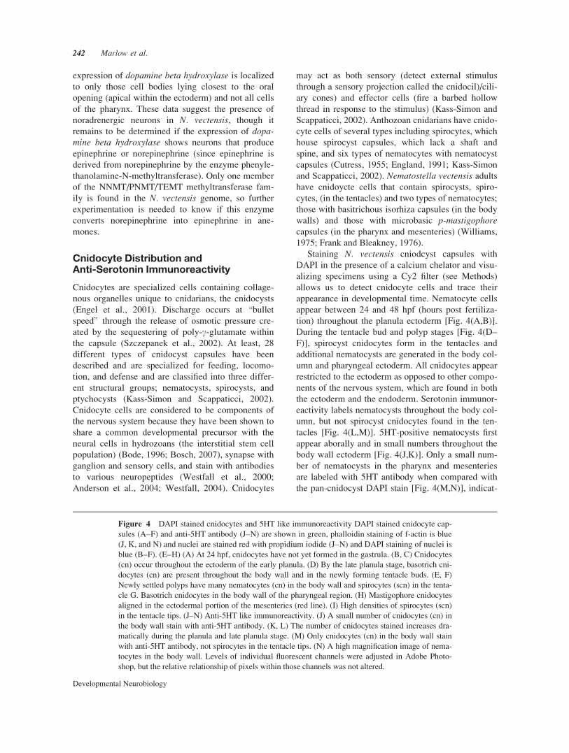
appearance in developmental time. Nematocyte cells
appear between 24 and 48 hpf (hours post fertiliza-
tion) throughout the planula ectoderm [Fig. 4(A,B)].
During the tentacle bud and polyp stages [Fig. 4(D–
F)], spirocyst cnidocytes form in the tentacles and
additional nematocysts are generated in the body col-
umn and pharyngeal ectoderm. All cnidocytes appear
restricted to the ectoderm as opposed to other compo-
nents of the nervous system, which are found in both
the ectoderm and the endoderm. Serotonin immunor-
eactivity labels nematocysts throughout the body col-
umn, but not spirocyst cnidocytes found in the ten-
tacles [Fig. 4(L,M)]. 5HT-positive nematocysts first
appear aborally and in small numbers throughout the
body wall ectoderm [Fig. 4(J,K)]. Only a small num-
ber of nematocysts in the pharynx and mesenteries
are labeled with 5HT antibody when compared with
the pan-cnidocyst DAPI stain [Fig. 4(M,N)], indicat-
Figure 4 DAPI stained cnidocytes and 5HT like immunoreactivity DAPI stained cnidocyte cap-
sules (A–F) and anti-5HT antibody (J–N) are shown in green, phalloidin staining of f-actin is blue
(J, K, and N) and nuclei are stained red with propidium iodide (J–N) and DAPI staining of nuclei is
blue (B–F). (E–H) (A) At 24 hpf, cnidocytes have not yet formed in the gastrula. (B, C) Cnidocytes
(cn) occur throughout the ectoderm of the early planula. (D) By the late planula stage, basotrich cni-
docytes (cn) are present throughout the body wall and in the newly forming tentacle buds. (E, F)
Newly settled polyps have many nematocytes (cn) in the body wall and spirocytes (scn) in the tenta-
cle G. Basotrich cnidocytes in the body wall of the pharyngeal region. (H) Mastigophore cnidocytes
aligned in the ectodermal portion of the mesenteries (red line). (I) High densities of spirocytes (scn)
in the tentacle tips. (J–N) Anti-5HT like immunoreactivity. (J) A small number of cnidocytes (cn) in
the body wall stain with anti-5HT antibody. (K, L) The number of cnidocytes stained increases dra-
matically during the planula and late planula stage. (M) Only cnidocytes (cn) in the body wall stain
with anti-5HT antibody, not spirocytes in the tentacle tips. (N) A high magnification image of nema-
tocytes in the body wall. Levels of individual fluorescent channels were adjusted in Adobe Photo-
shop, but the relative relationship of pixels within those channels was not altered.
242 Marlow et al.
Developmental Neurobiology
ing that some nematocysts are unlabeled. Spirocysts
of the tentacles are not labeled with the serotonin
antibody [Fig. 4(M)], even though neuro-spirocyte
synapses of the cnidarian Aiptasia pallida have been
shown to be serotonin immunoreactive (Westfall,
2004). In N. vectensis, the spirocysts of the late
Figure 4
Neural Development in N. vectensis 243
Developmental Neurobiology
planula and polyp stage show FMRFamide reactivity
[Fig. 2(L)], consistent with previous findings of
FMRFamide and RFamide immunoreactivity of cni-
docytes in the tentacles of many cnidarian species
(Anderson et al., 2004).
ELAV and Musashi Mark NeurogenicRegions of the Planula and Polyp
The RNA binding proteins (RNAbps), embryonic le-thal abnormal vision (ELAV), and musashi (msi) are
most notably expressed in the developing nervous
system in animals as distantly related as Drosophilaand vertebrates and have been implicated in neuro-
genesis and stem cell maintenance, respectively
(Robinow and White, 1991; Good, 1995; Sakakibara
and Okano, 1997; Akamatsu et al., 1999; Kawashima
et al., 2000; Koushika et al., 2000; Okabe et al.,
2001; Lowe et al., 2003; Okano et al., 2005; Denes et
al., 2007). Due to the widespread conservation of
expression and function of ELAV and msi in bilater-
ians, we investigated their potential role during em-
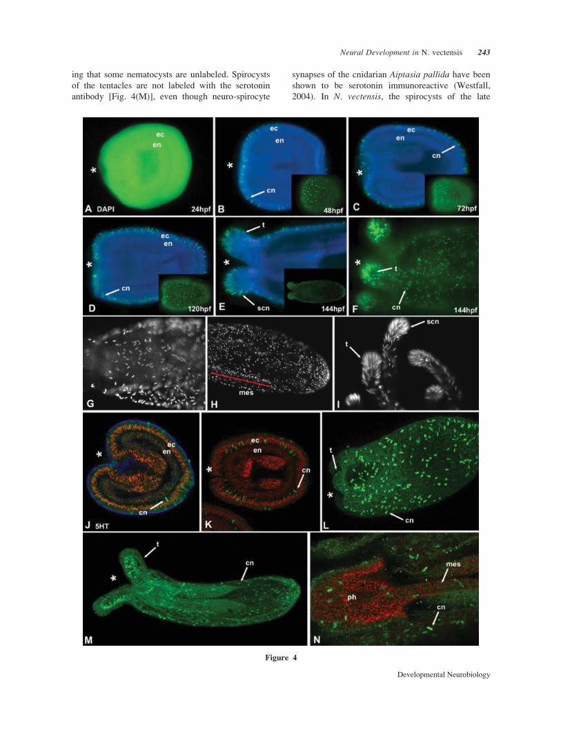
bryonic development in N. vectensis.Out of several related RNA-binding proteins found
in the N. vectensis genome, one NvMsi homolog falls
within a monophyletic group of previously identified
NvMsi proteins in Bayesian phylogenetic analyses
(Supp. Info. Fig. 3), and when aligned with other
NvMsi protein sequences clearly shares both RNA
binding motifs (RRMs) with them (Supp. Info. Fig.
5). RNA binding proteins like MSI and ELAV regu-
late gene expression post-transcriptionally by inter-
acting with the 30 UTR of mRNA and regulating the
stability, splicing, and translation of the targeted gene
(Sakakibara and Okano, 1997; Keene, 1999; Koush-
ika et al., 2000; Okano et al., 2002; Okano et al.,
2005; Siddall et al., 2006; Zhu et al., 2006). NvMsimRNA can be detected by in situ hybridization in
developing oral and tentacle bud ectoderm of the pla-
nula and later in pharyngeal ectoderm and scattered
cells of the tentacle ectoderm of the juvenile polyp
[Fig. 5(A–D)].
Two ELAV-like RNA-binding proteins identified
in N. vectensis cluster with ELAV/Hu genes identified
from other metazoans using phylogenetic analyses
(Supp. Info. Fig. 3). Predicted proteins of the
NvELAV1 and NvELAV2 genes both share the three
RRMs common to all ELAV/Hu family genes (Good,
1995; Yim et al., 2006) and do not appear to be recent
duplications as demonstrated by their relationship to
bilaterian ELAV genes (Supp. Info. Fig. 4). In situ
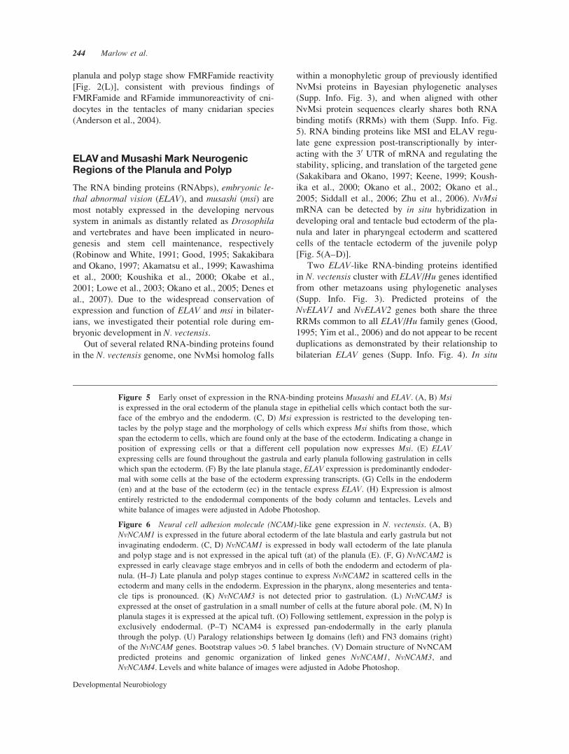
Figure 6 Neural cell adhesion molecule (NCAM)-like gene expression in N. vectensis. (A, B)
NvNCAM1 is expressed in the future aboral ectoderm of the late blastula and early gastrula but not
invaginating endoderm. (C, D) NvNCAM1 is expressed in body wall ectoderm of the late planula
and polyp stage and is not expressed in the apical tuft (at) of the planula (E). (F, G) NvNCAM2 is
expressed in early cleavage stage embryos and in cells of both the endoderm and ectoderm of pla-
nula. (H–J) Late planula and polyp stages continue to express NvNCAM2 in scattered cells in the
ectoderm and many cells in the endoderm. Expression in the pharynx, along mesenteries and tenta-
cle tips is pronounced. (K) NvNCAM3 is not detected prior to gastrulation. (L) NvNCAM3 is
expressed at the onset of gastrulation in a small number of cells at the future aboral pole. (M, N) In
planula stages it is expressed at the apical tuft. (O) Following settlement, expression in the polyp is
exclusively endodermal. (P–T) NCAM4 is expressed pan-endodermally in the early planula
through the polyp. (U) Paralogy relationships between Ig domains (left) and FN3 domains (right)
of the NvNCAM genes. Bootstrap values >0. 5 label branches. (V) Domain structure of NvNCAM
predicted proteins and genomic organization of linked genes NvNCAM1, NvNCAM3, and
NvNCAM4. Levels and white balance of images were adjusted in Adobe Photoshop.
Figure 5 Early onset of expression in the RNA-binding proteins Musashi and ELAV. (A, B) Msiis expressed in the oral ectoderm of the planula stage in epithelial cells which contact both the sur-
face of the embryo and the endoderm. (C, D) Msi expression is restricted to the developing ten-
tacles by the polyp stage and the morphology of cells which express Msi shifts from those, which
span the ectoderm to cells, which are found only at the base of the ectoderm. Indicating a change in
position of expressing cells or that a different cell population now expresses Msi. (E) ELAVexpressing cells are found throughout the gastrula and early planula following gastrulation in cells
which span the ectoderm. (F) By the late planula stage, ELAV expression is predominantly endoder-
mal with some cells at the base of the ectoderm expressing transcripts. (G) Cells in the endoderm
(en) and at the base of the ectoderm (ec) in the tentacle express ELAV. (H) Expression is almost
entirely restricted to the endodermal components of the body column and tentacles. Levels and
white balance of images were adjusted in Adobe Photoshop.
244 Marlow et al.
Developmental Neurobiology
Figure 5
Figure 6

hybridization detects NvELAV1 mRNA expression in
scattered ectodermal cells at the aboral pole shortly
after the onset of gastrulation [Fig. 5(E)]. This pre-
cedes the expression, but corresponds to the location
of the earliest expressed classical markers of neural
differentiation such as NvNCAM1 and NvNCAM3(see below), SoxB1 and SoxB2 (Magie et al., 2005).
Transcript expression expands to a large number of
endodermal cells and to ectodermal cells in the body
wall and tentacles [Fig. 5(F–H)] in the planula and
polyp stages. Although we were able to clone
NvELAV2 from mixed stage pools of N. vectensiscDNA, we were not able to detect its expression by
in situ hybridization.
Neural Cell Adhesion Molecule-LikeGenes of the Ig Superfamilyin N. vectensis
We have identified four immunoglobulin superfamily
(Ig) genes in N. vectensis. Members of the Ig super-
family share the conserved Ig domain (often
repeated) (Walsh and Doherty, 1997) and have been
identified throughout bilaterian animals (Mayford et
al., 1992; Vogel et al., 2003; Fusaoka et al., 2006),
and are found to serve in various aspects of neural de-
velopment (Rougon and Hobert, 2003; Maness and
Schachner, 2007). Phylogenetic analyses place all
four genes within the NCAM family to the exclusion
of other Ig families (Supp. Info. Fig. 3). Domain
architecture of three (NvNCAM1, NvNCAM3, and
NvNCAM4) of the four N. vectensis immunoglobulin
genes, consisting of 4–5 Ig domains followed by two
FnIII domains is consistent with placement within the
NCAM/Fasciclin II family [Fig. 6(V)]. These three
genes are found linked within a 75 kb region of scaf-
fold 47 in the recently sequenced N. vectensis ge-
nome (v1.0). The fourth gene (NvCAM2) contains
only one Ig domain and one FN3 domain and occu-
pies a different genomic location in relation to the
three NCAM-like genes.
NCAMs may be expressed in a variety of tissues
including muscle, glia, and neurons (Walsh and Doh-
erty, 1997), but are primarily expressed in the nerv-
ous system and play conserved roles in synapse
formation, neural migration, axon pathfinding, and
vessicle cycling (Walsh and Doherty, 1997; Rougon
and Hobert, 2003; Maness and Schachner, 2007).
NCAMs, as cell adhesion molecules, regulate cell–
cell and cell-ECM interactions, and through both het-
ero and homophilic interactions can activate second
messenger cascades (Walsh and Doherty, 1997; Rou-
gon and Hobert, 2003). Like the NCAMs of Aplysiaand vertebrates, NvNCAM1 has an mRNA expression
domain, which encompasses what we hypothesize to
be both neurons, and nonneural cells of the ectoderm
[Fig. 6(A–E)]. In contrast, NvNCAM3 is expressed in
the apical tuft, a neuralized chemosensory structure,
and in what are likely to be components of the endo-
dermal nerve net (based on immunostaining with
antibodies to neurotransmitters) [Fig. 6(K–O)]. Inter-
estingly, the expression of NvNCAM3 overlaps with
that of the FGF ligand NvFGF1a and the FGF recep-
tor NvFGFRa (Matus et al., 2007b). Components of
these two pathways are known to interact to regulate
the growth of developing neurons (Maness, 2007). In
contrast to NvNCAM1 and NvNCAM3, NvNCAM4 is
expressed pan-endodermally in a non-neurally re-
stricted manner [Fig. 6(P–T)]. NvNCAM1 and
NvNCAM4 are sister paralogs (Supp. Info. Fig. 3),
share an identical domain structure [Fig. 6(V)] and
their predicted proteins are more distantly related to
bilaterian NCAM proteins than the other NvNCAMs.
NvNCAM1 and NvNCAM4 also show the most wide-
spread expression. NvNCAM2 is expressed by the
early cleavage stage where it is localized in the nu-
cleus of cells and is expressed throughout the early
planula. Expression is later restricted to a large num-
ber of scattered cells in the endoderm as well as a few
ectodermal cells. Although its genomic location and
domain structure differ from NvNCAMs1, 3, and 4,
the expression of NvNCAM2, NvNCAM1, and
NvNCAM3, is consistent with a role in the nervous
system [Fig. 6(F–J)]. As three of the four NvNCAMs
are linked and posses similar domain architecture, we
hypothesize that NvNCAMs1, 3, and 4 originated as
the result of a duplication event from an ancestral
NCAM-like gene. NvNCAM1 and 3 maintain roles in
patterning domains of the nervous system and
NvNCAM4 has subsequently diverged in its expres-
sion and potential role in development.
Bilaterian Glial and Neural Determinantsin N. vectensis Development
Subsets of neural precursor cells in the developing
nervous systems of both vertebrates (Wakamatsu,
2004) and invertebrates (Hosoya et al., 1995) have
the potential to generate either neurons or glial cells.
It is plausbile that regulators of glial fate such as
GCM may have initially evolved as more general reg-
ulators of neural precursor differentiation as evi-
denced from studies in chick and fly (Soustelle et al.,
2007). To investigate the ancestral roles of bilaterian
glial-specific genes in neural patterning, we looked
for the expression of these genes in Nematostella.
Determination of the glial cell fate and subsequent
differentiation in flies is regulated by glial cells miss-
246 Marlow et al.
Developmental Neurobiology
ing (GCM) through the induction of glia-specific
genes such as pointed, tramtrack69, and repo in flies
(Hosoya et al., 1995; Yuasa et al., 2003; Lee and
Jones, 2005), whereas in vertebrates it appears that
notch/delta signaling in combination with glial induc-
ing genes such as neuregulin1 and olig2 preferen-
tially promote glial cell fates (Wakamatsu, 2004; Jes-
sen and Mirsky, 2005). In both systems, it appears
that a combination of positive regulation of glial fate
and the negative regulation of neural fate is required
for proper glial specification (Hosoya et al., 1995;
Wakamatsu, 2004).
One glial specific gene in flies that has been shown
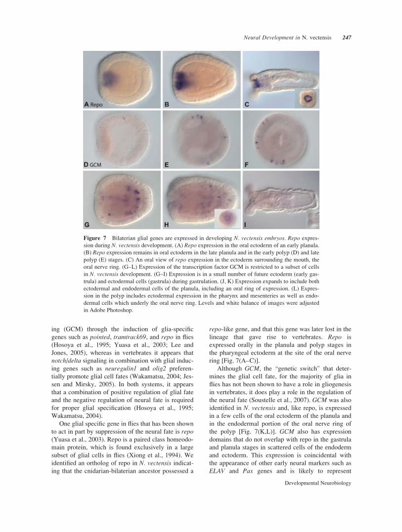
to act in part by suppression of the neural fate is repo(Yuasa et al., 2003). Repo is a paired class homeodo-
main protein, which is found exclusively in a large
subset of glial cells in flies (Xiong et al., 1994). We
identified an ortholog of repo in N. vectensis indicat-
ing that the cnidarian-bilaterian ancestor possessed a
repo-like gene, and that this gene was later lost in the
lineage that gave rise to vertebrates. Repo is
expressed orally in the planula and polyp stages in
the pharyngeal ectoderm at the site of the oral nerve
ring [Fig. 7(A–C)].
Although GCM, the \genetic switch" that deter-
mines the glial cell fate, for the majority of glia in
flies has not been shown to have a role in gliogenesis
in vertebrates, it does play a role in the regulation of
the neural fate (Soustelle et al., 2007). GCM was also
identified in N. vectensis and, like repo, is expressed
in a few cells of the oral ectoderm of the planula and
in the endodermal portion of the oral nerve ring of
the polyp [Fig. 7(K,L)]. GCM also has expression
domains that do not overlap with repo in the gastrula
and planula stages in scattered cells of the endoderm
and ectoderm. This expression is coincidental with
the appearance of other early neural markers such as
ELAV and Pax genes and is likely to represent
Figure 7 Bilaterian glial genes are expressed in developing N. vectensis embryos. Repo expres-
sion during N. vectensis development. (A) Repo expression in the oral ectoderm of an early planula.
(B) Repo expression remains in oral ectoderm in the late planula and in the early polyp (D) and late
polyp (E) stages. (C) An oral view of repo expression in the ectoderm surrounding the mouth, the
oral nerve ring. (G–L) Expression of the transcription factor GCM is restricted to a subset of cells
in N. vectensis development. (G–I) Expression is in a small number of future ectoderm (early gas-
trula) and ectodermal cells (gastrula) during gastrulation. (J, K) Expression expands to include both
ectodermal and endodermal cells of the planula, including an oral ring of expression. (L) Expres-
sion in the polyp includes ectodermal expression in the pharynx and mesenteries as well as endo-
dermal cells which underly the oral nerve ring. Levels and white balance of images were adjusted
in Adobe Photoshop.
Neural Development in N. vectensis 247
Developmental Neurobiology
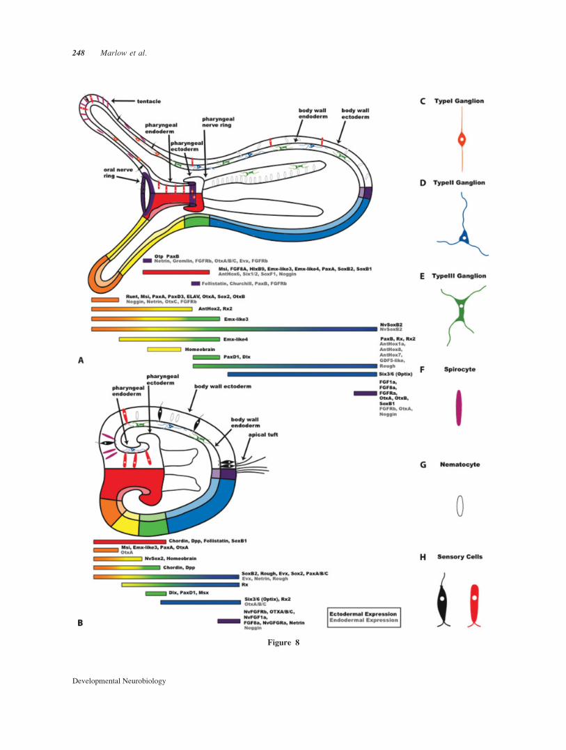
Figure 8
248 Marlow et al.
Developmental Neurobiology
expression in predifferentiated neurons. Examining
the role of GCM and repo in the early branching line-
age giving rise to cnidarians provides insight into the
ancestral roles of these genes. As glia have not been
identified in basal metazoans, the ancestral role of
GCM and repo are likely to be as regulators of differ-
entiation in subsets of neurons, consistent with their
expression in N. vectensis.
DISCUSSION
Here, we report a complex neural morphology for the
anthozoan anemone, N. vectensis. Individual neural
populations are organized along the oral–aboral axis
and in discrete structures that are readily correlated to
the behavior and life history of the animal. Cnidarians
and ctenophores are likely the first metazoans to have
evolved a recognizable nervous system. Anthozoan
cnidarians (corals, sea pens, and anemones) represent
the earliest branching class of cnidarians (Collins et
al., 2006). Consistent with previous studies of antho-
zoans, we find the neural anatomy of the anemone N.vectensis to be considerably more complex than that
of the classical model for cnidarians, Hydra. This
morphological organization is accompanied by the
similarly complex expression of molecular markers
for neural specification and patterning. In addition to
describing the neural anatomy, we report the origins
and development of neural structures over the course
of developmental time, information that has been of-
ten overlooked in investigations of Cnidaria. The sig-
nificant morphological and molecular complexity of
the neural system of N. vectensis, the absence of the
seemingly derived \i" cell populations identified as
progenitors in Hydra, and the evolutionary distance
(*550 my) between hydrozoans and anthozoans as
demonstrated by fossil (Chen et al., 2002) and molec-
ular (Putnam et al., 2007) evidence highlights the
necessity of this new developmental model and prom-
ises to lend considerable insight into evolution of
neural cell types and stem populations in both the cni-
darians and early metazoans.
This study indicates that N. vectensis posseses a
diverse assembly of neural elements, consisting of
multiple classes of neural cells and cnidocyte classes.
These can be identified based upon their morphology
and neuropeptide or neurotransmitter expression and
are localized to restricted domains in the endoderm
and ectoderm and arise at specific developmental
stages. Previous work, including the discovery of
both dense and clear core vesicles (Kass-Simon and
Pierobon, 2007), immunostaining, biochemical anala-
yses, and behavioral response studies (Westfall et al.,
2000; Grimmelikhuijzen et al., 2002; Kass-Simon
and Pierobon, 2007) has suggested that cnidarians
possess both chemical neurotransmitters (Grimmeli-
khuijzen and Graff, 1986; Westfall et al., 2000; Gillis
and Anctil, 2001), as well as an abundance of neuro-
peptides (Grimmelikhuijzen et al., 2002; Kass-Simon
and Pierobon, 2007). Here, we show evidence for the
presence of monamines (5HT and dopamine synthesis
pathway components), GABA, and neuropeptides in
the two main classes of previously identified cni-
darian neurons; sensory and ganglion cells, and in
cnidocytes (Grimmelikhuijzen et al., 2002; Koizumi,
2002). As in other cnidarians, these transmitters can
be associated with different subsets of the cnidocyte
assemblages (anti-5HT reactive nematocytes versus
anti-FMRFamide reactive spirocytes) as well as
GABA and FRMF-amide positive sensory and gan-
glion cells (Koizumi, 2002; Koizumi et al., 2004).
Further spatial restriction of the neurotransmitter syn-
thesis component dopamine beta hydroxylase pharyn-
geal ectoderm) and the specific RFamide family
member Antho-RFamide (oral region and physa/foot
region as in Hydra) suggests that expression profiles
differentiate many neurons that may appear indistin-
guishable morphologically. These distinct neural ele-
ments are differentially positioned along both the
oral–aboral axis and between the endoderm and ecto-
derm. Analyses of the genome of N. vectensis indi-
Figure 8 Developmental transcription factors, axial patterning genes and neural architecture
Expression domains of neural markers and developmental genes in N. vectensis planula and polyp
stages. (A) A polyp stage with neural morphology shown in the upper portion of the diagram. The
oral and pharyngeal nerve rings are highlighted in purple. Associated patterns of gene expression
are shown in the lower region of the diagram. (B) A planula stage with neural morphology shown
in the upper portion of the diagram and associated gene expression patterns in the lower region of
the diagram. (C–H) Individual neural and cnidocyte morphologies shown schematicaly. Three gan-
glion, two cnidocyte, and two sensory type cells are represented. Developmental gene expression is
drawn according to data presented in this paper and that previously published (Magie et al., 2005;
Matus et al., 2006a,b; Matus et al., 2007a,b; Mazza et al., 2007; Ryan et al., 2007; Rentzsch et al.,
2008), and currently unpublished data Emx-like genes (Matus et al., in prep) and Homeobrain and
Rx2 (Martindale et al., in prep).
Neural Development in N. vectensis 249
Developmental Neurobiology
cate the presence of cholinergic, andrenergic, and cat-
echolamine pathways as well as several characteristic
cnidarian propeptide families, and suggest an even
larger number of specialized cell types (Supp. Info.
Fig. 4).
Although no distinct CNS or ganglia are present in
anemones, N. vectensis has specialized neural struc-
tures with distinct populations of neurons located at
the pharyngeal nerve ring, the oral nerve ring, inner-
vating the mesenteries, and at the tentacle tips (see
Fig. 8). Furthermore, stage specific structures, such as
the apical tuft, possess additional neural subpopula-
tions. Each neural subset in N. vectensis is likely to
carry out its own function and may be related to behav-
iors such as feeding, coordination of swimming behav-
ior (in larvae), defense or contraction for locomotion
(burrowing and creeping) (Hand and Uhlinger, 1992;
Williams, 2003). Cnidarian nervous systems form
both local and multineuron circuits (Westfall, 2004)
that may regulate behaviors such as feeding. Morpho-
logically similar cells, such as ganglion cells, often
express different neurotransmitters or a combination
of transmitters (Grimmelikhuijzen et al., 2002; Koi-
zumi et al., 2004; Westfall, 2004). These findings indi-
cate that cnidarians do not possess a simple homoge-
neous nerve net but rather have complex subfunction-
alized cell types in both ectodermal and endodermal
tissue layers. This organization is dependent upon
both cell type specific and position specific develop-
mental cues for both an ectodermal and endodermal
nerve net. Such sub-functionalization of the nerve net
requires similarly complex molecular patterning.
Cnidarians have a central body axis (the oral–abo-
ral axis) and a nerve net, but no nerve cord or distinct
brain, yet many of the conserved bilaterian A-P neu-
ral patterning genes are expressed in distinct patterns
along the cnidarian oral–aboral axis (Kusserow et al.,
2005). These expression domains in many cases cor-
respond to what we have identified as distinct neural
structures (oral nerve ring, pharyngeal nerve ring, ap-
ical tuft, and tentacle tips).
Paired homeodomain genes, four classes of cnidar-
ian pax genes PaxA-D as well as an Rx gene have pre-
viously been identified in N. vectensis (Matus et al.,
2007a) and the coral Acropora millepora (Miller
et al., 2000). Expression patterns of four classes of
Pax genes in N. vectensis (which correspond to the
three bilaterian pax classes Pax2/5/8, Pox neuro, and
Pax3/7), demarcate distinct regions encompassing a
large portion of the oral–aboral body axis. PaxB (The
bilaterian Pax2/5/8 ortholog) is expressed in an endo-
dermal ring at the base of the pharynx (Matus et al.,
2007a) corresponding to the location of the pharyngeal
nerve ring. Pax genes, i.e., Pax 2/5/8, in bilaterian ani-
mals have been determined to be involved in the main-
tenance of boundaries in the developing nervous sys-
tem, such as the midbrain-hindbrain boundary in verte-
brates. The paired homeodomains repo and NvPaxBdemarcate discrete structures, the oral and pharyngeal
nerve rings, respectively [Fig. 8(A)].
TGF-beta and FGF signaling pathways are respon-
sible for the promotion of neural fate through regula-
tion of neural inhibitors. Expression of the TGF-betaantagonists follistatin (endodermal pharyngeal ring),
gremlin (endodermal oral ring), and noggin (tentacle
tips and pharynx) overlap with neuralized regions of
N. vectensis in both the planula and polyp stage. This
in addition to a conserved function of NvGremlin and
NvChordin to block the ventralizing activity of
NvDpp, respectively (Rentzsch et al., 2006). Expres-
sion of the TGF-beta antagonists and the dorsalizing
activity of NvChordin and NvGremlin indicate that
this pathway has a conserved role in axial (and possi-
bly neural) patterning along a central body axis. FGF
ligands and receptors are also localized in the main
planula sensory structure, the apical tuft (Matus et al.,
2007b; Rentzsch et al., 2008), and appear to regulate
the boundaries of this structure (Rentzsch et al.,
2008). NCAMs have been shown to interact with
FGF receptors and regulate growth of neural proc-
esses (Maness and Schachner, 2007), and we
hypothesize based on the role of FGFs for apical tuft
formation (Rentzsch et al., 2008), and the presence of
NCAM3 transcripts in this region that it is likely that
a similar interaction occurs in N. vectensis. Thus, it
appears likely that many of the bilaterian patterning
pathways not only existed in the cnidarian-bilaterian
ancestor, but were deployed to generate complexity
of neural elements.
The organization of elements of the N. vectensisnervous system change over developmental time.
Biphasic gene expression patterns are particularly
common in N. vectensis. This shift in expression can
be correlated with the change in the direction of loco-
motion of developmental stages: planula and newly
settled polyps swim with the aboral end forward,
whereas adult polyps creep with the aboral end trail-
ing (Hand and Uhlinger, 1992). Cnidarian HOX gene
expression (Finnerty et al., 2004) and the biphasic
expression patterns of genes, such as the OTX genes
(Mazza et al., 2007), are best considered with respect
to cnidarian-specific traits including the endodermal
component of the nerve net and the individual larval
(apical tuft) and adult (tentacle tips, pharyngeal, and
oral nerve rings) neural structures. It, therefore,
appears that many of the same genes required for cell
type specification are redeployed in distinct regions
at different life history stages.
250 Marlow et al.
Developmental Neurobiology
The developmental origin of neural stem cells
and neural progenitor cells is still an open question
in cnidarians. A significant amount of work sur-
rounding the stem cell population of hydrozoans (i
cells) has been conducted, their embryonic origin,
and the molecular signals directing their specifica-
tion and differentiation have yet to be worked out.
As a first attempt at identifying potential stem or
progenitor populations, we have utilized widely con-
served molecular markers of developing bilaterian
neurons. ELAV and Msi act as markers for early neu-
rons or progenitors even in cases when developmen-
tal processes and pathways leading to neural specifi-
cation among these organisms differ, making them
particularly promising candidates for the identifica-
tion of developing neural populations in many dis-
tantly related taxa (Robinow and White, 1991;
Good, 1995; Kawashima et al., 2000; Perrone-Biz-
zozero and Bolognani, 2002; Lowe et al., 2003).
GCM, while serving a dominant role in the decision
of a cell to become a neuron or a glia in bilaterian
taxa, also plays a role in determining whether a cell
will differentiate into a neuron or remain in an un-
differentiated state (Soustelle et al., 2007). It is
likely that regulators of glial fate such as GCM may
have initially evolved as more general regulators of
cell fate as can be extrapolated from its many roles
in vertebrate and fly development (Anson-Cart-
wright, 2000; Soustelle, 2007). In N. vectensis,ELAV1 and GCM undergo a marked shift in expres-
sion from their early ectodermal expression domains
in the gastrula to a predominantly endodermal pat-
tern in the late planula and polyp. This shift may
indicate that ectodermal neurons are specified prior
to endodermal populations or that ectodermal popu-
lations present in planula larvae are replaced by an
adult nervous system as has been recently described
in scyphozoan jellyfish (Nakanishi, 2008 no. 2192).
In contrast, Msi expression is limited to the ecto-
derm of tentacle precursors and the tips of tentacles
in polyps. Msi regulates the division of neural pre-
cursor cells through repression of the neural inducer
tramtrack (ttk) in Drosophila (Siddall et al., 2006),
and the maintenace of neural stem cells by repres-
sing the Notch signaling antagonist numb (Imai
et al., 2001). NvMsi expression, particularly in the
planula stage is coincident with expression with
NvNotch (Marlow and Martindale, in preparation),
and as ttk, the target of Msi in Drosophila, has not
been identified in the N. vectensis genome, we
hypothesize that Msi may act to maintain a special-
ized subset of neural precursors by way of a verte-
brate-like molecular mechanism. Functional analyses
are needed to test this model.
Nematostella vectensis has not been shown to pos-
ses a large number of migratory neurons in adult ani-
mals or an interstitially restricted stem population,
but rather displays expression of neural progenitor
markers throughout the body as well as in both ecto
and endodermal tissue. We propose that neurogenesis
occurs through a significantly different route than that
described for the model Hydra, wherein stem popula-
tions are spatially restricted, migration of precursors
occurs, and position dependent cues subsequently
determine identity. Examination of neural precursor
markers reveals a striking trend in the shift from ecto-
dermal expression early in development to endoder-
mal expression later in development (with the excep-
tion of a small Msi-regulated population). This pat-
tern is indicative of either a scenario in which neural
stem cells/progenitors are specified in the ectoderm
and later migrate locally to an endodermal position or
of a system in which two independent stem cell line-
ages (endodermal and ectodermal) are specified. To
test these models, additional experiments to test the
function of these stem cell markers as well as cell lin-
eage experiments to test for migratory cells in early
embryos are needed.
The authors thank Eric Roettinger for providing photos
of N. vectensis and the ctenophore illustration for Figure 1.
They also thank anonymous reviewers for their prompt
review as well as their suggestions which have improved
the manuscript.
REFERENCES
Akamatsu W, Okano HJ, Osumi N, Inoue T, Nakamura S,
Sakakibara S, Miura M, et al. 1999. Mammalian ELAV-
like neuronal RNA-binding proteins HuB and HuC pro-
mote neuronal development in both the central and the
peripheral nervous systems. Proc Natl Acad Sci USA
96:9885–9890.
Alexopoulos H, Bottger A, Fischer S, Levin A, Wolf A,
Fujisawa T, Hayakawa S, et al. 2004. Evolution of gap
junctions: The missing link? Curr Biol 14:R879–R880.
Anderson PA. 1985. Physiology of a bidirectional, excita-
tory, chemical synapse. J Neurophysiol 53:821–835.
Anderson PA, Grunert U. 1987. Three-dimensional struc-
ture of bidirectional, excitatory chemical synapses in the
jellyfish Cyanea capillata. Synapse 2:606–613.
Anderson PA, Spencer AN. 1989. The importance of cnidar-
ian synapses for neurobiology. J Neurobiol 20:435–457.
Anderson PA, Thompson LF, Moneypenny CG. 2004. Evi-
dence for a common pattern of peptidergic innervation of
cnidocytes. Biol Bull 207:141–146.
Anson-Cartwright L, Dawson K, Holmyard D, Fisher SJ,
Lazzarini RA, Cross JC. 2000. The glial cells missing-1
protein is essential for branching morphogenesis in the
chorioallantoic placenta. Nat Genet 25:311–314.
Neural Development in N. vectensis 251
Developmental Neurobiology
Bode HR. 1996. The interstitial cell lineage of hydra: A
stem cell system that arose early in evolution. J Cell Sci
109(Part 6):1155–1164.
Bosch TC. 2007. Symmetry breaking in stem cells of the
basal metazoan Hydra. Prog Mol Subcell Biol 45:61–78.
Brusca RC, Brusca GJ. 2003. Invertebrates, 2nd ed. Sunder-
land, Mass.: Sinauer Associates, p 936.
Chen JY, Oliveri P, Gao F, Dornbos SQ, Li CW, Bottjer
DJ, Davidson EH. 2002. Precambrian animal life: Proba-
ble developmental and adult cnidarian forms from South-
west China. Dev Biol 248:182–196.
Collins AG, Schuchert P, Marques AC, Jankowski T, Me-
dina M, Schierwater B. 2006. Medusozoan phylogeny
and character evolution clarified by new large and small
subunit rDNA data and an assessment of the utility of
phylogenetic mixture models. Syst Biol 55:97–115.
Cutress CE. 1955. An interpretation of the structure and dis-
tribution of cnidae in Anthozoa. Syst Zool 4:120–137.
Darling JA, Reitzel AR, Burton PM, Mazza ME, Ryan JF,
Sullivan JC, Finnerty JR. 2005. Rising starlet: The starlet
sea anemone. Nematostella vectensis. BioEssays 27:211–
221.
Denes AS, Jekely G, Steinmetz PR, Raible F, Snyman H,
Prud’homme B, Ferrier DE, et al. 2007. Molecular archi-
tecture of annelid nerve cord supports common origin of
nervous system centralization in bilateria. Cell 129:277–
288.
Dunn CW, Hejnol A, Matus DQ, Pang K, Browne WE,
Smith SA, Seaver E, et al. 2008. Broad phylogenomic
sampling improves resolution of the animal tree of life.
Nature 452:745–749.
Engel U, Pertz O, Fauser C, Engel J, David CN, Holstein
TW. 2001. A switch in disulfide linkage during mini-
collagen assembly in Hydra nematocysts. EMBO J
20:3063–3073.
England KW. 1991. Nematocysts of sea anemones (Acti-
niaria. Ceriantharia and Corallimorpharia: Cnidaria).
Hydrobiologia 216/217:691–697.
Finnerty JR, Pang K, Burton P, Paulson D, Martindale MQ.
2004. Origins of bilateral symmetry: Hox and dpp
expression in a sea anemone. Science 304:1335–1337.
Frank P, Bleakney JS. 1976. Histology and sexual repro-
duction of anemone Nematostella vectensis stephenson
1935. J Nat Hist 10:441–449.
Fritzenwanker JH, Technau U. 2002. Induction of gameto-
genesis in the basal cnidarian Nematostella vectensis(Anthozoa). Dev Genes Evol 212:99–103.
Fusaoka E, Inoue T, Mineta K, Agata K, Takeuchi K. 2006.
Structure and function of primitive immunoglobulin
superfamily neural cell adhesion molecules: A lesson
from studies on planarian. Genes Cells 11:541–555.
Gillis MA, Anctil M. 2001. Monoamine release by neurons
of a primitive nervous system: An amperometric study.
J Neurochem 76:1774–1784.
Good PJ. 1995. A conserved family of elav-like genes in
vertebrates. Proc Natl Acad Sci USA 92:4557–4561.
Greenspan RJ. 2007. An introduction to nervous systems.
In: Cold Spring Harbor. New York: Cold Spring Harbor
Laboratory Press, p 172.
Grimmelikhuijzen CJ, Graff D. 1986. Isolation of pyroGlu-
Gly-Arg-Phe-NH2 (Antho-RFamide), a neuropeptide
from sea anemones. Proc Natl Acad Sci USA 83:9817–
9821.
Grimmelikhuijzen CJP, Williamson M, Hansen GN. 2002.
Neuropeptides in cnidarians. Can J Zool 80:1690–1702.
Hand C, Uhlinger KR. 1992. The culture, sexual and asex-
ual reproduction, and growth of the sea anemone Nema-tostella vectensis. Biol Bull 182:169–176.
Hirth F, Kammermeier L, Frei E, Walldorf U, Noll M,
Reichert H. 2003. An urbilaterian origin of the tripartite
brain: Developmental genetic insights from Drosophila.
Development 130:2365–2373.
Hosoya T, Takizawa K, Nitta K, Hotta Y. 1995. Glial cells
missing: A binary switch between neuronal and glial
determination in Drosophila. Cell 82:1025–1036.
Huelsenbeck JP, Ronquist F. 2001. MRBAYES: Bayesian
inference of phylogenetic trees. Bioinformatics 17:754–
755.
Imai T, Tokunaga A, Yoshida T, Hashimoto M, Mikoshiba
K, Weinmaster G, Nakafuku M, et al. 2001. The neural
RNA-binding protein Musashi1 translationally regulates
mammalian numb gene expression by interacting with its
mRNA. Mol Cell Biol 21:3888–3900.
Jessen KR, Mirsky R. 2005. The origin and development of
glial cells in peripheral nerves. Nat Rev Neurosci 6:671–
682.
Kall L, Krogh A, Sonnhammer EL. 2004. A combined
transmembrane topology and signal peptide prediction
method. J Mol Biol 338:1027–1036.
Kass-Simon G, Pierobon P. 2007. Cnidarian chemical neu-
rotransmission, an updated overview. Comp Biochem
Physiol A Mol Integr Physiol 146:9–25.
Kass-Simon G, Scappaticci AA. 2002. The behavioral and
developmental physiology of nematocysts. Can J Zool
80:1772–1794.
Kawashima T, Murakami AR, Ogasawara M, Tanaka K,
Isoda R, Sasakura Y, Nishikata T, et al. 2000. Expression
patterns of musashi homologs of the ascidians. Halocyn-thia roretzi and Ciona intestinalis. Dev Genes Evol
210:162–165.
Keene JD. 1999. Why is Hu where? Shuttling of early-
response-gene messenger RNA subsets. Proc Natl Acad
Sci USA 96:5–7.
Koizumi O. 2002. Developmental neurobiology of hydra, a
model animal of cnidarians. Can J Zool 80:1678–1689.
Koizumi O, Sato N, Got C. 2004. Chemical anatomy of
hydra nervous system using antibodies against hydra
neuropeptides. Hydrobiologia 530/531:41–47.
Koushika SP, Soller M, White K. 2000. The neuron-
enriched splicing pattern of Drosophila erect wing is de-
pendent on the presence of ELAV protein. Mol Cell Biol
20:1836–1845.
Kusserow A, Pang K, Sturm C, Hrouda M, Lentfer J,
Schmidt HA, Technau U, et al. 2005. Unexpected com-
plexity of the Wnt gene family in a sea anemone. Nature
433:156–160.
Lee BP, Jones BW. 2005. Transcriptional regulation of the
Drosophila glial gene repo. Mech Dev 122:849–862.
252 Marlow et al.
Developmental Neurobiology
Lee PN, Kumburegama S, Marlow HQ, Martindale MQ,
Wikramanayake AH. 2007. Asymmetric developmental
potential along the animal-vegetal axis in the anthozoan
cnidarian, Nematostella vectensis, is mediated by Dishev-
eled. Dev Biol 310:169–186.
Lowe CJ, Wu M, Salic A, Evans L, Lander E, Stange-Tho-
mann N, Gruber CE, et al. 2003. Anteroposterior pattern-
ing in hemichordates and the origins of the chordate
nervous system. Cell 113:853–865.
Mackie GO. 1965. Conduction in the nerve-free epithelia of
Siphonophores. Am Zool 5:439–453.
Mackie GO. 1976. Propagated spikes and secretion in a
coelenterate glandular epithelium. J Gen Physiol 68:313–
325.
Mackie GO. 1980. Jellyfish neurobiology since Romanes.
Trends Neuorosci 3:13–16.
Mackie GO. 2004. Central neural circuitry in the jellyfish
Aglantha: A model ‘simple nervous system’. Neurosig-
nals 13:5–19.
Mackie GO, Meech RW. 2000. Central circuitry in the jel-
lyfish Aglantha digitale. III. The rootlet and pacemaker
systems. J Exp Biol 203:1797–1807.
Mackie GO, Passano LM. 1968. Epithelial conduction in
hydromedusae. J Gen Physiol 52:600–621.
Magie CR, Daly M, Martindale MQ. 2007. Gastrulation in
the cnidarian Nematostella vectensis occurs via invagina-
tion not ingression. Dev Biol 305:483–497.
Magie CR, Pang K, Martindale MQ. 2005. Genomic inven-
tory and expression of Sox and Fox genes in the cnidar-
ian Nematostella vectensis. Dev Genes Evol 215:618–
630.
Maness PF, Schachner M. 2007. Neural recognition mole-
cules of the immunoglobulin superfamily: Signaling
transducers of axon guidance and neuronal migration.
Nat Neurosci 10:19–26.
Martindale MQ, Pang K, Finnerty JR. 2004. Investigating
the origins of triploblasty: ‘Mesodermal’ gene expression
in a diploblastic animal, the sea anemone Nematostellavectensis (phylum. Cnidaria; class Anthozoa). Develop-
ment 131:2463–2474.
Matus DQ, Pang K, Daly M, Martindale MQ. 2007a.
Expression of Pax gene family members in the anthozoan
cnidarian, Nematostella vectensis. Evol Dev 9:25–38.
Matus DQ, Pang K, Marlow H, Dunn CW, Thomsen GH,
Martindale MQ. 2006a. Deep evolutionary roots for
bilaterality in the metazoa. Proc Natl Acad Sci USA,
in press.
Matus DQ, Thomsen GH, Martindale MQ. 2006b. Dorso/
ventral genes are asymmetrically expressed and involved
in germ-layer demarcation during cnidarian gastrulation.
Curr Biol 16:499–505.
Matus DQ, Thomsen GH, Martindale MQ. 2007b. FGF sig-
naling in gastrulation and neural development in Nema-tostella vectensis, an anthozoan cnidarian. Dev Genes
Evol 217:137–148.
Mayford M, Barzilai A, Keller F, Schacher S, Kandel ER.
1992. Modulation of an NCAM-related adhesion mole-
cule with long-term synaptic plasticity in Aplysia. Sci-
ence 256:638–644.
Mazza ME, Pang K, Martindale MQ, Finnerty JR. 2007.
Genomic organization, gene structure, and developmen-
tal expression of three clustered otx genes in the sea
anemone Nematostella vectensis. J Exp Zoolog B Mol
Dev Evol 308:494–506.
Meech RW, Mackie GO. 1993a. Ionic currents in giant
motor axons of the jellyfish. Aglantha digitale. J Neuro-
physiol 69:884–893.
Meech RW, Mackie GO. 1993b. Potassium channel family
in giant motor axons of Aglantha digitale. J Neurophy-
siol 69:894–901.
Miller DJ, Hayward DC, Reece-Hoyes JS, Scholten I, Catmull
J, Gehring WJ, Callaerts P, et al. 2000. Pax gene diversity
in the basal cnidarian Acropora millepora (Cnidaria. Antho-
zoa): Implications for the evolution of the Pax gene family.
Proc Natl Acad Sci USA 97:4475–4480.
Okabe M, Imai T, Kurusu M, Hiromi Y, Okano H. 2001.
Translational repression determines a neuronal potential in
Drosophila asymmetric cell division. Nature 411:94–98.
Okano H, Imai T, Okabe M. 2002. Musashi: A translational
regulator of cell fate. J Cell Sci 115:1355–1359.
Okano H, Kawahara H, Toriya M, Nakao K, Shibata S,
Imai T. 2005. Function of RNA-binding protein Musa-
shi-1 in stem cells. Exp Cell Res 306:349–356.
Pantin CFA. 1942. The excitation of nematocysts. J Exp
Biol 19:294–310.
Parker GH. 1919. The Elementary Nervous System. Phila-
delphia London: J.B. Lippincott company. 229 p.
Perrone-Bizzozero N, Bolognani F. 2002. Role of HuD and
other RNA-binding proteins in neural development and
plasticity. J Neurosci Res 68:121–126.
Ponting CP, Schultz J, Milpetz F, Bork P. 1999. SMART:
Identification and annotation of domains from signalling
and extracellular protein sequences. Nucleic Acids Res
27:229–232.
Putnam NH, Srivastava M, Hellsten U, Dirks B, Chapman
J, Salamov A, Terry A, et al. 2007. Sea anemone genome
reveals ancestral eumetazoan gene repertoire and
genomic organization. Science 317:86–94.
Ramsay JA. 1952. A Physiological Approach to the Lower
Animals. Cambridge [Eng.]: University Press. 148 p.
Rentzsch F, Anton R, Saina M, Hammerschmidt M, Hol-
stein TW, Technau U. 2006. Asymmetric expression of
the BMP antagonists chordin and gremlin in the sea
anemone Nematostella vectensis: Implications for the
evolution of axial patterning. Dev Biol 296:375–387.
Rentzsch F, Fritzenwanker JH, Scholz CB, Technau U.
2008. FGF signalling controls formation of the apical
sensory organ in the cnidarian Nematostella vectensis.Development 135:1761–1769.
Robinow S, White K. 1991. Characterization and spatial
distribution of the ELAV protein during Drosophila mel-
anogaster development. J Neurobiol 22:443–461.
Ronquist F, Huelsenbeck JP. 2003. MrBayes 3: Bayesian
phylogenetic inference under mixed models. Bioinfor-
matics 19:1572–1574.
Rougon G, Hobert O. 2003. New insights into the diversity
and function of neuronal immunoglobulin superfamily
molecules. Annu Rev Neurosci 26:207–238.
Neural Development in N. vectensis 253
Developmental Neurobiology
Ryan JF, Mazza ME, Pang K, Matus DQ, Baxevanis AD,
Martindale MQ, Finnerty JR. 2007. Pre-bilaterian origins
of the Hox cluster and the Hox code: Evidence from the
sea anemone. Nematostella vectensis. PLoS ONE 2:e153.
Sakakibara S, Okano H. 1997. Expression of neural RNA-
binding proteins in the postnatal CNS: Implications of
their roles in neuronal and glial cell development. J Neu-
rosci 17:8300–8312.
Sakarya O, Armstrong KA, Adamska M, Adamski M,
Wang IF, Tidor B, Degnan BM, et al. 2007. A post-syn-
aptic scaffold at the origin of the animal kingdom. PLoS
ONE 2:e506.
Satterlie RA. 2002. Neuronal control of swimming in
jellyfish: A comparative story. Can J Zool 80:1654–
1669.
Satterlie RA, Spencer AN. 1987. Organization of conduct-
ing systems in simple invertebrates: Porifera. Cnidaria
and Ctenophora. NATO ASI Ser. A, Life Sci 141:213–
264.
Siddall NA, McLaughlin EA, Marriner NL, Hime GR.
2006. The RNA-binding protein Musashi is required
intrinsically to maintain stem cell identity. Proc Natl
Acad Sci USA 103:8402–8407.
Soustelle L, Trousse F, Jacques C, Ceron J, Cochard P,
Soula C, Giangrande A. 2007. Neurogenic role of Gcm
transcription factors is conserved in chicken spinal cord.
Development 134:625–634.
Szczepanek S, Cikala M, David CN. 2002. Poly-gamma-
glutamate synthesis during formation of nematocyst cap-
sules in Hydra. J Cell Sci 115:745–751.
Vogel C, Teichmann SA, Chothia C. 2003. The immuno-
globulin superfamily in Drosophila melanogaster and
Caenorhabditis elegans and the evolution of complexity.
Development 130:6317–6328.
Wakamatsu Y. 2004. Understanding glial differentiation in
vertebrate nervous system development. Tohoku J Exp
Med 203:233–240.
Walsh FS, Doherty P. 1997. Neural cell adhesion molecules
of the immunoglobulin superfamily: Role in axon growth
and guidance. Annu Rev Cell Dev Biol 13:425–456.
Westfall JA. 2004. Neural pathways and innervation of cni-
docytes in tentacles of sea anemones. Hydrobiologia
530/531:117–121.
Westfall JA, Elliott SR, MohanKumar PS, Carlin W. 2000.
Immunocytochemical evidence for biogenic amines and
immunogol labeling of serotonergic synapses in tentacles
of Aiptasia pallida (Cnidaria. Anthozoa). Invert Biol
119:370–378.
Westfall JA, Yamataka S, Enos PD. 1971. Ultrastructural
evidence of polarized synapses in the nerve net of Hydra.
J Cell Biol 51:318–323.
Wikramanayake AH, Hong M, Lee PN, Pang K, Byrum
CA, Bince JM, Xu R, et al. 2003. An ancient role for nu-
clear beta-catenin in the evolution of axial polarity and
germ layer segregation. Nature 426:446–450.
Williams RB. 1975. A redescription of the brackis-
water sea anemone Nematostella vectensis Stephenson,
with an appraisal of congeneric species. J Nat Hist
9:51–64.
Williams RB. 2003. Locomotory behaviour and functional
morphology of Nematostella vectensis (Anthozoa: Acti-
niaria: Edwardsiidae). Zool Verhandel 345:437–484.
Xiong WC, Okano H, Patel NH, Blendy JA, Montell C.
1994. repo encodes a glial-specific homeo domain pro-
tein required in the Drosophila nervous system. Genes
Dev 8:981–994.
Yim SJ, Lee YS, Lee JA, Chang DJ, Han JH, Kim H, Park
H, et al. 2006. Regulation of ApC/EBP mRNA by the
Aplysia AU-rich element-binding protein. Ap ELAV,
and its effects on 5-hydroxytryptamine-induced long-
term facilitation. J Neurochem 98:420–429.
Yuasa Y, Okabe M, Yoshikawa S, Tabuchi K, Xiong WC,
Hiromi Y, Okano H. 2003. Drosophila homeodomain
protein REPO controls glial differentiation by cooperat-
ing with ETS and BTB transcription factors. Develop-
ment 130:2419–2428.
Zhu H, Hasman RA, Barron VA, Luo G, Lou H. 2006.
A nuclear function of Hu proteins as neuron-specific
alternative RNA processing regulators. Mol Biol Cell
17:5105–5114.
254 Marlow et al.
Developmental Neurobiology