Embed Size (px)
Citation preview

ANATOMY AND PHYSIOLOGY
OF THE EXTERNAL EAR,
MIDDLE EAR AND
INNER EAR
MUDr. Pavel Hermann
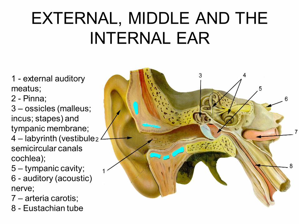
EXTERNAL, MIDDLE AND THE
INTERNAL EAR
1 - external auditory
meatus;
2 - Pinna;
3 – ossicles (malleus;
incus; stapes) and
tympanic membrane;
4 – labyrinth (vestibule
semicircular canals
cochlea);
5 – tympanic cavity;
6 - auditory (acoustic)
nerve;
7 – arteria carotis;
8 - Eustachian tube
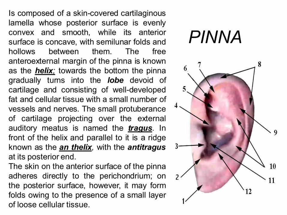
PINNA
Is composed of a skin-covered cartilaginous
lamella whose posterior surface is evenly
convex and smooth, while its anterior
surface is concave, with semilunar folds and
hollows between them. The free
anteroexternal margin of the pinna is known
as the helix; towards the bottom the pinna
gradually turns into the lobe devoid of
cartilage and consisting of well-developed
fat and cellular tissue with a small number of
vessels and nerves. The small protuberance
of cartilage projecting over the external
auditory meatus is named the tragus. In
front of the helix and parallel to it is a ridge
known as the an thelix, with the antitragus
at its posterior end.
The skin on the anterior surface of the pinna
adheres directly to the perichondrium; on
the posterior surface, however, it may form
folds owing to the presence of a small layer
of loose cellular tissue.

External Ear
• Auricle - framework of elastic
cartilage covered by skin
• Ear canal - about 3,5 cm
long, consists of outer
cartilaginous part and inner
bony part
• The cartilaginous part is
curved and lies at an angle
to the bony part, it also
narrows medially - ear drum
is protected from trauma
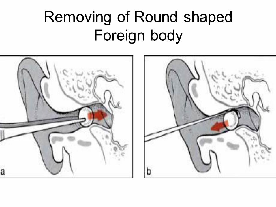
Removing of Round shaped
Foreign body
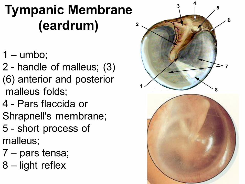
1 – umbo;
2 - handle of malleus; (3)
(6) anterior and posterior
malleus folds;
4 - Pars flaccida or
Shrapnell's membrane;
5 - short process of
malleus;
7 – pars tensa;
8 – light reflex
Tympanic Membrane
(eardrum)

Interconnected Auditory Ossicles
1 - malleus handle;
2 - head and neck of malleus;
3- Malleus-incudal joint;
4- body of incus
5 - short process of incus;
6 - long process of incus;
7 - head of stapes;
8 – base of stapes;
9 - cruses of stapes
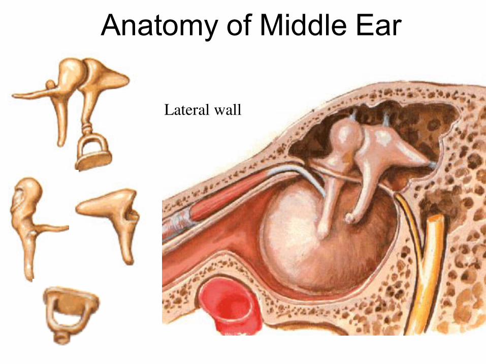
Anatomy of Middle Ear
Lateral wall
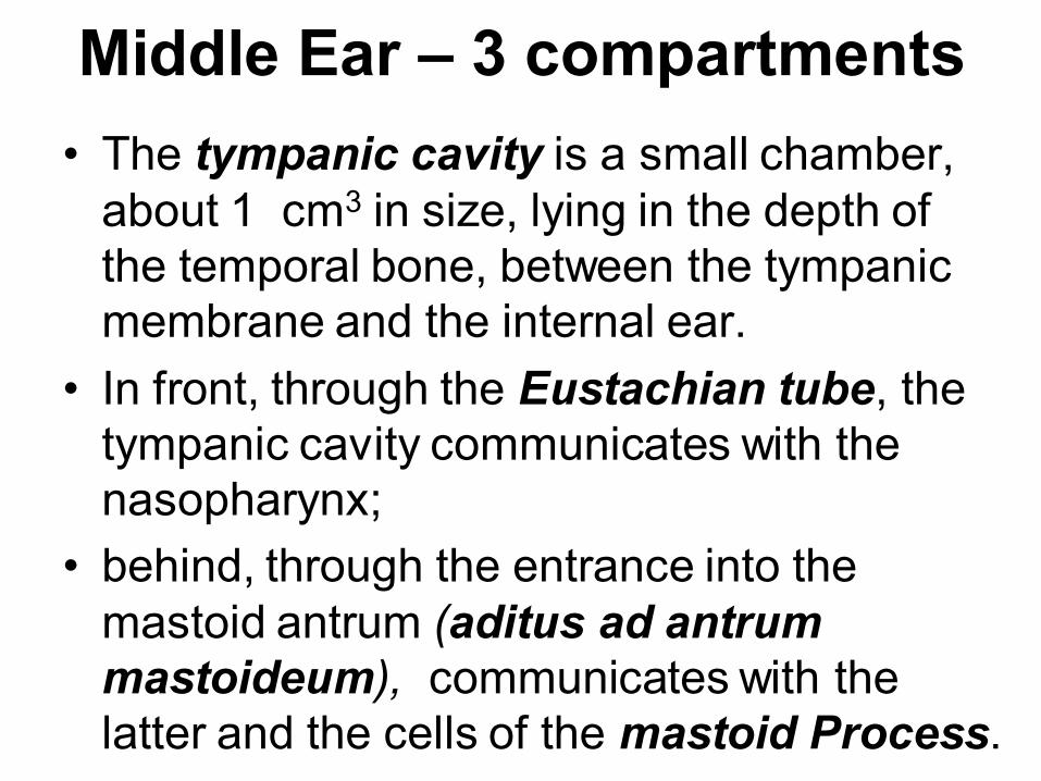
Middle Ear – 3 compartments
• The tympanic cavity is a small chamber,
about 1 cm3 in size, lying in the depth of
the temporal bone, between the tympanic
membrane and the internal ear.
• In front, through the Eustachian tube, the
tympanic cavity communicates with the
nasopharynx;
• behind, through the entrance into the
mastoid antrum (aditus ad antrum
mastoideum), communicates with the
latter and the cells of the mastoid Process.
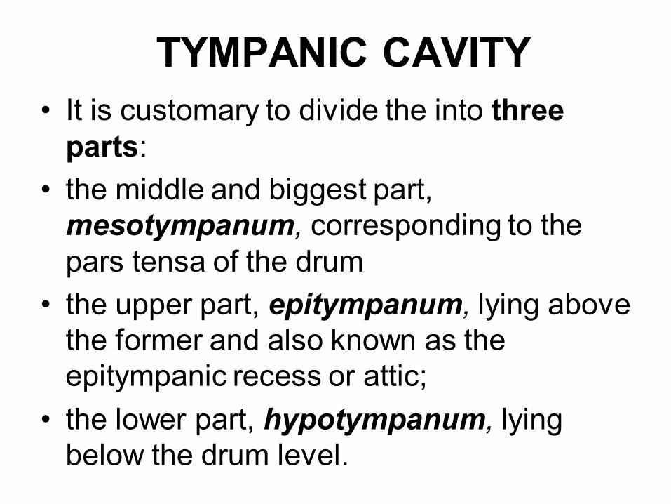
TYMPANIC CAVITY
• It is customary to divide the into three
parts:
• the middle and biggest part,
mesotympanum, corresponding to the
pars tensa of the drum
• the upper part, epitympanum, lying above
the former and also known as the
epitympanic recess or attic;
• the lower part, hypotympanum, lying
below the drum level.
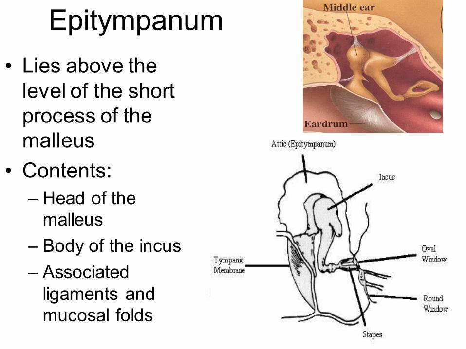
Epitympanum
• Lies above the
level of the short
process of the
malleus
• Contents:
– Head of the
malleus
– Body of the incus
– Associated
ligaments and
mucosal folds
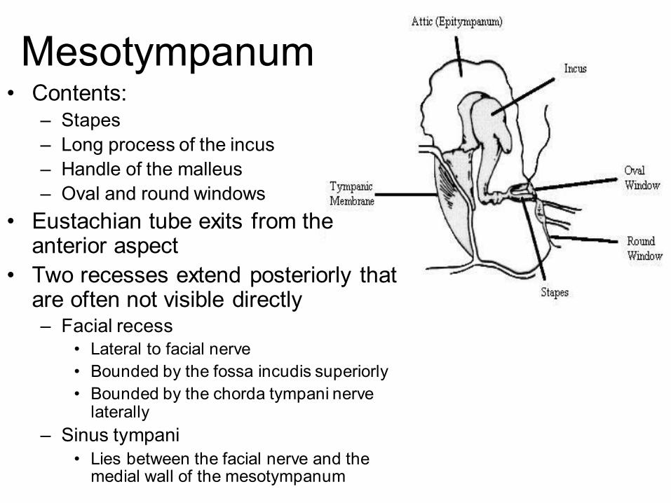
Mesotympanum • Contents:
– Stapes
– Long process of the incus
– Handle of the malleus
– Oval and round windows
• Eustachian tube exits from the anterior aspect
• Two recesses extend posteriorly that are often not visible directly – Facial recess
• Lateral to facial nerve
• Bounded by the fossa incudis superiorly
• Bounded by the chorda tympani nerve laterally
– Sinus tympani
• Lies between the facial nerve and the medial wall of the mesotympanum

Hypotympanum
• Lies inferior and
medial to the
floor of the bony
ear canal
• Irregular bony
groove that is
seldom involved
by cholesteatoma
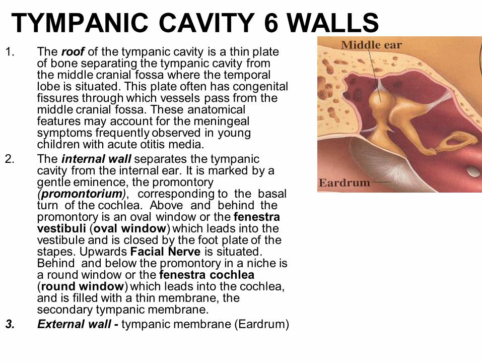
TYMPANIC CAVITY 6 WALLS 1. The roof of the tympanic cavity is a thin plate
of bone separating the tympanic cavity from the middle cranial fossa where the temporal lobe is situated. This plate often has congenital fissures through which vessels pass from the middle cranial fossa. These anatomical features may account for the meningeal symptoms frequently observed in young children with acute otitis media.
2. The internal wall separates the tympanic cavity from the internal ear. It is marked by a gentle eminence, the promontory (promontorium), corresponding to the basal turn of the cochlea. Above and behind the promontory is an oval window or the fenestra vestibuli (oval window) which leads into the vestibule and is closed by the foot plate of the stapes. Upwards Facial Nerve is situated. Behind and below the promontory in a niche is a round window or the fenestra cochlea (round window) which leads into the cochlea, and is filled with a thin membrane, the secondary tympanic membrane.
3. External wall - tympanic membrane (Eardrum)
1. The inferior wall or floor of the tympanic cavity is separated from the jugular bulb by a fairly thick bony plate. Bone fissures in this wall are rarely found.
2. The Eustachian tube begins with an opening in the anterior wall separating the tympanic cavity from the internal carotid canal.
3. An opening in the upper part of the posterior wall leads to the mastoid antrum (aditus ad antrum mastoideum).
TYMPANIC MUSCLES • There are two muscles in the tympanic cavity:
• (1) The tensor tympani muscle which stretches the tympanic membrane. It lies in the bony canal above the Eustachian tube, and is attached to the handle of the malleus.
• (2) The stapedius muscle which arises from the posterior wall of the tympanic cavity and is attached to the head of the stapes by a slender tendon. The tensor tympani is innervated by a branch of the trigeminal nerve, and the stapedius muscle by a branch of the facial nerve.
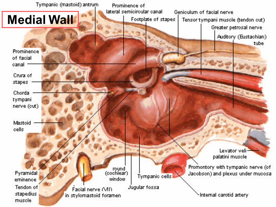
Medial Wall

Eustachian or auditory tube • About 3.5 cm in length connects
the tympanic cavity with the naso-pharynx. The upper third of this tube, adjoining the tympanic cavity, has bony walls, while the remaining lower portion leading into the nasopharynx is made up of membrane and cartilage.
• The movement of the cilia of the ciliated epithelium lining the Eustachian tube is towards the nasopharynx.
• At rest, the Eustachian tube is in a collapsed state, but with each swallowing movement it opens by contraction of the soft palatal muscles attached to it, to let air into the tympanic cavity.

MASTOID
PROCESS
• Is located just behind the external auditory meatus is a bony structure protruding downwards with the sternocleidomastoid muscle attached to it. In young children, the mastoid process is not fully developed and represents a bony tubercle behind the osseous tympanic ring.
• The antrum communicates with the tympanic cavity and the air-filled cells of the mastoid process. The superior wall or roof of the antrum separates it from the middle cranial fossa.
• The following types of structure are to be found in the mastoid process: the pneumatic or large-celled,
• diploic ". In the case of pneumatic structures, the cavity of the mastoid process is divided by thin bony partitions into a lattice of larger and smaller cells. The diploic structure has tiny cells resembling a diploetic bone;
• the most frequent is the mixed form of mastoid structure where smaller cells are to be found alongside bigger ones. In compact structures the bone is indurated and the cells are very few; this structure frequently occurs as a result of chronic suppurative otitis media.
•
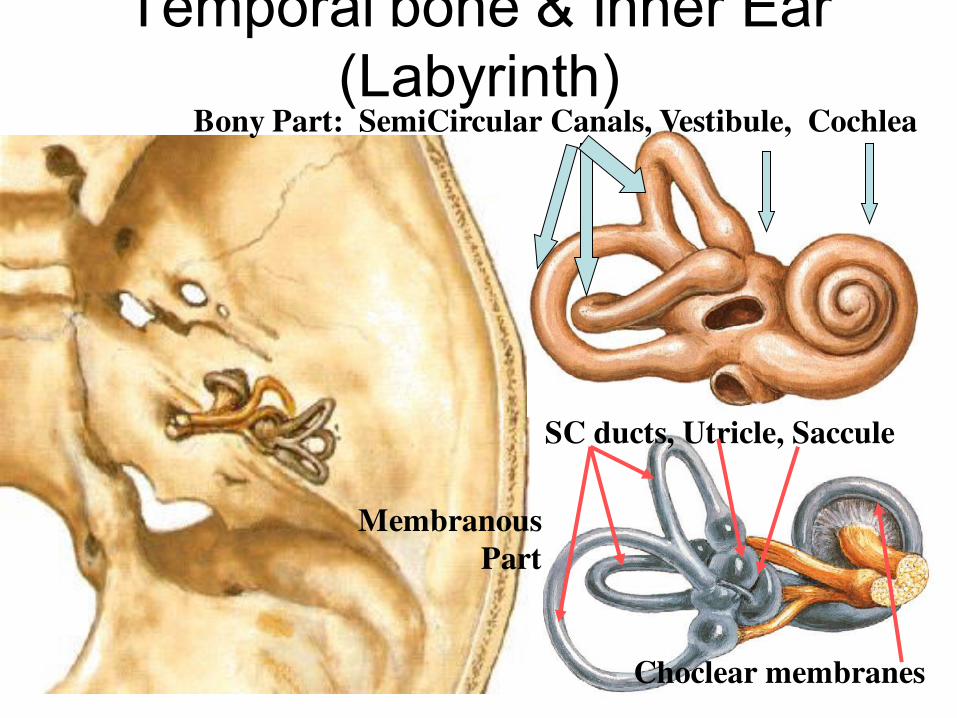
Temporal bone & Inner Ear
(Labyrinth) Bony Part: SemiCircular Canals, Vestibule, Cochlea
SC ducts, Utricle, Saccule
Choclear membranes
Membranous
Part
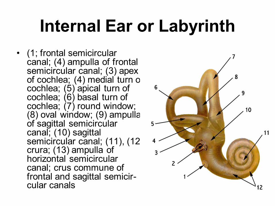
Internal Ear or Labyrinth
• (1; frontal semicircular canal; (4) ampulla of frontal semicircular canal; (3) apex of cochlea; (4) medial turn of cochlea; (5) apical turn of cochlea; (6) basal turn of cochlea; (7) round window; (8) oval window; (9) ampulla of sagittal semicircular canal; (10) sagittal semicircular canal; (11), (12) crura; (13) ampulla of horizontal semicircular canal; crus commune of frontal and sagittal semicir-cular canals
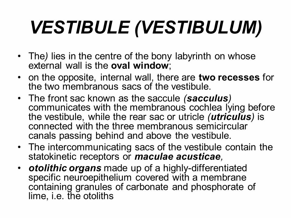
VESTIBULE (VESTIBULUM)
• The) lies in the centre of the bony labyrinth on whose external wall is the oval window;
• on the opposite, internal wall, there are two recesses for the two membranous sacs of the vestibule.
• The front sac known as the saccule (sacculus) communicates with the membranous cochlea lying before the vestibule, while the rear sac or utricle (utriculus) is connected with the three membranous semicircular canals passing behind and above the vestibule.
• The intercommunicating sacs of the vestibule contain the statokinetic receptors or maculae acusticae,
• otolithic organs made up of a highly-differentiated specific neuroepithelium covered with a membrane containing granules of carbonate and phosphorate of lime, i.e. the otoliths

SEMICIRCULAR CANALS
• The are set at right angles to each other and represent the three planes of space.
• They are three in number: the external or horizontal, the superior or frontal, and the posterior or sagittal. One end of each canal opens out into a larger space known as ampulla, the other end is even. The frontal and sagittal canals have a common even stem (crus commune).
• The ampulla of each membranous canal contains a ridge.
• the crista ampullaris, which is a receptor, i.e. a nerve ending consisting ot a highly-differentiated neuroepithelium or hair and supporting cells.
• The free surface of the hair cells is covered with hairs which respond to the slightest displacement or pressure of the endolymph.
• The receptors of the vestibule and semicircular canals are the peripheral nerve endings of the vestibular analysator.

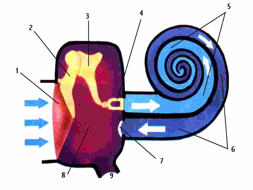
Modiolus the external wall and also turning
round the former, divides the tube lumen into
two directions, the upper or scala vestibuli
and the lower or scala tympani which
communicate at the apex of the cochlea through a small opening known as the
helicotrema. Both channels are filled with
perilymph. The scala vestibuli communicates
with the vestibule, while the scala tympani
borders on the tympanic cavity through the round window covered by the secondary tym-
panic membrane.
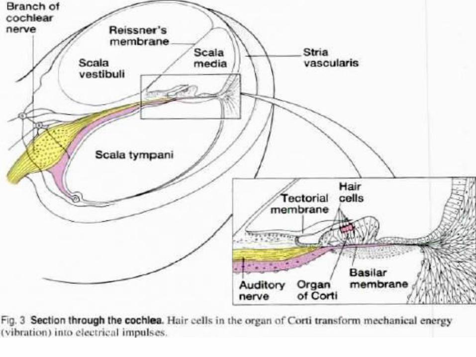
The scala vestibuli of the cochlea contains
the thin Reisner's membrane which extends
from the osseous spirn lamina to cut off a small membranous canal of trianguli section
filled with endolymph and known as the
cochles duct or ductus cochlearis.
COCHLEA
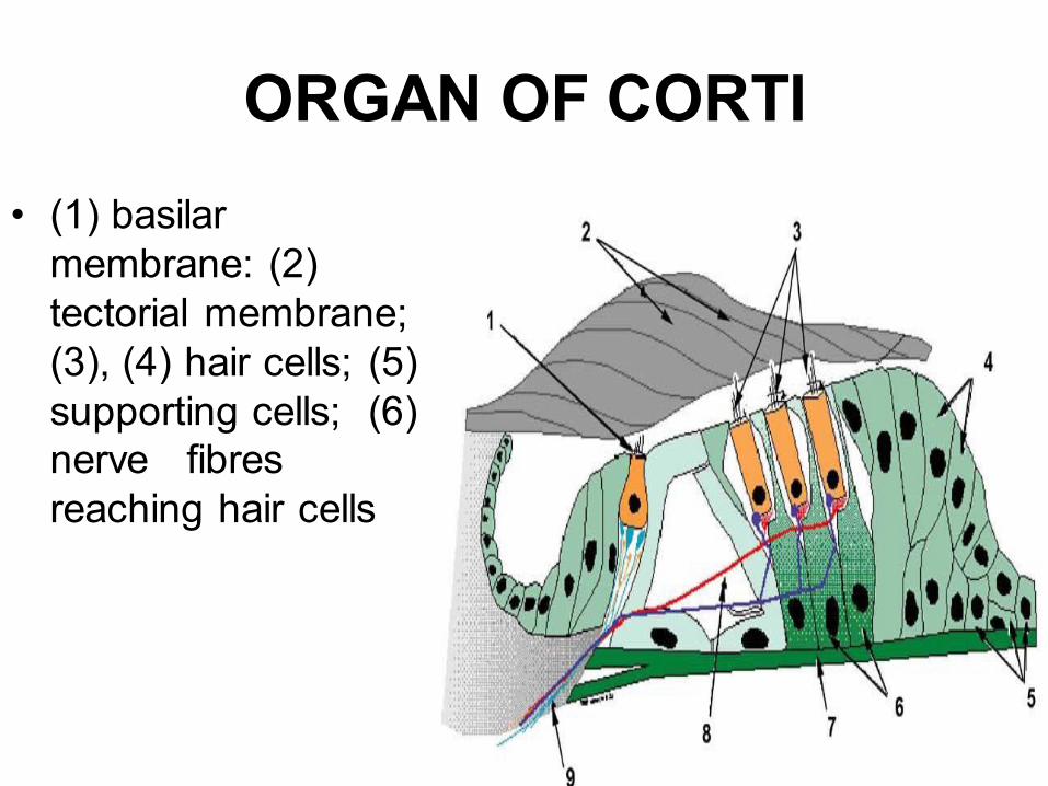
ORGAN OF CORTI
• (1) basilar
membrane: (2)
tectorial membrane;
(3), (4) hair cells; (5)
supporting cells; (6)
nerve fibres
reaching hair cells
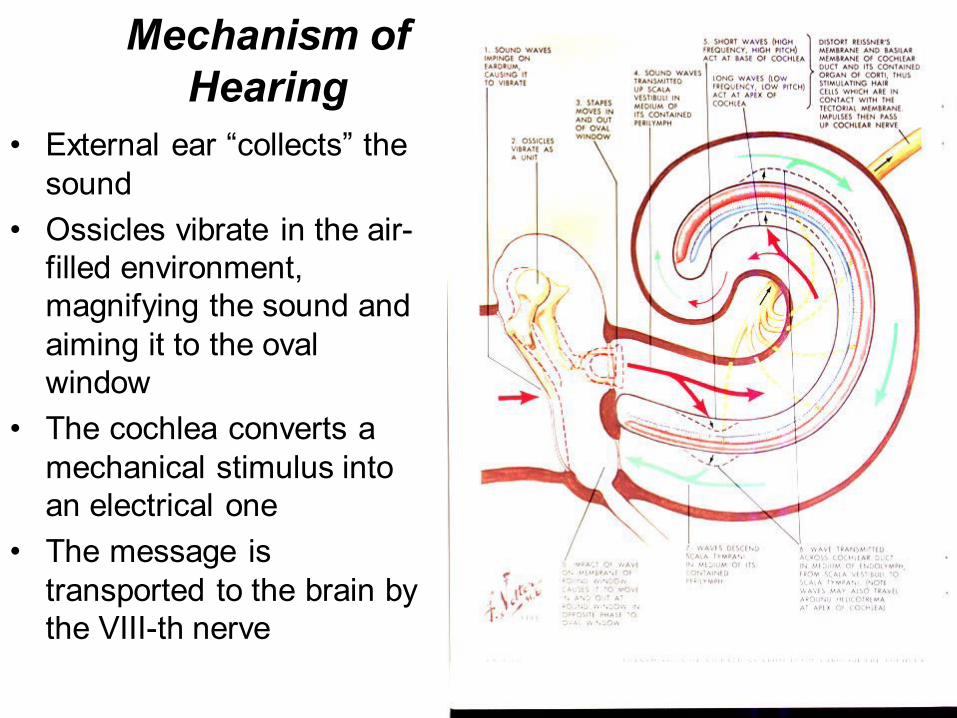
Mechanism of
Hearing
• External ear “collects” the sound
• Ossicles vibrate in the air-
filled environment,
magnifying the sound and
aiming it to the oval
window
• The cochlea converts a
mechanical stimulus into
an electrical one
• The message is
transported to the brain by
the VIII-th nerve
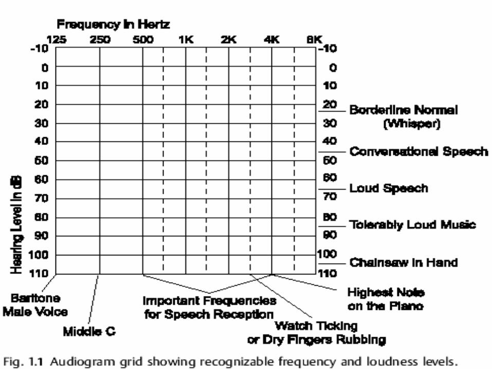
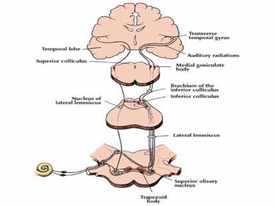
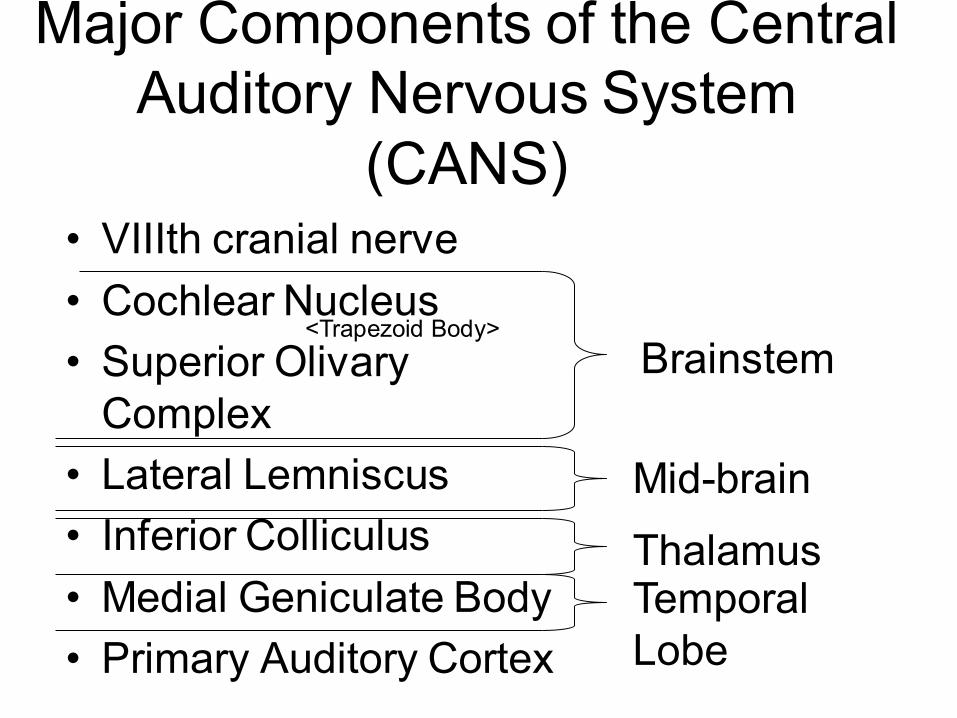
Major Components of the Central
Auditory Nervous System
(CANS)
• VIIIth cranial nerve
• Cochlear Nucleus
• Superior Olivary
Complex
• Lateral Lemniscus
• Inferior Colliculus
• Medial Geniculate Body
• Primary Auditory Cortex
Brainstem
Thalamus
Mid-brain
Temporal
Lobe
<Trapezoid Body>