Embed Size (px)
Citation preview

Anatomy, Imaging, and Common Anatomy, Imaging, and Common Pain-Generating Degenerative Pain-Generating Degenerative
Pathologies of the SpinePathologies of the Spine
R2 R2 김현석김현석
ANTOMYANTOMY
Osseous spinal columnOsseous spinal column 7 cervical, 12 thoracic, 5 lumbar and 5 fused sacral segments7 cervical, 12 thoracic, 5 lumbar and 5 fused sacral segments
C1 (atlas) : anterior arch, posterior arch, and paired lateral masses *C1 (atlas) : anterior arch, posterior arch, and paired lateral masses * C2 (axis) : bony projection which articulate with C1 (odontoid process C2 (axis) : bony projection which articulate with C1 (odontoid process
or dens)or dens) C3 ~ C7 : dorsolateral margin of the superior endplate (uncinate procC3 ~ C7 : dorsolateral margin of the superior endplate (uncinate proc
ess)ess) • typical cervical, thoracic, and lumbar vertebra : anterior body, pairetypical cervical, thoracic, and lumbar vertebra : anterior body, paire
d pedicles, articular pillars and laminae, and a single dorsal midlind pedicles, articular pillars and laminae, and a single dorsal midline spinous processe spinous process
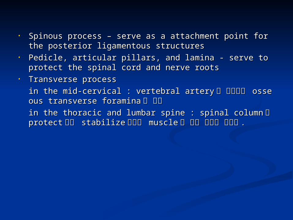
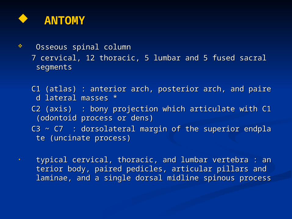
• Spinous process – serve as a attachment point for the posterior ligaSpinous process – serve as a attachment point for the posterior ligamentous structuresmentous structures
• Pedicle, articular pillars, and lamina - serve to protect the spinal cord Pedicle, articular pillars, and lamina - serve to protect the spinal cord and nerve rootsand nerve roots
• Transverse process Transverse process in the mid-cervical : vertebral arteryin the mid-cervical : vertebral artery 가 통과하는 가 통과하는 osseous transverse osseous transverse foraminaforamina 을 형성을 형성in the thoracic and lumbar spine : spinal columnin the thoracic and lumbar spine : spinal column 을 을 protectprotect 하고 하고 stastabilizebilize 시키는 시키는 musclemuscle 이 붙는 지점을 제공함이 붙는 지점을 제공함 ..
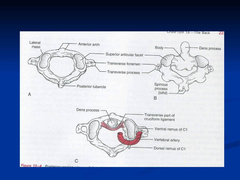
JointsJoints• atlanto-occipital : occipital condyle ~ lateral mass of C1atlanto-occipital : occipital condyle ~ lateral mass of C1• Atlantoaxial : ventral dens ~ dorsal surface of C1 anterior archAtlantoaxial : ventral dens ~ dorsal surface of C1 anterior arch• Uncovertebral : dorsolateral margin of the superior endplate of the Uncovertebral : dorsolateral margin of the superior endplate of the
C3~C7C3~C7• Costovertebra and Costotransverse : rib ~ vertebral body or transvCostovertebra and Costotransverse : rib ~ vertebral body or transv
erse process of the thoracic spineerse process of the thoracic spine• zygoapophyseal (facet) joints : the most prevalent joints , superior zygoapophyseal (facet) joints : the most prevalent joints , superior
and inferior articular process *and inferior articular process *

Transverse foramen, Intervertebral foramen, and Nerve rootsTransverse foramen, Intervertebral foramen, and Nerve roots• Contents of Transverse foramen – vertebral artery, vertebral venous Contents of Transverse foramen – vertebral artery, vertebral venous
plexus, sympathetic chainplexus, sympathetic chain• Intervertebral foramen Intervertebral foramen
bounded by the pedicle, vertebral body, disc and superior articular prbounded by the pedicle, vertebral body, disc and superior articular process *ocess *- Adamkiewicz artery : supply to the lower two-thirds of the spinal cor- Adamkiewicz artery : supply to the lower two-thirds of the spinal cord , enter the spinal canal via an intervertebral foramend , enter the spinal canal via an intervertebral foramen-> transforaminal or periganglionic intervention-> transforaminal or periganglionic intervention 시 시 damagedamage 에 주의에 주의
• Intervertebral Discs : thicker in the cervical and lumbar region and thiIntervertebral Discs : thicker in the cervical and lumbar region and thicker anteriorly than posteriorly cker anteriorly than posteriorly - axial loading - axial loading 의 의 absorb, flexibilityabsorb, flexibility 제공제공- nucleus pulposus, annulus fibrosis, cartilaginous endplate- nucleus pulposus, annulus fibrosis, cartilaginous endplate

- nucleus pulposus : type II collagen, hyaluronic acid, glycoaminoglyc- nucleus pulposus : type II collagen, hyaluronic acid, glycoaminoglycan *an *- annulus fibrosis : outer dense circumferential fibrous band and inne- annulus fibrosis : outer dense circumferential fibrous band and inner fibrocartilagenous layerr fibrocartilagenous layer
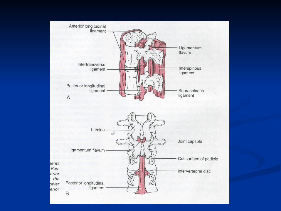
Ligament – stability, flexion, extension, rotationLigament – stability, flexion, extension, rotation- ALL, PLL, ligamentum flavum, interspinous ligament, supraspinous l- ALL, PLL, ligamentum flavum, interspinous ligament, supraspinous ligamentigament- ALL : vertebral body and intervertebral disc- ALL : vertebral body and intervertebral disc- PLL : annulus fibrosis but does not contact the posterior vertebral - PLL : annulus fibrosis but does not contact the posterior vertebral marginmargin- ligamentum flavum : laminar segment - ligamentum flavum : laminar segment 사이로 사이로 extend, spinal canalextend, spinal canal의 의 dorsolateral margindorsolateral margin 을 을 definedefine- interspinous ligament, supraspinous lig. : spinous process - interspinous ligament, supraspinous lig. : spinous process 사이를 사이를 연결연결
IMAGING OVERVIEWIMAGING OVERVIEW
Conventional Radiographs (X-rays)Conventional Radiographs (X-rays)- quick, inexpensive, easy to perform, execellent spatial resolution- quick, inexpensive, easy to perform, execellent spatial resolution- important information : alignment, structure, mineralization- important information : alignment, structure, mineralization- unstable spine : dynamic, weight bearing upright flextion and exten- unstable spine : dynamic, weight bearing upright flextion and extension viewsion view- foraminal stenosis, spondylolysis : oblique projection - foraminal stenosis, spondylolysis : oblique projection - vertebra alignment evaluate : lateral projection - vertebra alignment evaluate : lateral projection 에서 에서 three longitudithree longitudinal curve ( anterior and posterior spinal line = anterior and posterior lnal curve ( anterior and posterior spinal line = anterior and posterior longitudinal ligament ongitudinal ligament 의 의 course , spinolaminar line = ligamentum flacourse , spinolaminar line = ligamentum flavum vum 의 의 course ) course ) 과 과 pediclepedicle 의 관계에서 의 관계에서 rotational malalignmentrotational malalignment 가 가 보여짐보여짐 . *. *
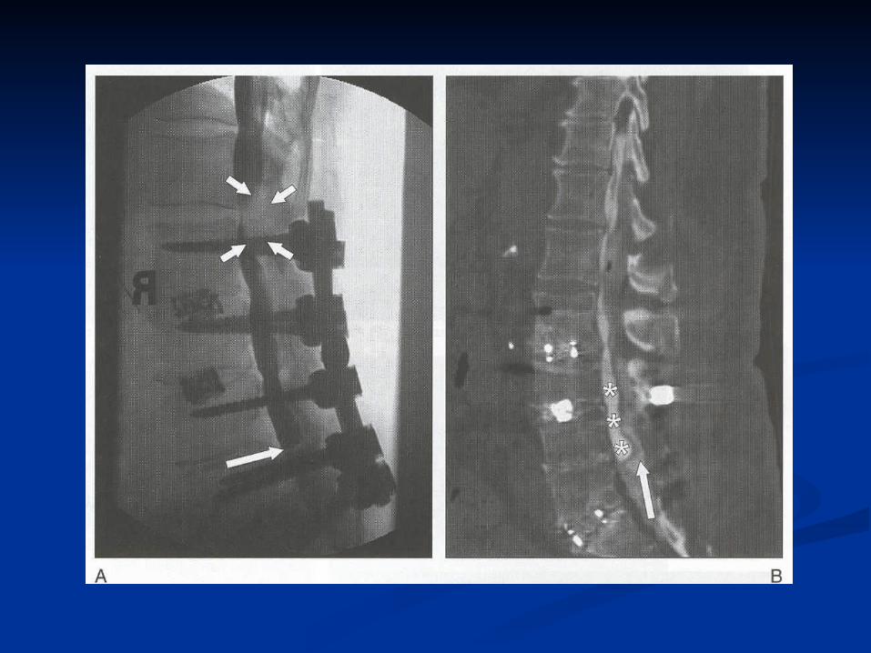
Myelography and postmyelography CT scanMyelography and postmyelography CT scan- non-ionic, water-soluble, radiographically dense iodinated contrast - non-ionic, water-soluble, radiographically dense iodinated contrast materialmaterial 을 을 subarachnoid spacesubarachnoid space 에 넣어서 에 넣어서 spinal canalspinal canal 의 의 contentcontent를 를 evaluateevaluate 하는 하는 radiographic technique.radiographic technique.- disc abnormality, ligament thickening, hypertrophic facet degenerati- disc abnormality, ligament thickening, hypertrophic facet degenerative change, spinal stenosis, nerve root impingement, redundant thickve change, spinal stenosis, nerve root impingement, redundant thickened nerve root, arachnoiditis can be detectedened nerve root, arachnoiditis can be detected- postmyelography CT scan : better definition of anatomic relationshi- postmyelography CT scan : better definition of anatomic relationship of the contents of the spinal canal to the surrounding structurep of the contents of the spinal canal to the surrounding structure
- invasiveness and non-invasive imaging tool(CT, MRI)- invasiveness and non-invasive imaging tool(CT, MRI) 의 의 availabilityavailability로 인해 사용이 줄어듬로 인해 사용이 줄어듬 ..- Cx. : positional headache(m/c), contrast-related seizure, infection - Cx. : positional headache(m/c), contrast-related seizure, infection **

Computer-assisted tomography (CAT or CT scan)Computer-assisted tomography (CAT or CT scan)- osseous structure- osseous structure 의 의 definitiondefinition 에 가장 우수에 가장 우수 ..- x-ray beam- x-ray beam 의 의 diffenrential attenuationdiffenrential attenuation 에 기초한 것으로에 기초한 것으로 , bone, lig, bone, ligament, disc material, CSF ament, disc material, CSF 등의 등의 radiographic densityradiographic density 차이가 차이가 있으므로있으므로 , disc herniation, ligamentous disorder, disc herniation, ligamentous disorder 등을 진단하는데 등을 진단하는데 사용될 수 있다사용될 수 있다 ..- spinal rods, transpedicular screw, laminar wire/hook, intervertebral - spinal rods, transpedicular screw, laminar wire/hook, intervertebral cagecage 등의 등의 surgical metallic implantsurgical metallic implant 에 의한 에 의한 artifactartifact 가 생김가 생김 ..- children, pregnant female, other young adult- children, pregnant female, other young adult 에서 더 에서 더 sensitivesensitive하므로 적절한 하므로 적절한 carecare 하에 시행되야 함하에 시행되야 함 ..
Magnetic resonance imaging(MRI)Magnetic resonance imaging(MRI)- hydrogen atom(proton)- hydrogen atom(proton) 의 의 amount and stateamount and state 에 기초하여 에 기초하여 tissuetissue 를 를 localizelocalize 하기 위해 하기 위해 gradient fieldgradient field 와 와 radiofrequencyradiofrequency 를 사용를 사용- radiation- radiation 은 없지만은 없지만 , electrical and metal implant, electrical and metal implant 에 의한 에 의한 riskrisk 와 와 fefetustus 에 대한 에 대한 unknown riskunknown risk 가 있다가 있다 ..- soft tissue contrast resolution- soft tissue contrast resolution 이 우수하여 이 우수하여 spinal disorderspinal disorder 의 의 diagdiagnostic imaging modalitynostic imaging modality 로 유용함로 유용함 ..- bone marrow, muscle, ligament, disc material, nerve root- bone marrow, muscle, ligament, disc material, nerve root 등의 등의 tisstissue typeue type 의 구분이 우수하고의 구분이 우수하고 , extradural, intradural, extramedullary, in, extradural, intradural, extramedullary, intramedullary pathologytramedullary pathology 의 구분이 정확함의 구분이 정확함 ..- medullary bone evaluation- medullary bone evaluation 에도 우수하여 에도 우수하여 marrow edemamarrow edema 나 나 marromarrow replacementw replacement 를 일으키는 를 일으키는 osseous conditionosseous condition 을 알 수 있으나을 알 수 있으나 , den, dense cortical bone, sclerotic lesion, osteophytese cortical bone, sclerotic lesion, osteophyte 에 대한 정확성은 에 대한 정확성은 CTCT에 비해 낮다에 비해 낮다 . .

- standard MRI protocol : sagittal and axial with T1- and T2- weighted - standard MRI protocol : sagittal and axial with T1- and T2- weighted sequencesequence- T1 high signal intensity – fat (fatty bone marrow, subcu. Fat)- T1 high signal intensity – fat (fatty bone marrow, subcu. Fat)
low signal intensity – fluid (CSF, bone marrow edema, low signal intensity – fluid (CSF, bone marrow edema, normal nucleus pulposus) *normal nucleus pulposus) *
- STIR(short-tau inversion recovery): fat depressed T2-weighted sequ- STIR(short-tau inversion recovery): fat depressed T2-weighted sequenceence 로 로 fluidfluid 에 에 sensitivesensitive 하여 하여 traumatic injury, malignancy, infectiotraumatic injury, malignancy, infectionn 등에서 등에서 edemaedema 를 를 detectdetect 하는데 유용하는데 유용- GRE(gradient recalled echo) T2-weighted imaging- GRE(gradient recalled echo) T2-weighted imaging 은 은 blood product blood product and calciumand calcium 에 에 sensitivesensitive 하여 하여 spinal traumaspinal trauma 에 유용에 유용- infection, multiple sclerosis, intramedullary neoplasm, metastatic di- infection, multiple sclerosis, intramedullary neoplasm, metastatic disease, postoperative scarring evaluation – IV gadolinium contrast masease, postoperative scarring evaluation – IV gadolinium contrast material administration sagittal and axial T1-weighted imageterial administration sagittal and axial T1-weighted image
- C/Ix. : cardiac pacemaker, metallic foreign body, metallic surgical i- C/Ix. : cardiac pacemaker, metallic foreign body, metallic surgical implant(ex. cerebral aneurysm clip and heart valve)mplant(ex. cerebral aneurysm clip and heart valve)- MRI- MRI 시행시 시행시 m/c problemm/c problem 은 은 claustrophobiaclaustrophobia 이고 이고 sedationsedation 에 의해에 의해overcomeovercome 되지만되지만 , , 마취과가 필요한 경우도 있음마취과가 필요한 경우도 있음 ..
DEGERATIVE DISC DISEASEDEGERATIVE DISC DISEASE
OverviewOverview• Acute or chronic back painAcute or chronic back pain 을 일으키는 을 일으키는 uncertain etiology, aginguncertain etiology, aging 에 에
의한 의한 pathologic processpathologic process• Conventional radiographic finding : disc space narrowing, vacuum disConventional radiographic finding : disc space narrowing, vacuum dis
c, endplate sclerosis, ostephyte formationc, endplate sclerosis, ostephyte formation• MRI : excellent soft tissue contrast and multiplanar capabilityMRI : excellent soft tissue contrast and multiplanar capability
-> disc degeneration evaluate-> disc degeneration evaluate 의 의 modality of choicemodality of choice
Disc dehydration and NarrowingDisc dehydration and Narrowing• T1 : hydrated and nonhydrated disc : homogenousT1 : hydrated and nonhydrated disc : homogenous
T2 : discT2 : disc 의 의 water contentwater content 가 가 bright signal -> central bright nucleus bright signal -> central bright nucleus pulposus and dark annular fibrosispulposus and dark annular fibrosis
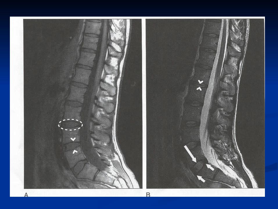

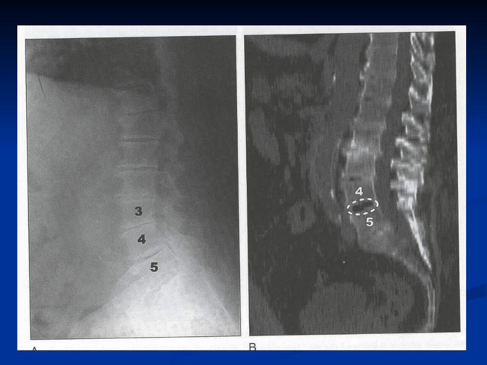
Degenertive disease -> disc desiccation -> disc signalDegenertive disease -> disc desiccation -> disc signal 감소감소 -> intradiscal gas accumulation -> intradiscal gas accumulation
-> proton -> proton 이 없어서 이 없어서 T1- and T2-T1- and T2- 에서 에서 hypointense sequensehypointense sequense(vacuum disc phenomenon)(vacuum disc phenomenon)
Disc heightDisc height 는 는 normal or diminishednormal or diminished 이어서 이어서 disc degenerationdisc degeneration 의 의 inindicatordicator 가 못되나가 못되나 , decreased height, decreased height 는 는 intervertebral foramina sizeintervertebral foramina size의 감소이고 의 감소이고 nerve rootnerve root 의 의 compressioncompression 과 관련됨과 관련됨 ..
Annular fissure / tearsAnnular fissure / tears-On T2-weighted image, linear hyperintense signal-On T2-weighted image, linear hyperintense signal
• Three type of annular degeneration – concentric fissuring, transverse Three type of annular degeneration – concentric fissuring, transverse tear, radial teartear, radial tear- concentric fissuring : collagen fiber delamination – T2- concentric fissuring : collagen fiber delamination – T2 에서 에서 disc madisc marginrgin 에 에 parellelparellel 한 한 high signal intensityhigh signal intensity

- transverse tear : vertebral body ring apophyses- transverse tear : vertebral body ring apophyses 와 와 Sharpey’s fiberSharpey’s fiber의 의 junctionjunction 에서 에서 small foci of T2 hyperintensitysmall foci of T2 hyperintensity- radial tear : primary failure of the annulus and full-thickness disrupt- radial tear : primary failure of the annulus and full-thickness disruption of the annulus *ion of the annulus *outer third of annulus fibrosis and PLL are innervated by nociceptive outer third of annulus fibrosis and PLL are innervated by nociceptive nerve ending -> discogenic back pain nerve ending -> discogenic back pain
Subchondral marrow changesSubchondral marrow changes- vertebral end plate degenerative disease- vertebral end plate degenerative disease 의 의 three typethree type: T1- and T2- weighted signal charateristic: T1- and T2- weighted signal charateristic 에 따라에 따라- Type I change : T1- Type I change : T1 감소감소 , T2, T2 증가 – 증가 – vascularized marrowvascularized marrow- Type II change : T1- Type II change : T1 증가증가 , T2, T2 증가 증가 or isointense – bone marrowor isointense – bone marrow 의 의 fatty replacementfatty replacement- Type III change : T1, T2 both low signal – subchondral sclerosis - Type III change : T1, T2 both low signal – subchondral sclerosis

DISC HERNIATIONDISC HERNIATION
OverviewOverview- disc herniation evaluation : imaging modality of choice is MRI (due t- disc herniation evaluation : imaging modality of choice is MRI (due to its excellent soft tissue resolution)o its excellent soft tissue resolution)
Disc contourDisc contour• Disc herniation : Intervertebral disc spaceDisc herniation : Intervertebral disc space 를 넘어선 를 넘어선 localized disc mlocalized disc m
aterial displacementaterial displacement• Circumferential bulge : vertebral body’s ring apophysis edgeCircumferential bulge : vertebral body’s ring apophysis edge 의 의 50~150~1
00%00% 를 넘어서는 를 넘어서는 disc material bulgingdisc material bulging- 25% - 25% 넘지않는 넘지않는 localized herniated disc materiallocalized herniated disc material 은 “은 “ focal” , 25~5focal” , 25~50% 0% 는 “는 “ broad-based”broad-based” 라는 라는 termterm- vertebral endplate- vertebral endplate 로 로 focal disc herniation : Schmorl’s nodefocal disc herniation : Schmorl’s node

Disc marginDisc margin 과 과 herniated disc fragmentherniated disc fragment 의 의 shapeshape 에 따라 에 따라 protrusion protrusion and extrusionand extrusion 으로 으로 describe.describe.- protrusion : base- protrusion : base 가 가 wider than apexwider than apex- extrusion : base- extrusion : base 에서 에서 widthwidth 보다 보다 herniated fragmentherniated fragment 가 더 가 더 awayaway- sequestated or free-fragment disc herniation : parent disc- sequestated or free-fragment disc herniation : parent disc 에서 에서 cocompletely seperatedmpletely seperated
Disc migration in the cranial or caudal direction is best evaluated in tDisc migration in the cranial or caudal direction is best evaluated in the sagittal planehe sagittal plane- - 주로 주로 posterior extrusionposterior extrusion 은 은 PLL PLL 내에 있고내에 있고 , inferiorly migrate, inferiorly migrate 됨됨
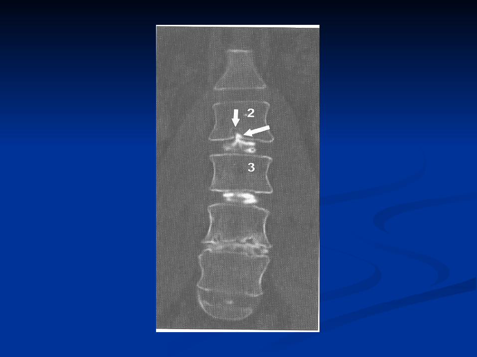
Disc herniation positionDisc herniation position• Disc herniationDisc herniation 의 위치는 의 위치는 anatomic landmarkanatomic landmark 를 사용하여 를 사용하여 describe.describe.
- extraforaminal zone : pedicle- extraforaminal zone : pedicle 의 의 lateral aspectlateral aspect 의 의 parasagittal lineparasagittal line을 넘어서는 을 넘어서는 zone.zone.- lateral recess : pedicle- lateral recess : pedicle 의 의 medial bordermedial border 를 따르는 를 따르는 area, disc level area, disc level and superior vertebral endplateand superior vertebral endplate 의 이하의 이하
• On sagittal imageOn sagittal image- suprapedicular level : pedicle- suprapedicular level : pedicle 의 바로 위 의 바로 위 ~ superior end plate~ superior end plate- pedicle level : pedicle- pedicle level : pedicle 의 의 superior and inferior edgesuperior and inferior edge 사이사이- infrapedicle level : pedicle- infrapedicle level : pedicle 의 의 inferior edgeinferior edge 아래 아래 ~ inferior end plate~ inferior end plate
• Herniated discHerniated disc 의 위치에 따라 의 위치에 따라 compresscompress 하는 하는 nerve rootnerve root 결정결정- in the Cervical spine : central or paramedian herniation -> descendi- in the Cervical spine : central or paramedian herniation -> descending nerve rootng nerve rootex) C3-4 paramedian disc extrusion -> C5 nerve rootex) C3-4 paramedian disc extrusion -> C5 nerve root

foraminal disc -> foraminal disc -> 같은 같은 levellevel 의 의 nerve rootnerve rootex) C3-4 foraminal disc -> C4 nerve rootex) C3-4 foraminal disc -> C4 nerve root- in the thoracic and lumbar spine(existing root is associated with su- in the thoracic and lumbar spine(existing root is associated with superior level) : paramedian disc T3-4 -> T4 rootperior level) : paramedian disc T3-4 -> T4 rootforaminal disc T3-4 -> T3 rootforaminal disc T3-4 -> T3 root
Neural compressionNeural compression 의 정도는 의 정도는 herniated discherniated disc 에 의한 에 의한 normally rounnormally round or oval configurationd or oval configuration 의 의 spinal cord, nerve root, root ganglionspinal cord, nerve root, root ganglion 의 의 변화의 정도에 따라 변화의 정도에 따라 graded.graded.- mild compression : normal diameter- mild compression : normal diameter 의 의 75~99% 75~99% 유지유지- moderate : 50~74%, severe : <50%- moderate : 50~74%, severe : <50%

FACET JOINTFACET JOINT
OverviewOverview- low back pain- low back pain 의 의 sourcesource 가 되지만가 되지만 , clinically primary cause, clinically primary cause 인지 인지 감별하기는 어렵다감별하기는 어렵다 ..- facet joint syndrome : degenerative facet joint- facet joint syndrome : degenerative facet joint 와 해부학적으로 와 해부학적으로 관련된 관련된 focal or referred painfocal or referred pain 을 말하는 을 말하는 controversial diagnosiscontroversial diagnosis
ImagingImaging- facet joint arthropathy : hypertrophic osteophytic overgrowth, subch- facet joint arthropathy : hypertrophic osteophytic overgrowth, subchondral sclerosis, bone marrow edema, joint space narrowing/wideninondral sclerosis, bone marrow edema, joint space narrowing/widening, joint stiffness, periarticular soft tissue edema…g, joint stiffness, periarticular soft tissue edema…- osteophytosis and subchondral sclerosis : T1- and T2- - osteophytosis and subchondral sclerosis : T1- and T2- 에서 에서 hypoinhypointensetense- bone marrow and periarticular soft tissue edema : T1- bone marrow and periarticular soft tissue edema : T1 에서 에서 hypointhypointense and T2-ense and T2- 에서 에서 hyperintensehyperintense 보임보임 ..- joint space widening : joint space- joint space widening : joint space 의 의 effusion, T2-hyperintenseeffusion, T2-hyperintense

- facet joint arthropathy- facet joint arthropathy 는 는 intrinsic abnormalityintrinsic abnormality 로 로 painpain 일으킬 수 일으킬 수 있고있고 , lateral recess, lateral recess 에서 에서 descending nerve rootdescending nerve root 나 나 intervertebral forintervertebral foramenamen 에서 에서 existing nerve rootexisting nerve root 를 를 compressioncompression 하여 하여 painpain 만듬만듬 ..- facet joint osteoarthritis : CT scanning- facet joint osteoarthritis : CT scanning 으로 정확히 진단으로 정확히 진단 .. cervical spinecervical spine 에서 애매한 에서 애매한 sclerotic change and osteophytesclerotic change and osteophyte 는 는 MRMRII 보다 더 쉽게 진단보다 더 쉽게 진단 ..

INTRASPINAL FACET CYSTSINTRASPINAL FACET CYSTS OverviewOverview
- intraspinal facet cyst : facet joint - intraspinal facet cyst : facet joint 에서 에서 originorigin 한 한 smooth bordersmooth border 의 의 fluid-filled rounded structurefluid-filled rounded structure- lining of cyst : synovial epithelial cell(synovial cyst) or fibrous wall s- lining of cyst : synovial epithelial cell(synovial cyst) or fibrous wall surrounding myxoid material(ganglion cyst)urrounding myxoid material(ganglion cyst)- radiologically both type- radiologically both type 이 같게 보이고이 같게 보이고 , treatment, treatment 도 감압술로 도 감압술로 같아서 같아서 typetype 의 감별이 임상적으로 중요치 않다의 감별이 임상적으로 중요치 않다 . *. *- synovial cyst(- synovial cyst( 통칭통칭 )) 는 거의 항상 는 거의 항상 degenerated facet jointdegenerated facet joint 에서 에서 생기고생기고 , joint, joint 의 의 dorsal surfacedorsal surface 에서 나온 것은 에서 나온 것은 soft tissuesoft tissue 로 로 protruprotrudingding 하지만 하지만 neural structureneural structure 를 를 compressioncompression 하지는 않고하지는 않고 , ventral s, ventral surfaceurface 에서 생기고 에서 생기고 intervertebral joint, lateral recess, lateral spinal cintervertebral joint, lateral recess, lateral spinal canalanal 로 로 protrudingprotruding 한 것은 한 것은 locationlocation 에 따라 에 따라 existing nerve root(in thexisting nerve root(in the foramen) or descending nerve root(in the lateral recess or lateral se foramen) or descending nerve root(in the lateral recess or lateral spinal canal)pinal canal) 를 를 compression compression 함함- nociceptive synovial lining -> intrinsically painful- nociceptive synovial lining -> intrinsically painful 할 수도 있음할 수도 있음 ..
ImagingImaging- on CT, uncomplicated synovial cyst : isodense to CSF, occasionally - on CT, uncomplicated synovial cyst : isodense to CSF, occasionally has a calcified wallhas a calcified wall
proteinaceous material or blood within cyst : isodense toproteinaceous material or blood within cyst : isodense to muscle or ligamentmuscle or ligament
- mass effect or stenosis related to intraspinal or foaminal synovial c- mass effect or stenosis related to intraspinal or foaminal synovial cyst : CT myelography *yst : CT myelography *- MRI : T1- and T2- prolongation, isodense to CSF- MRI : T1- and T2- prolongation, isodense to CSF
proteinaceous or hemorrhagic material : T1 hyperintensityproteinaceous or hemorrhagic material : T1 hyperintensity: wall: wall 은 은 fibrous materialfibrous material 로 구성로 구성 , calcified, calcified
- juxta-articular cyst- juxta-articular cyst 와 와 disc extrusion D/Dx. : short term follow up Mdisc extrusion D/Dx. : short term follow up MRIRI 에서 에서 disc fragmentdisc fragment 는 는 resolutionresolution 되고되고 , cyst, cyst 는 는 no changeno change- treatment : conservative management, percutaneous decompressio- treatment : conservative management, percutaneous decompression, sugical removal -> successful outcomen, sugical removal -> successful outcome

SPINAL STENOSISSPINAL STENOSIS
OverviewOverview- CT- CT 는 는 bony abnormality, bulging or herniated discbony abnormality, bulging or herniated disc 로 인한 로 인한 spinal stspinal stenosisenosis 를 효과적으로 를 효과적으로 evaluationevaluation- MRI : GRE T2 image in the cervical spine and conventional or fast s- MRI : GRE T2 image in the cervical spine and conventional or fast spin echo T2-weighted image in the T-L spine -> canalpin echo T2-weighted image in the T-L spine -> canal 내에서 내에서 CSF floCSF floww 로 인한 로 인한 artifactartifact 없이 없이 central canal and intervertebral foramencentral canal and intervertebral foramen 을 을 evaluationevaluation 할 수 있다할 수 있다 ..- surgical hardware- surgical hardware 있는 경우엔있는 경우엔 , conventional T2-weighted image, conventional T2-weighted image 로 로 artifactartifact 를 최소화 할 수 있다를 최소화 할 수 있다 ..
Grading spinal stenosisGrading spinal stenosis- AP dimension- AP dimension 에 의해 에 의해 mild stenosismild stenosis 는 는 normal levelnormal level 의 의 spinal canspinal canalal 의 의 AP diameterAP diameter 의 의 75~99%75~99% 유지유지 , moderate and severe, moderate and severe 는 는 50~750~74%, <50%4%, <50%- intervertebral foramen- intervertebral foramen 의 의 AP and craniocaudal dimensionAP and craniocaudal dimension 으로 으로 evalevaluateuate
- mild foraminal stenosis- mild foraminal stenosis 는 는 bulging disc or hypertrophic superior artibulging disc or hypertrophic superior articular processcular process 에 의해 에 의해 foramenforamen 의 의 inferior partinferior part 의 의 narrowingnarrowing- moderate narrowing- moderate narrowing 은 은 nerve rootnerve root 를 따르는 를 따르는 fatfat 이 줄어듬이 줄어듬 ..- severe stenosis- severe stenosis 은 은 no fat, nerve root clearly compressedno fat, nerve root clearly compressed+ + 이런 변화는 이런 변화는 sagittal T1-weighted sequencesagittal T1-weighted sequence 에서 에서 most sensitivemost sensitive
SPONDYLOLYSIS AND SPONDYLOLYSIS AND SPONDYLOLISTHESISSPONDYLOLISTHESIS OverviewOverview
- spondylolysis : articular pillar- spondylolysis : articular pillar 의 의 pars intra-articularispars intra-articularis 에서의 에서의 discodiscontinuity, ntinuity, 원인은 불확실하지만원인은 불확실하지만 , chronic microtrauma, chronic microtrauma 와 관련된걸로 와 관련된걸로 보임보임 ..- bilateral pars fracture- bilateral pars fracture 있으면있으면 , vertebral body, vertebral body 는 앞으로 는 앞으로 slipslip-> spondylolisthesis-> spondylolisthesis- mild and moderate slip : canal- mild and moderate slip : canal 이 이 not narrow, paradoxically enlargenot narrow, paradoxically enlarge- severe spondylolisthesis : spinal canal- severe spondylolisthesis : spinal canal 의 의 AP diameter elongate anAP diameter elongate and sagittal planed sagittal plane 에서 에서 spinal canal narrowingspinal canal narrowing 시킴시킴 ..
ImagingImaging- CT : test of choice to diagnose, pars- CT : test of choice to diagnose, pars 의 의 sclerosis and fracturesclerosis and fracture- MRI : - MRI : 유사한 소견이지만유사한 소견이지만 , actual fracture, actual fracture 를 파악하기 어려운 를 파악하기 어려운 때가 있다때가 있다 ..
- MRI- MRI 는 는 foraminal stenosisforaminal stenosis 와 와 nerve root compressionnerve root compression 은 잘 보임은 잘 보임 pars fracturepars fracture 부위에서의 부위에서의 cartilagenous overgrowth cartilagenous overgrowth 보임보임 ..- plain film- plain film 은 은 oblique projectionoblique projection 에서 에서 spondylolisthesisspondylolisthesis 를 쉽게 를 쉽게 진단할 수 있고진단할 수 있고 , bone detail, bone detail 과 과 MRI examMRI exam 의 관계보는데도 효과적의 관계보는데도 효과적
![Role of Peripheral Nerve Stimulation in Degenerative Lumbar Spine Pathologies … · 2017. 7. 22. · pathologies[6] with neurodeficit like Prolapsed Intervertebral Disc or Lumbar](https://img.pdfslide.net/doc/110x75/5fc9de7116d6441b5078b9e4/role-of-peripheral-nerve-stimulation-in-degenerative-lumbar-spine-pathologies-2017.jpg)


![[IGC 2016] 골드로쉬 김현석 - 왜 항상 기획자는 욕을 들어야만 하는 걸까? –게임 기획의 포지션 변화-](https://img.pdfslide.net/doc/110x75/587690da1a28abab2f8b5859/igc-2016--591a76c13e86a.jpg)