Embed Size (px)
Citation preview

1
ANATOMY OF MUSCULOSKELETAL SYSTEM
S asanthy KusumaningtyasFMUI
2
Anatomical aspect of MS
• The musculoskeletal system is consist of: – Musculo=muscleactive– S keletal=bonepasive– Nervous system mechanism of
movement
3
The skeleton
• The skeleton consist of bones, cartilage, joints and ligaments.
• Function of the skeleton (bones):– S upport– Protection– Movement– Mineral storage – Blood-cell formation
4
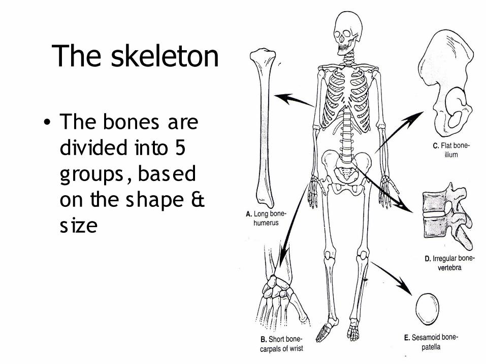
• The bones are divided into 5 groups, based on the shape & size
The skeleton
5
The skeleton
• The human skeleton are grouped into:– The axial skeleton– The appendicular skeleton (upper limb and
lower limb)
6
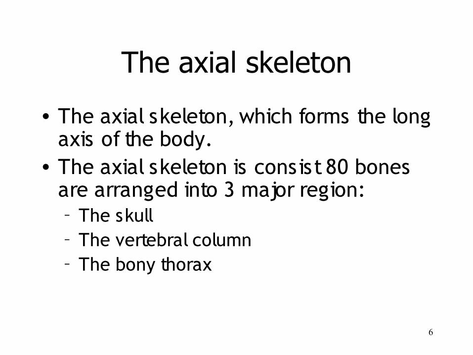
The axial skeleton
• The axial skeleton, which forms the long axis of the body.
• The axial skeleton is consist 80 bones are arranged into 3 major region:– The skull– The vertebral column– The bony thorax
7
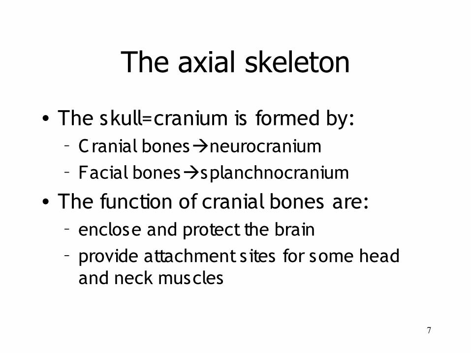
The axial skeleton
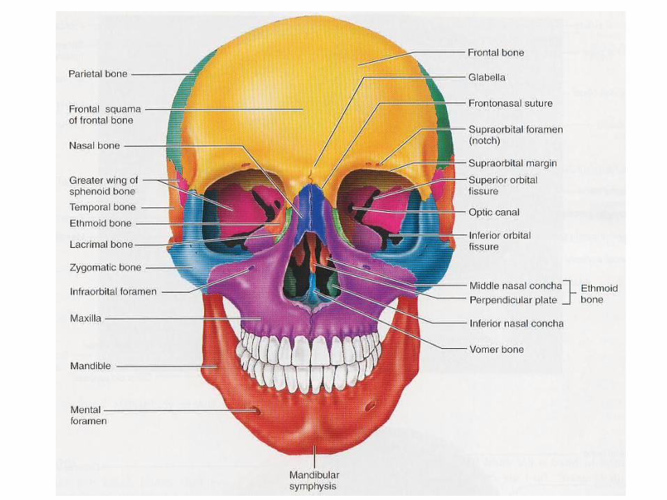
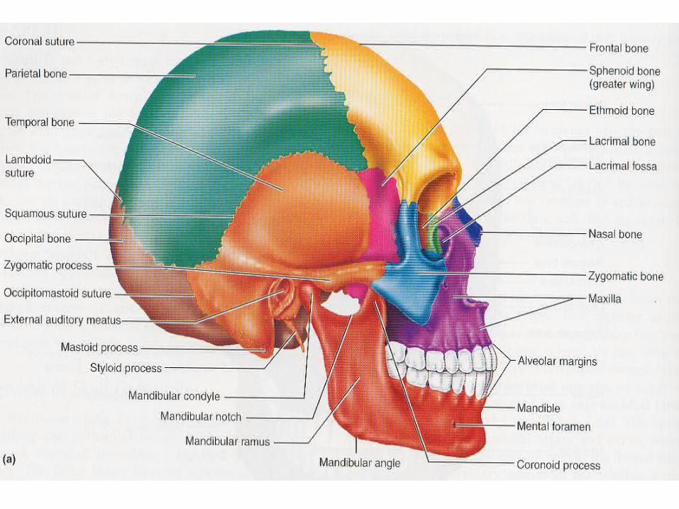
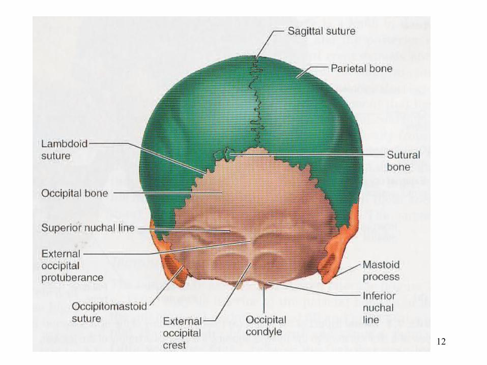
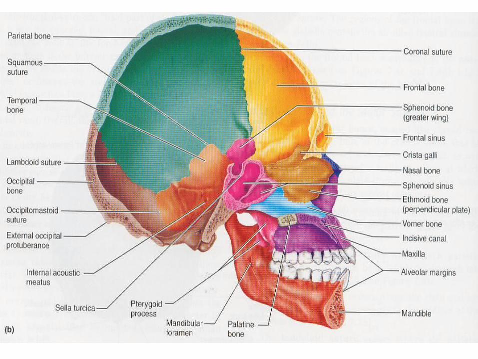
• The skull=cranium is formed by:– Cranial bonesneurocranium– Facial bonessplanchnocranium
• The function of cranial bones are: – enclose and protect the brain – provide attachment sites for some head
and neck muscles
8
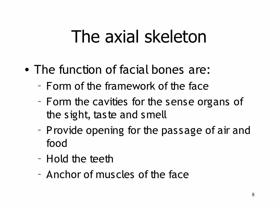
The axial skeleton
• The function of facial bones are:– Form of the framework of the face– Form the cavities for the sense organs of
the sight, taste and smell– Provide opening for the passage of air and
food– Hold the teeth– Anchor of muscles of the face
9
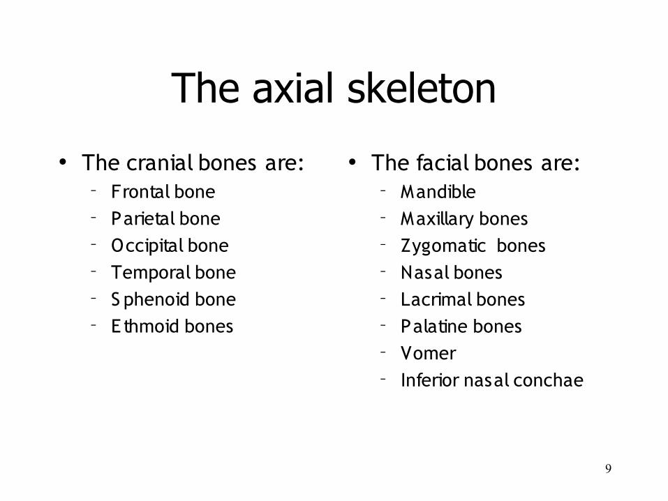
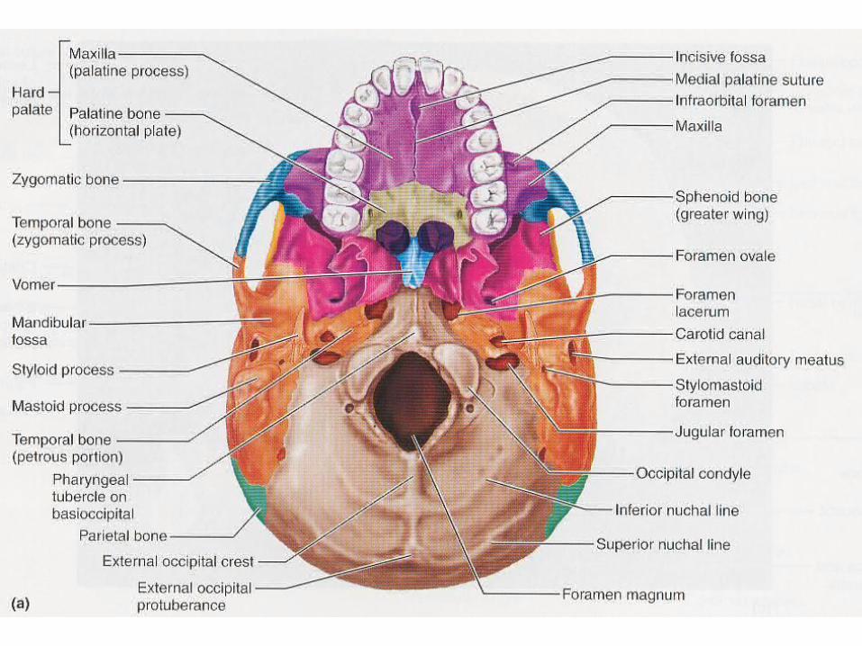
The axial skeleton
• The cranial bones are:– Frontal bone– Parietal bone– Occipital bone– Temporal bone– S phenoid bone– Ethmoid bones
• The facial bones are:– Mandible– Maxillary bones– Zygomatic bones– Nasal bones– Lacrimal bones– Palatine bones– Vomer– Inferior nasal conchae
10
11
12
13

14
15
16
The vertebral column
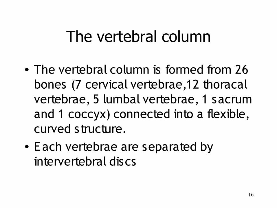
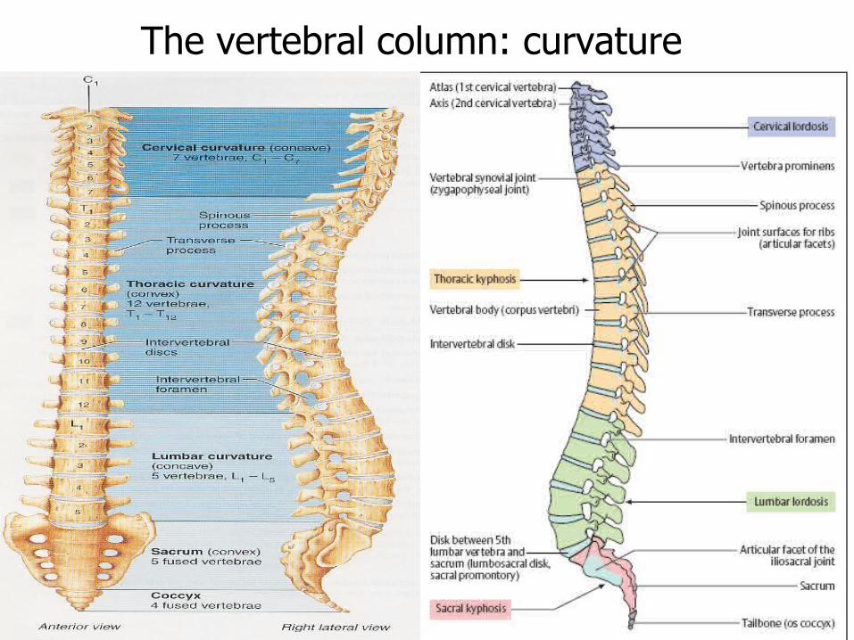
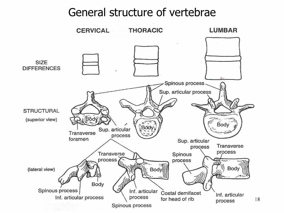
• The vertebral column is formed from 26 bones (7 cervical vertebrae,12 thoracal vertebrae, 5 lumbal vertebrae, 1 sacrum and 1 coccyx) connected into a flexible, curved structure.
• Each vertebrae are separated by intervertebral discs
17
The vertebral column: curvature
18
General structure of vertebrae
19
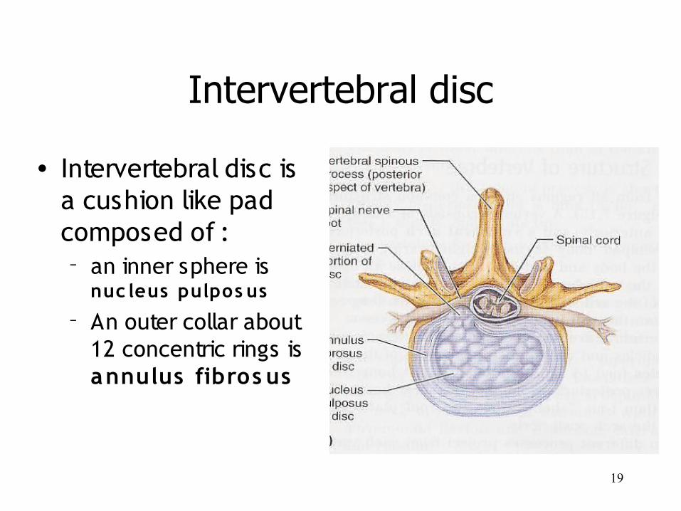
Intervertebral disc
• Intervertebral disc is a cushion like pad composed of :– an inner sphere is
nuc leus pulpos us
– An outer collar about 12 concentric rings is annulus fibros us
20
The appendicular skeleton
• The appendicular skeleton consists of: limb bones and their girdle– The upper limb bones & the pectoral girdle – The lower limb bones & the pelvic girdle
21
The appendicular skeleton
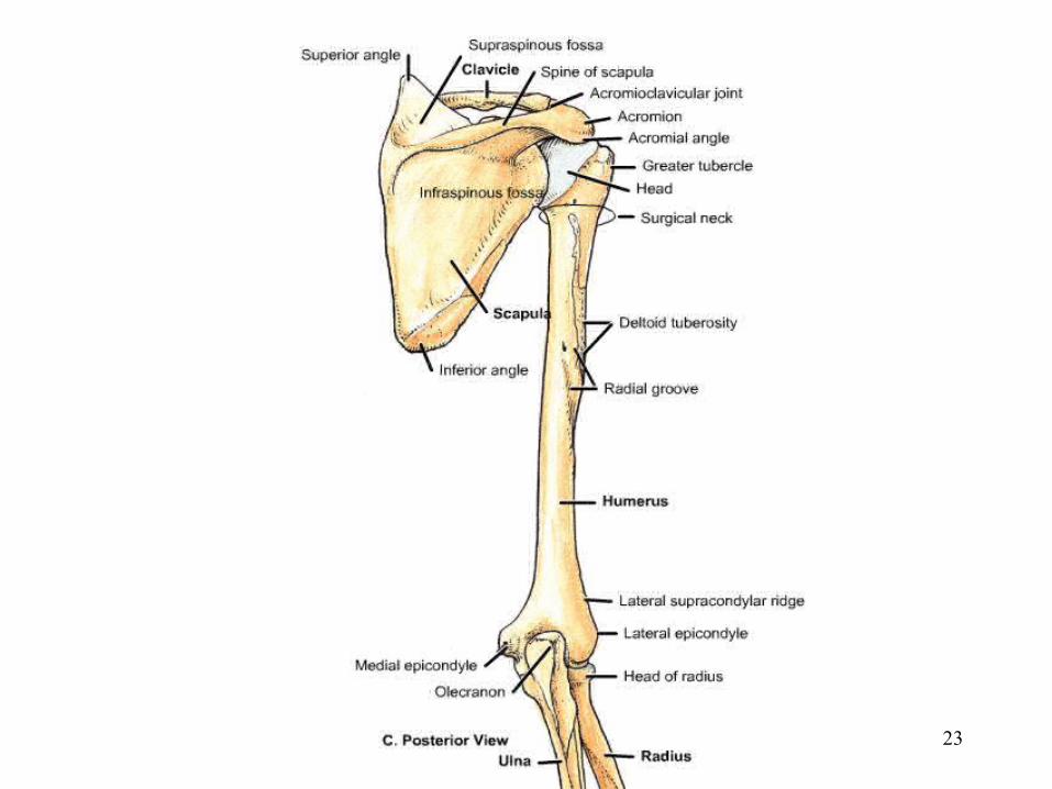
The upper limb:• The pectoral girdle consists of a clavicle
and scapula the paired pectoral girdle and their associated muscles shoulder
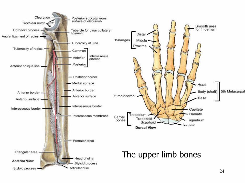
• The Upper limb bones consists of:• Arm humerus• Fore arm radius and ulna• Handcarpal, metacarpal and phalanges

22
23
24
The upper limb bones
25
The appendicular skeleton
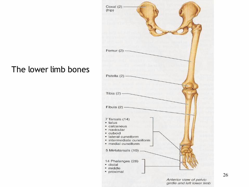
The lower limb:• The pelvic girdle consists of paired hip
bones or coxae (ilium, ischium and pubis bones)
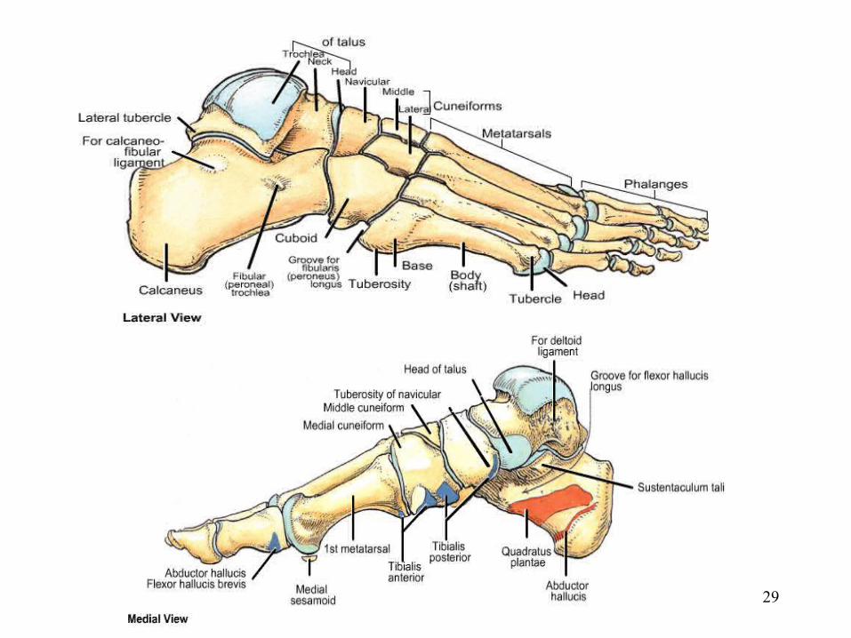
• The lower limb consists of:– Thigh femur– Leg tibia and fibula– Foot tarsal, metatarsal and phalanges
26
The lower limb bones
27
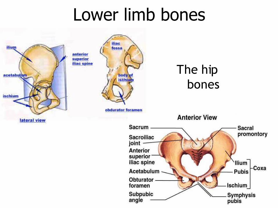
Lower limb bones
The hip bones
28
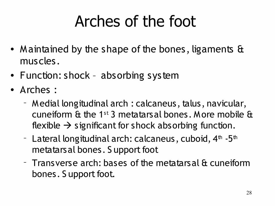
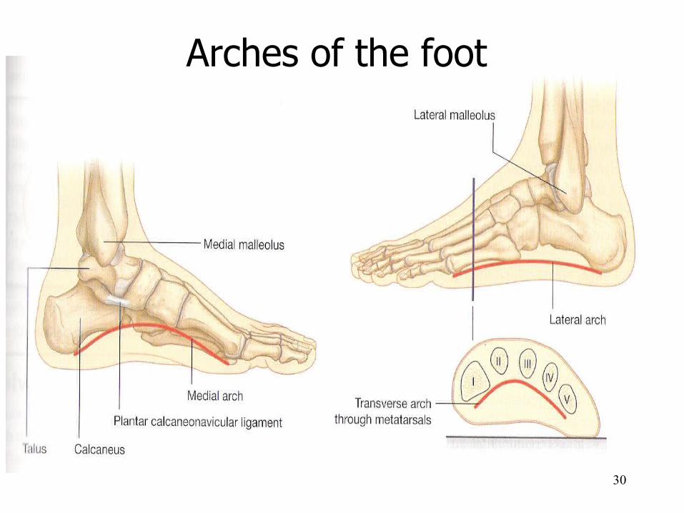
• Maintained by the shape of the bones, ligaments & muscles.
• Function: shock – absorbing system• Arches :
– Medial longitudinal arch : calcaneus, talus, navicular, cuneiform & the 1st 3 metatarsal bones. More mobile & flexible significant for shock absorbing function.
– Lateral longitudinal arch: calcaneus, cuboid, 4th -5th metatarsal bones. S upport foot
– Transverse arch: bases of the metatarsal & cuneiform bones. S upport foot.
Arches of the foot
29
30
Arches of the foot
31
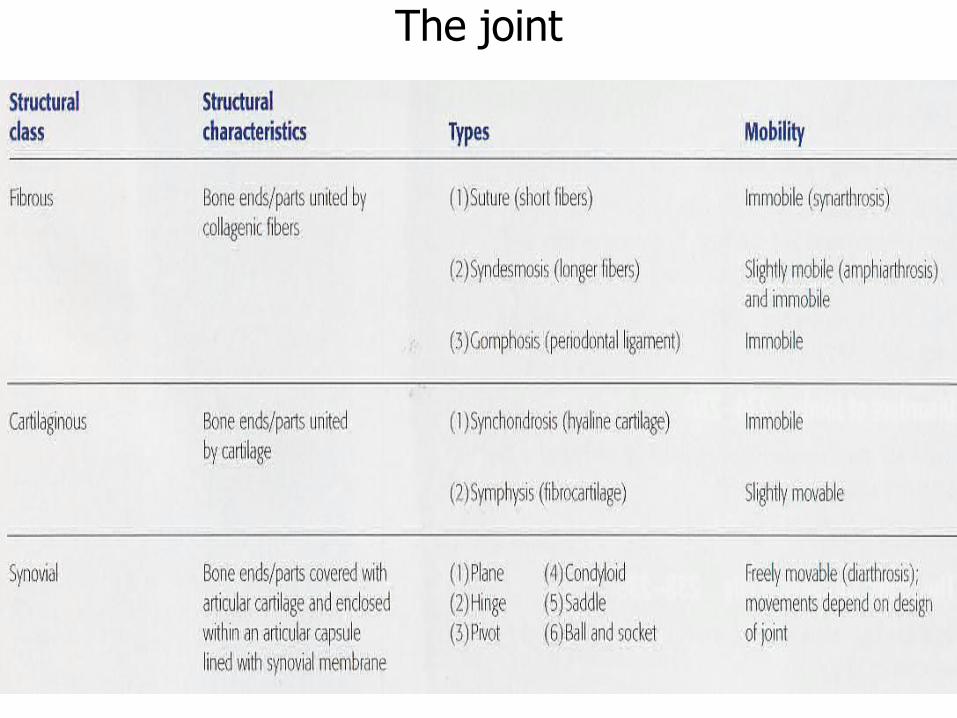
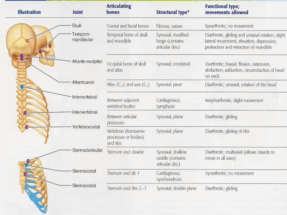
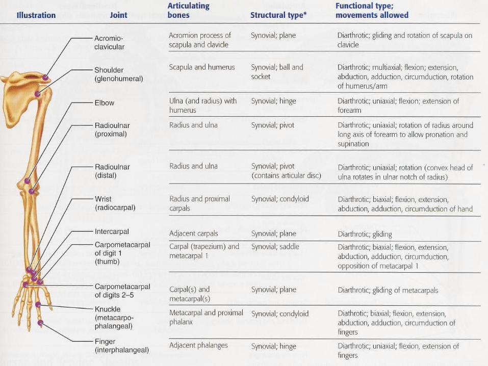
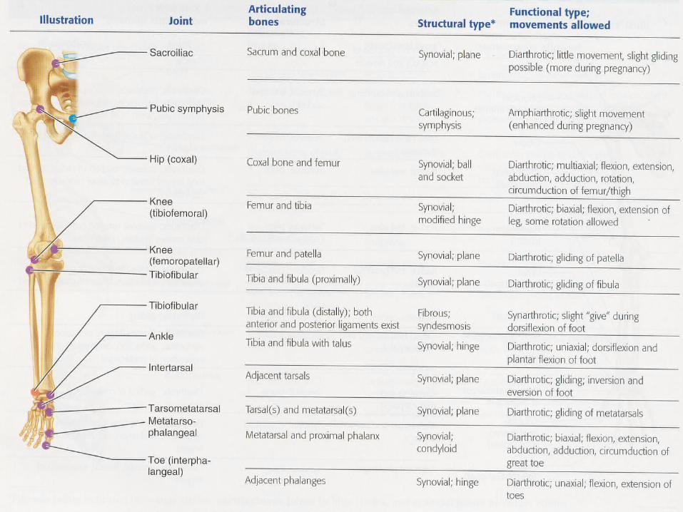
The joint
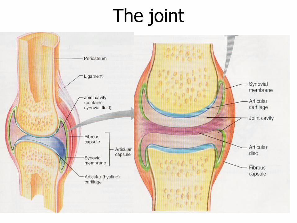
32
The joint
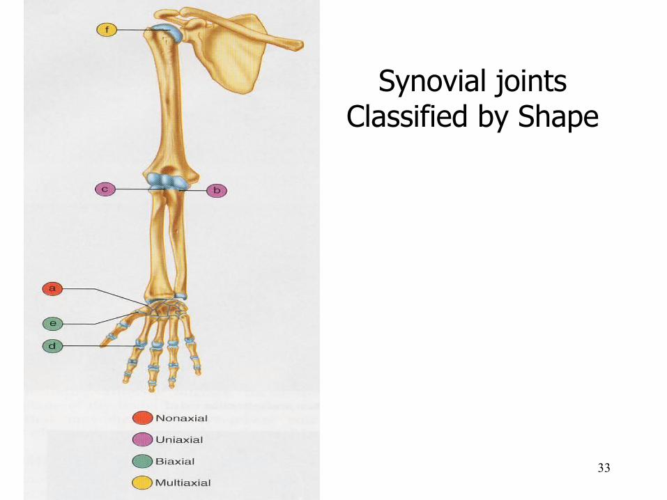
33
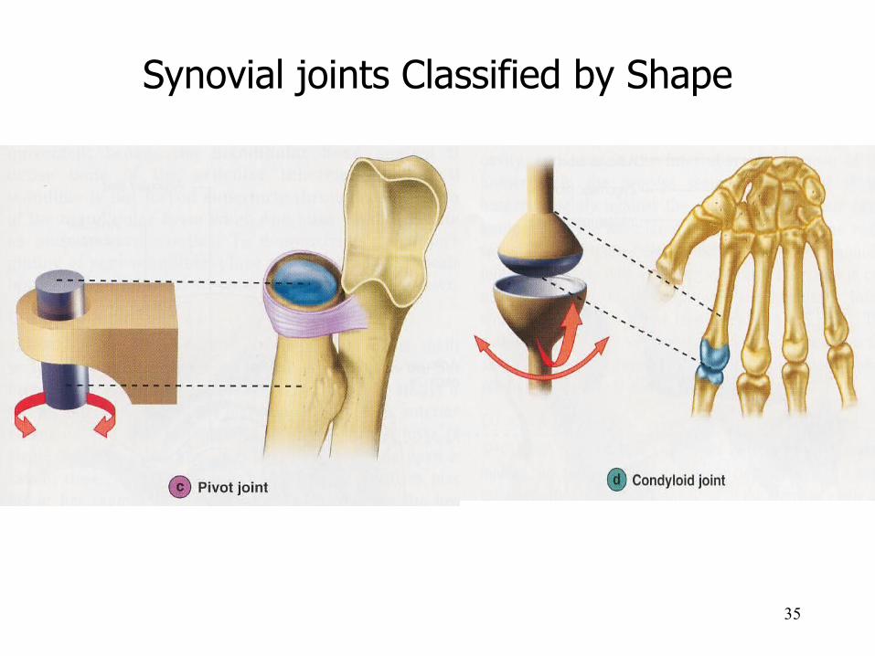
Synovial joints Classified by Shape
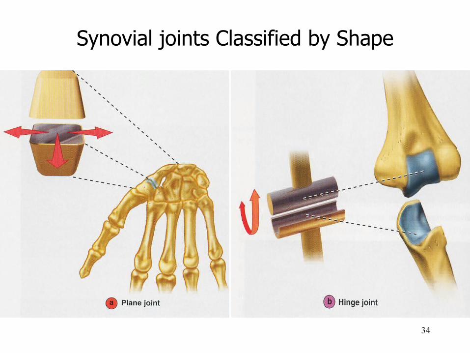
34
Synovial joints Classified by Shape
35
Synovial joints Classified by Shape
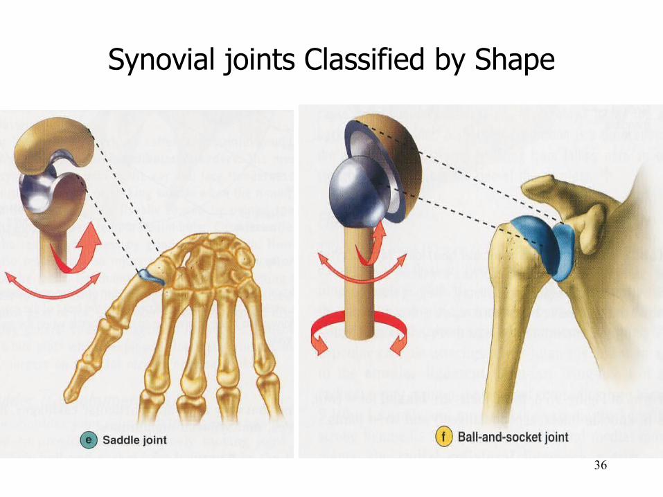
36
Synovial joints Classified by Shape
37
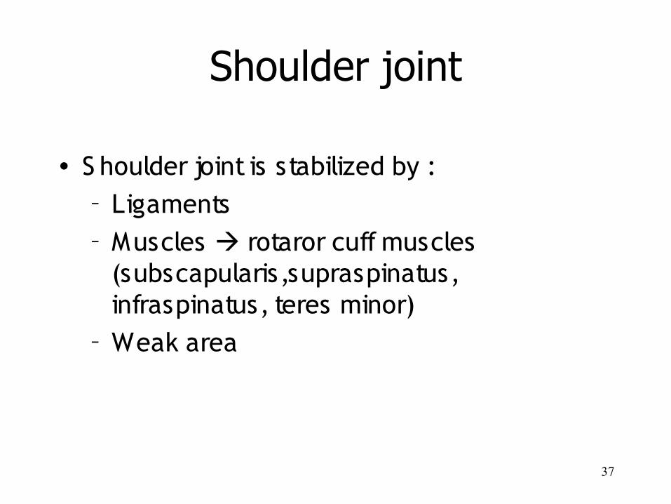
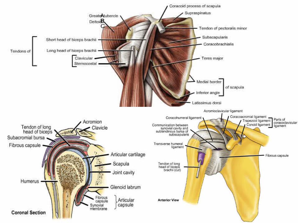
Shoulder joint
• S houlder joint is stabilized by :– Ligaments– Muscles rotaror cuff muscles
(subscapularis,supraspinatus, infraspinatus, teres minor)
– Weak area
38
39
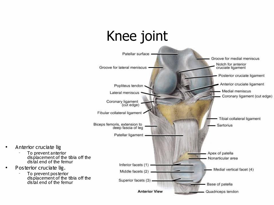
Knee joint
• Anterior cruciate lig– To prevent anterior
displacement of the tibia off the distal end of the femur
• Posterior cruciate lig.– To prevent posterior
displacement of the tibia off the distal end of the femur
40
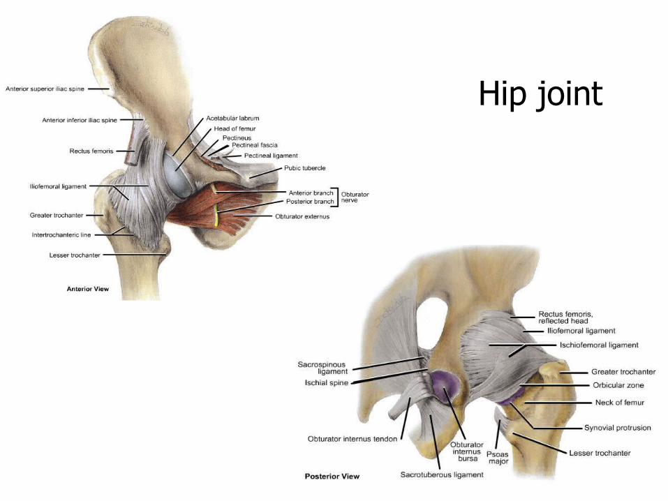
Hip joint
41
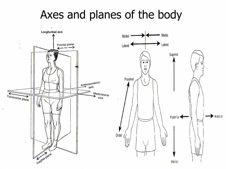
Axes and planes of the body
42
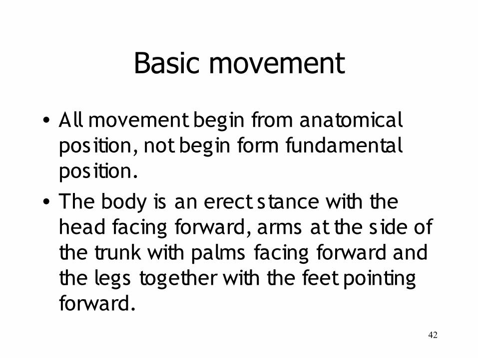
Basic movement
• All movement begin from anatomical position, not begin form fundamental position.
• The body is an erect stance with the head facing forward, arms at the side of the trunk with palms facing forward and the legs together with the feet pointing forward.
43
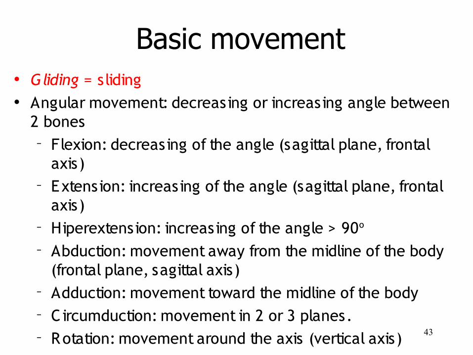
• Gliding = sliding• Angular movement: decreasing or increasing angle between
2 bones– Flexion: decreasing of the angle (sagittal plane, frontal
axis)– Extension: increasing of the angle (sagittal plane, frontal
axis)– Hiperextension: increasing of the angle > 90o
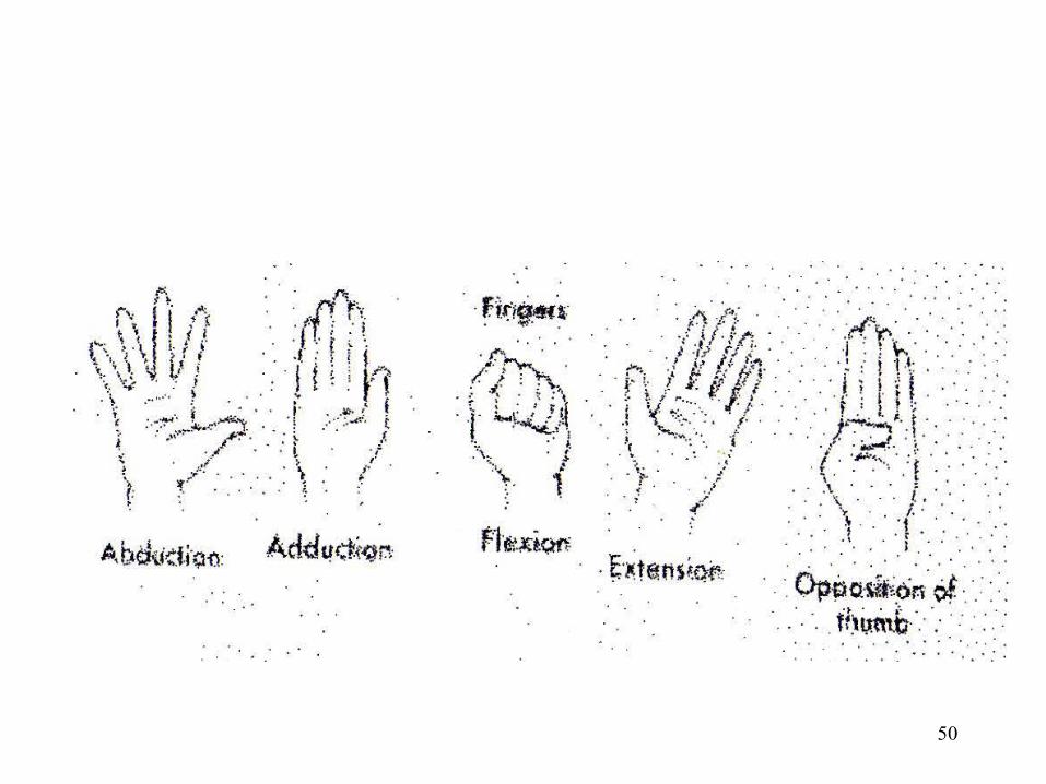
– Abduction: movement away from the midline of the body (frontal plane, sagittal axis)
– Adduction: movement toward the midline of the body– C ircumduction: movement in 2 or 3 planes.– Rotation: movement around the axis (vertical axis)
Basic movement
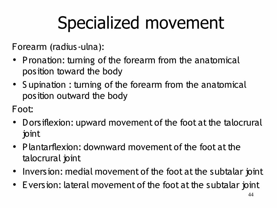
44
Forearm (radius-ulna): • Pronation: turning of the forearm from the anatomical
position toward the body• S upination : turning of the forearm from the anatomical
position outward the bodyFoot: • Dorsiflexion: upward movement of the foot at the talocrural
joint• Plantarflexion: downward movement of the foot at the
talocrural joint• Inversion: medial movement of the foot at the subtalar joint• Eversion: lateral movement of the foot at the subtalar joint
Specialized movement
45
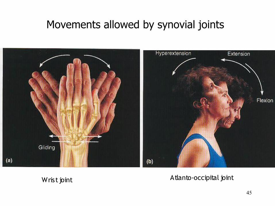
Movements allowed by synovial joints
Wrist joint Atlanto-occipital joint
46
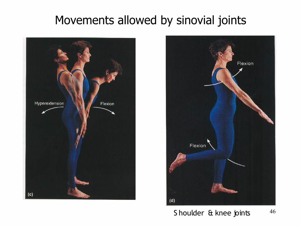
Movements allowed by sinovial joints
S houlder & knee joints
47
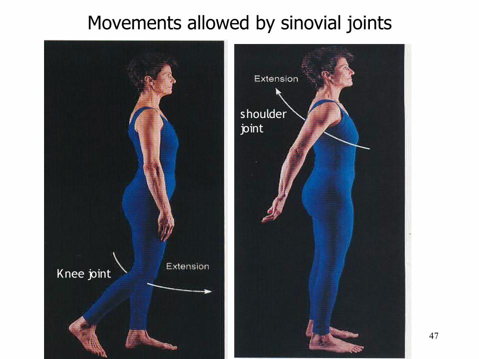
Movements allowed by sinovial joints
Knee joint
shoulder joint
48
Movements allowed by sinovial joints
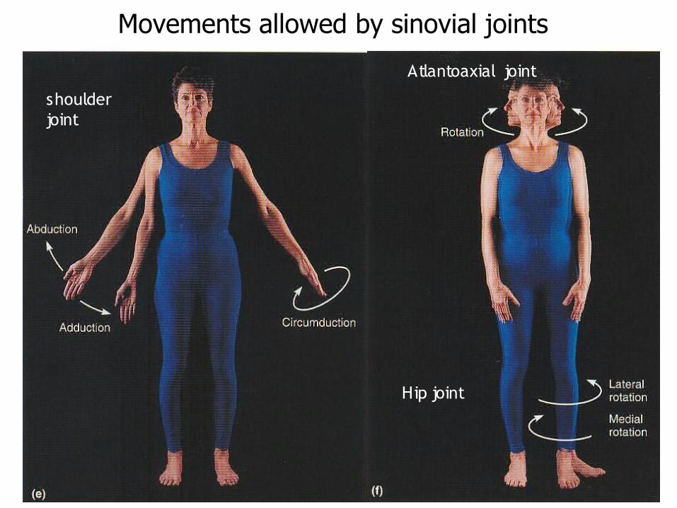
shoulder joint
Hip joint
Atlantoaxial joint
49
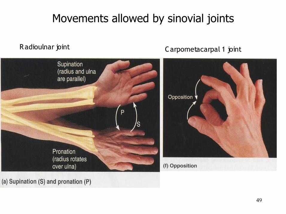
Movements allowed by sinovial joints
Radioulnar joint Carpometacarpal 1 joint
50

51
Movements allowed by sinovial joints
Talocrural joint tibiofibular & tibiotalar joints
S ubtalar joint (talus-calcaneus)
52
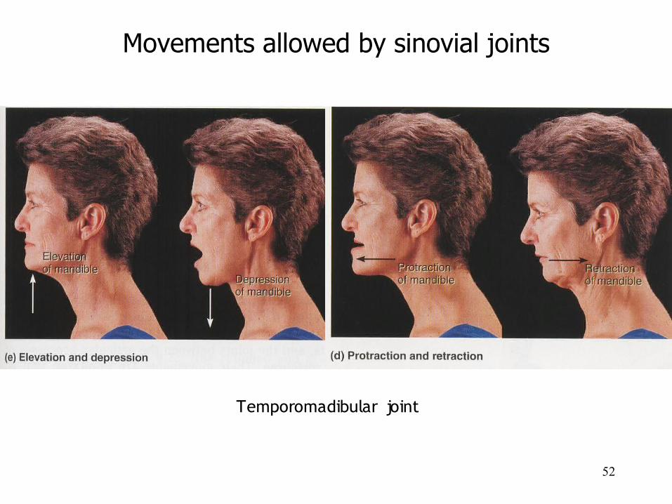
Movements allowed by sinovial joints
Temporomadibular joint
53
54
55
56
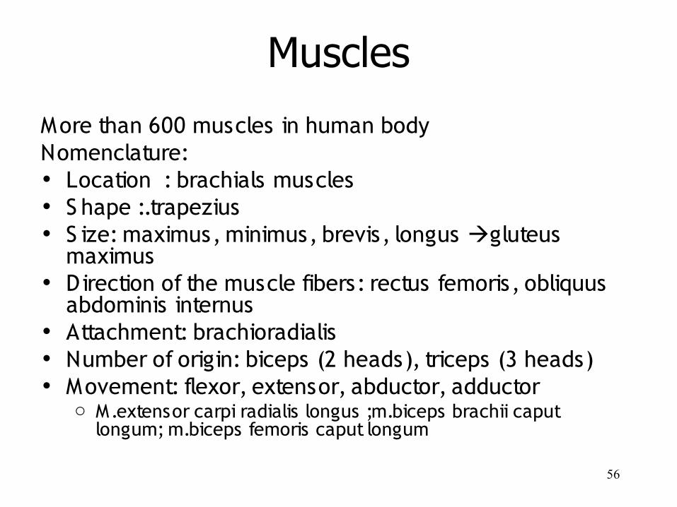
Muscles
More than 600 muscles in human bodyNomenclature:• Location : brachials muscles• S hape :.trapezius• S ize: maximus, minimus, brevis, longus gluteus
maximus• Direction of the muscle fibers: rectus femoris, obliquus
abdominis internus• Attachment: brachioradialis• Number of origin: biceps (2 heads), triceps (3 heads)• Movement: flexor, extensor, abductor, adductor
o M.extensor carpi radialis longus ;m.biceps brachii caput longum; m.biceps femoris caput longum
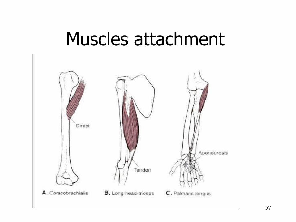
57
Muscles attachment
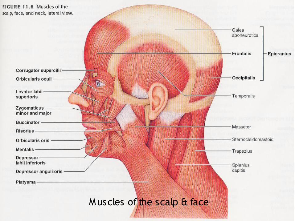
58Muscles of the scalp & face
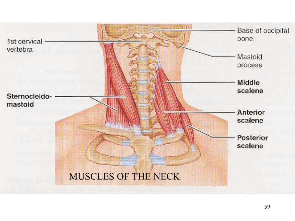
59
MUSCLES OF THE NECK
60
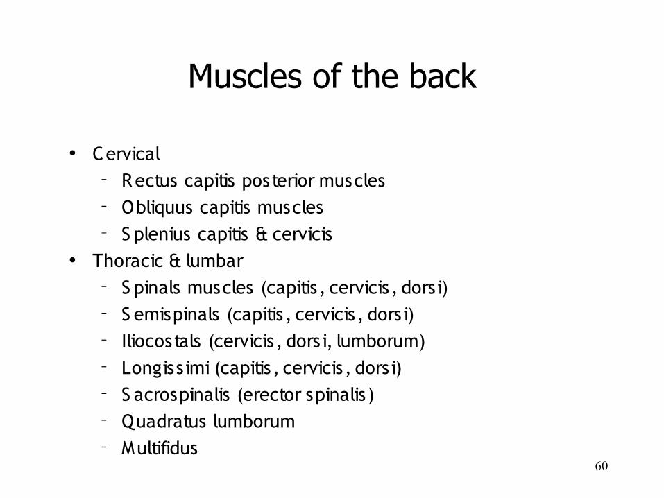
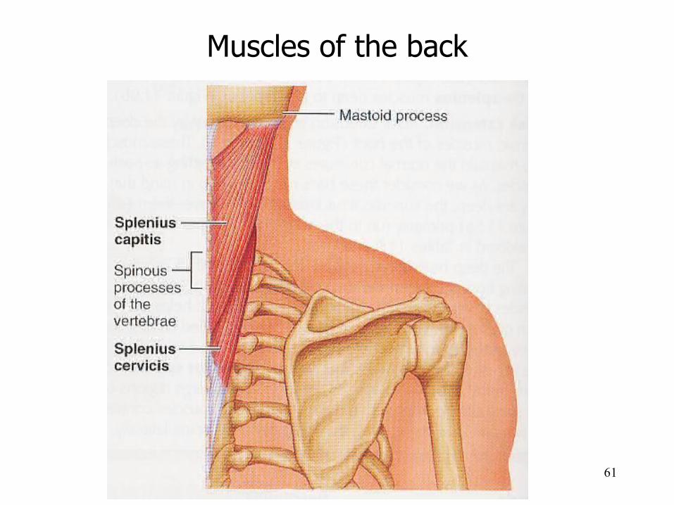
Muscles of the back
• Cervical – Rectus capitis posterior muscles– Obliquus capitis muscles– S plenius capitis & cervicis
• Thoracic & lumbar– S pinals muscles (capitis, cervicis, dorsi)– S emispinals (capitis, cervicis, dorsi)– Iliocostals (cervicis, dorsi, lumborum)– Longissimi (capitis, cervicis, dorsi)– S acrospinalis (erector spinalis)– Quadratus lumborum– Multifidus
61
Muscles of the back
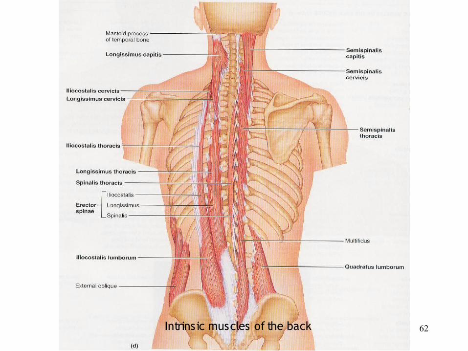
62Intrinsic muscles of the back
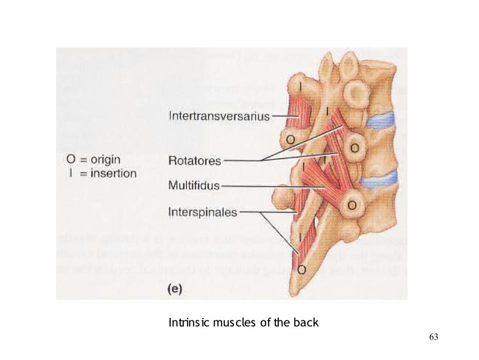
63Intrinsic muscles of the back
64
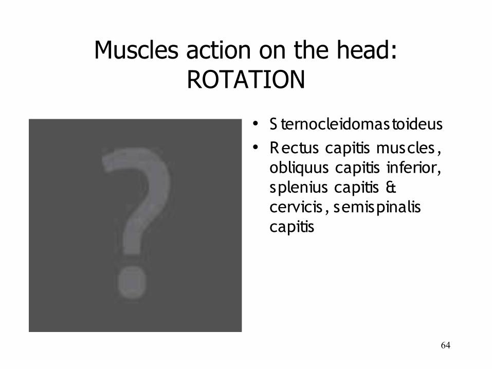
Muscles action on the head:ROTATION
• S ternocleidomastoideus• Rectus capitis muscles,
obliquus capitis inferior, splenius capitis & cervicis, semispinalis capitis
65
Muscles action on the head:LATERAL FLEXION
• S ternocleidomastoideus, rectus capitis anterior muscles, scalenus anteror & posterior• Rectus capitis posterior muscles, splenius capitis & cervicis
66
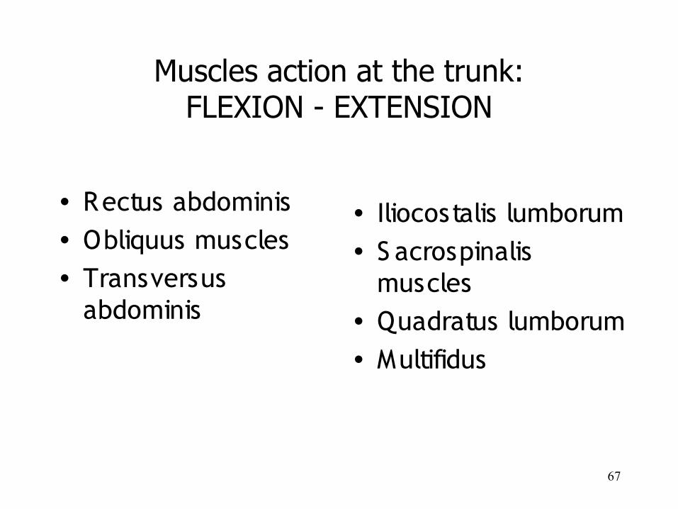
Muscles action at the trunk:LATERAL FLEXION
67
• Rectus abdominis• Obliquus muscles• Transversus
abdominis
• Iliocostalis lumborum• S acrospinalis
muscles• Quadratus lumborum• Multifidus
Muscles action at the trunk:FLEXION - EXTENSION
68
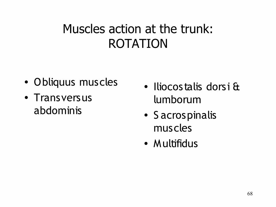
• Obliquus muscles• Transversus
abdominis
• Iliocostalis dorsi & lumborum
• S acrospinalis muscles
• Multifidus
Muscles action at the trunk:ROTATION
69
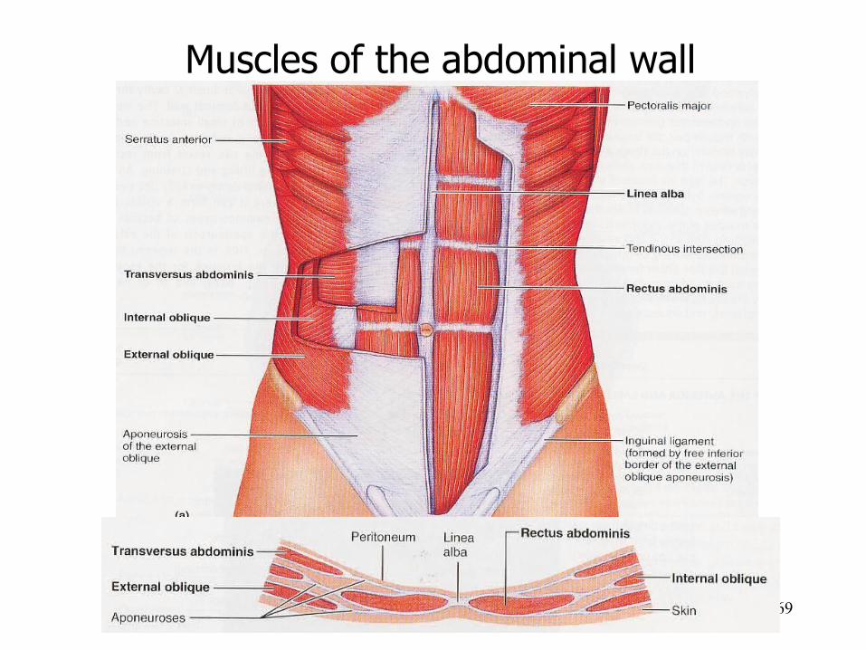
Muscles of the abdominal wall
70
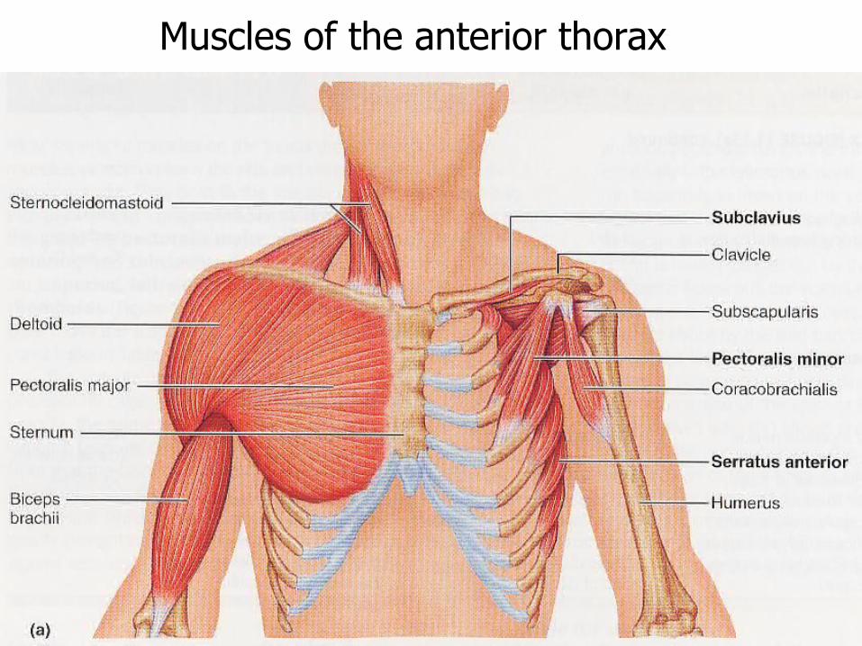
Muscles of the anterior thorax
71
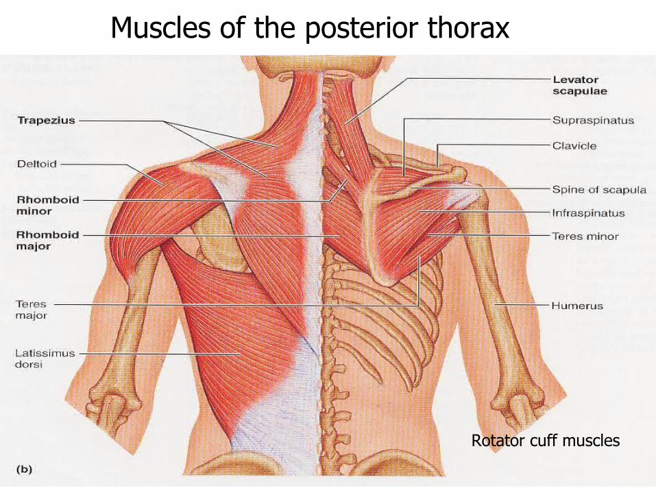
Muscles of the posterior thorax
Rotator cuff muscles
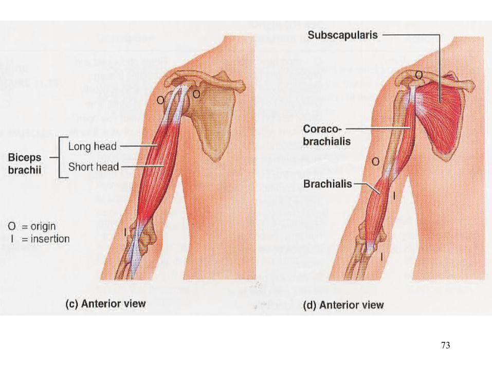
72
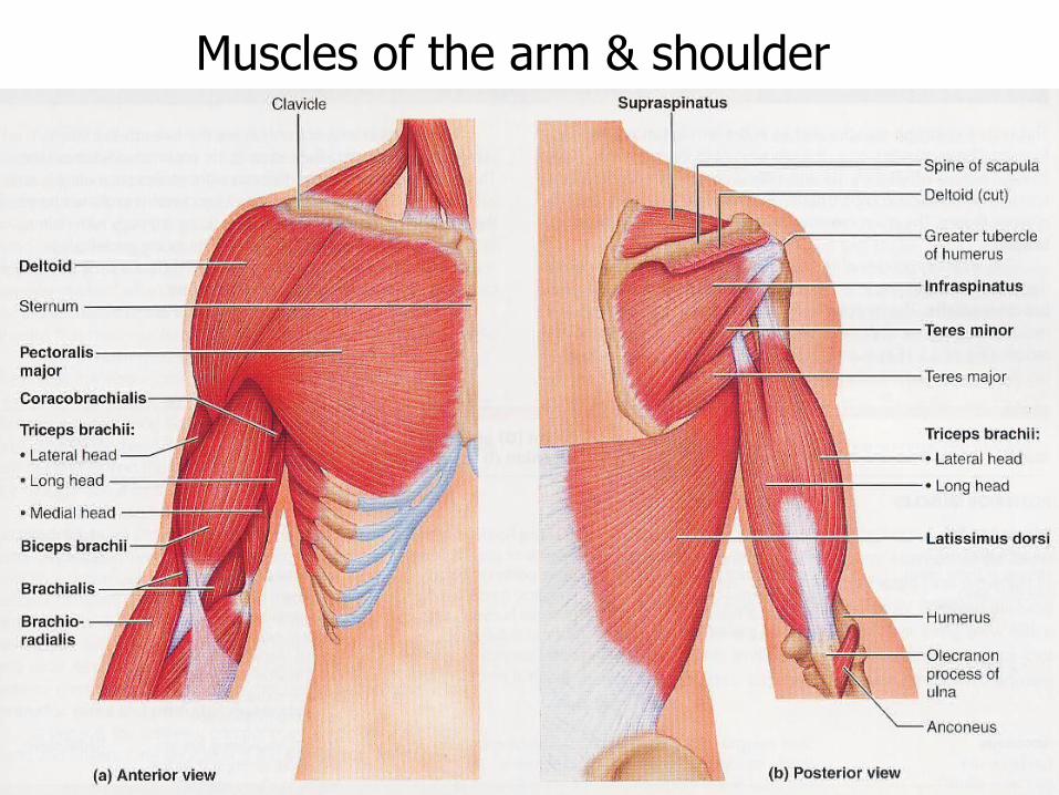
Muscles of the arm & shoulder
73
74
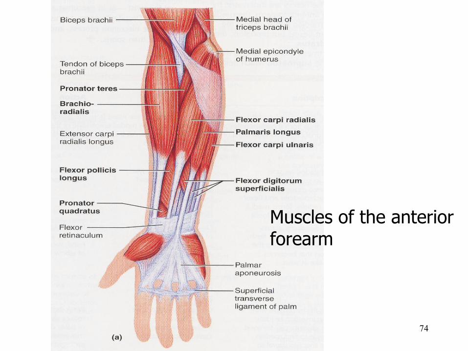
Muscles of the anterior forearm
75
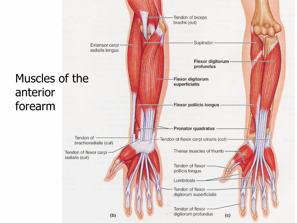
Muscles of the anterior forearm
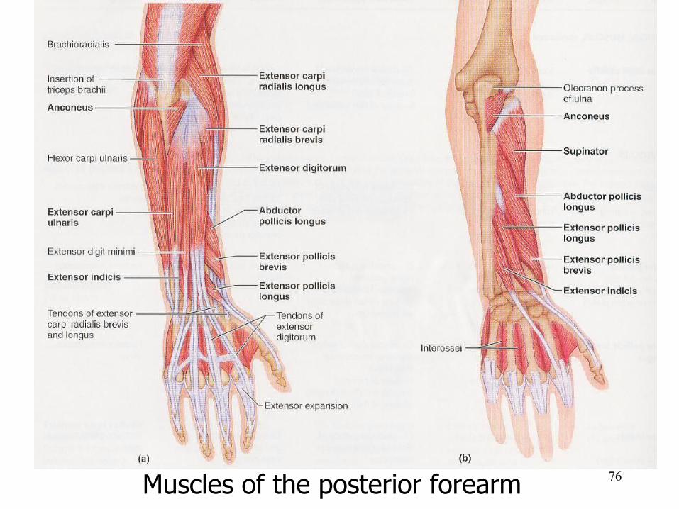
76Muscles of the posterior forearm
77
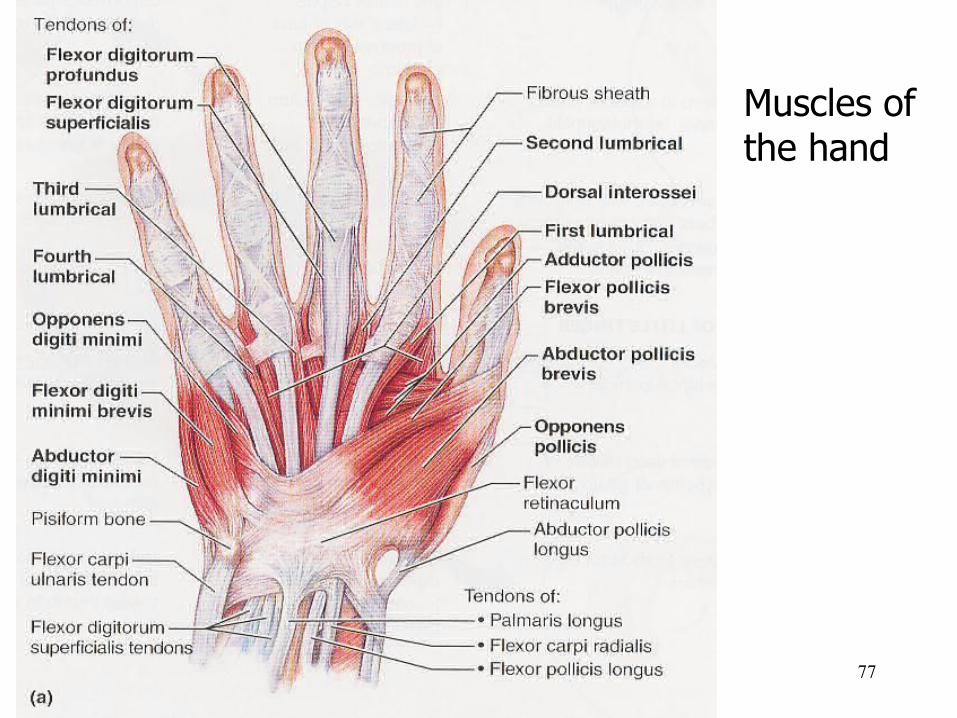
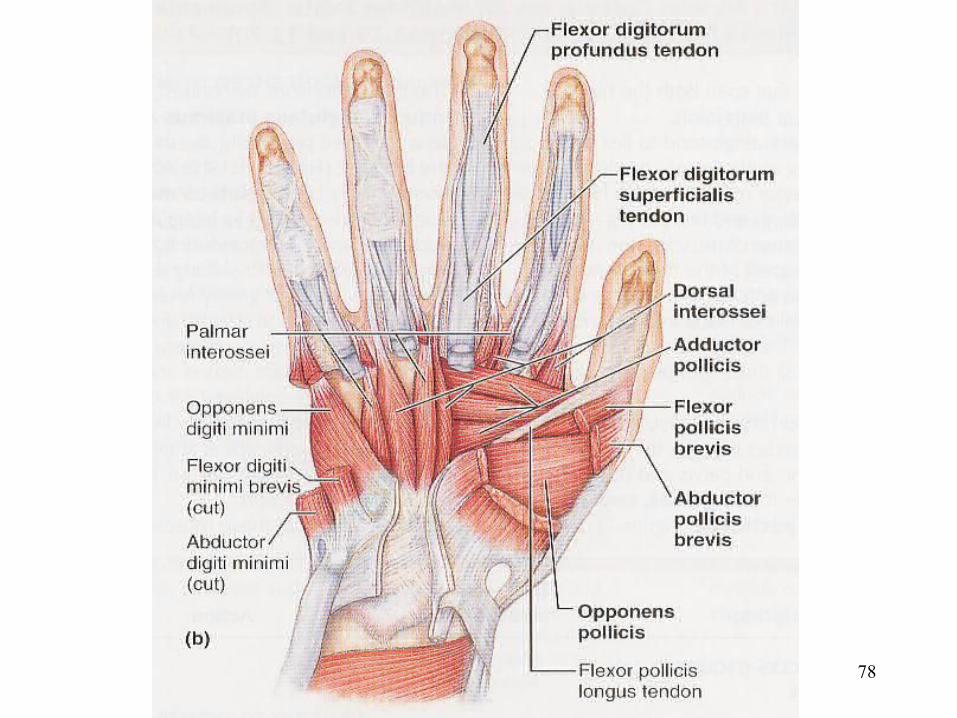
Muscles of the hand
78
79
80
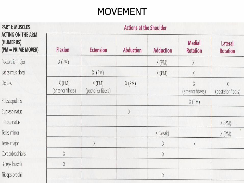
MOVEMENT
81
Muscles action on the scapula:ELEVATION - DEPRESION
• trapezius (upper part), rhomboids, levator scapulae
82
Muscles action on the shoulder joint FLEXION - EXTENSION
• Pectoralis major• Latissimus dorsi
83
Muscles action on the shoulder joint ABDUCTION - ADDUCTION
• Deltoid• Pectoralis major & latissimus dorsi
84
Muscles action on the shoulder joint MEDIAL ROTATION – LATERAL ROTATION
• S ubscapular• Infraspinatus & teres minor
85
86
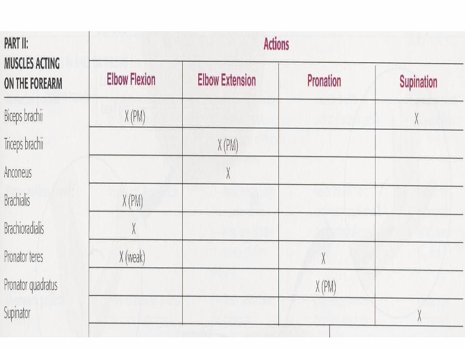
Muscles action on the elbow joint FLEXION - EXTENSION
• Biceps bracii & brachialis• Triceps brachii
87
Muscles action on the elbow joint PRONATION - SUPINATION
• Pronator quadratus• S upinator
88
89
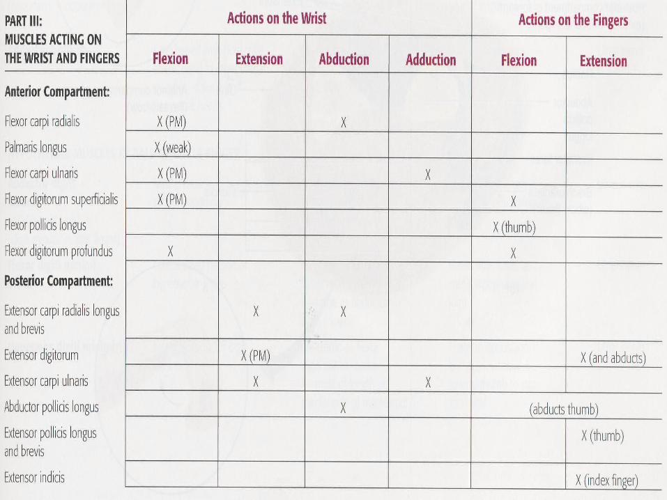
Muscles action on the wrist joint FLEXION – EXTENSION
• Flexor carpi radialis, flexor carpi ulnaris & flexor digitorum superficialis• Extensor digitorum
90
Muscles action on the wrist joint ABDUCTION - ADDUCTION
• Flexor carpi radialis, extensor carpi radialis longus & brevis, abductor pollicis longus• Flexor carpi ulnaris & extensor carpi ulnaris
equal
91
Muscles action on the finger: FLEXION – EXTENSION
• Flexor digitorum superficialis & flexor digitorum profundus; flexor pollicis longus & brevis (thumb)• Extensor digitorum; extensor pollicis longus & brevis (thumb);extensor indicis (index)
92
Muscles action on the finger:FLEXION – EXTENSION
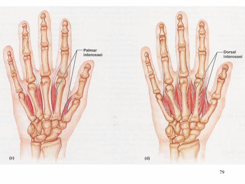
• Dorsal & palmar interossei• Lumbricals
93
Muscles action on the finger:ABDUCTION – ADDUCTION
• Dorsal interossei• Palmar interossei
94
Mucles action on the thumb:ADDUCTION - ABDUCTION
• Adductor pollicis• Abductor pollicis longus & brevis
95
Mucles action on the thumb: OPPOSITION
• Opponens pollicis• Opponens digiti quinti (minimi)
96
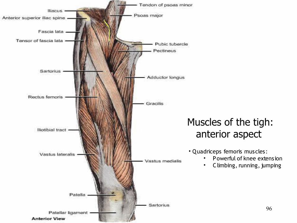
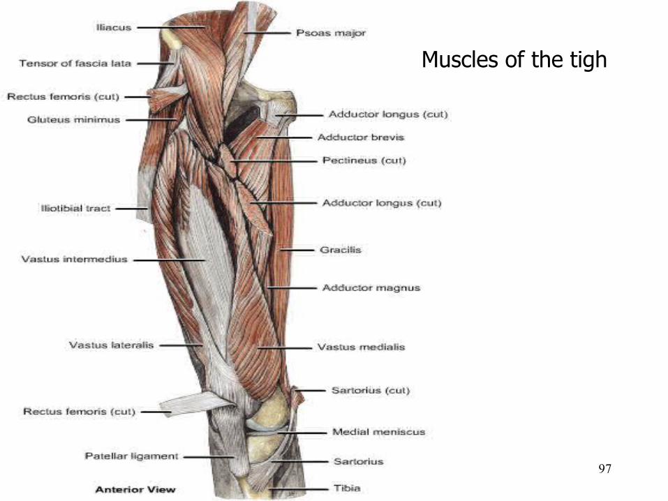
Muscles of the tigh: anterior aspect
• Quadriceps femoris muscles:• Powerful of knee extension• Climbing, running, jumping
97
Muscles of the tigh
98
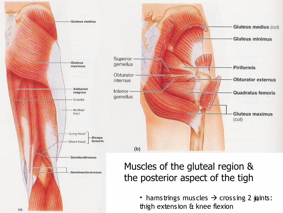
Muscles of the gluteal region & the posterior aspect of the tigh
• hamstrings muscles crossing 2 joints: thigh extension & knee flexion
99
100
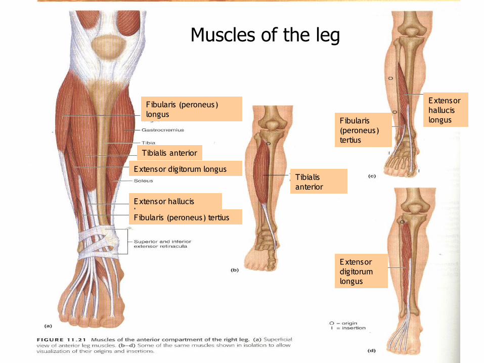
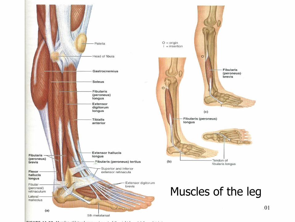
Fibularis (peroneus) longus
Tibialis anterior
Extensor digitorum longus
Extensor hallucis longusFibularis (peroneus) tertius
Tibialis anterior
Extensor hallucis longusFibularis
(peroneus) tertius
Extensor digitorum longus
Muscles of the leg
101
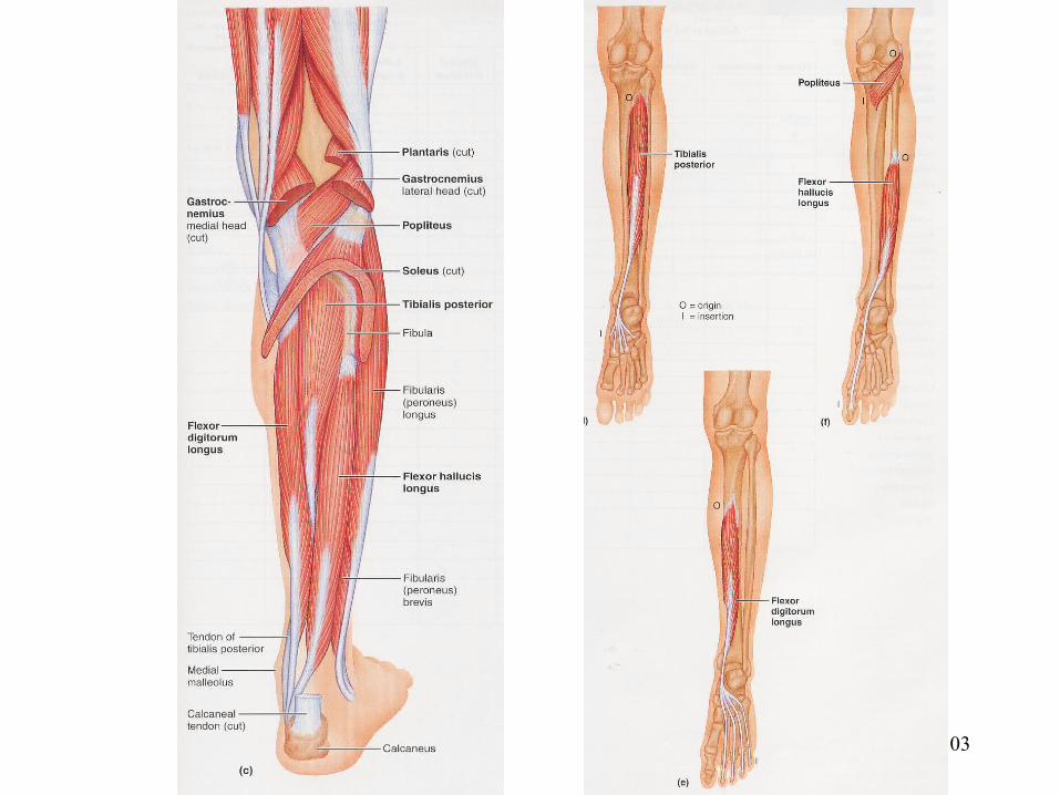
Muscles of the leg
102
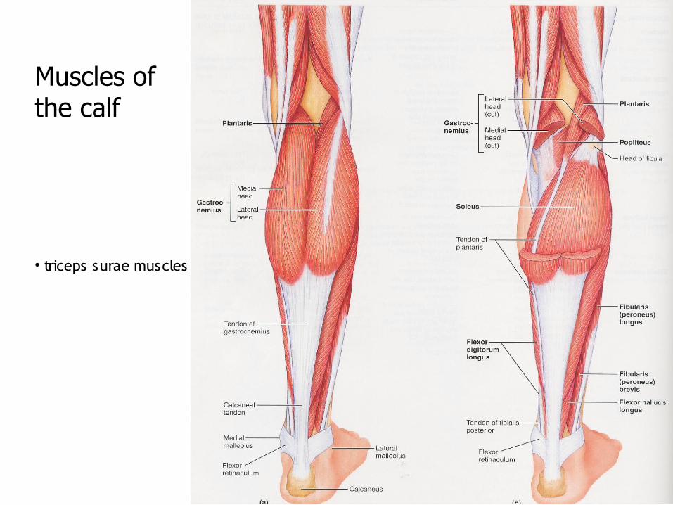
• triceps surae muscles
Muscles of the calf
103
104
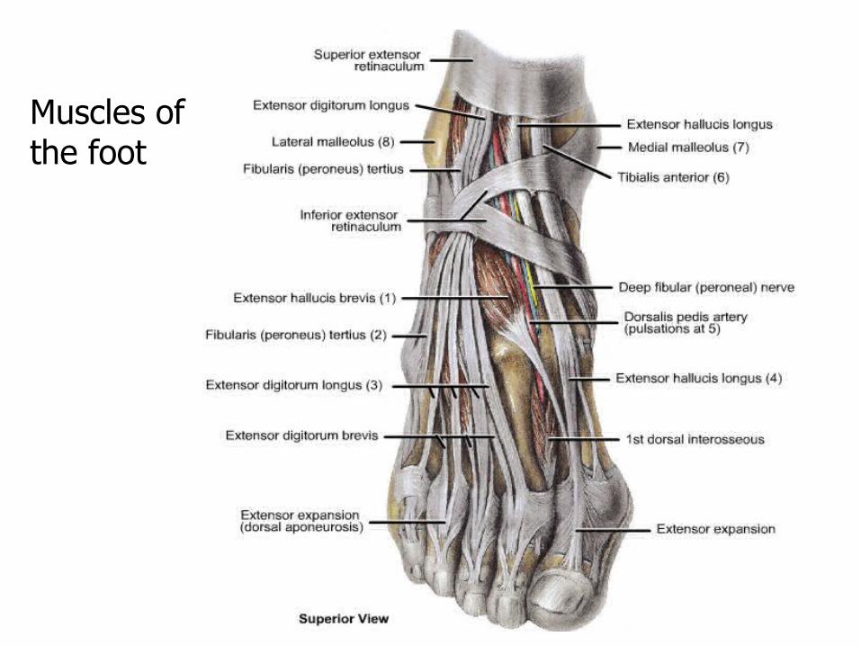
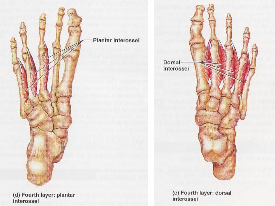
Muscles of the foot
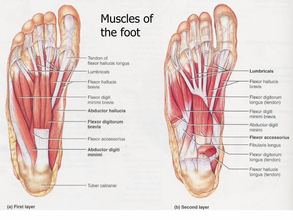
105
Muscles of the foot

106
Muscles of the foot
107
108
109
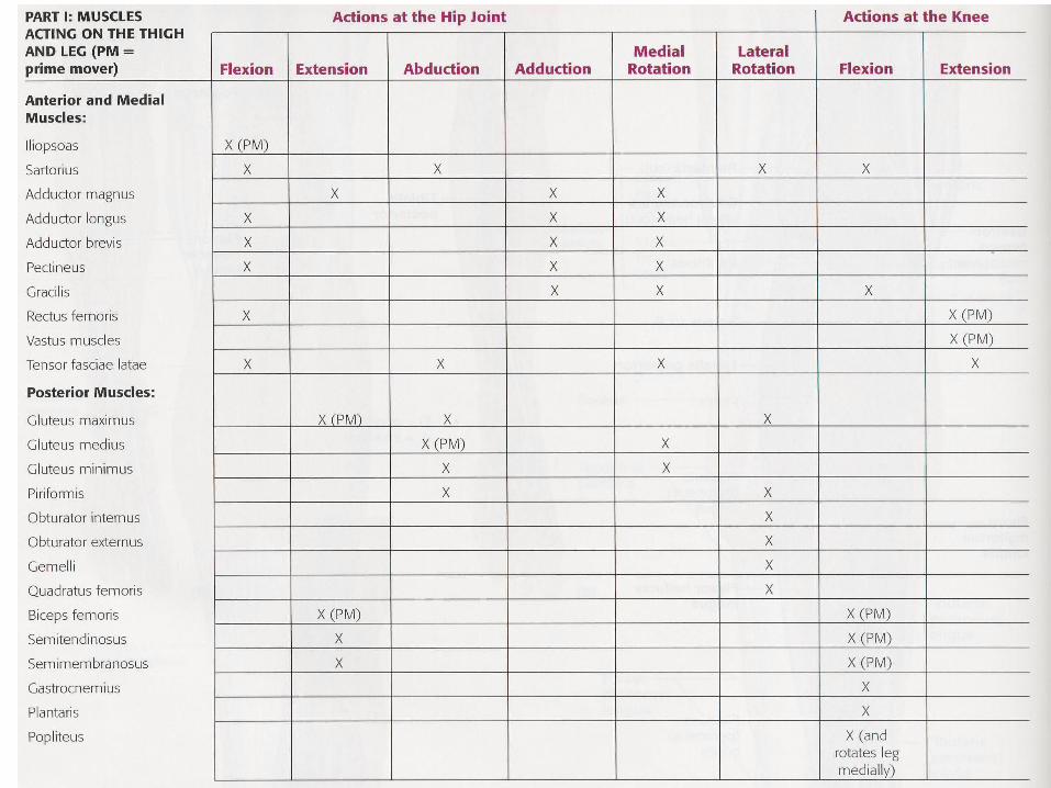
Muscles action at the hip jointFLEXION - EXTENSION
• Iliopsoas• Gluteus maximus & biceps femoris
110
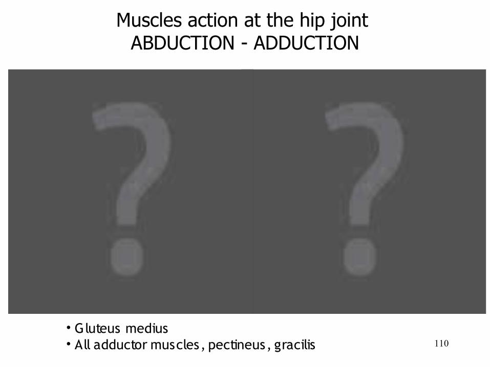
Muscles action at the hip joint ABDUCTION - ADDUCTION
• Gluteus medius• All adductor muscles, pectineus, gracilis
111
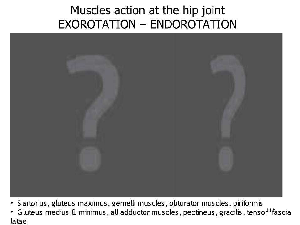
Muscles action at the hip jointEXOROTATION – ENDOROTATION
• S artorius, gluteus maximus, gemelli muscles, obturator muscles, piriformis • Gluteus medius & minimus, all adductor muscles, pectineus, gracilis, tensor fascia latae
112
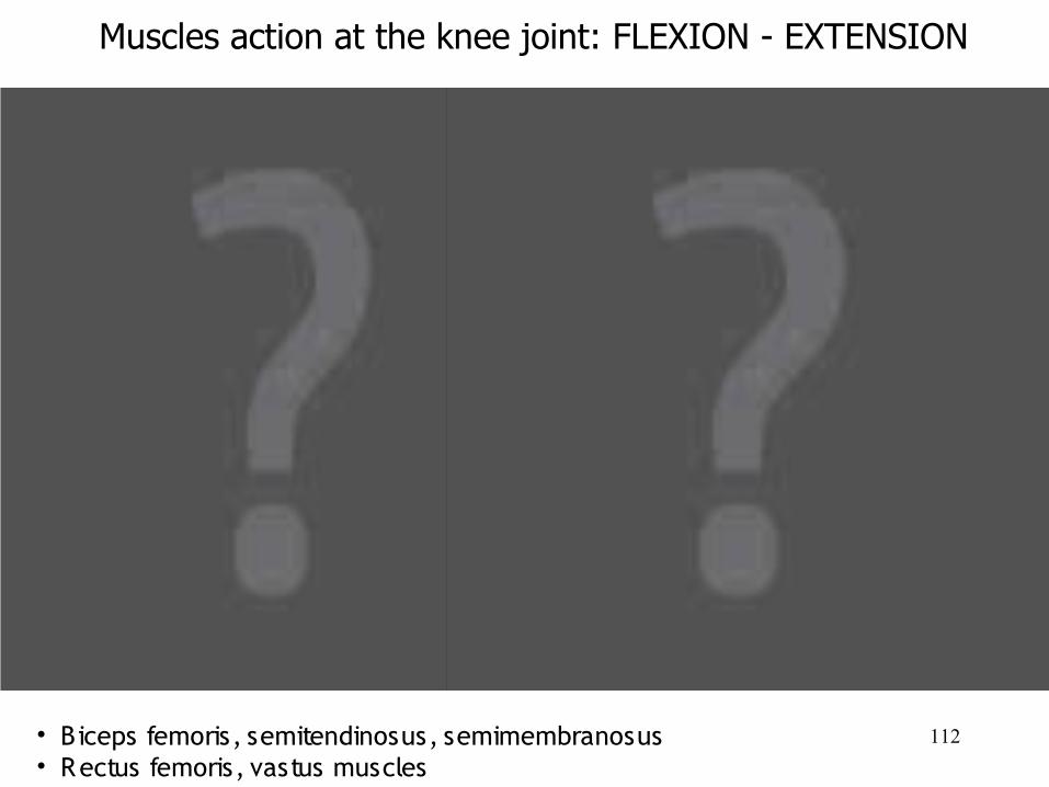
Muscles action at the knee joint: FLEXION - EXTENSION
• Biceps femoris, semitendinosus, semimembranosus• Rectus femoris, vastus muscles
113
114
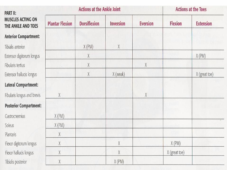
Muscles action at the ankle jointPANTARFLEXION & DORSIFLEXION
• Gastrocnemius & soleus• Tibialis anterior
115
Muscles action at the ankle joint:INVERSION - EVERSION
• Tibialis posterior• Tibialis anterior
116
Muscles action at the toesFLEXION - EXTENSION
• Flexor digitorum longus• Extensor digitorum longus
117
Muscles action at the toesABDUCTION - ADDUCTION
• Dorsal interossei• Palmar interossei
118
Reference
• Behnke,R .S . Kinetic Anatomy. 2nd ed. Human Kinetics.2006.
• Dynamic Human Anatomy 2.0. • Hillman, S .K. Interactive functional anatomy.
3rd ed. Primal Picture, Ltd. 2006 (DVD)• Marieb, E .N., & J. Mallat. Human Anatomy.
3rd ed. Benjamin Cummings.2001.