Embed Size (px)
Citation preview
2/9/13 Anatomy of Orbit – ENT SCHOLAR
entscholar.com/?articles=anatomy-of-orbit 1/15
Anatomy of OrbitOtolaryngologist's perspective
February 9, 2013 · Rhinology
A careful study of anatomy of orbit is very important to an ENT surgeon because of its proximity to the
para nasal sinuses. A comprehensive knowlege of orbital and peri orbital anatomy is necessary to
understand the various disorders of this region and in its surgical mangement. Current day
otolaryngologists venture into other unchartered territories like orbit, lacrimal sac etc. Anatomical
knowledge of this area will help otolaryngologists to avoid complications during surgical procedures
involving this area. This article attempts to explore this topic from otolaryngologist’s perspective.
Introduction:
Orbit supports the eye and ensures that this organ functions in an optimal manner. It also protects this
vital structure. The shape of the orbit resembles a four sided pyramid to begin with but as one goes
posterior it becomes three sided towards the apex. The volume of the orbital cavity in an adult is
roughly about 30cc. The rim of orbit in an adult measures about 40mm horizontally and 35 mm
vertically. The medial walls of orbit are roughly parallel and are about 25 mm apart in an adult. The
lateral walls of orbit angles about 90 degrees from each other. This is actually a fixed cavity with no
scope for enlargement, hence a small increase in ocular pressure can lead to disastrous
consequences.
Osteology:
Seven bones join together to form the orbit . These include:
1. Frontal bone
2. Lacrimal bone
3. Zygoma
4. Maxilla
5. Ethmoid
6. Sphenoid
7. Palate
The orbital rim is more or less spiral with its two ends overlapping medially on either side of lacrimal
fossa. The inferior orbital rim is formed by the maxillary bone medially and zygomatic bone laterally.
The zygomatic bone forms the lateral orbital rim, while the frontal bone forms the superior orbital rim.
The superior rim is commonly indented by a small notch known as the supra orbital notch. This notch
is invariably present at the junction of medial and lateral 1/3. The supra ortbital nerve and artery pass
Abstract
Anatomy of orbit
1
Author
Professor Balasubramanian Thiagarajan Balasubramanian Thiagarajan
2/9/13 Anatomy of Orbit – ENT SCHOLAR
entscholar.com/?articles=anatomy-of-orbit 2/15
through this notch to reach the forehead.
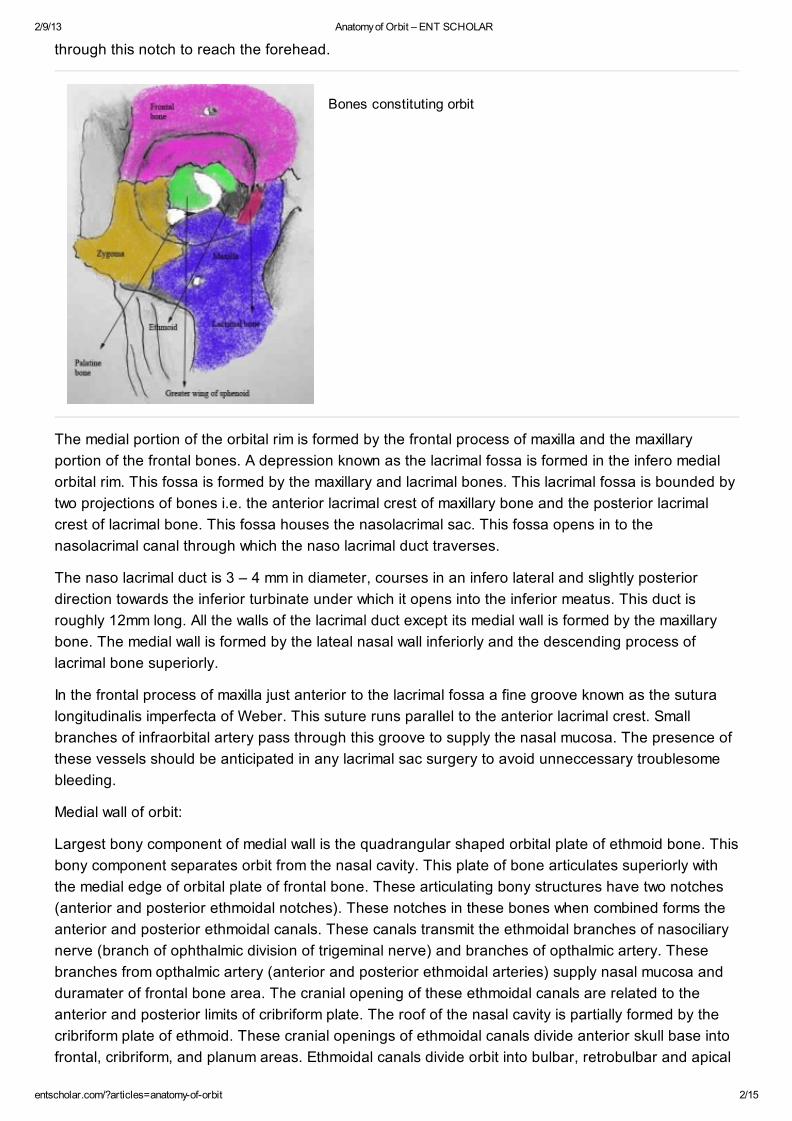
Bones constituting orbit
The medial portion of the orbital rim is formed by the frontal process of maxilla and the maxillary
portion of the frontal bones. A depression known as the lacrimal fossa is formed in the infero medial
orbital rim. This fossa is formed by the maxillary and lacrimal bones. This lacrimal fossa is bounded by
two projections of bones i.e. the anterior lacrimal crest of maxillary bone and the posterior lacrimal
crest of lacrimal bone. This fossa houses the nasolacrimal sac. This fossa opens in to the
nasolacrimal canal through which the naso lacrimal duct traverses.
The naso lacrimal duct is 3 – 4 mm in diameter, courses in an infero lateral and slightly posterior
direction towards the inferior turbinate under which it opens into the inferior meatus. This duct is
roughly 12mm long. All the walls of the lacrimal duct except its medial wall is formed by the maxillary
bone. The medial wall is formed by the lateal nasal wall inferiorly and the descending process of
lacrimal bone superiorly.
In the frontal process of maxilla just anterior to the lacrimal fossa a fine groove known as the sutura
longitudinalis imperfecta of Weber. This suture runs parallel to the anterior lacrimal crest. Small
branches of infraorbital artery pass through this groove to supply the nasal mucosa. The presence of
these vessels should be anticipated in any lacrimal sac surgery to avoid unneccessary troublesome
bleeding.
Medial wall of orbit:
Largest bony component of medial wall is the quadrangular shaped orbital plate of ethmoid bone. This
bony component separates orbit from the nasal cavity. This plate of bone articulates superiorly with
the medial edge of orbital plate of frontal bone. These articulating bony structures have two notches
(anterior and posterior ethmoidal notches). These notches in these bones when combined forms the
anterior and posterior ethmoidal canals. These canals transmit the ethmoidal branches of nasociliary
nerve (branch of ophthalmic division of trigeminal nerve) and branches of opthalmic artery. These
branches from opthalmic artery (anterior and posterior ethmoidal arteries) supply nasal mucosa and
duramater of frontal bone area. The cranial opening of these ethmoidal canals are related to the
anterior and posterior limits of cribriform plate. The roof of the nasal cavity is partially formed by the
cribriform plate of ethmoid. These cranial openings of ethmoidal canals divide anterior skull base into
frontal, cribriform, and planum areas. Ethmoidal canals divide orbit into bulbar, retrobulbar and apical
2/9/13 Anatomy of Orbit – ENT SCHOLAR
entscholar.com/?articles=anatomy-of-orbit 3/15
portions. This intricate knowledge of orbital anatomy helps during advanced endoscopic skull base
surgical procedures .
The medial wall of the orbit is formed from anterior to posterior by :
1. frontal process of maxilla
2. lacrimal bone
3. ethmoid bone
4. lesser wing of sphenoid bone
The thinnest portion of the medial wall is the lamina papyracea which separates the ethmoidal sinuses
from the orbit. It is one of the components of ethmoid bone. Infections from ethmoidal sinus can easily
breach this paper thin bone and affect the orbital contents. The medial wall of the orbit is thicker
posterior where the sphenoid bone is present and anteriorly where the posterior lacrimal crest is
present.
The fronto ethmoidal suture line marks the approximate level of ethmoidal sinus roof, hence any
dissestion above this line may expose the cranial cavity. The anterior and posterior ethmoidal
foramina through which branches of ophthalmic artery (anterior and posterior ethmoidal arteries) and
branches of naso ciliary nerve passes are present in this suture. The anterior ethmoidal foramen is
located at a distance of 24 mm from the anterior lacrimal crest, while the posterior ethmoidal foramen
is located at a distance of 36 mm from the anterior lacrimal crest.
A vertical suture that runs between the anterior and posterior lacrimal crests is the anastomotic area
between the maxillary and the lacrimal bone. If this suture is located more anteriorly it indicates a
predominance of lacrimal bone, while a more posteriorly placed suture line indicates a predominance
of maxillary bone in the anastomotic relationship. The lacrimal bone at the level of lacrimal fossa is
pretty thin (106 micrometer). This bone can be easily penetrated during dacryocystorhinostomy
surgery. If the maxillary component is predominant it becomes difficult to perform the osteotomy in this
area to access the sac because the maxillary bone is pretty thick. Hence lacrimal bone predominance
makes it easy to expose the sac during dacryocystorhinostomy .
Applied anatomy of medial wall of orbit:
This wall is aligned parallel to the antero posterior axis and is very fragile because of its proximity to
anterior ethmoidal air cells. Disruption of this wall due to trauma causes hypertelorism (Traumatic
Hypertelorism). Lateral displacement of frontal process of maxilla will cause traumatic telecanthus
because the medial palpebral ligament is attached here. Both hypertelorism and telecanthus can be
caused due to trauma.
Contribution of ethmoid bone:
Ethmoid bone forms the medial boundary of orbit. It is separated from obital contents by a paper thin
bone (Lamina papyracea). This bone can be breached due to diseases involving ethmoids or during
nasal surgeries allowing infections to reach the orbital cavity. Inferiorly ethmoid bone articulates with
the orbital plate of maxilla. Posteriorly the ethmoid bone articulates with the body of sphenoid
completing the medial bony wall of orbital cavity.
Sphenoid bone:
Sphenoid bone contributes to the formation of bony orbit by its greater and lesser wings. The lesser
wings of sphenoid articulates with orbital plate of frontal bone to form the roof of orbit. The greater
wings of sphenoid articulates laterally with the orbital plate of zygoma forming the lateral wall of bony
2
3
2/9/13 Anatomy of Orbit – ENT SCHOLAR
entscholar.com/?articles=anatomy-of-orbit 4/15
orbit.
Lateral wall of orbit:
Understanding this wall of the orbit is vital from the surgeon’s point of view. Two components are
involved in the formation of this wall. The greater wing of sphenoid faces the orbit on its exocranial
side and its endocranial surface forms the anterior limit of middle cranial fossa. The zygomatic bone
on the contrary does not have cerebral surface / endocranial surface. It virtually faces the orbit while
its opposite surface froms the anterior limit of infratemporal fossa. This anatomical relationship
provides lateral access to the orbit without resorting to craniotomy. In the lateral orbital approach, the
contents of the orbit can be reached just by displacing the temporal bone and performing zygomatic
osteotomy.
The recurrent meningeal branch of middle meningeal artery may be seen coursing through a foramen
in the suture line between the frontal and sphenoid bones. This artery forms a anastomosis between
the external and internal carotid arterial systems. Roughly 4 – 5 mm behind the lateral orbital rim and
1 cm inferior to the frontozygomatic suture is the lateral tubercle of Whitnall. The following structures
gets attached to this tubercle:
1. Lateral canthal tendon
2. Lateral rectus check ligament
3. Suspensory ligament of lower eyelid (Lockwoods ligament).
4. Orbital septum
5. Lacrimal gland fascia.
Lateral canthal tendon:
The pretarsal muscles join laterally to form the lateral canthal tendon. This tendon inserts into the
periosteum of Whitnall’s tubercle about 5 mm behind the infraorbital rim.
Lateral rectus check ligament:
This is a fibrous membrane arising from the lateral rectus muscle and gets attached to the zygomatic
tubercle, posterior aspect of lateral palpebral ligament and the lateral conjunctival fornix.
Being most prone for injury this wall of the orbit happens to be the thickest. It is very strong at the
orbital margin. Behind this thick portion of lateral wall comes the somewhat thinner portion, behind this
thin portion the wall again becomes thick. Posterior most portion of this lateral wall is thin (about 1 mm)
nearly translucent.
This wall is further weakend by the presence of superior orbital fissure between lateral and superior
walls of orbit. The presence of inferior orbital fissure between lateral and inferior walls of orbit creates
another area of weakness.
Diagram showing lateral wall of orbit: GW (Greater wing of sphenoid)
2/9/13 Anatomy of Orbit – ENT SCHOLAR
entscholar.com/?articles=anatomy-of-orbit 5/15
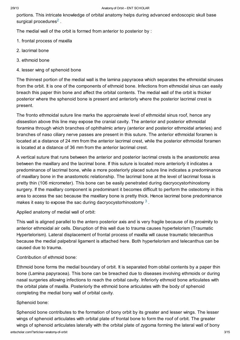
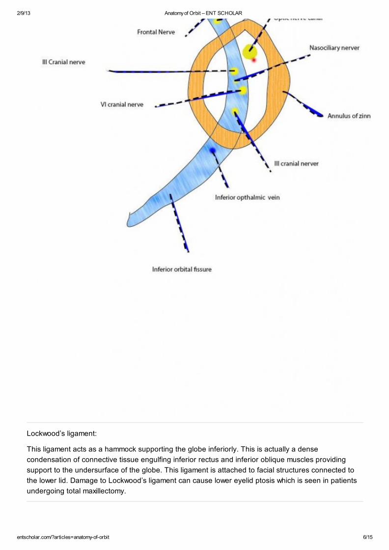
Superior orbital fissure:
This is also known as sphenoidal fissure because it lies between lesser and greater wings of
sphenoid. This space is closed laterally by the frontal bone. This fissure lies between the lateral wall
and roof of orbit. At its medial end it is slightly wider. At this point it lies below the optic foramen. This
fissure gradually reduces in size as it reaches its lateral extremity. Superior orbital fissure hence
should be considered to have a narrow lateral and a wide medial part. This fissure is about 22 mm
long and is the largest communication between the orbit and the middle cranial fossa. Its tip is situated
about 30-40 mm from the frontozygomatic suture line. Its medial end is separated from optic foramen
by the posterior root of lesser wing of sphenoid. This portion of sphenoid bone has a small tubercle
known as infraoptic tubercle. The annulus of Zinn from which all the intraocular muscles originate
spans the superior orbital fissure between its medial wide and lateral narrow portions. Annulus of Zinn
surrounds the optic nerve at its entrance into the orbit.
The following structures pass through the annulus of Zinn:
1. Superior division of 3rd nerve
2. Nasociliary nerve
3. Sympathetic root of cervical ganglion
4. Inferior division of 3rd nerve
5. 6th nerve
6. Opthalmic vein (superior opthalmic)
This is the rough order of structures passing through the annulus from above downwards.
Inferior opthalmic vein passes below the annulus.
2/9/13 Anatomy of Orbit – ENT SCHOLAR
entscholar.com/?articles=anatomy-of-orbit 6/15
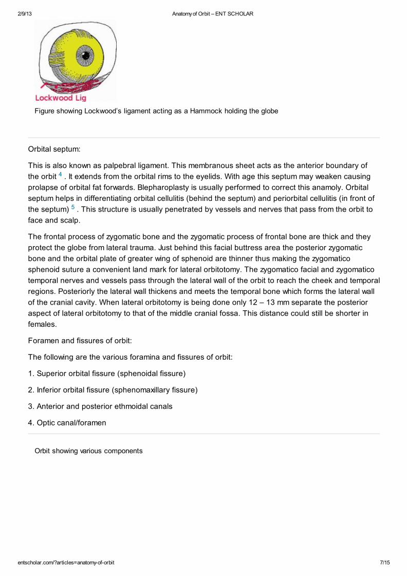
Lockwood’s ligament:
This ligament acts as a hammock supporting the globe inferiorly. This is actually a dense
condensation of connective tissue engulfing inferior rectus and inferior oblique muscles providing
support to the undersurface of the globe. This ligament is attached to facial structures connected to
the lower lid. Damage to Lockwood’s ligament can cause lower eyelid ptosis which is seen in patients
undergoing total maxillectomy.
2/9/13 Anatomy of Orbit – ENT SCHOLAR
entscholar.com/?articles=anatomy-of-orbit 7/15
Figure showing Lockwood’s ligament acting as a Hammock holding the globe
Orbital septum:
This is also known as palpebral ligament. This membranous sheet acts as the anterior boundary of
the orbit . It extends from the orbital rims to the eyelids. With age this septum may weaken causing
prolapse of orbital fat forwards. Blepharoplasty is usually performed to correct this anamoly. Orbital
septum helps in differentiating orbital cellulitis (behind the septum) and periorbital cellulitis (in front of
the septum) . This structure is usually penetrated by vessels and nerves that pass from the orbit to
face and scalp.
The frontal process of zygomatic bone and the zygomatic process of frontal bone are thick and they
protect the globe from lateral trauma. Just behind this facial buttress area the posterior zygomatic
bone and the orbital plate of greater wing of sphenoid are thinner thus making the zygomatico
sphenoid suture a convenient land mark for lateral orbitotomy. The zygomatico facial and zygomatico
temporal nerves and vessels pass through the lateral wall of the orbit to reach the cheek and temporal
regions. Posteriorly the lateral wall thickens and meets the temporal bone which forms the lateral wall
of the cranial cavity. When lateral orbitotomy is being done only 12 – 13 mm separate the posterior
aspect of lateral orbitotomy to that of the middle cranial fossa. This distance could still be shorter in
females.
Foramen and fissures of orbit:
The following are the various foramina and fissures of orbit:
1. Superior orbital fissure (sphenoidal fissure)
2. Inferior orbital fissure (sphenomaxillary fissure)
3. Anterior and posterior ethmoidal canals
4. Optic canal/foramen
Orbit showing various components
4
5
2/9/13 Anatomy of Orbit – ENT SCHOLAR
entscholar.com/?articles=anatomy-of-orbit 8/15
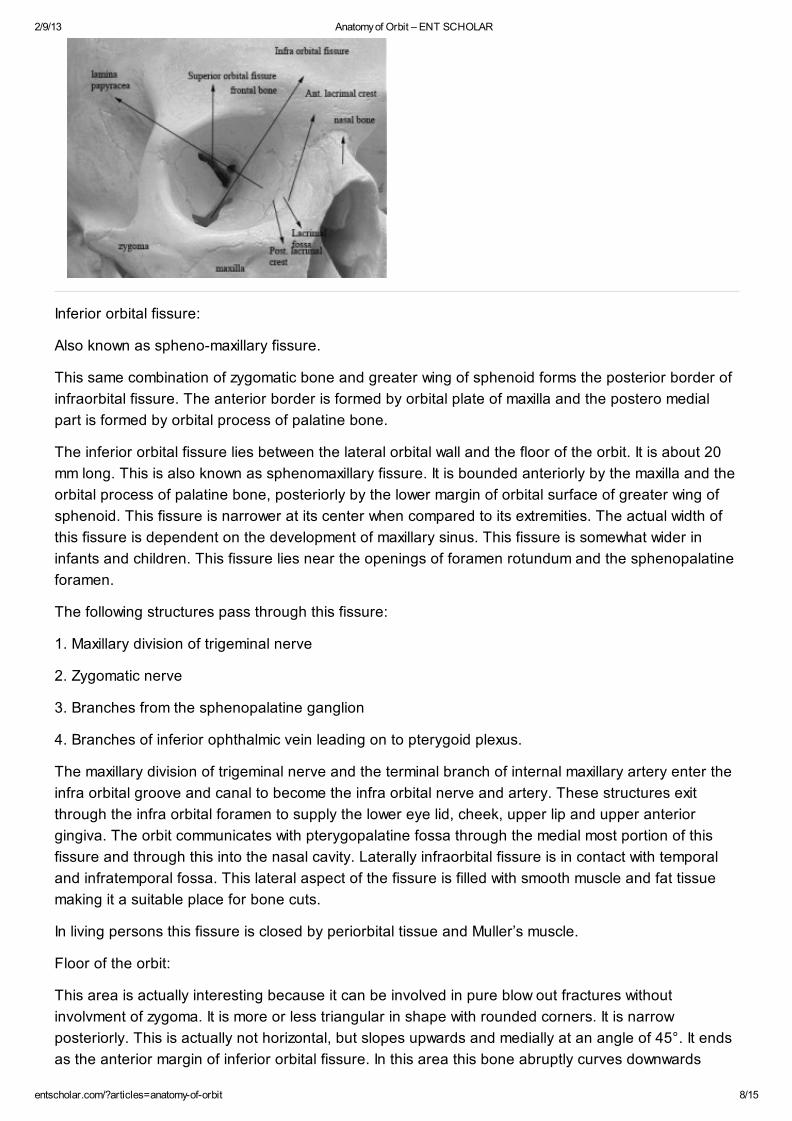
Inferior orbital fissure:
Also known as spheno-maxillary fissure.
This same combination of zygomatic bone and greater wing of sphenoid forms the posterior border of
infraorbital fissure. The anterior border is formed by orbital plate of maxilla and the postero medial
part is formed by orbital process of palatine bone.
The inferior orbital fissure lies between the lateral orbital wall and the floor of the orbit. It is about 20
mm long. This is also known as sphenomaxillary fissure. It is bounded anteriorly by the maxilla and the
orbital process of palatine bone, posteriorly by the lower margin of orbital surface of greater wing of
sphenoid. This fissure is narrower at its center when compared to its extremities. The actual width of
this fissure is dependent on the development of maxillary sinus. This fissure is somewhat wider in
infants and children. This fissure lies near the openings of foramen rotundum and the sphenopalatine
foramen.
The following structures pass through this fissure:
1. Maxillary division of trigeminal nerve
2. Zygomatic nerve
3. Branches from the sphenopalatine ganglion
4. Branches of inferior ophthalmic vein leading on to pterygoid plexus.
The maxillary division of trigeminal nerve and the terminal branch of internal maxillary artery enter the
infra orbital groove and canal to become the infra orbital nerve and artery. These structures exit
through the infra orbital foramen to supply the lower eye lid, cheek, upper lip and upper anterior
gingiva. The orbit communicates with pterygopalatine fossa through the medial most portion of this
fissure and through this into the nasal cavity. Laterally infraorbital fissure is in contact with temporal
and infratemporal fossa. This lateral aspect of the fissure is filled with smooth muscle and fat tissue
making it a suitable place for bone cuts.
In living persons this fissure is closed by periorbital tissue and Muller’s muscle.
Floor of the orbit:
This area is actually interesting because it can be involved in pure blow out fractures without
involvment of zygoma. It is more or less triangular in shape with rounded corners. It is narrow
posteriorly. This is actually not horizontal, but slopes upwards and medially at an angle of 45°. It ends
as the anterior margin of inferior orbital fissure. In this area this bone abruptly curves downwards
2/9/13 Anatomy of Orbit – ENT SCHOLAR
entscholar.com/?articles=anatomy-of-orbit 9/15
towards the infratemporal fossa forming the posterior wall of maxilla.
Components of the floor of the orbit:
1. Orbital plate of maxilla (largest component)
2. Orbital plate of zygomatic bone (antero lateral part)
3. Orbital process of palatine bone (forms a small portion behind the maxilla)
The floor of the orbit is traversed by inferior orbital fissure. This fissure infact weakens the floor. Most
of blowout fractures occur medial to this fissure. Fracture line can cause entrapment of infraorbital
nerve leading on to anesthesia of cheek area of that side. Infraorbital canal formed from this fissure
sinks anteriorly and opens into the infraorbital foramen.
The roof of the orbit slopes down medially. In fact this slope continues up to fronto ethmoidal suture to
form the roof of the ethmoid sinus. This is otherwise known as fovea ethmoidalis.
The anatomical relationship between the anterior ethmoidal air cells and the lacrimal fossa should be
borne in mind to avoid confusion between the ethmoid and nasal cavities during
dacryocystorhinostomy surgery.
Ethmoidal foramen:
These foramina lie between the roof and medial wall of orbit. These foramina invariably lie within the
frontoethmoidal suture line or in the frontal bone. These openings form canals known as anterior and
posterior ethmoidal canals. These canals are formed by frontal bone to a great extent with minor
contributions from ethmoids.
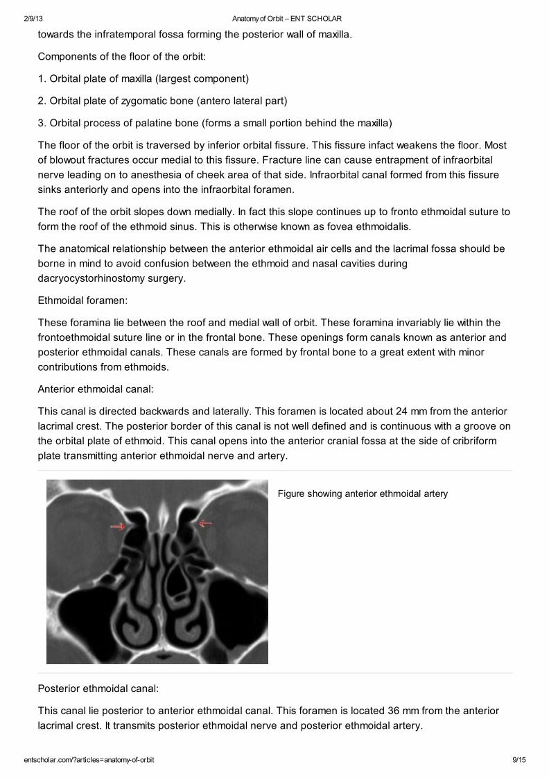
Anterior ethmoidal canal:
This canal is directed backwards and laterally. This foramen is located about 24 mm from the anterior
lacrimal crest. The posterior border of this canal is not well defined and is continuous with a groove on
the orbital plate of ethmoid. This canal opens into the anterior cranial fossa at the side of cribriform
plate transmitting anterior ethmoidal nerve and artery.
Figure showing anterior ethmoidal artery
Posterior ethmoidal canal:
This canal lie posterior to anterior ethmoidal canal. This foramen is located 36 mm from the anterior
lacrimal crest. It transmits posterior ethmoidal nerve and posterior ethmoidal artery.
2/9/13 Anatomy of Orbit – ENT SCHOLAR
entscholar.com/?articles=anatomy-of-orbit 10/15
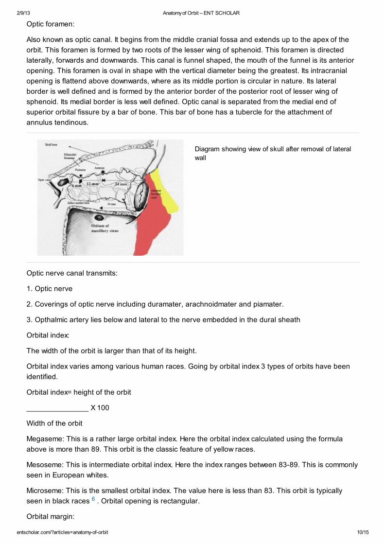
Optic foramen:
Also known as optic canal. It begins from the middle cranial fossa and extends up to the apex of the
orbit. This foramen is formed by two roots of the lesser wing of sphenoid. This foramen is directed
laterally, forwards and downwards. This canal is funnel shaped, the mouth of the funnel is its anterior
opening. This foramen is oval in shape with the vertical diameter being the greatest. Its intracranial
opening is flattend above downwards, where as its middle portion is circular in nature. Its lateral
border is well defined and is formed by the anterior border of the posterior root of lesser wing of
sphenoid. Its medial border is less well defined. Optic canal is separated from the medial end of
superior orbital fissure by a bar of bone. This bar of bone has a tubercle for the attachment of
annulus tendinous.
Diagram showing view of skull after removal of lateral
wall
Optic nerve canal transmits:
1. Optic nerve
2. Coverings of optic nerve including duramater, arachnoidmater and piamater.
3. Opthalmic artery lies below and lateral to the nerve embedded in the dural sheath
Orbital index:
The width of the orbit is larger than that of its height.
Orbital index varies among various human races. Going by orbital index 3 types of orbits have been
identified.
Orbital index= height of the orbit
_______________ X 100
Width of the orbit
Megaseme: This is a rather large orbital index. Here the orbital index calculated using the formula
above is more than 89. This orbit is the classic feature of yellow races.
Mesoseme: This is intermediate orbital index. Here the index ranges between 83-89. This is commonly
seen in European whites.
Microseme: This is the smallest orbital index. The value here is less than 83. This orbit is typically
seen in black races . Orbital opening is rectangular.
Orbital margin:
6
2/9/13 Anatomy of Orbit – ENT SCHOLAR
entscholar.com/?articles=anatomy-of-orbit 11/15
This is made up of three bones.
Frontal
Zygomatic
Maxilla
Superior orbtial margin:
This is entirely formed by frontal bone. This portion of frontal bone is also known as orbital arch. This
margin is sharp in lateral 2/3 and rounded in medial third. At the junction of these two portions about
25 mm from midline is situated the supraorbital notch. This notch transmits supraorbital vessels and
nerves. This notch is converted into a foramen due to ossification of the ligament which lies inferior to
this notch. This notch can easily be palpated in the living.
Arnold’s notch:
This is rarely seen medial to supraorbital notch. This notch is also known as the Arnold’s notch. This
notch transmits the medial branches of supraorbital vessels and nerves.
Lateral orbtial margin:
This margin is the most exposed one and is the strongest of the orbital margins. It is formed by the
zygomatic process of frontal bone and zygomatic bone. The lateral orbital rim is recessed to
accomodate lacrimal gland. This recess may be involved in segmental frature in this region. The
narrowest and weakes part of this rim is the frontozygomatic suture line. Separation of this suture line
is a common feature of trauma in this region.
Inferior orbtial margin:
This margin is raised sligthly above the floor of the orbit. This margin is formed by zygomatic bone and
maxilla in equal proportions. The infraorbital margin is clearly defined at its lateral margin and is easily
palpable. Inner portion of the rim is rounded and is not easily palpable.
Medial margin:
This margin is formed by anterior lacrimal crest present on the frontal process of maxilla and the
posterior lacrimal crest on the lacrimal bone. The medial margin is hence not a continuous ridge.
Age changes in orbit:
Anatomical changes involving orbit depends on the development of facial skeleton and the
neighbouring paranasal sinuses.
At birth the orbital margins are sharp and completely ossified. This helps in protecting the eyes during
the stressful event of parturition. At about the age of 7 the orbital margins but for the superior margin
become fairly rounded and less sharp. At this age the superomedial and inferolateral angles are well
marked than other angles. This causes the orbit to be triangular.
Infant’s orbit look more laterally than adults.
Orbital fissures are large in a child when compared with that of adults. This is because of the narrow
orbtial surface of greater wing of sphenoid.
The orbital index is higher in a child when compared to adults. The vertical diameter is the same as
that of horizontal diameter. As growth progresses the transverse diameter increases more than the
vertical..
2/9/13 Anatomy of Orbit – ENT SCHOLAR
entscholar.com/?articles=anatomy-of-orbit 12/15
The interorbital distance is rather small in children. This mimiks squint.
The roof of the orbit is much larger than the floor at birth.
In infants optic canal is not a canal. It is just a foramen. As the infant grows this foramen elongates to
become the optic canal.
The periorbita is thicker and stronger at birth than in adults.
Old age changes occuring in the orbit are actually due to bone absorption. The roof of the orbit in
elderly person may actually contain holes which causes periorbita to come into direct contact with
dura. Parts of lacrimal bone too can be absorbed due to ageing process.
Soft tissues of orbit:
Orbital septum is the anterior soft tissue boundary of the orbit. It acts as a physical barrier against
pathogens. This is a thin multilayered fibrous tissue derived from the mesodermal layer of eyelid. This
septum is covered anteriorly by the preseptal orbicularis oculi muscle.
Periorbita: is the periosteal lining of orbital walls. The periorbita is attached to the suture lines, fissures
and foramina of the orbit. Posteriorly the periorbita is continuous with the optic nerve sheath.
Orbital fat: Adipose tissue present in the orbit has a cushioning effect on the contents of orbit.
The extra ocular muscles of orbit arise from the annulus of zinn and are responsible for the movement
of the globe. These muscles are:
lateral and medial rectus
Superior and inferior rectus
Superior and inferior oblique
The four recti muscles arise from the annulus of zinn. The annulus of zinn actually has two tendons.
The lower tendon of annulus of zinn is attached to the medial end of superior orbital fissure enclosing
the optic foramen. This tendon gives origin to parts of medial and lateral recti. It also gives attachment
of entire inferior rectus muscle. The upper tendon of the annulus of zinn also known as tendon of
Lockwood arises from the body of sphenoid. This tendon gives origin to part of medial and lateral recti
and all of the superior rectus muscle. The attachments of superior and medial recti muscles are close
to the dural sheath of optic nerve. This fact explains the pain caused during extremes of eye
movements in retrobulbar neuritis.
Medial rectus:
This is the largest of the ocular muscles. It is also stronger than the lateral rectus. From its origin from
the annulus of Zinn it inserts into the globe medially 5.5 mm from the limbus. Its blood supply is
derived from the inferior muscular branch of opthalmic artery and anterior ciliary arteries. It derives its
motor innervation from the third cranial nerve on its lateral surface at the junction of middle and
posterior thirds. It is a pure adductor.
Inferior rectus:
This is the shortest of all recti muscles. From its origin in the annulus of zinn close to optic foramen it
inserts into the globe inferiorly 6.5 mm from the limbus. It is also attached to the lower eye lid via its
facial expansion. It derives its blood supply from the inferior muscular branch of ophthalmic artery,
infraorbital artery and anterior ciliary vessels. It derives its motor innervation from the inferior division
of third nerve on its upper aspect at the junction of its middle and posterior thirds. It moves the eye
2/9/13 Anatomy of Orbit – ENT SCHOLAR
entscholar.com/?articles=anatomy-of-orbit 13/15
downwards and medially / rotates it laterally (extorsion). It can also depress the lower eye lid by its
facial sling which inserts into it. Its principal action is depression of out turned eye. Infact it is the only
depressor of the abducted eye.
Lateral rectus:
From its origin from the annulus of zinn it is inserted laterally into the globe about 6.9 mm from the
limbus. It receives blood supply from lacrimal artery. It is the only ocular muscle with single source of
blood supply. It is innervated by the 6th cranial nerve in its medial aspect. It is a pure abductor making
the eye to look directly laterally.
Superior rectus muscle:
Arising from the superior portion of annulus of zinn it is inserted in to the bulb superiorly about 7.7 mm
from the limbus. It receives its blood supply from the superior muscular branch of ophthalmic artery
and anterior ciliary arteries. It is innervated by superior division of oculomotor nerve. This nerve
enters the undersurface of the muscle at the junction of middle and posterior thirds. It helps in
upwards and medial rotation of the eye and is also capable of intorting the eye ball.
Superior oblique muscle:
This is the longest and thinnest of ocular muscles. It arises medial to the optic foramen and gets
inserted into the trochlea on the orbital rim (on the anterosuperior portion of the medial wall of orbit).
Its tendon gets inserted on to the temporal aspect of the eye behind the equator. The superior
muscular branch of opthalmic artery and ciliary arteries supply this muscle. It moves the eye
downwards and laterally. It is the only muscle that can depress the eye in adducted position. It is
supplied by the 4th nerve.
Inferior oblique:
This is the only extrinsic muscle to take origin from the front of the orbit. This muscle arises from the
orbital floor in a depression near the orbital rim. Some of its fibres may also arise from the fascia
covering lacrimal sac. It is inserted into the posterior inferior temporal quadrant at the level of macula.
It derives its blood supply from the inferior branch of ophthalmic artery and infra orbital artery. It is
innervated by the inferior division of oculomotor nerve. This nerve enters the muscle from its upper
surface. This muscle helps the eye to look upwards and laterally and in extorsion of orbit. This is the
only muscle that elevates the eye in the adducted position.
Levator palpebrae superioris:
This striated muscle elevates the eyelid. This muscle arises from the under surface of lesser wing of
sphenoid just above and in front of optic foramen, and usually it is blended with the origin of superior
rectus muscle. From this attachment this ribbon like muscle passes forwards below the roof on top of
the superior rectus muscle. It gets inserted into the skin of the upper eyelid, and upper tarsal plate. It
receives its nerve supply from the superior divison of 3rd cranial nerve. This muscle by its elevating
action raises the upper eyelid, thus uncovers the cornea and portions of sclera. The action of this
muscle is antogonized by orbicularis oculi muscle inneravated by facial nerve.
Muller’s muscle:
This smooth muscle acts as an eyelid elevator. It arises from the inferior aspect of levator palpebrae.
This muscle is inserted into the upper edge of tarsal plate. It is innervated by sympathetic fibers. The
action of this muscle accounts for the presence of upper lid elevation in patients with 3rd cranial nerve
palsy.
2/9/13 Anatomy of Orbit – ENT SCHOLAR
entscholar.com/?articles=anatomy-of-orbit 14/15
The lacrimal system:
The main lacrimal gland is located in the supero temporal portion of orbit. It lies in the shallow lacrimal
fossa of the frontal bone. The gland is composed of numerous secretory units known as acini which
progressively drain in to small and larger ducts. The gland measures 20 mm by 12 mm. A fibrous band
incompletely devides the lacrimal gland into two lobes i.e. posterior larger orbital lobe and a smaller
anterior palpebral lobe. 2 – 6 ducts from the orbital lobe pass through the palpebral lobe joining with
the ducts from the palpebral lobe to form 6 – 12 tubules to empty into the superio lateral conjunctiva.
Hence damage to the palpebral lobe may block drainage from the entire gland. About 20 – 40
accessory lacrimal glands of Krause are located in the superior conjuctival fornix, about half this
number is located over the lower fornix.
The lacrimal gland is innervated by branches from 5th and 7th cranial nerves, sympathetic supply to
lacrimal gland is via the nerves from the superior cervical ganglion. The parasympathetic fibers are
supplied via the 6th nerve. Sensory supply is via the branches of trigeminal nerve.
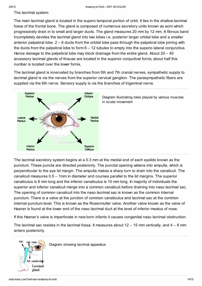
Diagram illustrating roles played by various muscles
in ocular movement
The lacrimal excretory system begins at a 0.3 mm at the medial end of each eyelids known as the
punctum. These puncta are directed posteriorly. The punctal opening widens into ampulla, which is
perpendicular to the eye lid margin. The ampulla makes a sharp turn to drain into the canaliculi. The
canaliculi measures 0.5 – 1mm in diameter and courses parallel to the lid margins. The superior
canaliculus is 8 mm long and the inferior canaliculus is 10 mm long. In majority of individuals the
superior and inferior canaliculi merge into a common canaliculi before draining into naso lacrimal sac.
The opening of common canaliculi into the naso lacrimal sac is known as the common internal
punctum. There is a valve at the junction of common canaliculus and lacrimal sac at the common
internal punctum level. This is known as the Rosenmuller valve. Another valve known as the valve of
Hasner is found at the lower end of the naso lacrimal duct at the level of inferior meatus of nose.
If this Hasner’s valve is imperforate in new born infants it causes congenital naso lacrimal obstruction.
The lacrimal sac resides in the lacrimal fossa. It measures about 12 – 15 mm vertically, and 4 – 8 mm
antero posteriorly.
Diagram showing lacrimal apparatus
2/9/13 Anatomy of Orbit – ENT SCHOLAR
entscholar.com/?articles=anatomy-of-orbit 15/15
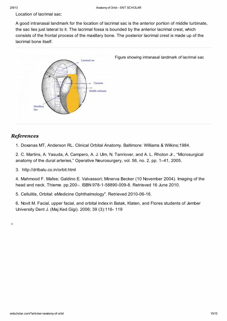
Location of lacrimal sac:
A good intranasal landmark for the location of lacrimal sac is the anterior portion of middle turbinate,
the sac lies just lateral to it. The lacrimal fossa is bounded by the anterior lacrimal crest, which
consists of the frontal process of the maxillary bone. The posterior lacrimal crest is made up of the
lacrimal bone itself.
Figure showing intranasal landmark of lacrimal sac
1. Doxanas MT, Anderson RL. Clinical Orbital Anatomy. Baltimore: Williams & Wilkins;1984.
2. C. Martins, A. Yasuda, A. Campero, A. J. Ulm, N. Tanriover, and A. L. Rhoton Jr., “Microsurgical
anatomy of the dural arteries,” Operative Neurosurgery, vol. 56, no. 2, pp. 1–41, 2005.
3. http://drtbalu.co.in/orbit.html
4. Mahmood F. Mafee; Galdino E. Valvassori; Minerva Becker (10 November 2004). Imaging of the
head and neck. Thieme. pp.200–. ISBN 978-1-58890-009-8. Retrieved 16 June 2010.
5. Cellulitis, Orbital: eMedicine Ophthalmology". Retrieved 2010-06-16.
6. Novit M. Facial, upper facial, and orbital index in Batak, Klaten, and Flores students of Jember
University Dent J. (Maj.Ked.Gigi). 2006; 39 (3):116- 119
References