Embed Size (px)
Citation preview

Anatomy of the Anatomy of the Cardiovascular Cardiovascular
SystemSystemChapter 18Chapter 18
pages 677 - 724pages 677 - 724
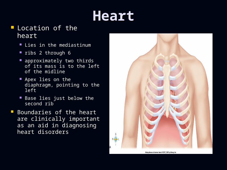
HeartHeart Location of the heartLocation of the heart
Lies in the mediastinumLies in the mediastinum ribs 2 through 6ribs 2 through 6 approximately two thirds approximately two thirds
of its mass is to the left of of its mass is to the left of the midlinethe midline
Apex lies on the Apex lies on the diaphragm, pointing to the diaphragm, pointing to the leftleft
Base lies just below the Base lies just below the second ribsecond rib
Boundaries of the heart Boundaries of the heart are clinically important are clinically important as an aid in diagnosing as an aid in diagnosing heart disordersheart disorders
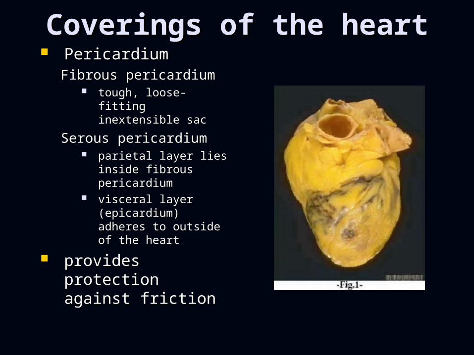
Coverings of the heartCoverings of the heart PericardiumPericardium
Fibrous pericardiumFibrous pericardium tough, loose-fitting tough, loose-fitting
inextensible sacinextensible sac
Serous pericardiumSerous pericardium parietal layer lies parietal layer lies
inside fibrous inside fibrous pericardiumpericardium
visceral layer visceral layer (epicardium) (epicardium) adheres to outside adheres to outside of the heartof the heart
provides provides protection against protection against frictionfriction
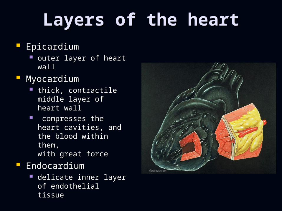
Layers of the heartLayers of the heart EpicardiumEpicardium
outer layer of heart outer layer of heart wallwall
MyocardiumMyocardium thick, contractile thick, contractile
middle layer of heart middle layer of heart wallwall
compresses the heart compresses the heart cavities, and the cavities, and the blood within them,blood within them,with great force with great force
EndocardiumEndocardium delicate inner layer of delicate inner layer of
endothelial tissueendothelial tissue

Chambers of the heartChambers of the heart
Atria (2)Atria (2) ““receiving receiving
chamberschambers”” because they because they
receive blood receive blood from veinsfrom veins
Myocardial Myocardial wall of each wall of each atrium is not atrium is not very thickvery thick
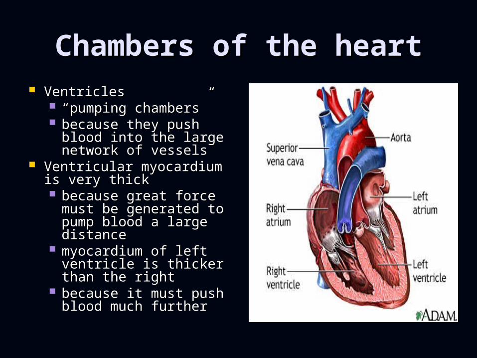
Chambers of the heartChambers of the heart VentriclesVentricles
““pumping chamberspumping chambers”” because they push because they push
blood into the large blood into the large network of vesselsnetwork of vessels
Ventricular myocardium is Ventricular myocardium is very thickvery thick because great force because great force
must be generated to must be generated to pump blood a large pump blood a large distancedistance
myocardium of left myocardium of left ventricle is thicker than ventricle is thicker than the rightthe right
because it must push because it must push blood much furtherblood much further

Heart ValvesHeart ValvesAtrioventricular (AV) valvesAtrioventricular (AV) valves
prevent blood from flowing prevent blood from flowing back into the atria from the back into the atria from the ventricles when the ventricles when the ventricles contractventricles contract
Tricuspid valve (right AV Tricuspid valve (right AV valve)valve) guards the right guards the right
atrioventricular orificeatrioventricular orifice free edges of three flaps of free edges of three flaps of
endocardium are attached endocardium are attached to papillary muscles by to papillary muscles by chordae tendineaechordae tendineae
Bicuspid, or mitral, valve (left Bicuspid, or mitral, valve (left AV valve)AV valve) similar in structure to similar in structure to
tricuspid valve except only tricuspid valve except only two flaps presenttwo flaps present

Heart ValvesHeart Valves
Semilunar (SL) valvesSemilunar (SL) valves Half-moon shaped Half-moon shaped
valves that prevent valves that prevent back flow into the back flow into the heartheart
Pulmonary Pulmonary semilunar valvesemilunar valve at entrance of at entrance of
pulmonary arterypulmonary artery Aortic semilunar Aortic semilunar
valvevalve at entrance of aortaat entrance of aorta

Blood supply of heart Blood supply of heart tissuetissue Myocardial cells Myocardial cells
receive blood from receive blood from right and left right and left coronary arteriescoronary arteries
First branches to come off First branches to come off aortaaorta
Ventricles receive blood Ventricles receive blood from branches of both right from branches of both right and left coronary arteriesand left coronary arteries
Each ventricle receives Each ventricle receives blood only from a small blood only from a small branch of corresponding branch of corresponding coronary arterycoronary artery
Most abundant blood Most abundant blood supply goes to myocardium supply goes to myocardium of left ventricleof left ventricle
The right coronary artery The right coronary artery is dominant in is dominant in approximately 50% of all approximately 50% of all hearts and the left in about hearts and the left in about 20%; in approximately 20%; in approximately 30%, neither coronary 30%, neither coronary artery is dominantartery is dominant
http://www.youtube.com/watch?v=TS0Je1m9Q8A&feature=player_detailpage

Blood supply of heart Blood supply of heart tissuetissue
Veins of the Veins of the coronary circulationcoronary circulation As a rule, veins As a rule, veins
follow a course that follow a course that closely parallels that closely parallels that of coronary arteriesof coronary arteries
After going through After going through cardiac veins, blood cardiac veins, blood enters coronary enters coronary sinus to drain into sinus to drain into right atriumright atrium
Several veins drain Several veins drain directly into right directly into right atriumatrium

Conduction system of Conduction system of the heartthe heart Key ComponentsKey Components
Sinoatrial nodeSinoatrial node hundreds of cells in right hundreds of cells in right
atrial wall near opening of atrial wall near opening of superior vena cavasuperior vena cava
Atrioventricular nodeAtrioventricular node small mass of special small mass of special
cardiac muscle in right cardiac muscle in right atrium along lower part of atrium along lower part of interatrial septuminteratrial septum
Atrioventricular bundleAtrioventricular bundle AV bundle originates in AV AV bundle originates in AV
node, extends by two node, extends by two branches down the two branches down the two sides of the interventricular sides of the interventricular septumseptum
Purkinje fibersPurkinje fibers extend out to papillary extend out to papillary
muscles and lateral walls of muscles and lateral walls of ventriclesventricles
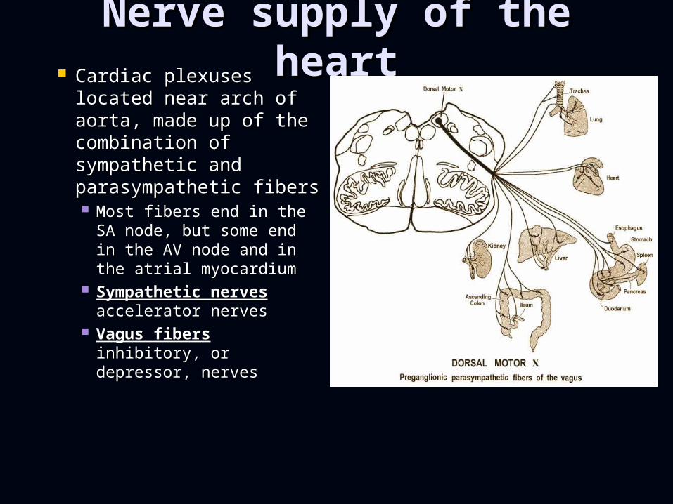
Nerve supply of the Nerve supply of the heartheart Cardiac plexuses Cardiac plexuses
located near arch of located near arch of aorta, made up of the aorta, made up of the combination of combination of sympathetic and sympathetic and parasympathetic fibersparasympathetic fibers Most fibers end in the SA Most fibers end in the SA
node, but some end in node, but some end in the AV node and in the the AV node and in the atrial myocardiumatrial myocardium
Sympathetic nervesSympathetic nerves accelerator nervesaccelerator nerves
Vagus fibersVagus fibers inhibitory, inhibitory, or depressor, nervesor depressor, nerves

Blood Vessels Blood Vessels
ArteriesArteries Carry blood away Carry blood away
from heartfrom heart VeinsVeins
Carry Blood toward Carry Blood toward the heartthe heart
CapillariesCapillaries Carry blood to cells Carry blood to cells
of the bodyof the body

ArteriesArteries Elastic arteriesElastic arteries
Able to stretch Able to stretch without injurywithout injury
Accommodate surge Accommodate surge of blood when heart of blood when heart contracts and able to contracts and able to recoil when recoil when ventricles relaxventricles relax
aorta and its major aorta and its major branchesbranches
Muscular Muscular (distributing) (distributing) arteriesarteries Smaller in diameter Smaller in diameter
than elastic arteriesthan elastic arteries Muscular layer is Muscular layer is
thickthick brachial, gastricbrachial, gastric
Arterioles Arterioles (resistance (resistance vessels)vessels)
Smallest arteriesSmallest arteries Important in Important in
regulating blood regulating blood flow to end-organsflow to end-organs
MetarteriolesMetarterioles Short connecting Short connecting
vessel between vessel between true arteriole and true arteriole and 20 to 100 20 to 100 capillariescapillaries
Encircled by Encircled by precapillary precapillary sphincterssphincters
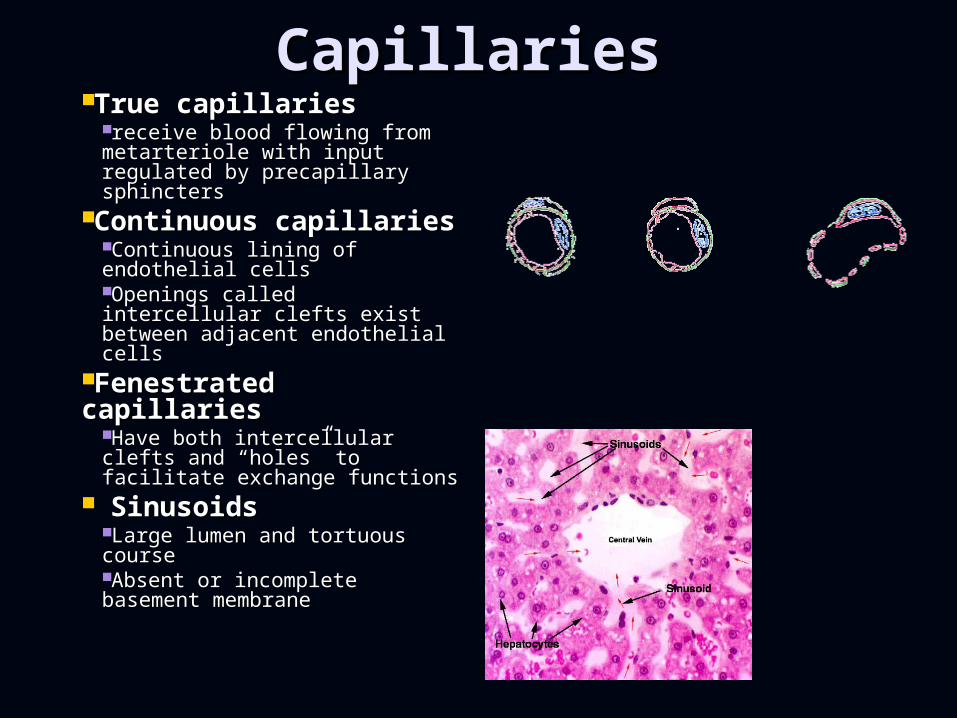
CapillariesCapillariesTrue capillariesTrue capillaries
receive blood flowing from receive blood flowing from metarteriole with input metarteriole with input regulated by precapillary regulated by precapillary sphincterssphincters
Continuous Continuous capillariescapillaries
Continuous lining of Continuous lining of endothelial cellsendothelial cellsOpenings called intercellular Openings called intercellular clefts exist between adjacent clefts exist between adjacent endothelial cellsendothelial cells
Fenestrated Fenestrated capillariescapillaries
Have both intercellular clefts Have both intercellular clefts and and ““holesholes”” to facilitate to facilitate exchange functions exchange functions
SinusoidsSinusoidsLarge lumen and tortuous Large lumen and tortuous coursecourseAbsent or incomplete Absent or incomplete basement membranebasement membrane

VeinsVeins
VeinsVeins Carry blood toward Carry blood toward
the heartthe heart Act as collectors Act as collectors
and as reservoir and as reservoir vesselsvessels
capacitance vesselscapacitance vessels TypesTypes
VeinulesVeinules VeinsVeins

Blood Vessel LayersBlood Vessel Layers
Three Main layersThree Main layers Tunica adventitiaTunica adventitia
found in arteries found in arteries and veinsand veins
Tunica mediaTunica media found in arteries found in arteries
and veinsand veins Tunica intimaTunica intima
found in all blood found in all blood vesselsvessels
only layer present in only layer present in capillariescapillaries

Building Blocks of Blood Building Blocks of Blood VesselsVessels
Lining endothelial Lining endothelial cellscells Only lining found in Only lining found in
capillarycapillary Line entire vascular Line entire vascular
treetree Provide a smooth luminal Provide a smooth luminal
surfacesurface——protects against protects against intravascular coagulationintravascular coagulation
Capable of secreting a Capable of secreting a number of substancesnumber of substances
Capable of Capable of reproductionreproduction

Building Blocks of Blood Building Blocks of Blood VesselsVessels
Collagen fibersCollagen fibers Exhibit woven Exhibit woven
appearanceappearance Have only a limited Have only a limited
ability to stretch ability to stretch (2% to 3%) under (2% to 3%) under physiological physiological conditionsconditions
Function to Function to strengthen and strengthen and keep lumen of keep lumen of vessel openvessel open

Building Blocks of Building Blocks of Blood VesselsBlood Vessels Elastic fibersElastic fibers
Fibers can stretch over Fibers can stretch over 100% under 100% under physiological conditionsphysiological conditions
Play important role in Play important role in creating passive tension creating passive tension to help regulate blood to help regulate blood pressure throughout pressure throughout cardiac cyclecardiac cycle
Smooth muscle fibersSmooth muscle fibers Present in all segments Present in all segments
of vascular system of vascular system except capillariesexcept capillaries
Most numerous in elastic Most numerous in elastic and muscular arteriesand muscular arteries
Exert active tension in Exert active tension in vessels when contractingvessels when contracting

Circulatory routesCirculatory routes Systemic circulationSystemic circulation
Left sided pumpLeft sided pump blood flows from the blood flows from the
left ventricle of the left ventricle of the heart through blood heart through blood vessels to all parts of vessels to all parts of the bodythe body
except gas except gas exchange tissues exchange tissues of lungsof lungs
back to right atriumback to right atrium

Circulatory routesCirculatory routes
Pulmonary circulationPulmonary circulation Right Sided pumpRight Sided pump venous blood moves venous blood moves
from right atrium to from right atrium to right ventricle to right ventricle to pulmonary artery to pulmonary artery to lung arterioles and lung arterioles and capillaries capillaries
gases are exchangedgases are exchanged oxygenated blood oxygenated blood
returns to left atrium returns to left atrium via pulmonary veinsvia pulmonary veins

Systemic circulationSystemic circulation
Arterial anastomosis Arterial anastomosis open into other open into other
branches of the same branches of the same or other arteries or other arteries
Arteriovenous Arteriovenous anastomoses anastomoses shunts occur when shunts occur when
blood flows from an blood flows from an artery directly into a artery directly into a veinvein

Systemic circulationSystemic circulation
Superior Vena CavaSuperior Vena Cava Venous blood from Venous blood from
the head, neck, the head, neck, upper extremities, upper extremities, and thoracic cavityand thoracic cavity
except lungs except lungs Inferior vena cavaInferior vena cava
Venous blood from Venous blood from lower extremities lower extremities and abdomenand abdomen

Fetal circulationFetal circulation Two umbilicalTwo umbilical arteriesarteries
extensions of internal extensions of internal iliac arteries;iliac arteries;
carry fetal blood to carry fetal blood to placentaplacenta
PlacentaPlacenta attached to uterine wallattached to uterine wall where exchange of where exchange of
oxygen and other oxygen and other substances between the substances between the separated maternal and separated maternal and fetal blood occurs fetal blood occurs
Umbilical veinUmbilical vein returns oxygenated returns oxygenated
blood from placenta to blood from placenta to fetusfetus
enters body through enters body through umbilicus continues as umbilicus continues as ductus venosusductus venosus
Ductus venosus Ductus venosus continuation of continuation of
umbilical vein, drains umbilical vein, drains into inferior vena cavainto inferior vena cava
Foramen ovaleForamen ovale opening in septum opening in septum
between right and left between right and left atriaatria
Ductus arteriosusDuctus arteriosus small vessel small vessel
connecting pulmonary connecting pulmonary artery with descending artery with descending thoracic aortathoracic aorta

Diagram of Fetal Blood Diagram of Fetal Blood FlowFlow

Fetal circulationFetal circulation Changes in circulation at Changes in circulation at
birthbirth When umbilical cord is cut, the When umbilical cord is cut, the
two umbilical arteries, the two umbilical arteries, the placenta and the umbilical vein placenta and the umbilical vein no longer functionno longer function
Umbilical vein within the babyUmbilical vein within the baby’’s s body becomes the round ligament body becomes the round ligament of the liverof the liver
Ductus venosus becomes the Ductus venosus becomes the ligamentum venosum of the liverligamentum venosum of the liver
Foramen ovaleForamen ovale functionally closed shortly after a functionally closed shortly after a
newbornnewborn’’s first breath and s first breath and pulmonary circulation is establishedpulmonary circulation is established
structural closure takes structural closure takes approximately 9 monthsapproximately 9 months
Ductus arteriosusDuctus arteriosus contracts with establishment of contracts with establishment of
respiration, becomes ligamentum respiration, becomes ligamentum arteriosumarteriosum

Changes over timeChanges over time BirthBirth
change from placenta-dependent change from placenta-dependent systemsystem
Childhood to adulthoodChildhood to adulthood Exercise thickens myocardium Exercise thickens myocardium increases blood supplyincreases blood supply
Adulthood through later Adulthood through later adulthoodadulthood degenerative changesdegenerative changes
AtherosclerosisAtherosclerosis blockage or weakening of critical blockage or weakening of critical
arteriesarteries Heart valves and myocardial Heart valves and myocardial
tissue degenerate - reduces tissue degenerate - reduces pumping efficiencypumping efficiency