Embed Size (px)
Citation preview
Anatomy of the Human Eye
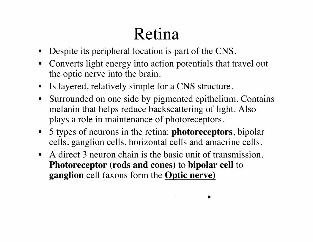
Retina• Despite its peripheral location is part of the CNS.• Converts light energy into action potentials that travel out
the optic nerve into the brain.• Is layered, relatively simple for a CNS structure.• Surrounded on one side by pigmented epithelium. Contains
melanin that helps reduce backscattering of light. Alsoplays a role in maintenance of photoreceptors.
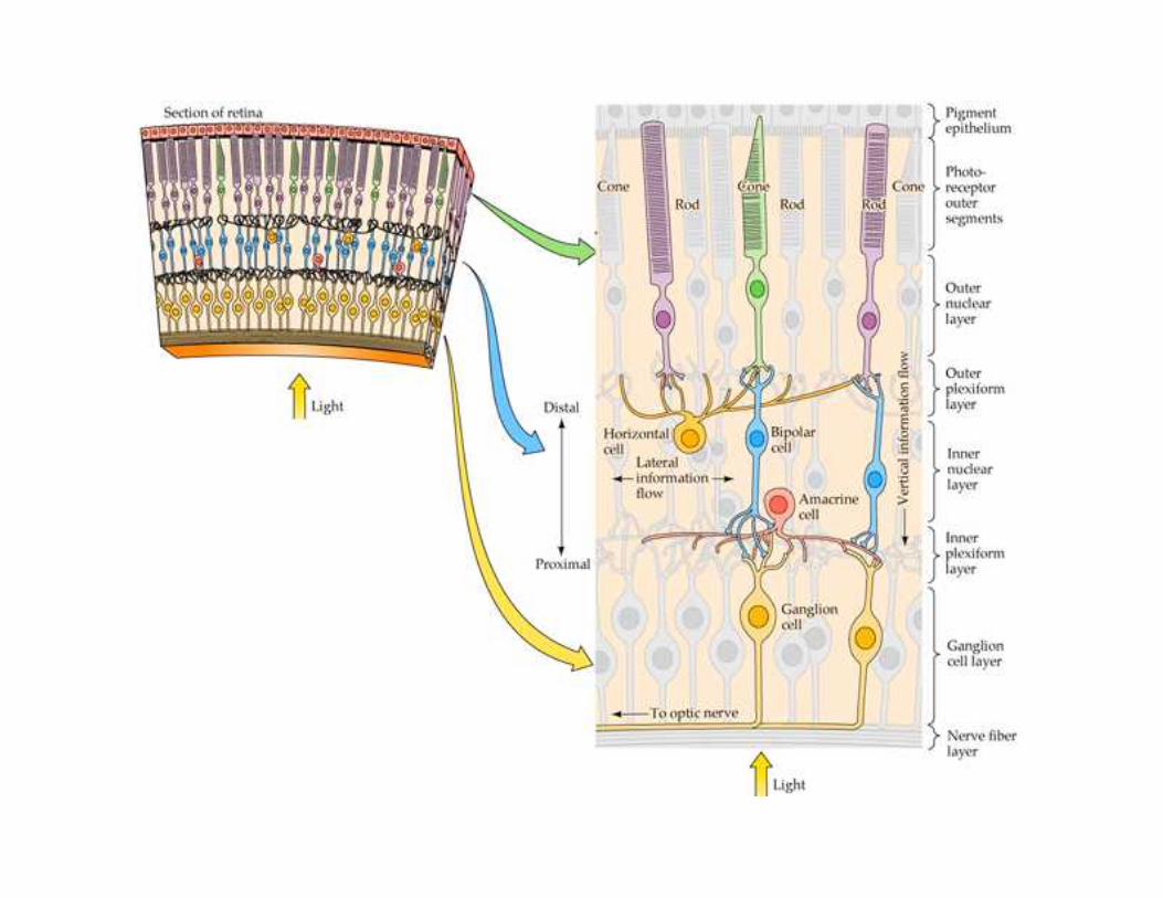
• 5 types of neurons in the retina: photoreceptors, bipolarcells, ganglion cells, horizontal cells and amacrine cells.
• A direct 3 neuron chain is the basic unit of transmission.Photoreceptor (rods and cones) to bipolar cell toganglion cell (axons form the Optic nerve)
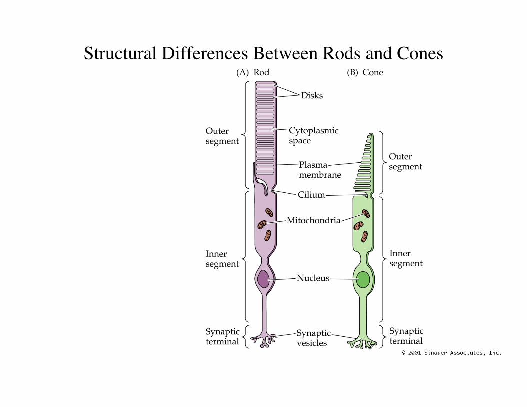
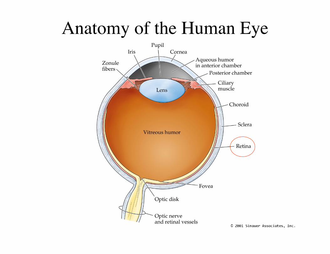
Structural Differences Between Rods and Cones
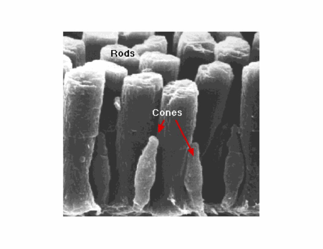
rods and cones are distinguished by:
• shape• type of photopigment they contain• distribution across the retina• pattern of synaptic connections• specialized for different aspects of visionRod system-low spatial resolution but extremely
sensitive to lightCone system- high spatial resolution but is relatively
insensitive to light.
Phototransduction
• Photoreceptors do not exhibit action potentials-light causes a graded change in membranepotential that changes the rate at whichneurotransmitter is released.– The NT is thought to be glutamate
• Light absorption leads to hyperpolarization ofthe neuron. This leads to less release ofneurotransmitter to post-synaptic cell.
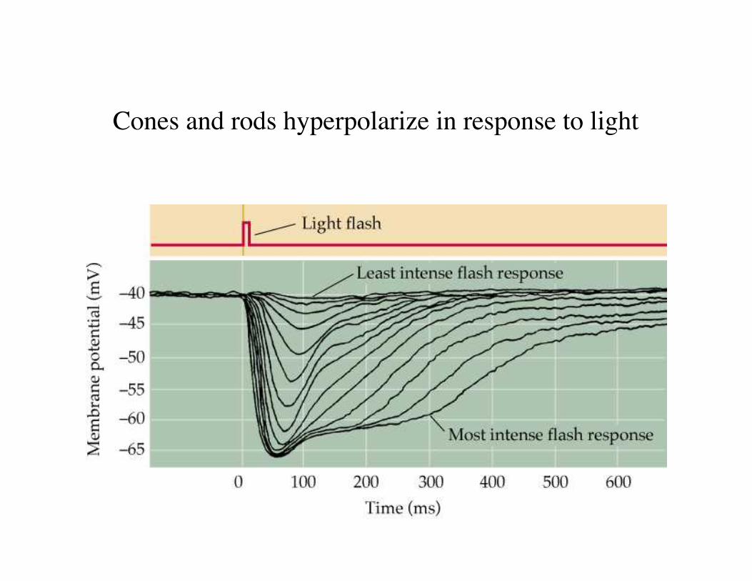
Cones and rods hyperpolarize in response to light
What does light do?
• In the dark, resting potential of the photoreceptoris - 40 mv.
• Light shining onto outer segment leads to thehyperpolarization of the photoreceptor andreduction of neurotransmitter released.
• In the dark the number of Ca++ channels open atthe synaptic terminal is high, and rate ofneurotransmitter release is high.
• In the light number of open channels is reducedand rate of neurotransmitter release is reduced.
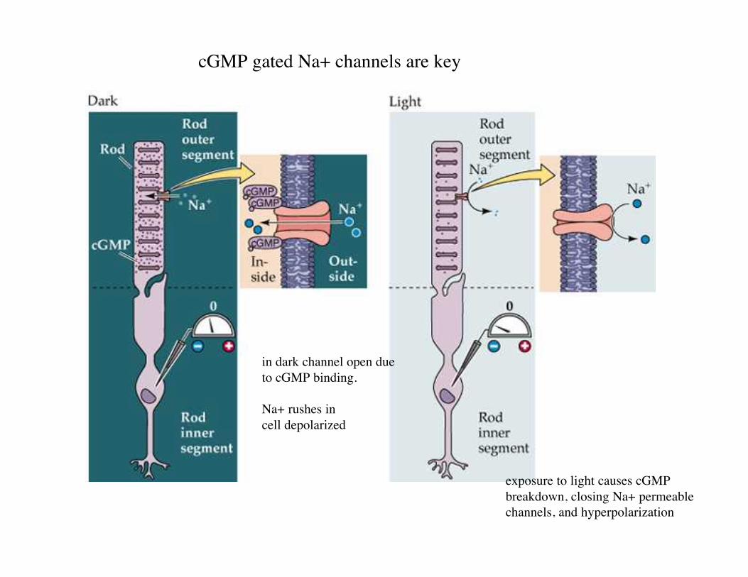
cGMP gated Na+ channels are key
in dark channel open dueto cGMP binding.
Na+ rushes incell depolarized
exposure to light causes cGMPbreakdown, closing Na+ permeablechannels, and hyperpolarization
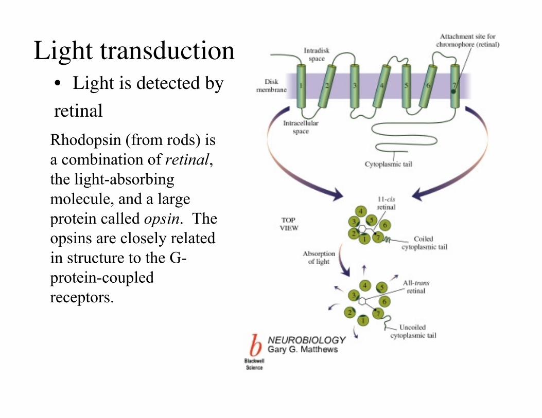
Light transduction• Light is detected byretinalRhodopsin (from rods) isa combination of retinal,the light-absorbingmolecule, and a largeprotein called opsin. Theopsins are closely relatedin structure to the G-protein-coupledreceptors.
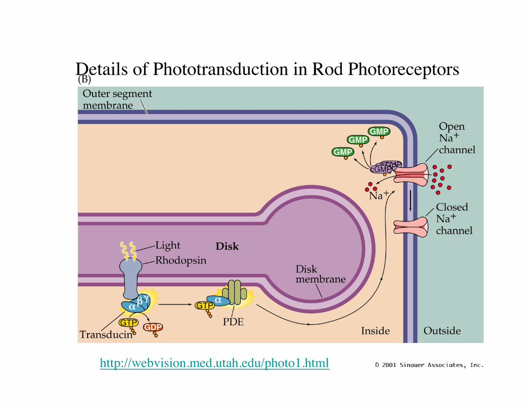
Details of Phototransduction in Rod Photoreceptors
http://webvision.med.utah.edu/photo1.html
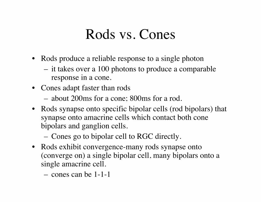
Rods vs. Cones• Rods produce a reliable response to a single photon
– it takes over a 100 photons to produce a comparableresponse in a cone.
• Cones adapt faster than rods– about 200ms for a cone; 800ms for a rod.
• Rods synapse onto specific bipolar cells (rod bipolars) thatsynapse onto amacrine cells which contact both conebipolars and ganglion cells.– Cones go to bipolar cell to RGC directly.
• Rods exhibit convergence-many rods synapse onto(converge on) a single bipolar cell, many bipolars onto asingle amacrine cell.– cones can be 1-1-1
Rods and cones are not distributedequally in the retina
• Human - 91 million rods, 4.5 million cones.• In most places the density of rods exceeds that of cones.• Cones increase in density 200 fold in the fovea, become
highly packed. Center of the fovea is rod free.• Gives high visual acuity, which decreases rapidly away
from the fovea.• Reason why we constantly move our eyes toward what we
want to look at.– And why it it best to see a dim object by looking away from it.
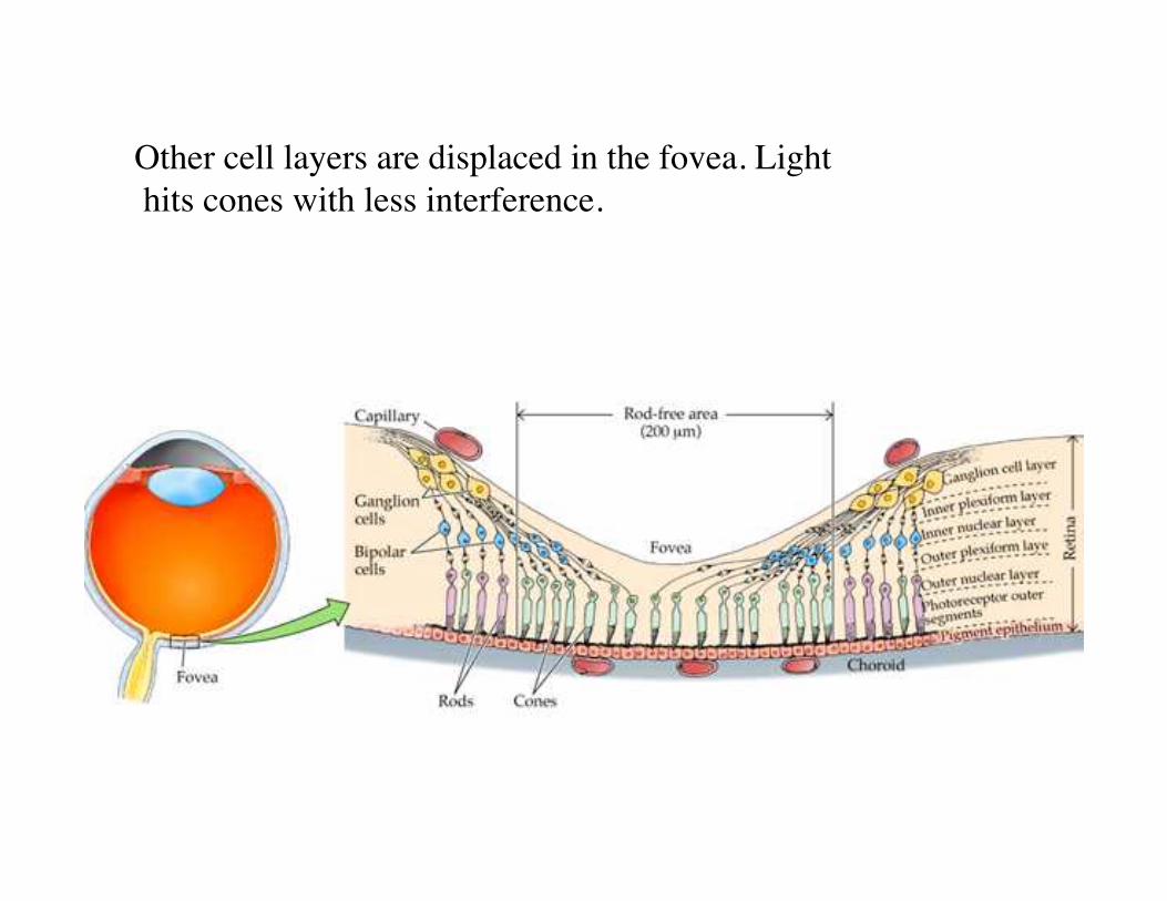
Other cell layers are displaced in the fovea. Light hits cones with less interference.
cones and color vision
• 3 types of cone, each having different absorptionspectra- called blue, green, and red opsin.
• Most people can match any color by changing theintensities of these three colors (RGB).
• 5-6% of males are color blind- due to mutations inred and green opsins. The pigment genes are X-linked and near each other.
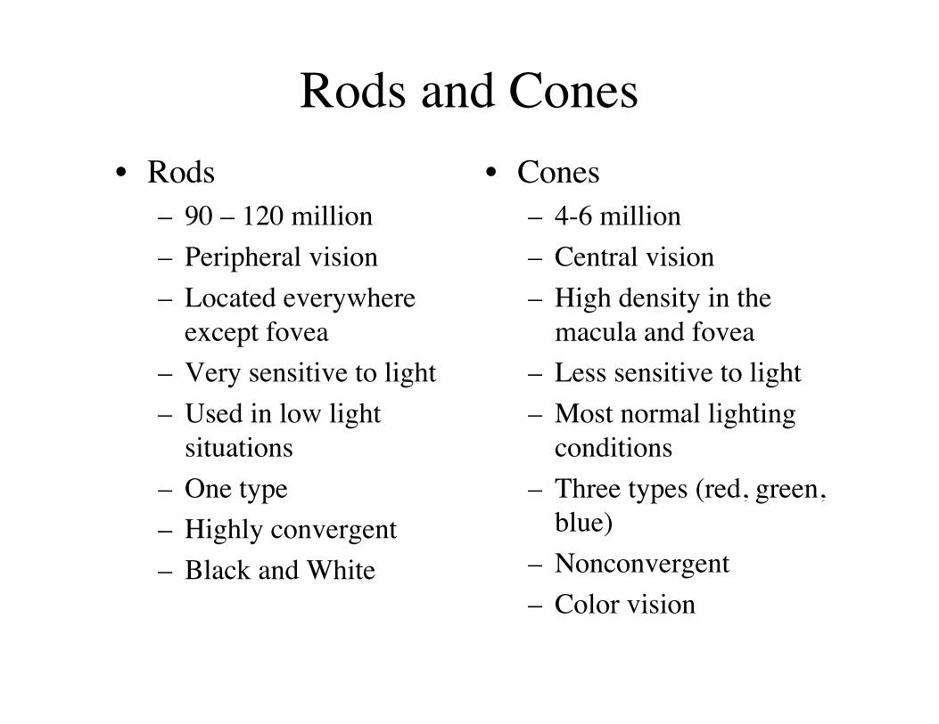
Rods and Cones• Rods
– 90 – 120 million– Peripheral vision– Located everywhere
except fovea– Very sensitive to light– Used in low light
situations– One type– Highly convergent– Black and White
• Cones– 4-6 million– Central vision– High density in the
macula and fovea– Less sensitive to light– Most normal lighting
conditions– Three types (red, green,
blue)– Nonconvergent– Color vision
Retinal ganglion cells (RGC)• Record from an RGC and shine light onto different
photoreceptors. Find:• Even in the dark RGCs are spontaneously active.• Receptive fields of RGCs are circular. Smaller in the
center and bigger in the periphery• The receptive fields of RGCs overlap so that multiple
RGCs see each point of space.
• http://webvision.med.utah.edu/IPL.html
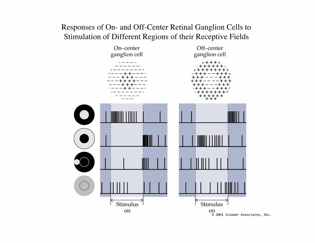
Responses of On- and Off-Center Retinal Ganglion Cells toStimulation of Different Regions of their Receptive Fields
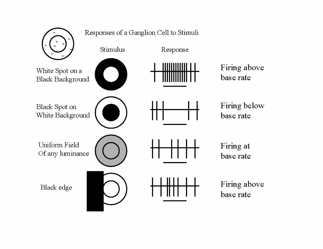
Summary- Retinal GanglionCells• For an on-center RGC, a point of light that just fills the
center will give maximal stimulation (increased actionpotentials).
• Light that crosses into surround will yield intermediateresponse depending on the relative amounts.
• Both center and surround illuminated is similar to being inthe dark (background levels).
• Ganglion cells fire depending on contrast, not by absolutelight intensity.
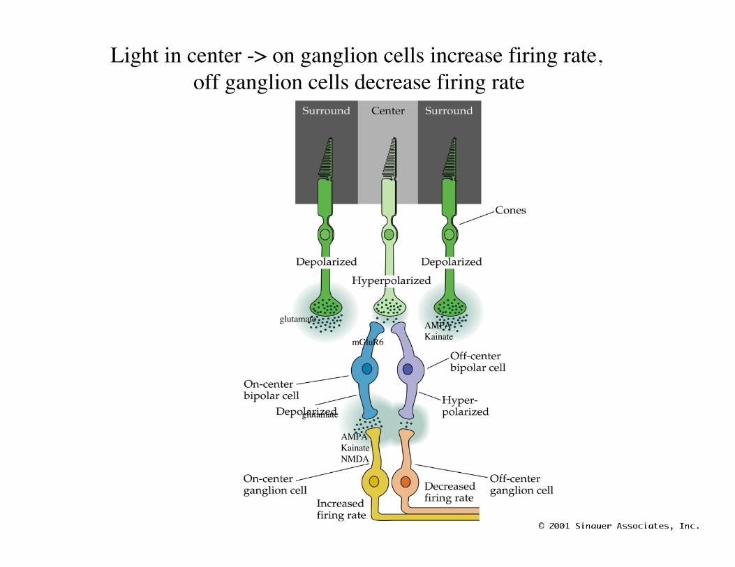
Light in center -> on ganglion cells increase firing rate, off ganglion cells decrease firing rate
glutamate
mGluR6
AMPAKainate
glutamate
AMPAKainateNMDA
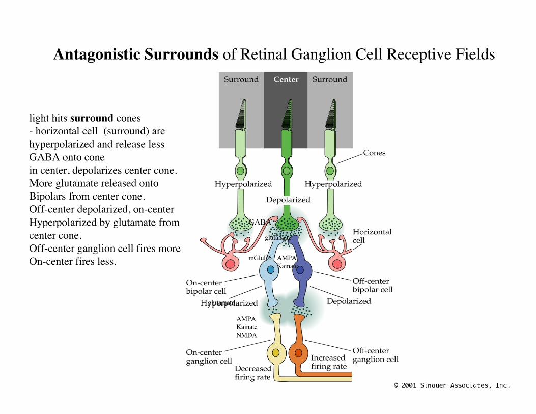
Antagonistic Surrounds of Retinal Ganglion Cell Receptive Fields
glutamate
mGluR6 AMPAKainate
glutamate
AMPAKainateNMDA
GABA
light hits surround cones- horizontal cell (surround) arehyperpolarized and release lessGABA onto conein center, depolarizes center cone.More glutamate released ontoBipolars from center cone.Off-center depolarized, on-centerHyperpolarized by glutamate fromcenter cone.Off-center ganglion cell fires moreOn-center fires less.
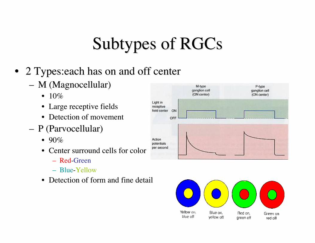
Subtypes of RGCsSubtypes of RGCs•• 2 Types:each has on and off center2 Types:each has on and off center
–– M (M (MagnocellularMagnocellular))•• 10%10%•• Large receptive fieldsLarge receptive fields•• Detection of movementDetection of movement
–– P (P (ParvocellularParvocellular))•• 90%90%•• Center surround cells for colorCenter surround cells for color
–– RedRed--GreenGreen–– BlueBlue--YellowYellow
•• Detection of form and fine detailDetection of form and fine detail
Summary
• Light falls on photopigment, that is transformed toaction potentials that ganglion cells convey to thebrain.
• Phototransduction occurs in rods and cones thathave different properties that meet the conflictingdemands of sensitivity and acuity.
• RGCs have a center-surround arrangement ofreceptive fields that makes them good at contrastdetection and relatively insensitive to backgroundillumination.
Central Visual Pathways:Retinal Targets
• The retina (axons of RGCs) projects to multiple areas.Each area is specialized for different functions.– Lateral geniculate nucleus (LGN)- in the thalamus-
receives input from retina and sends it to visual cortex.Most important visual projection for perception.
– Pretectum-located at midbrain-thalamus boundary.Responsible for pupillary light reflex.
– Superior colliculus-in midbrain, coordinates head andeye movements.
– Suprachiasmatic nucleus- in hypothalamus-involvedin day/night cycles.
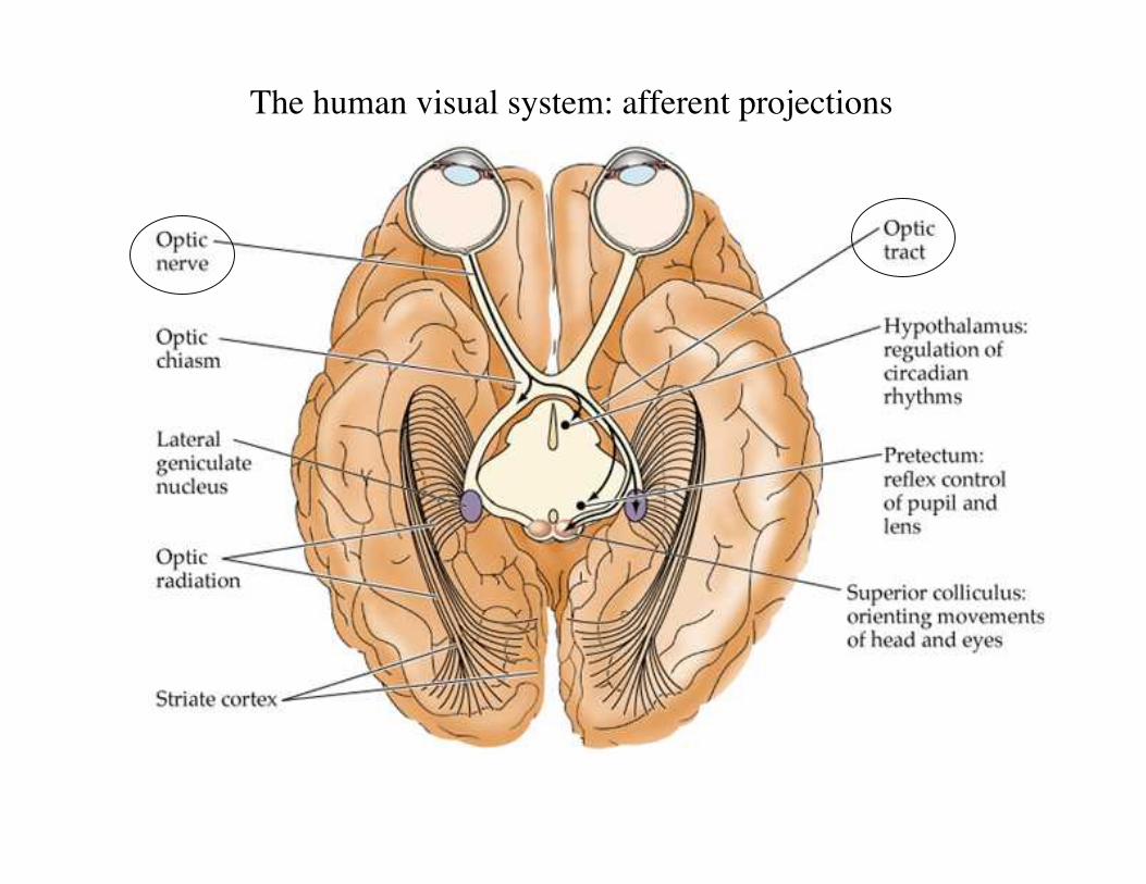
The human visual system: afferent projections
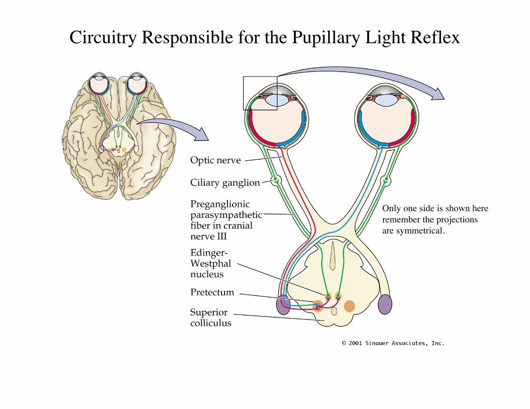
Circuitry Responsible for the Pupillary Light Reflex
Only one side is shown hereremember the projectionsare symmetrical.
The spatial relationships among theRGCs are maintained in their targets.• Organized in maps.• Images are inverted and left-right reversed as they are
projected onto the retina.• The visual field can be divided into nasal, temporal,
inferior (ventral) superior (dorsal). Fixation point is wherethe foveas align.
• The left half of the visual world is represented in theright half of the brain and vice versa. (not left eye toright side of brain, but left visual field)
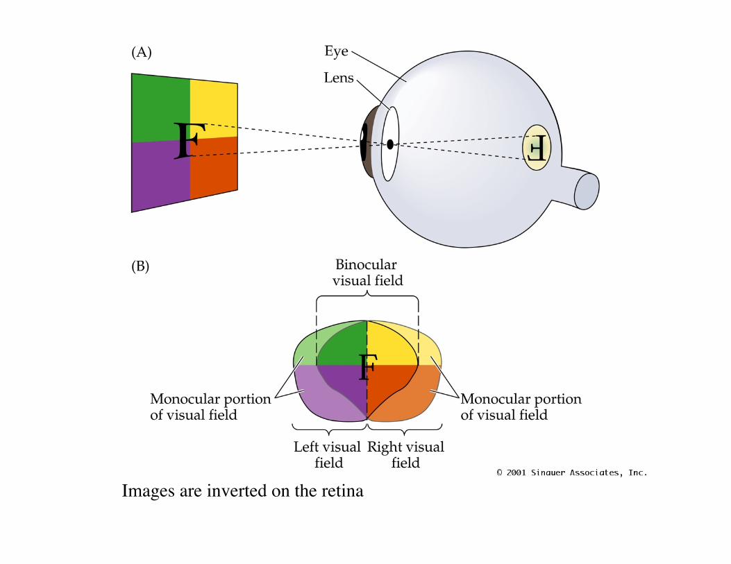
Projection of the Visual Fieldsonto the Retinas
• PN12041.JPG
Images are inverted on the retina
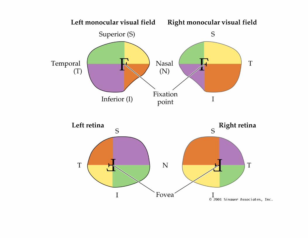
Projection of the Visual Fieldsonto the Retinas
• PN12042.JPG
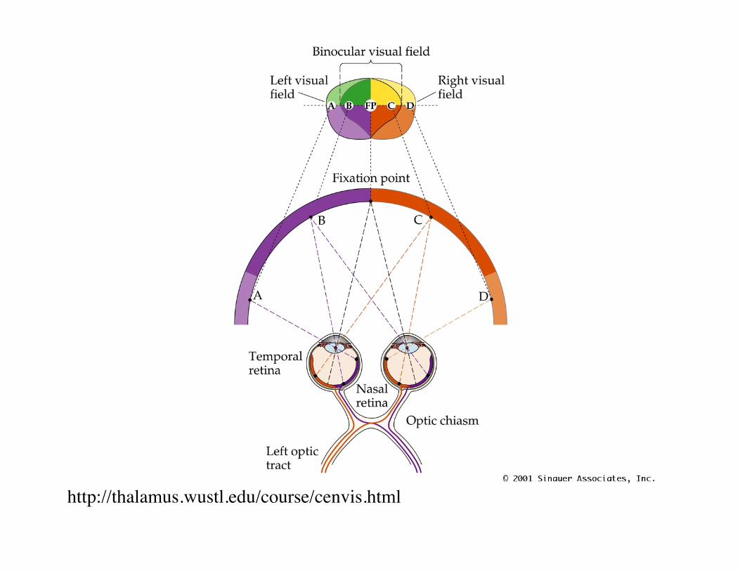
Projection of the Binocular Fieldof View Relates to Crossing of
Fibers in Optic Chiasm• PN12050.JPG
http://thalamus.wustl.edu/course/cenvis.html
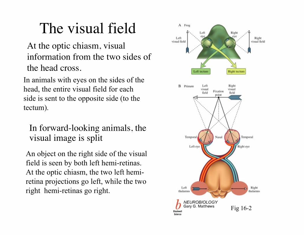
The visual fieldAt the optic chiasm, visualinformation from the two sides ofthe head cross.
In animals with eyes on the sides of thehead, the entire visual field for eachside is sent to the opposite side (to thetectum).
Fig 16-2
In forward-looking animals, thevisual image is split
An object on the right side of the visualfield is seen by both left hemi-retinas.At the optic chiasm, the two left hemi-retina projections go left, while the tworight hemi-retinas go right.
Visual field defects
• Because the spatial relationships in theretina are maintained in the brain, a carefulanalysis of the visual fields of a patient canoften indicate where brain damage islocated.
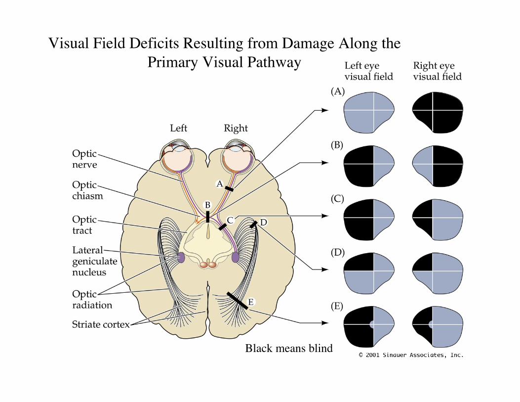
Visual Field Deficits Resulting from Damage Along thePrimary Visual Pathway
Black means blind
LGN
• 90% of retinal axons go to Lateral GeniculateNucleus (LGN).
• LGN projects to visual cortex (striate cortex).• Contains 6 layers, specific to eye (ipsi vs contra)
and with type of ganglion cell, magnocellular(gross shape and movement) or parvocellular(form and color.)
• Layers align in order of visual fields.
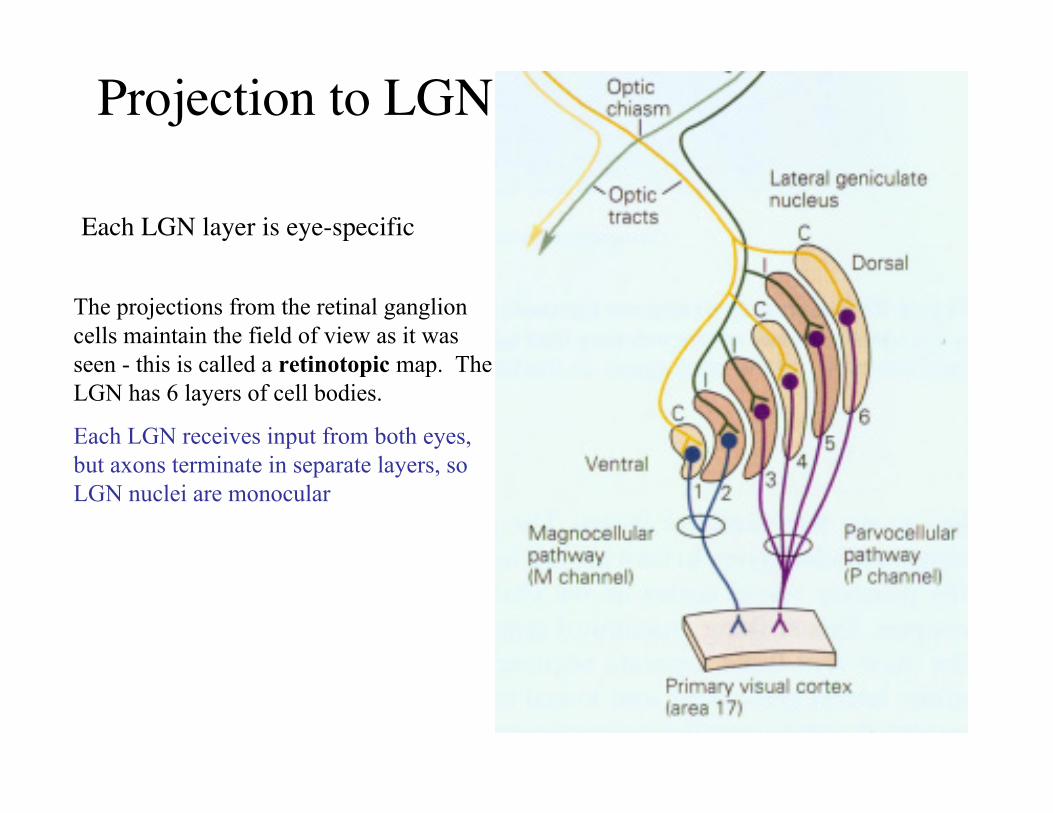
Projection to LGN
Each LGN layer is eye-specific
The projections from the retinal ganglioncells maintain the field of view as it wasseen - this is called a retinotopic map. TheLGN has 6 layers of cell bodies.
Each LGN receives input from both eyes,but axons terminate in separate layers, soLGN nuclei are monocular
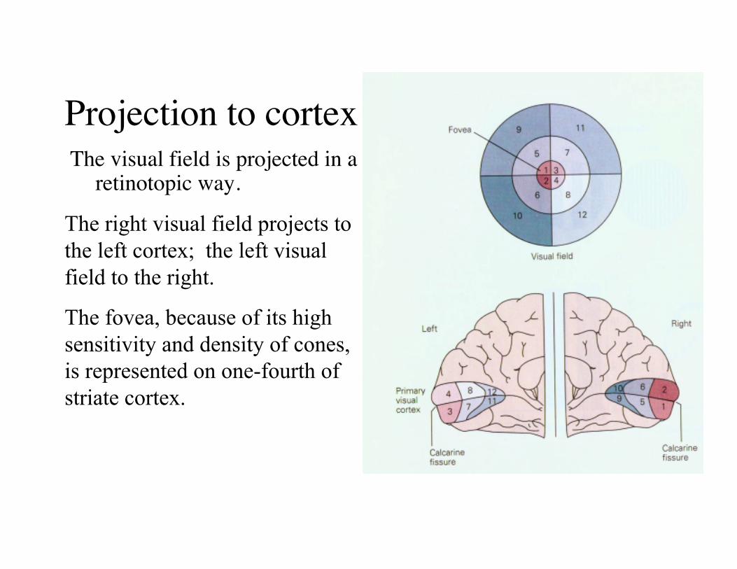
Projection to cortexThe visual field is projected in a
retinotopic way.
The right visual field projects tothe left cortex; the left visualfield to the right.
The fovea, because of its highsensitivity and density of cones,is represented on one-fourth ofstriate cortex.
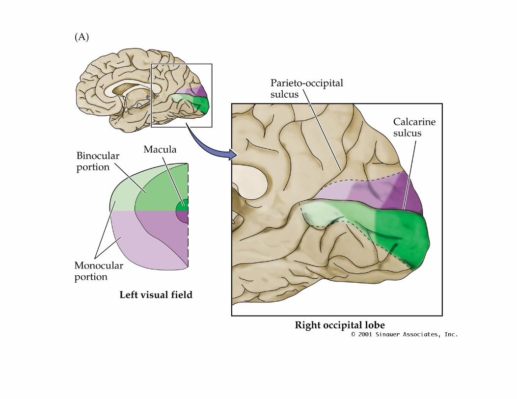
Visuotopic Organization in theRight Occipital Lobe
• PN12060.JPG
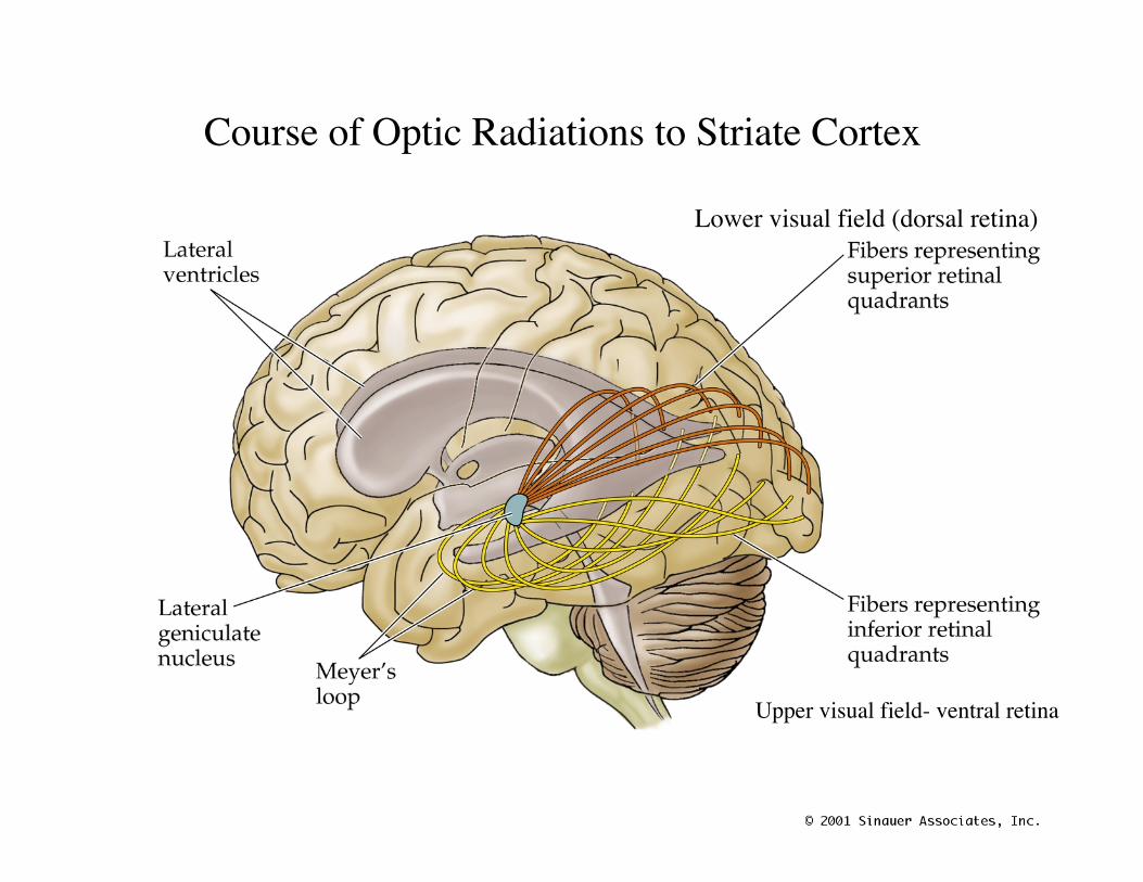
Course of Optic Radiations to Striate Cortex
Lower visual field (dorsal retina)
Upper visual field- ventral retina
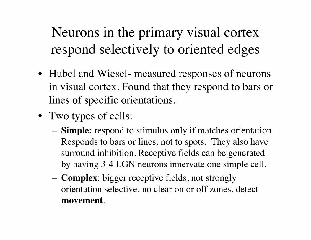
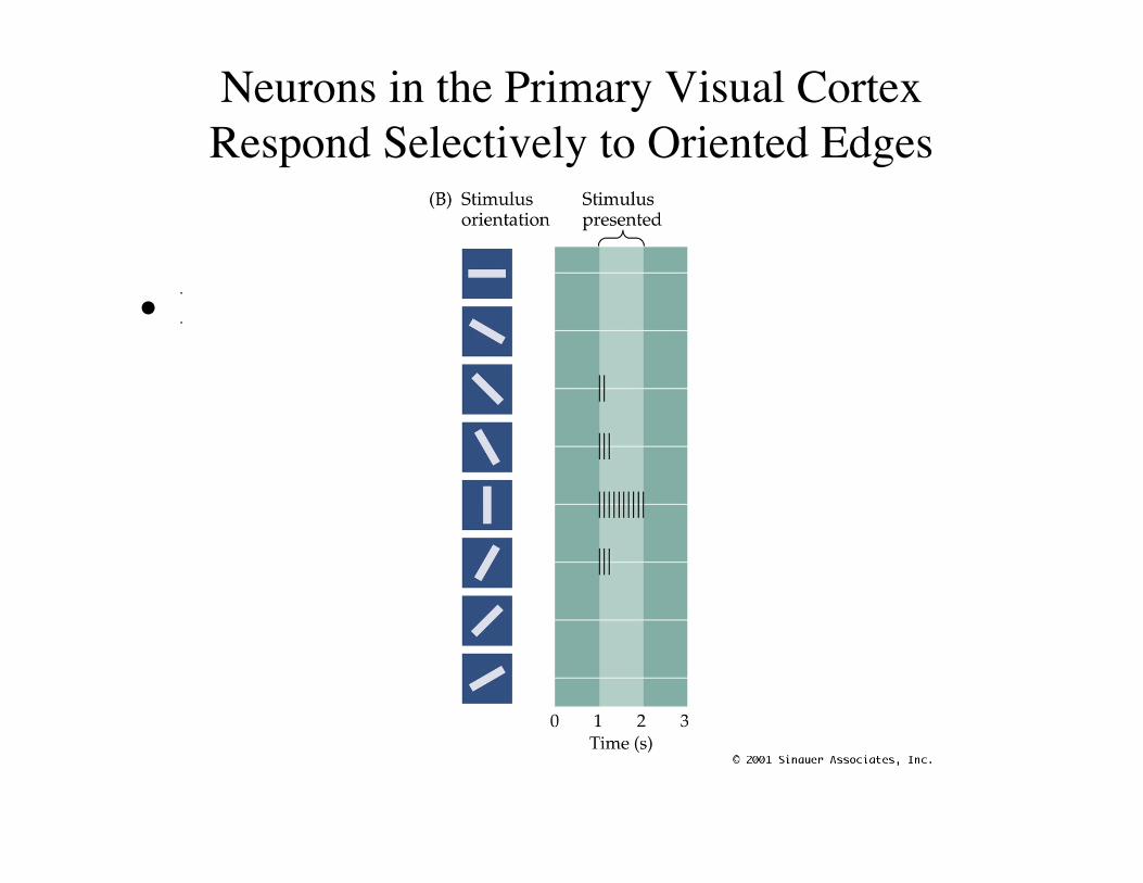
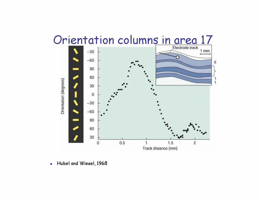
Neurons in the primary visual cortexrespond selectively to oriented edges
• Hubel and Wiesel- measured responses of neuronsin visual cortex. Found that they respond to bars orlines of specific orientations.
• Two types of cells:– Simple: respond to stimulus only if matches orientation.
Responds to bars or lines, not to spots. They also havesurround inhibition. Receptive fields can be generatedby having 3-4 LGN neurons innervate one simple cell.
– Complex: bigger receptive fields, not stronglyorientation selective, no clear on or off zones, detectmovement.
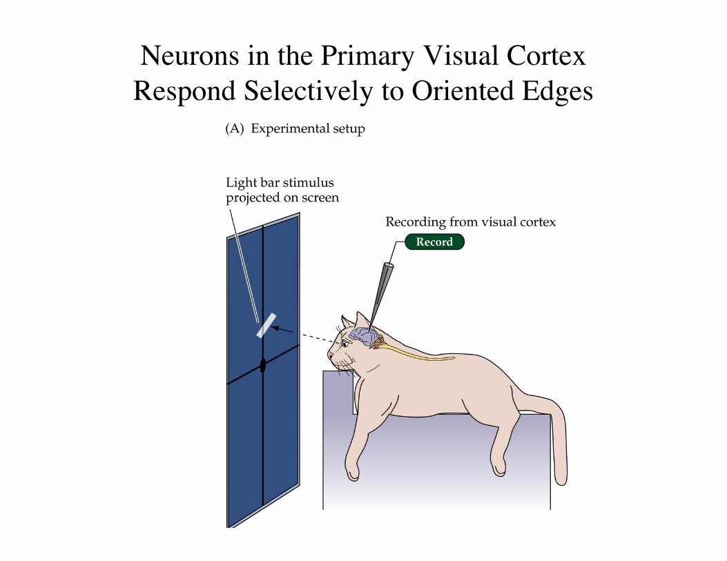
Neurons in the Primary Visual CortexRespond Selectively to Oriented Edges
• PN12092.JPG
Neurons in the Primary Visual CortexRespond Selectively to Oriented Edges
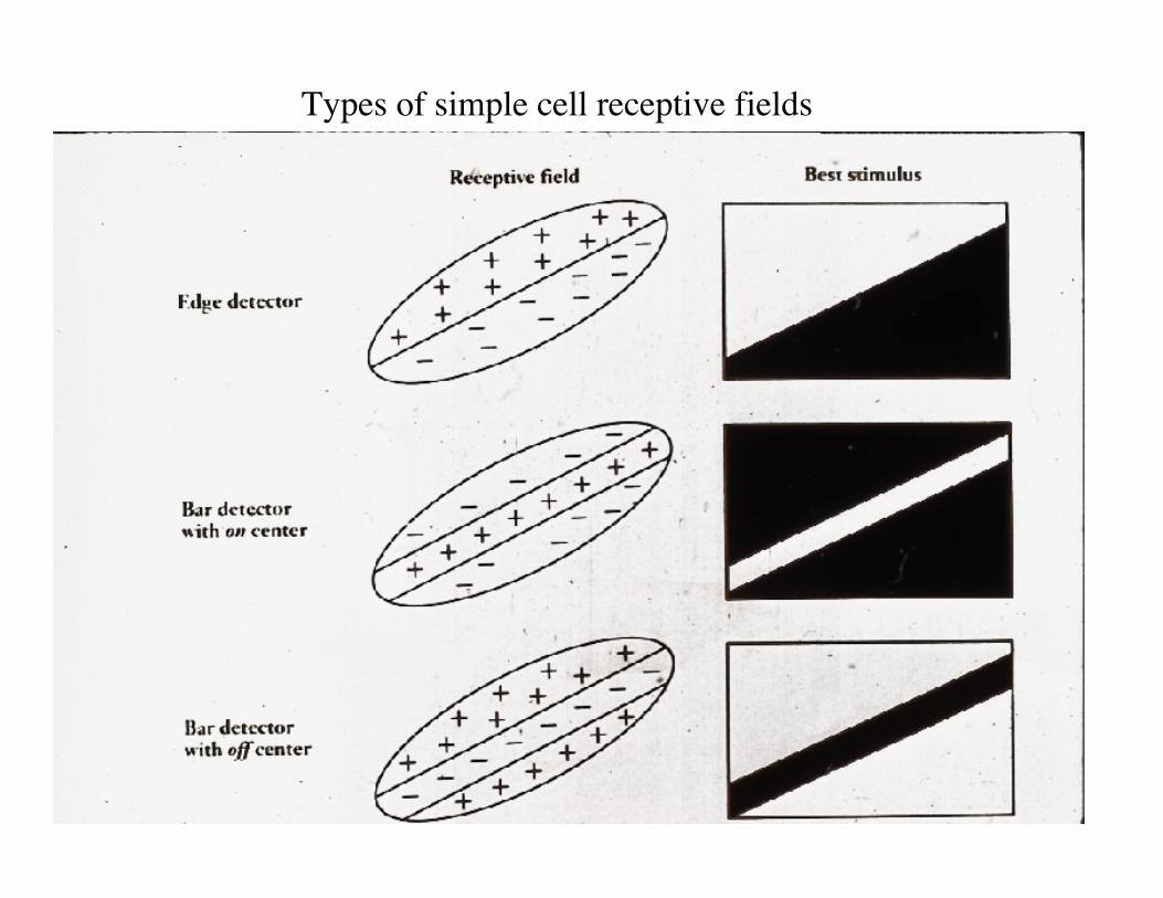
Types of simple cell receptive fields
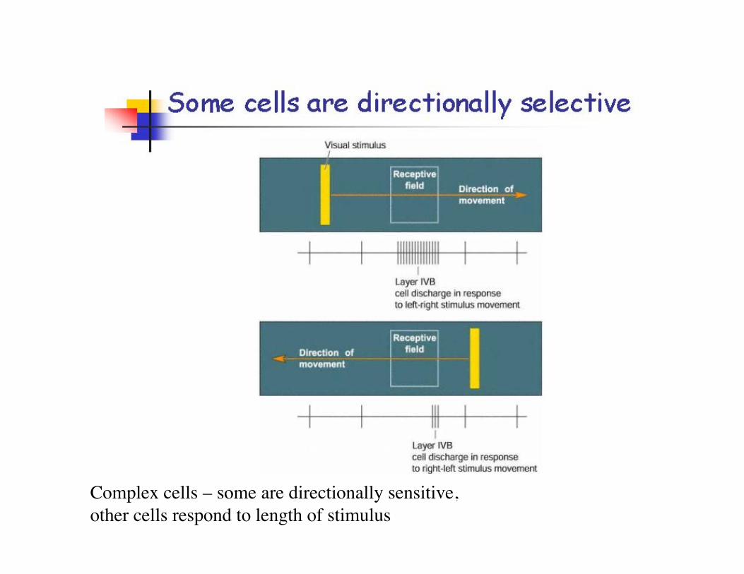
Complex cells – some are directionally sensitive, other cells respond to length of stimulus
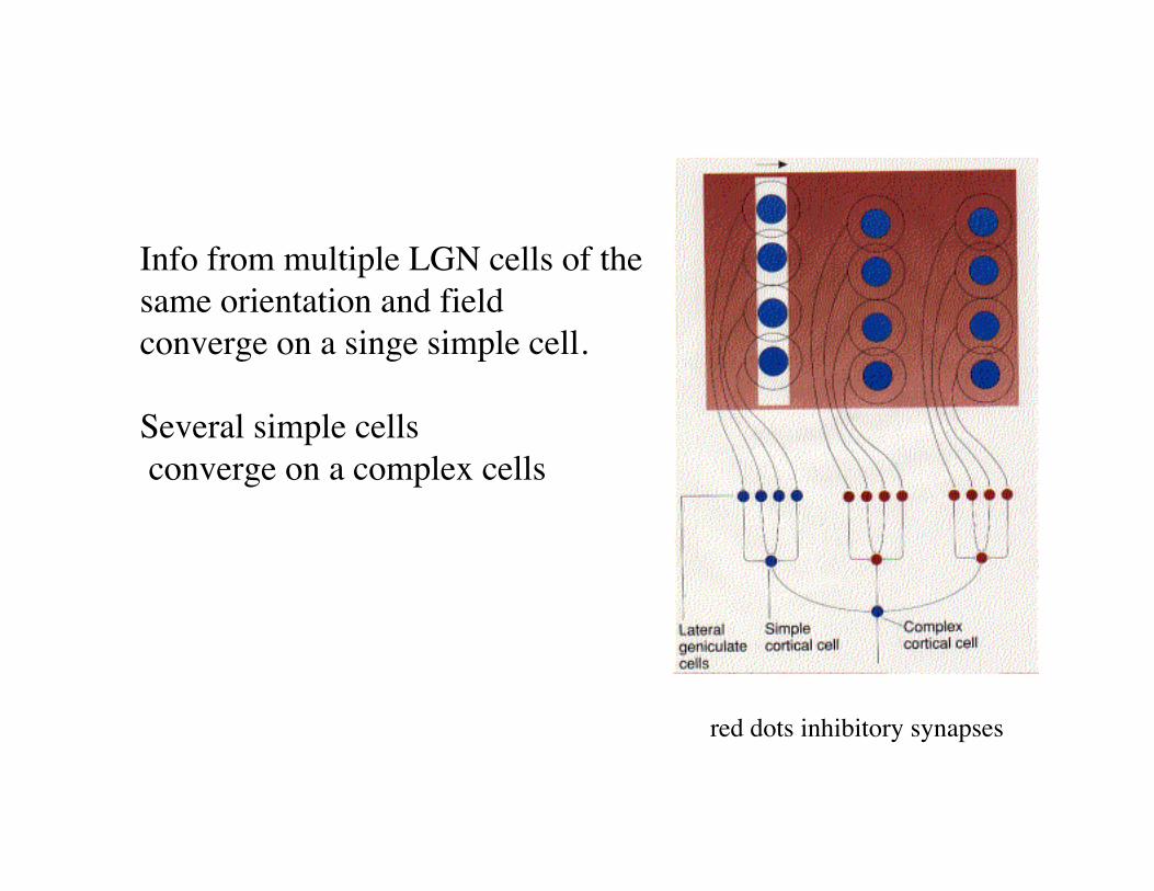
Info from multiple LGN cells of the same orientation and field converge on a singe simple cell.
Several simple cells converge on a complex cells
red dots inhibitory synapses
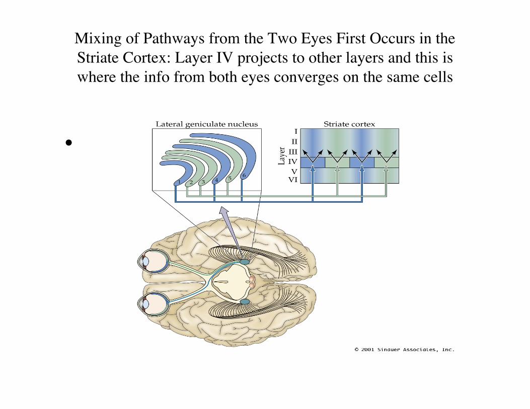
Mixing of Pathways from the Two Eyes First Occurs in theStriate Cortex: Layer IV projects to other layers and this iswhere the info from both eyes converges on the same cells
• PN12100.JPG
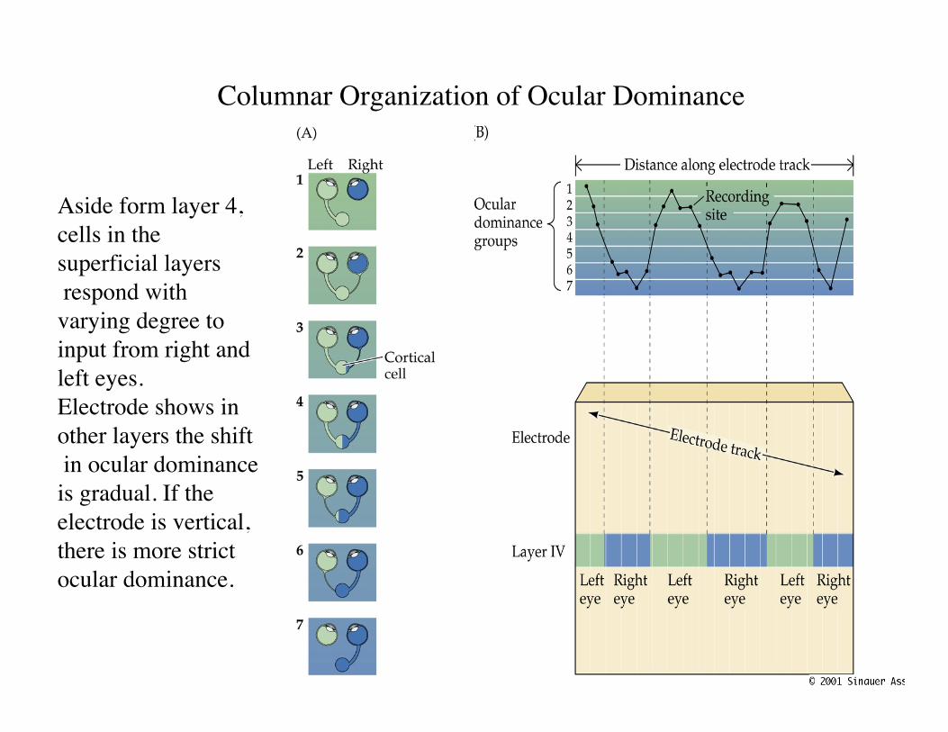
Columnar Organization of Ocular Dominance
Aside form layer 4,cells in thesuperficial layers respond withvarying degree toinput from right andleft eyes.Electrode shows inother layers the shift in ocular dominanceis gradual. If theelectrode is vertical,there is more strictocular dominance.
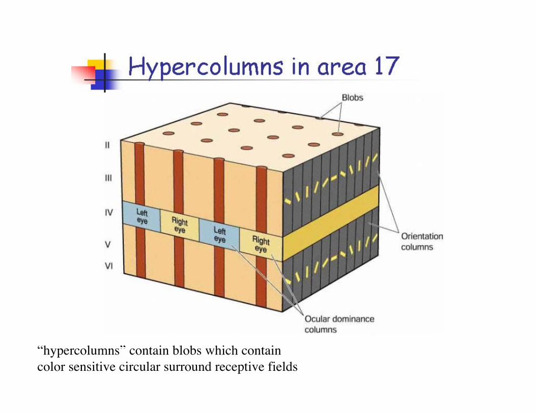
“hypercolumns” contain blobs which contain color sensitive circular surround receptive fields
• PN12120.JPG
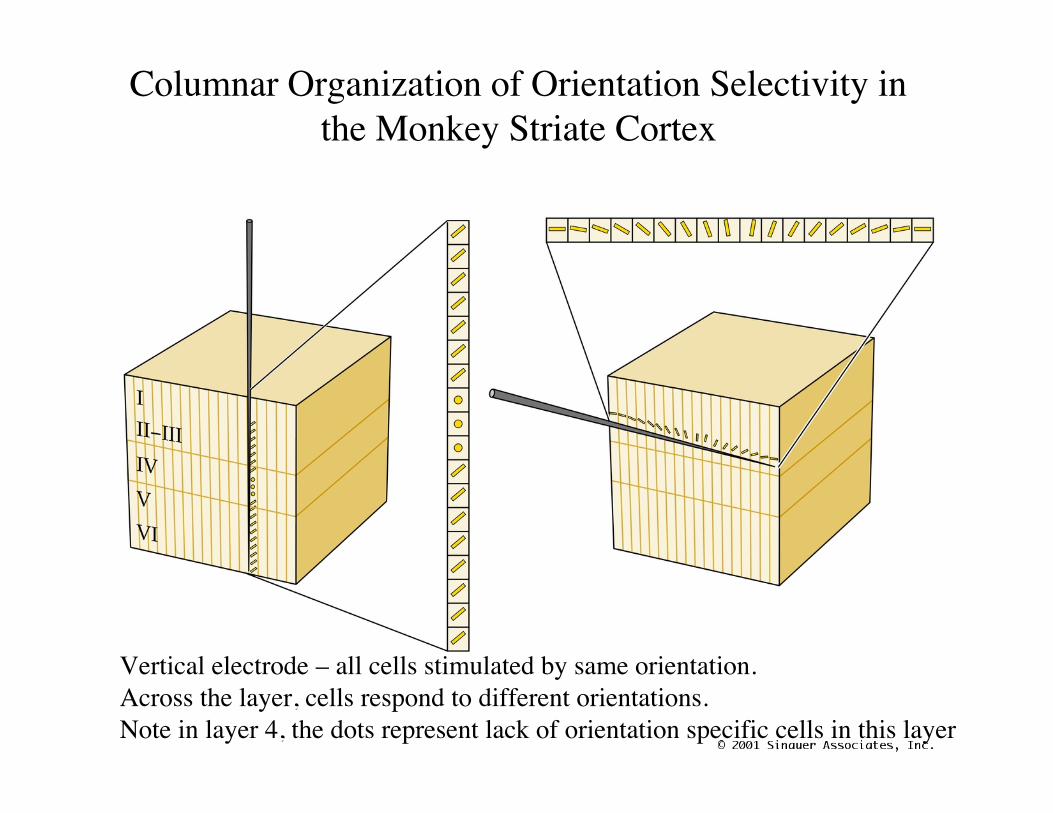
Columnar Organization of Orientation Selectivity inthe Monkey Striate Cortex
Vertical electrode – all cells stimulated by same orientation. Across the layer, cells respond to different orientations. Note in layer 4, the dots represent lack of orientation specific cells in this layer
Parallel processing in the visual system
• Separate pathways for color (Parvocellular) andmovement (Magnocellular).
• M and P ganglion cells in retina. M cells bigger,larger receptive fields, faster conduction velocities,and respond transiently to visual stimulation. Pcells smaller, respond in a sustained fashion.
• M and P go to different layers in the LGN andproject to different populations of cells in layer 4of V1.
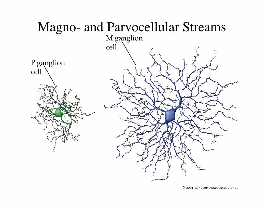
Magno- and Parvocellular Streams
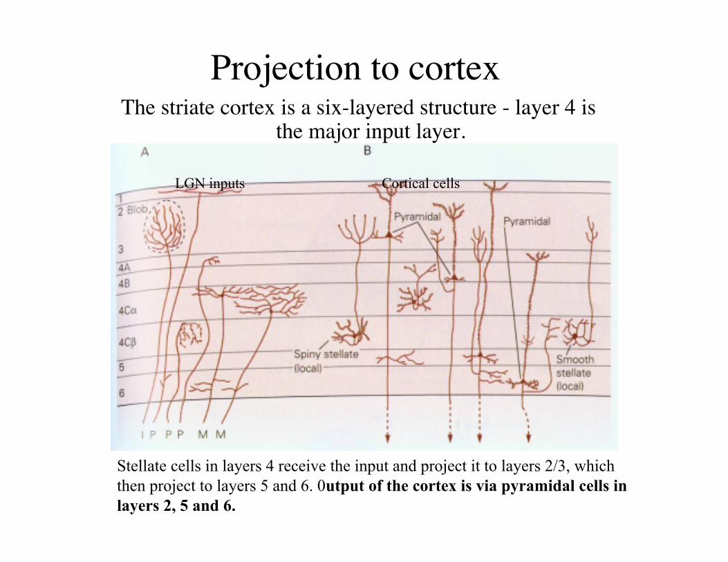
Projection to cortexThe striate cortex is a six-layered structure - layer 4 is
the major input layer.
LGN inputs Cortical cells
Stellate cells in layers 4 receive the input and project it to layers 2/3, whichthen project to layers 5 and 6. 0utput of the cortex is via pyramidal cells inlayers 2, 5 and 6.
• There are many other areas of the brain that process visualinformation; each gets info from primary visual cortex(V1).
• Specialized for different functions.• Middle temporal area (MT), respond to direction of a
moving edge without regard to its color.• V4 responds to color of a stimulus without regard to form.• 10 different visual areas, each with a topographic map.• Damage in these areas can give weird experiences.
Extrastriate visual areas
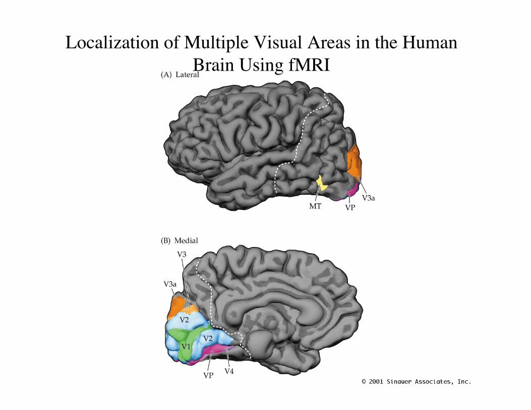
Localization of Multiple Visual Areas in the HumanBrain Using fMRI
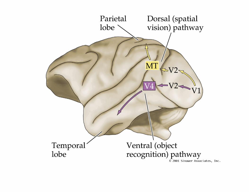
Organization of the Dorsal andVentral Visual Pathways
• PN12170.JPG
Weird visual defects• Disruptions in V1 cause blindness, but some people can
“guess” what an object is. Implies that other projectionsfrom eye to brain can somehow compensate.
• Cerebral achromatopsia- do not see in color-only black andwhite. Legions in extrastriate cortex regions like V4 or inventral stream.
• Lesions in MT region cause defects in detecting motion.Hard to pour drinks accurately.