Embed Size (px)
Citation preview

Anatomy of the Muscular System
Lindsey BilyAnatomy & Physiology
Austin High School

Muscular System Is composed of the large mass of skeletal
muscle that moves the framework of the body.
When we do anything that we “will” (walk, talk, breathe, run, etc.) we do so by contraction of our skeletal muscles.
More than 600 skeletal muscles. Make up 40% to 50% of our body weight.
MUSCLE WEIGHS MORE THAN FAT!


Skeletal Muscle Structure Skeletal muscle
cells are also called muscle fibers
Each fiber is striated and has multiple nuclei
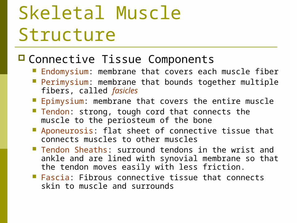
Skeletal Muscle Structure Connective Tissue Components
Endomysium: membrane that covers each muscle fiber Perimysium: membrane that bounds together multiple
fibers, called fasicles Epimysium: membrane that covers the entire muscle Tendon: strong, tough cord that connects the muscle to
the periosteum of the bone Aponeurosis: flat sheet of connective tissue that
connects muscles to other muscles Tendon Sheaths: surround tendons in the wrist and
ankle and are lined with synovial membrane so that the tendon moves easily with less friction.
Fascia: Fibrous connective tissue that connects skin to muscle and surrounds


Size, Shape, and Fiber Arrangement Skeletal muscles are organs. Contain muscle tissue, connective tissue, and
nervous tissue. Fibers can arrange themselves into different
patterns. Their pattern determines their function.
Parallel: fibers run parallel to the long axis of the muscle. Convergent: fibers converge to a narrow attachment. Pennate: fibers run obliquely, think of a feather. Bipennate: double feathered. Curved: fibers are curved.

Shape and Fiber Arrangement
Note: You do not need to know fusiform.

Attachment of Muscles Most muscles span at least one joint and
attach to both articulating bones. When contraction occurs, one bone
remains fixed and the other moves. Origin: the point of attachment that does
not move when the muscle contracts. Insertion: the point of attachment that
moves when the muscle contracts.

Muscle Actions Skeletal muscles almost always act in groups. Some relax while
others contract. We have functional classifications of muscles, based on their
movements. Prime mover or agonist: muscle or group of muscles that performs the
specific movement. (ex. Biceps brachii are the prime mover for flexion of the forearm)
Antagonist: muscle or group that relax when the prime movers are contracting. (ex. Triceps brachii are the antagonists when the biceps brachii are flexing the forearm)
If both the agonist and antagonist contract at the same time, you will rigidity and lack of movement.
Synergists: muscles that contract at the same time as the prime mover (agonist). The help the prime mover so that the movement is more effective. (ex. Brachialis is deep to the biceps brachii and helps to flex the elbow)
Fixator muscles: act as joint stabilizers. Help to maintain posture or balance during the contraction of the prime mover. (The trapezius and rhomboids stabilize the scapula so that it doesn’t move when the forearm is flexed)


How Muscles Are Named Muscles can be named using Latin or
English names Location (brachialis (arm)) Function (adductors of the thigh) Shape (deltoid refers to the Greek letter Delta
which is a triangle) Direction of Fibers (rectus abdominus) Number of Heads or Divisions (biceps brachii) Points of Attachment (sternocleidomastoid) Size of Muscle (teres major/teres minor)
Now starts the muscles you need to know….Muscles of Facial Expression
Occipitofrontalis: Raises the eyebrows and wrinkles the forehead horizontally.
Orbicularis oculi: encircles and closes the eye (blinking)
Orbicularis oris and the buccinator: pucker the mouth (kissing) and press the lips and cheeks against the teeth.

Muscles of Mastication Mastication means “to chew”. There are 3 muscles, but you only need to
know the…
Masseter: closes the jaw
The buccinator helps to keep food in the mouth as you chew.

Muscles that Move the Head There are 4 total muscles, but you only
need to know… Sternocleidomastoid: You have one on
each side of your body. Together they flex the head. Each one separately turns the head the opposite direction.
Splenius capitis: extends the head

Splenius capitis

Muscles of the Thorax External Intercostals: elevate ribs (breathe
in) Internal Intercostals: depress ribs (breathe
out) Diaphragm: enlarges the thorax. When
you breathe in, the diaphragm flattens out increasing the size and volume of the thorax so you can hold the air.


Muscles of the Abdominal Wall The muscles of the anterior and lateral
abdominal wall are arranged in 3 layers, with the fibers of each layer running in different directions.
This causes the wall to be very strong to cover and support the internal organs.

Muscles of the Abdominal Wall There are 5 total muscles, but you only need to
know…
External oblique: origin-ribs; insertion- pelvis: Maintains posture, rotates the trunk laterally. When the muscles lose their tone, you get poor posture and a protruding abdomen.
Rectus abdominus: Runs down the midline of the abdomen from the thorax to the pubis. Flexes the spinal column.

Abdominal Wall Muscles

Muscles Acting on the Shoulder Girdle There are 6 muscles that pass from the axial
skeleton to the shoulder girdle that attach the upper extremity to the body.
You need to know 2 of them… Trapezius: raises and lowers the shoulders to
“shrug” them. Serratus anterior: helps hold the scapula against
the thorax to prevent “winging” and is a strong abductor that is useful in pushing or punching movements.

Muscles Acting on the Shoulder Girdle
Trapezius

Muscles That Move the Upper Arm There are 9 muscles that move the upper arm
since it has such a wide range of motion. You need to know 6 of them…
Pectoralis major: origin- clavicle, sternum, and costal cartilage of the true ribs. Insertion is the humerus. Flexes the upper arm.
Latissimus Dorsi: origin- lower thoracic, lumbar and sacral vertebrae and the upper pelvis. Insertion-humerus. Extends the upper arm and adducts the upper arm posteriorly.
Muscles That Move the Upper Arm Deltoid: origin- clavicle and scapula.
Insertion- humerus. Abducts the upper arm.
Teres minor: origin- scapula. Insertion- humerus. Rotates the arm outward.
Teres major: origin- scapula. insertion-humerus. Assists in extension, adduction, and medial rotation of the arm.
Infraspinatus: origin- scapula. Insertion- humerus. Rotates the arm outward.

Muscles That Move the Upper Arm

Muscles That Move the Forearm There are 7 muscles that move the
forearm. You will need to know 2 of them…
Biceps brachii: origin- scapula. Insertion-proximal end of the radius. Flexes supinated forearm.
Triceps brachii: origin- scapula and humerus. Insertion- ulna. Extends the forearm.

Muscles that Move the Forearm

Muscles that Move the Wrist, Hand and Fingers Flexor carpi radialis: origin- humerus.
Insertion- second metacarpal. Flexes the hand and forearm.
Palmaris longus: origin-humerus. Insertion- fascia of palm. Flexes the hand.
Flexor carpi ulnaris: origin-humerus and ulna. Insertion- pisiform bone in the wrist. Flexes the hand.
Extensor digitorum: origin-humerus. Insertion- phalanges 2-5. Extends the fingers.

Muscles That Move the Wrist, Hand and Fingers

Carpal Tunnel Syndrome Tenosynovitis: inflammation of the tendon
sheath around the wrist . The inflammation presses on the median
nerve that connects the palm and thumb side of the hand.
Symptoms are pain, tingling in the hand and all the way up to the shoulder.
Control inflammation through anti-inflammatory agents or surgery by removing the inflamed tissue around the nerve.

Carpal Tunnel Syndrome

Muscles that Move the Thigh Rectus Femoris: origin-anterior pelvis. Insertion-tibia by way
of the patellar tendon. Flexes the thigh and extends the lower leg.
Gluteus Maximus: origin-posterior, superior pelvis, sacrum, & coccyx. Insertion-femur and iliotibial tract. Extends the thigh and rotates it outward.
Tensor faciae latae: origin-anterior surface of pelvis. Insertion- tibia by way of the iliotibial tract. Abducts the thigh and tightens the iliotibial tract.
Adductors of thigh: insertion-pubic bone. Insertion- femur. Adducts the thigh.
Gracialis- insertion-just below the pubic symphysis. Insertion- tibia. Adducts the thigh and flexes the lower leg.

Muscles that Move the Thigh

Muscles that Move the Thigh
Adductors of the Thigh
Gracialis

Iliotibial Tract You may have heard it being called the IT
Band. A thick band of fibrous tissue that runs
from the hip to the outside of the tibia just below the knee.
It helps to stabilize the knee. It can become inflamed and very painful
around the knee. Seen frequently in endurance athletes.

Iliotibial Tract
Stretches

Muscles That Move the Lower Leg Quadriceps Femoris Group
Includes Vastus Lateralis, Vastus Medialus, Rectus Femoris
Hamstring Group Includes Biceps Femoris, Semitendinosus, and
Semimembranosus Sartorius: origin-anterior side of hip.
Insertion-tibia. Adducts and flexes the leg.
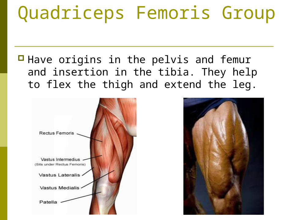
Quadriceps Femoris Group
Have origins in the pelvis and femur and insertion in the tibia. They help to flex the thigh and extend the leg.
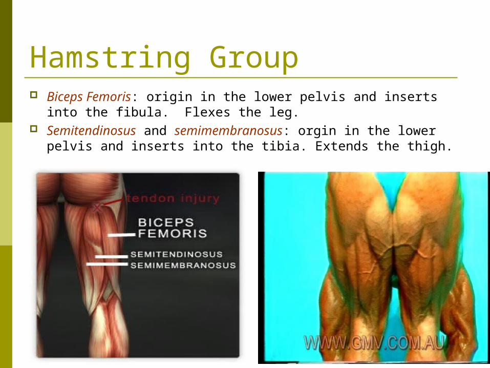
Hamstring Group Biceps Femoris: origin in the lower pelvis and inserts into
the fibula. Flexes the leg. Semitendinosus and semimembranosus: orgin in the lower
pelvis and inserts into the tibia. Extends the thigh.

Sartorius Origin on the hip and insertion on the tibia.
Adducts and flexes the leg and permits crossing of the legs Indian Style.

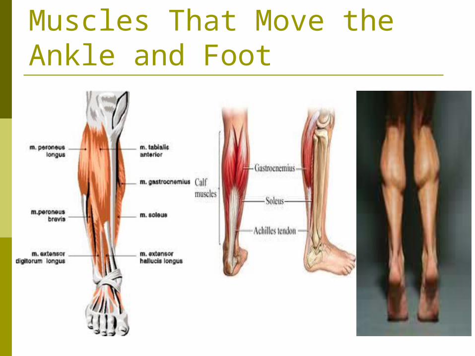
Muscles that Move the Ankle and Foot Tibialis Anterior: origin-tibia. Insertion-
tarsal. Flexes the foot. “Shin splints” Gastrocnemius: origin-femur. Insertion-
tarsal. Extends the foot. Forms part of the calf.
Soleus: origin-tibia (underneath the gastrocnemius). Insertion- tarsal. Plantar flexion. Forms part of the calf.
Muscles That Move the Ankle and Foot

Calcaneal Tendon Also known as the “Achilles Tendon”. The gastrocnemius and the soleus share
this common tendon distally that inserts into the calcaneal bone (heel bone).