Embed Size (px)
Citation preview
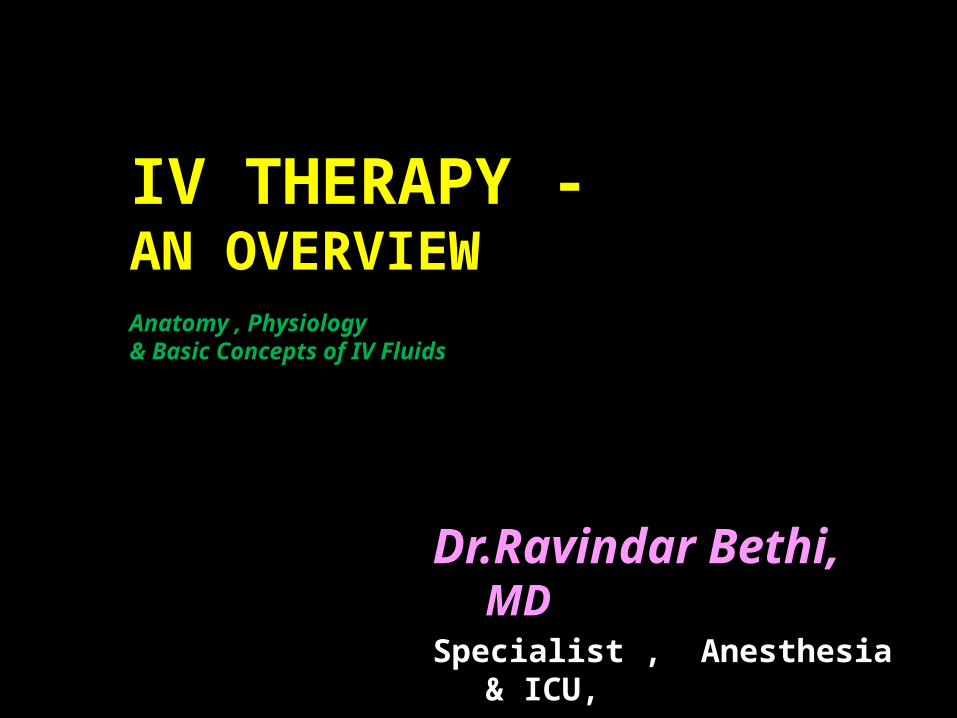
Anatomy , Physiology& Basic Concepts of IV Fluids
Dr.Ravindar Bethi, MDSpecialist , Anesthesia & ICU,Al Rass General Hospital, KSA.
IV THERAPY - AN OVERVIEW

IV THERAPY - AN OVERVIEW
Intravenous therapy or IV therapy is the giving of liquid substances
directly into a vein.

IV THERAPY - AN OVERVIEW
Compared with other routes of administration, the intravenous route is
the fastest way to deliver fluids and medications throughout the body.

Before we get started…Safe work aids project…
• http://www.friendtofriend.org/drugs/vein-care.html
• Harm reduction (or less commonly known as harm minimisation) refers to a range of public health policies designed to reduce the harmful consequences associated with recreational drug use and other high risk activities. Harm reduction is put forward as a useful perspective alongside the more conventional approaches of demand and supply reduction.[1]
• Many advocates argue that prohibitionist laws criminalize people for suffering from a disease and cause harm, for example by obliging drug addicts to obtain drugs of unknown purity from unreliable criminal sources at high prices, increasing the risk of overdose and death.[2] Its critics are concerned that tolerating risky or illegal behaviour sends a message to the community that these behaviours are acceptable
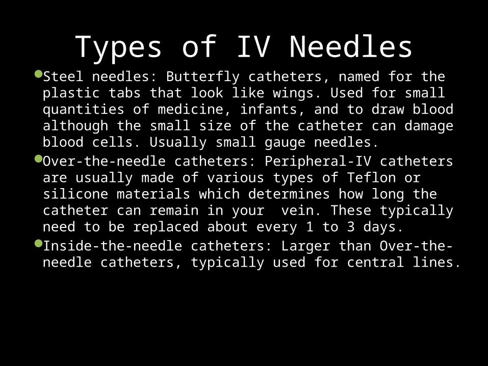
Types of IV NeedlesSteel needles: Butterfly catheters, named for the plastic tabs
that look like wings. Used for small quantities of medicine, infants, and to draw blood although the small size of the catheter can damage blood cells. Usually small gauge needles.
Over-the-needle catheters: Peripheral-IV catheters are usually made of various types of Teflon or silicone materials which determines how long the catheter can remain in your vein. These typically need to be replaced about every 1 to 3 days.
Inside-the-needle catheters: Larger than Over-the-needle catheters, typically used for central lines.

Gauges
• Needles & Catheters are sized by diameters which are called gauges.
• Smaller diameter = larger gauge• IE: 22-gauge catheter is smaller than a 14-gauge• Larger diameter = more fluid able to be
delivered• If you need to deliver a large amount of fluid,
typically 14- or 16-gauge catheters are used.

Intravenous Catheter Complications
• About 25 million Americans have intravenous catheters placed each year. Intravenous catheters (or IVs) are a very important part of medical treatment for acute illnesses, cancer, surgery, anesthesia, and trauma, allowing medications to reach as quickly and effectively as possible, via the bloodstream, the parts of the body where they work.
Intravenous Catheter Complications
• IV catheters can be placed in a hand, arm or leg. These are known as "peripheral" IVs. IVs placed in the central circulation, like the internal jugular vein (neck) or subclavian vein (just beneath the collar bone), are known as "central lines". The rest of this article will deal with peripheral IVs, the most common type of IV.

Intravenous Catheter Complications
• During the placement of an IV, a needle is inserted through the skin and into an accessible blood vessel. A Teflon (plastic) cannula is then slid over the needle, which is withdrawn. No needle remains in your body. (So-called "butterfly" needles are an exception to this). Some healthcare providers use a little bit of local anesthetic beforehand, with a very tiny needle, to numb the area of skin where the IV is inserted. Local anesthetic cream is sometimes applied 45-60 minutes beforehand to achieve the same effect. This is particularly helpful in the care of children.

Complications
• Local Infection • In any case where there is an open wound on the body,
disrupting the protective lining of skin, an infection can occur. A microscopic organism may use the tiny hole in the skin created by the IV catheter to find its way into the body, and cause an infection. Common signs of local infection ("abscess") include a large lump that is painful and hot to touch.
• Treatment - If you suspect an infection, see your healthcare provider immediately. Antibiotics may be used to control the bacterial infection.

Complications
• Infiltration • This occurs when the catheter unintentionally enters the tissue
surrounding the blood vessel. In this case the IV fluid and associated medications will go into the tissues and there will be a lump where the IV has been inserted. It would be cool to touch (this differentiates it from inflammation due to infection, which is warm to the touch).
• Treatment - If you notice this inform your healthcare professional and they will administer appropriate care immediately. Infiltrated IVs are not a big problem usually unless the medication being administered is very irritant, such as certain chemotherapy and circulatory medicines. The intravenous infusion must be stopped, obviously, to avoid putting any more fluid or medication into the tissues. Another IV may need to be started elsewhere.

Complications
• Hematoma • A hematoma is a collection of blood caused by internal
bleeding. This happens when the catheter punctures through the vein and causes a hematoma. Hematomas generally occur with unsuccessful IV insertion but can also happen when an IV is taken out. This is why pressure must be applied to the insertion site, to try to make the hematoma as small as possible.
• A hematoma may appear as a visible bruise or a lump. • Treatment - A hematoma normally recovers over time (a
few hours or days) without treatment.

Complications
• Nerve Damage • It is also possible for the IV needle to penetrate and injure a nerve, and for
bruising and bleeding to irritate a nerve. Nerves are invisible from the skin surface so it?s easy to understand how this could happen. If you feel a sudden sharp pain radiating along your arm as the IV is inserted, let your healthcare provider know immediately as this may be a sign that the needle has come into contact with a nerve.
• A 1996 study of 419,000 blood donations showed that 1 in every 6300 donors had a nerve injury. Fortunately, most got better within a month. The symptoms included excessive or radiating pain, and loss of arm or hand strength. Fifty-two of 56 donors achieved a full recovery, and 4 other donors had only a mild, localized, residual numbness.
• Treatment - Nerve damage tends to repair itself in a few weeks to a few months. If you suspect a nerve injury contact your doctor. In rare instances (such as persistent weakness) specific treatment, even surgery, may become necessary.

In classical terms, thrombosis is caused by abnormalities in one or more of the following (Virchow's triad):
• The composition of the blood (hypercoagulability) • Quality of the vessel wall (endothelial cell injury) • Nature of the blood flow
The formation of a thrombus is usually caused by Virchow's triad. To elaborate, the pathogenesis includes: an injury to the vessel's wall (such as by trauma, infection, or turbulent flow at bifurcations); by the slowing or stagnation of blood flow past the point of injury (which may occur after long periods of sedentary behavior—for example, sitting on a long airplane flight); by a blood state of hypercoagulability (caused for example, by genetic deficiencies or autoimmune disorders).

IV THERAPY - AN OVERVIEW
It is commonly referred to as a drip because it employs a
drip chamber, which prevents
air entering the blood stream (air embolism)
and allows an estimate of flow rate.
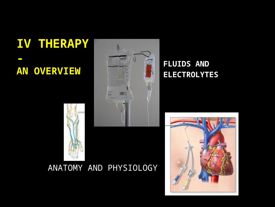
FLUIDS AND ELECTROLYTES
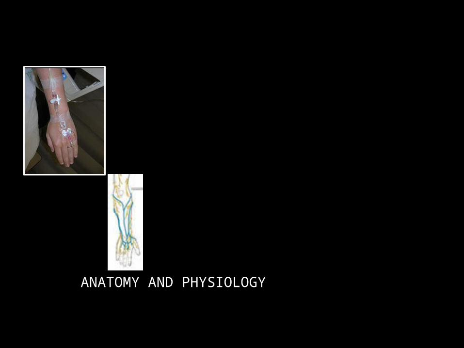
ANATOMY AND PHYSIOLOGY
IV THERAPY - AN OVERVIEW

ANATOMY AND PHYSIOLOGY
Dorsal venous arch

ANATOMY AND PHYSIOLOGY
Basilic vein

ANATOMY AND PHYSIOLOGY
Cephalic vein

ANATOMY AND PHYSIOLOGY
dorsal veins of forearm
ANATOMY AND PHYSIOLOGY

ANATOMY AND PHYSIOLOGY
Medial cubital vein

ANATOMY AND PHYSIOLOGY
Medial cubital vein
Brachial artery

ANATOMY AND PHYSIOLOGY
Medial cubital vein
Brachial artery
Median Nerve

ANATOMY AND PHYSIOLOGY
Dorsal venous arch
Great Saphenous Vein
Femoral Vein

ANATOMY AND PHYSIOLOGY
Scalp Veins

…the new access site has to be proximal to the "blown" area to prevent extravasation of medications through the damaged vein…
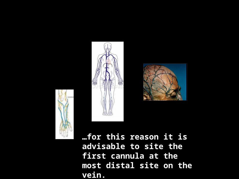
…for this reason it is advisable to site the first cannula at the most distal site on the vein.
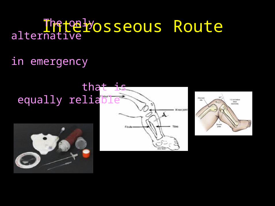
Interosseous Route The only alternative in emergency that is equally
reliable

ANATOMY AND PHYSIOLOGY
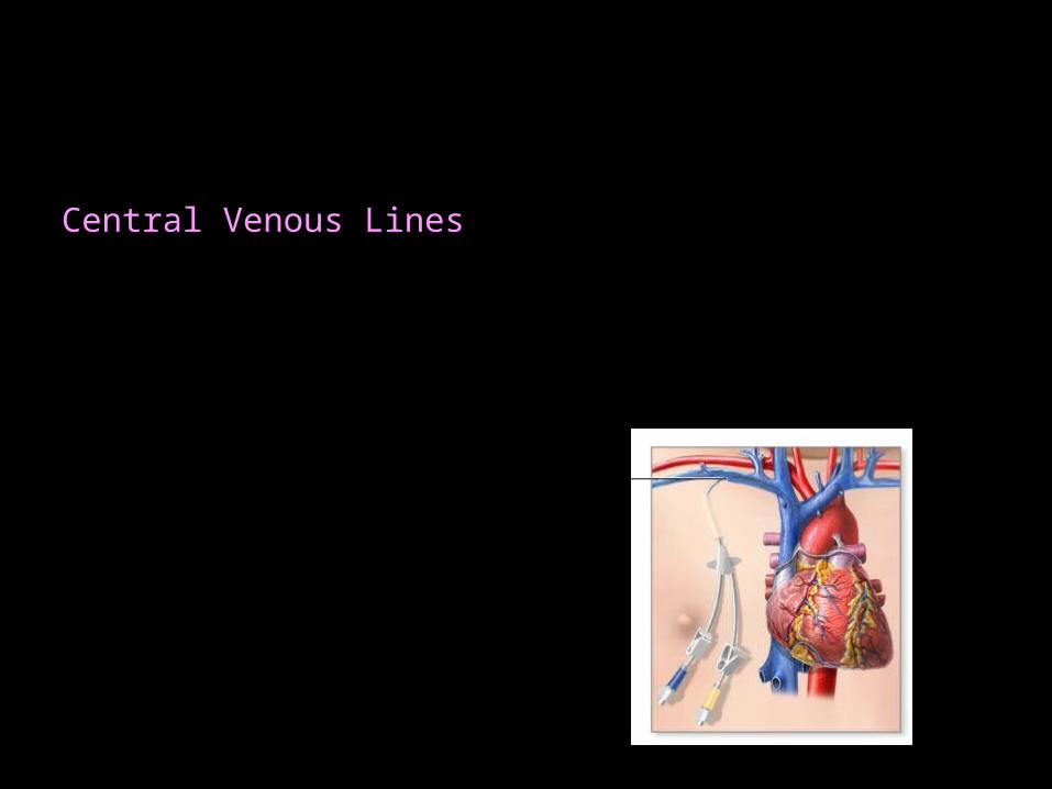
Central Venous LinesCentral Lines flow through a catheter with its tip
within a large vein, usually the superior vena cava or inferior vena cava, or within
the right atrium of the heart.

Central Venous LinesCentral Lines flow through a catheter with its tip
within a large vein, usually the superior vena cava or inferior vena cava, or within
the right atrium of the heart.

Central Venous LinesCentral Lines flow through a catheter with its tip
within a large vein, usually the superior vena cava or inferior vena cava, or within
the right atrium of the heart.
ADVANTAGES• Fluids irritating to peripheral veins can
be given• Chemotherapy• Total parenteral nutrition
• Medications reach the heart
immediately, and are quickly distributed to the rest of the body.
• Central venous pressure can be
measured

Central Venous LinesCentral Lines flow through a catheter with its tip
within a large vein, usually the superior vena cava or inferior vena cava, or within
the right atrium of the heart.
DISADVANTAGES
• Risks of bleeding, infection, air embolism.
• Technically difficult– • needs experienced clinician
knowing the appropriate landmarks and/or
• using an ultrasound probe to safely locate and enter the vein.
• Pleura and carotid artery are at risk of damage with the potential for pneumothorax or puncture/ cannulation of the artery.

Central Venous LinesCentral Lines flow through a catheter with its tip
within a large vein, usually the superior vena cava or inferior vena cava, or within
the right atrium of the heart.
INTERNAL JUGULAR• Nursing care• Be cautious with potassium

Central Venous LinesCentral Lines flow through a catheter with its tip
within a large vein, usually the superior vena cava or inferior vena cava, or within
the right atrium of the heart.
SUBCLAVIAN• Nursing care is easier• Open even in shock• Incompressible

Central Venous LinesCentral Lines flow through a catheter with its tip
within a large vein, usually the superior vena cava or inferior vena cava, or within
the right atrium of the heart.
FEMORAL
• Emergency situations where it is difficult to cannulate Internal jugular vein or Subclavian vein
• High risk of infection
• Preferred for potassium infusions

Central Venous LinesCentral Lines flow through a catheter with its tip
within a large vein, usually the superior vena cava or inferior vena cava, or within
the right atrium of the heart.

Central Venous Line Vs Pulmonary Artery Catheter

Central Venous LinesCentral Lines flow through a catheter with its tip
within a large vein, usually the superior vena cava or inferior vena cava, or within
the right atrium of the heart.
Peripherally inserted central catheterADVANTAGES
• Safer to insert with a relatively low risk of uncontrollable bleeding no risks of damage to the lungs or major blood vessels.
• With proper hygiene, care, can be left in place for several weeks for patients who require extended treatment.
Some special types of

Central Venous LinesCentral Lines flow through a catheter with its tip
within a large vein, usually the superior vena cava or inferior vena cava, or within
the right atrium of the heart.
Peripherally inserted central catheterDISADVANTAGES
• Must travel through a relatively small peripheral vein which can take a less predictable course on the way to the superior vena cava . Hence, more technically difficult to place in some patients.
• Travels through the axilla.
Hence, can become kinked causing poor function.
Some special types of

Central Venous LinesCentral Lines flow through a catheter with its tip
within a large vein, usually the superior vena cava or inferior vena cava, or within
the right atrium of the heart.
Tunneled LinesHickman line or Broviac catheter
• “Tunneled" under the skin to emerge a short distance away. from the central vein
• Reduced risk of infection, since bacteria from the skin surface are not able to travel directly into the vein;
• Catheters are also made of materials that resist infection and clotting.
A Hickman line in a leukemia patient.
It is tunneled under the skin to the jugular vein
Some special types of

Central Venous LinesCentral Lines flow through a catheter with its tip
within a large vein, usually the superior vena cava or inferior vena cava, or within
the right atrium of the heart.
Implantable ports• Silicone rubber reservoir, implanted
under the skin. • Medication is injected via its silicone
cover, into the reservoir. • The cover can accept several
hundreds of needle sticks during its lifetime. It is possible to leave the ports in the patient's body for years.
Some special types of

Central Venous LinesCentral Lines flow through a catheter with its tip
within a large vein, usually the superior vena cava or inferior vena cava, or within
the right atrium of the heart.
Implantable ports
• Needs regular maintenance. If it is plugged a thrombus can form with the accompanying risk of embolisation
• Commonly used for patients on long-term intermittent treatment.
Some special types of
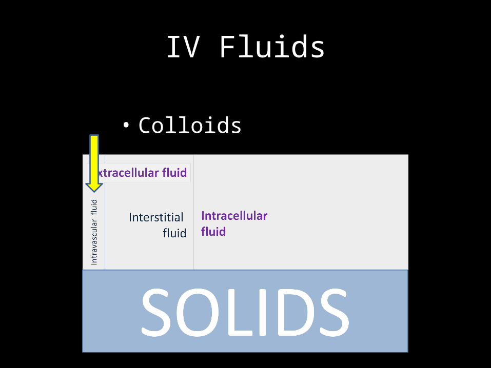
IV Fluids
• Colloids

IV Fluids
• Crystalloids

IV Fluids
• Colloids
• Contain larger insoluble molecules, such as albumen.
• Preserve a high colloid osmotic pressure in the blood
• Blood itself is a colloid.

IV Fluids
• Colloids

IV Fluids
• Crystalloids
• Aqueous solutions of water-soluble molecules.
• The most commonly used crystalloid fluid is normal saline=, a solution of sodium chloride at 0.9% concentration, which is close to the concentration in the blood (isotonic).
• What is isotonic? • What is Iso-osmolar ?

IV Fluids
• Crystalloids

IV Fluids
• Crystalloids

IV Fluids
• Crystalloids
• Fluid of choice in multiple situations• Trauma• Metabolic alkalosis
• Not to be given in hyperchloremic acidosis
isotonic

IV Fluids
• Crystalloids
hypotonic

IV Fluids
• Crystalloids
? Isotonic/ Hypotonic
• Isotonic in vitro• Hypotonic in vivo
• Iso-osmolar , compared to Normal Saline • Hypotonic to the human cells due to Insulin
• Hypertonic in insulin deficiency

IV Fluids
• Crystalloids
? Isotonic/ Hypertonic ?

IV Fluids
• Crystalloids
Contains calcium, potassium and Lactate
• Don’t give in alkalosis• Don’t give in hyperkalemia• Don’t give with Blood• Mind its Calcium content, when
giving with Mg therapy
Nearly Isotonic

• Don’t give potassium therapy with Dextrose containing solutions
IV Fluids
• Crystalloids
• When giving Dextrose containing solutions, add KCl to prevent hypokalemia
• When giving KCl in the treatment of hypokalemia, don’t add it to solutions containing Dextrose.

Distribution of fluid in human body
Colloids stay here
Crystalloids move up to
here

Risks and complications of IV THERAPY
1. Infection2. Phlebitis3. Infiltration and extravasation4. Embolism5. Fluid overload6. Electrolyte Imbalance

Electrolytes
• Sodium 135 – 145 mmol/L
• Potassium 3.5 – 5.0 mmol/L
• Calcium 2.12 – 2.75 mmol/L ( Ionised calcium 1.0-1.3 mmol/L) • Magnesium 1.5 – 2.2 m Eq/L
• Phosphorous 0.81 – 1.20 mmol/L

Electrolytes
• Sodium 135 – 145 mmol/L
• Potassium 3.5 – 5.0 mmol/L
• Calcium 2.12 – 2.75 mmol/L • Magnesium 1.5 – 2.2 m Eq/L
• Phosphorous 0.81 – 1.20 mmol/L
Low sodium – lower osmolality
High sodium – higher osmolality

• Sodium 135 – 145 mmol/L
• Potassium 3.5 – 5.0 mmol/L
• Calcium 2.12 – 2.75 mmol/L ( Ionised calcium 1.0-1.3 mmol/L) • Magnesium 1.5 – 2.2 m Eq/L
• Phosphorous 0.81 – 1.20 mmol/L
Electrolytes
Hypokalemia
Hyperkalemia

Hyperkalemia
• Sodium 135 – 145 mmol/L
• Potassium 3.5 – 5.0 mmol/L
• Calcium 2.12 – 2.75 mmol/L ( Ionised calcium 1.0-1.3 mmol/L) • Magnesium 1.5 – 2.2 m Eq/L
• Phosphorous 0.81 – 1.20 mmol/L
BE GOOD IN CLINICALSKILLS
KEEPDRUGSAWAY
• Bicarbonate• Glucose +• Insulin• Calcium• Sorbitol
• Keyexalate• Dialysis• Albuterol
ACLS - 2006

Electrolytes
• Sodium 135 – 145 mmol/L
• Potassium 3.5 – 5.0 mmol/L
• Calcium 2.12 – 2.75 mmol/L ( Ionised calcium 1.0-1.3 mmol/L) • Magnesium 1.5 – 2.2 m Eq/L
• Phosphorous 0.81 – 1.20 mmol/L

THANK YOURAVINDAR BETHI

![Yeshwanth Ravi Theja Bethi arXiv:1912.08519v1 [cs.CV] 18 Dec … · 2019-12-19 · Yeshwanth Ravi Theja Bethi Department of Electronics and Systems Engineering Indian Institue of](https://img.pdfslide.net/doc/110x75/5f329fc86f20316ff519d60d/yeshwanth-ravi-theja-bethi-arxiv191208519v1-cscv-18-dec-2019-12-19-yeshwanth.jpg)