Embed Size (px)
Citation preview
http://www.diva-portal.org
This is the published version of a paper published in Journal of Infectious Diseases.
Citation for the original published paper (version of record):
Tissera, H., Rathore, A P., Leong, W Y., Pike, B L., Warkentien, T E. et al. (2017)Chymase Level Is a Predictive Biomarker of Dengue Hemorrhagic Fever in Pediatricand Adult Patients.Journal of Infectious Diseases, 216(9): 1112-1121https://doi.org/10.1093/infdis/jix447
Access to the published version may require subscription.
N.B. When citing this work, cite the original published paper.
Permanent link to this version:http://urn.kb.se/resolve?urn=urn:nbn:se:umu:diva-143606
The Journal of Infectious Diseases
1112 • JID 2017:216 (1 November) • Tissera et al
The Journal of Infectious Diseases® 2017;216:1112–21
Chymase Level Is a Predictive Biomarker of Dengue Hemorrhagic Fever in Pediatric and Adult PatientsHasitha Tissera,1,2 Abhay P. S. Rathore,3,a Wei Yee Leong,4 Brian L. Pike,5 Tyler E. Warkentien,5 Farouk S. Farouk,7 Ayesa Syenina,3 Eng Eong Ooi,3 Duane J. Gubler,3 Annelies Wilder-Smith,4,9 and Ashley L. St. John3,6,8
1Epidemiology Unit, Ministry of Health, and 2National Dengue Control Unit, Colombo, Sri Lanka; 3Programme in Emerging Infectious Diseases, Duke–National University of Singapore, 4Lee Kong Chian School of Medicine, Nanyang Technological University, 5US Naval Medical Research Center–Asia, and 6Department of Microbiology and Immunology, Young Loo Lin School of Medicine, National University of Singapore, Singapore; 7Graduate Program in Global Health, Duke University, and 8Department of Pathology, Duke University Medical Center, Durham, North Carolina; and 9Department of Public Health and Clinical Medicine, Epidemiology and Global Health, Umeå University, Sweden
Background. Most patients with dengue experience mild disease, dengue fever (DF), while few develop the life-threatening diseases dengue hemorrhagic fever (DHF) or dengue shock syndrome (DSS). No laboratory tests predict DHF or DSS. We evaluated whether the serum chymase level can predict DHF or DSS in adult and pediatric patients and the influence of preexisting conditions (PECs) on chymase levels.
Methods. Serum chymase levels were measured in patients presenting with undifferentiated fever to hospitals in Colombo District, Sri Lanka. The value of serum the chymase concentration and clinical signs and symptoms as predictors of DHF and/or DSS was evaluated by multivariate analysis. We assessed the influence of age, PECs, and day after fever onset on the robustness of the chymase level as a biomarker for DHF and/or DSS.
Results. An elevated chymase level in acute phase blood samples was highly indicative of later diagnosis of DHF or DSS for pedi-atric and adult patients with dengue. No recorded PECs prevented an increase in the chymase level during DHF. However, certain PECs (obesity and cardiac or lung-associated diseases) resulted in a concomitant increase in chymase levels among adult patients with DHF.
Conclusions. These results show that patients with acute dengue who present with high levels of serum chymase consistently are at greater risk of DHF. The chymase level is a robust prognostic biomarker of severe dengue for adult and pediatric patients.
Keywords. dengue virus; dengue fever; DHF/DSS; prognosis; biomarker; chymase; mast cells; pediatric dengue.
Dengue is an arboviral infection spread by mosquitoes in the tropical and subtropical regions of the world [1]. It is caused by dengue virus (DENV), which has 4 distinct sero-types. Between 50 million and 400 million DENV infections occur yearly [2]. In DENV-endemic countries, such as the site of this study, Sri Lanka [3], many patients experience multi-ple DENV infections in a lifetime. An incubation period of approximately 4–7 days occurs before fever onset [4, 5]. Signs such as leukopenia, thrombocytopenia, and rash and symp-toms such as headache, vomiting, and retro-orbital eye pain are common during acute disease and usually resolve within
3–7 days for those who experience mild disease, diagnosed as dengue fever (DF) [6, 7]. Mild hemorrhagic manifestations such as petechiae and purpura are also common during this time [6, 7]. Resolution of the febrile response usually occurs around days 4–7, when most patients completely recover, but other individuals can suddenly experience complications [6]. Warning signs have been identified to aid in recogniz-ing patients on course to develop dengue hemorrhagic fever (DHF) or dengue shock syndrome (DSS), also known as severe dengue [8]. Warning signs include mucosal bleeding, liver enlargement, abdominal pain, rapid hematocrit increase, persistent vomiting, and clinical fluid accumulation [8]. Many of these signs are subjective and present very late. DHF and DSS are extremely difficult to predict, and there is an urgent need to identify risk factors and biomarkers of severe dengue. Although some studies associated high serum viral titers with DHF and DSS [9, 10], others have reported conflicting infor-mation [11–13]. Multiple factors could explain these discrep-ancies, including faster clearance of viremia during secondary infection [14, 15]. There is an unmet need for diagnostic tests to identify severe DENV during the acute phase of disease, since clinical symptoms often fail to predict it with sufficient warning, and to understand whether those tests are robust in multiple patient populations.
M A J O R A R T I C L E
© The Author 2017. Published by Oxford University Press for the Infectious Diseases Society of America. This is an Open Access article distributed under the terms of the Creative Commons Attribution-NonCommercial-NoDerivs licence (http://creativecommons.org/licenses/ by-nc-nd/4.0/), which permits non-commercial reproduction and distribution of the work, in any medium, provided the original work is not altered or transformed in any way, and that the work is properly cited. For commercial re-use, please contact [email protected]: 10.1093/infdis/jix447
Received 1 July 2017; editorial decision 20 August 2017; accepted 22 August 2017; published online August 25, 2017.
aPresent affiliation: Department of Pathology, Duke University Medical Center, Durham, North Carolina.
Correspondence: A. L. St. John, PhD, Program in Emerging Infectious Diseases, Duke–National University of Singapore Graduate Medical School, 8 College Rd, Level 9, Singapore ([email protected]).
Downloaded from https://academic.oup.com/jid/article-abstract/216/9/1112/4093871by Umea university library, Medical library useron 04 January 2018
Chymase Level Is a Predictive Biomarker of DHF • JID 2017:216 (1 November) • 1113
Differences in the severity of DENV disease between adult and pediatric cases have been identified [16, 17]. For example, in recent outbreaks in the Philippines, the symptomatic disease burden primarily involved children [18]. In Thailand, signs such as epistaxis, oliguria, and liver enlargement were more prevalent among children than among adults [17]. It is unclear whether there may be differences in the pathophysiological mechanisms in pediatric and adult patients with dengue or whether prior immunity in the adult population can account for the observed differences in severity. One study suggested that children have an inherently higher capillary fragility, possibly explaining why small children present more often with DSS than adults do [19]. In contrast, in some populations, including residents of Singapore, morbidity and mortality are more common in adult and elderly patients, potentially because of the increased inci-dence of comorbidities [20, 21]. Preexisting conditions (PECs) such as diabetes [22] and obesity [23] have also been associ-ated with severe disease in some studies. The observation in the CYD-TDV (Dengvaxia) trials that a greater percentage of chil-dren aged 2–5 years than those >5 years required hospitaliza-tion within 3 years of infection emphasizes the importance of evaluating DENV diagnostic tests in multiple age groups [24].
In humans, immune pathology is thought to underlie severe disease [7]. One immune cell type, the mast cell (MC), is criti-cal to the host response to DENV [25–27]. MCs are granulated cells that regulate the host vasculature during inflammation and homeostasis [28]. DENV activation of MCs prompts them to degranulate, which involves the release of their presynthesized inflammatory mediators from intracellular stores [26]. Most granule-associated mediators are proteases, including chymase, which is highly specific to MCs [29]. Chymase is a serine pro-tease and angiotensin-converting enzyme [30]. MC degranula-tion occurs rapidly. During anaphylaxis, MC-derived mediators are the underlying cause of shock and death and can be detected in the serum as biomarkers [31]. Our prior studies showed that DENV activates MCs, resulting in chymase release, and that MC activation is augmented by DENV-specific antibodies, as are present during secondary infections [25–27, 32]. This led to the observation in a retrospective study of adult DENV-infected patients in Singapore that chymase levels were elevated in acute-phase serum samples from patients with DHF [25].
Here, we questioned whether the MC-derived protease chymase is a robust predictor of DHF and/or DSS in multi-ple patient populations and we assessed whether PECs atten-uate the predictive capacity of chymase. Febrile patients were recruited at study sites in Colombo District, Sri Lanka [3, 33]. Patients’ signs and symptoms were recorded, and their disease severity was documented. Acute-phase serum samples were used to measure the chymase level to assess its potential to pre-dict DHF and/or DSS. Our analysis reveals that the chymase level is a prognostic biomarker for DHF and DSS in pediatric and adult patients, including those with PECs.
METHODS
Recruitment of and Collection of Samples From Patients With Dengue
Samples were prospectively obtained by the Ministry of Health in Sri Lanka and provided by the Dengue Tools Project [3, 33]. The Dengue Tools Project recruited patients with undifferenti-ated febrile illness with a duration of <7 days, the onset of which was self-reported and may have been subjective. The number recruited was capped at 60 patients per week, with 10 from each study site. All patients provided blood samples with informed consent, which were anonymized. Samples from 347 patients were used, from 6 clinical sites in the Colombo district of Sri Lanka (3 government hospitals and 3 general practitioner’s clinics). Ethical approval was obtained from the Ethics Review Committee, Faculty of Medicine, University of Colombo, Sri Lanka, and the National University of Singapore Institutional Review Board. Parental consent was obtained for participants aged <18 years. Serum samples collected from patients at the time of recruitment were selected for chymase testing if the patient had had fever for <6 days and a clear discharge diagno-sis of either DF or DHF (based on World Health Organization guidelines [34]), which was available for patients who were hos-pitalized but not for all patients who were not. DENV confir-mation was previously described in detail by Tissera et al [3]. Serum was isolated and stored at −80°C.
Serum Chymase Quantification
Serum chymase levels were measured using the Human Mast Cell Chymase I (CMA-I) ELISA Kit (BlueGene Biotech) accord-ing to the manufacturer’s instructions.
Data Analysis
SPSS (IBM) was used to generate demographic statistics and perform relative operating characteristic (ROC) curve analysis. For multivariate and univariate analyses, logistic regression was performed, with diagnosis (DF or DHF) as a dependent vari-able, using STATA, version 14. Prism software was used to com-pare mean chymase levels among groups by 1- or 2-way analysis of variance. Results were considered statistically significant if P values were <.05. G*Power software was used for post hoc anal-yses, to verify an observed power of >0.8.
RESULTS
Elevated Serum Chymase Level, a Predictor of DHF
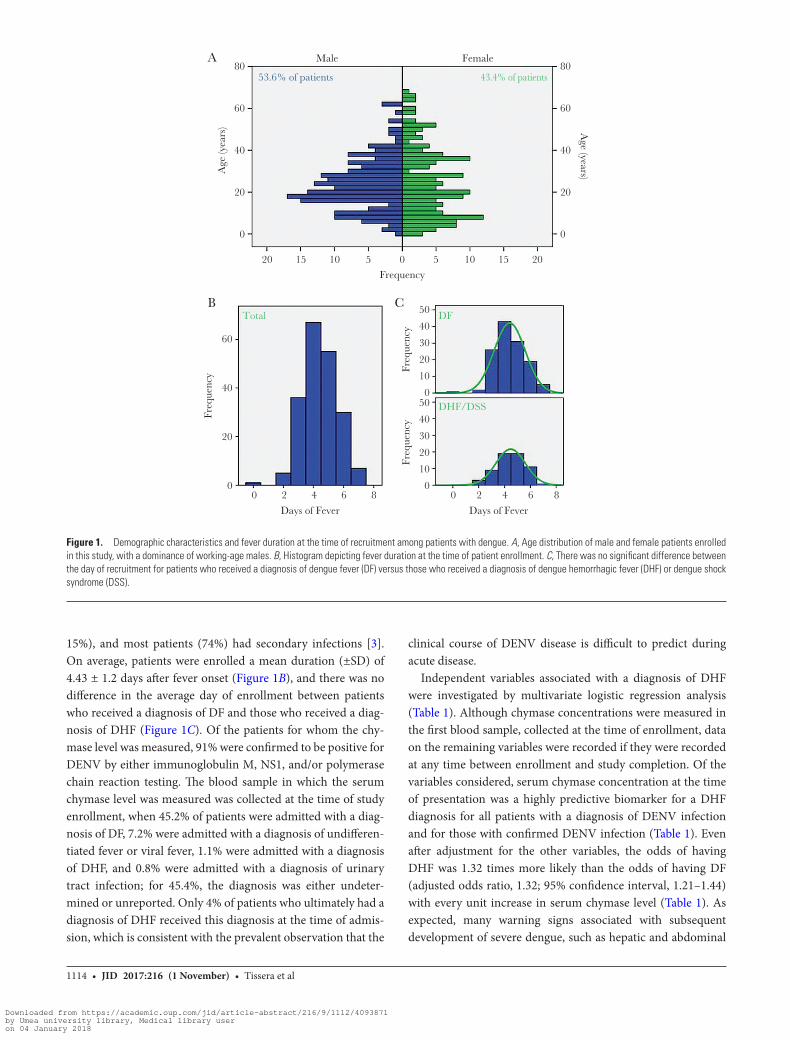
We measured the levels of chymase in the sera of 347 DENV-infected patients (mean age [±SD], 22.7 ± 14.8 years; Figure 1A) who had had fever for ≤6 days at the time of enrollment. Of this cohort, 99 (28.6%) received a diagnosis of DHF as a final discharge diagnosis. Among patients with DF, 15 of 223 (6.7%) also experienced mild hemorrhagic manifestations that did not meet the diagnosis criteria for DHF, which was termed “DF with bleeding” (DFWB). The dominant serotype of infection was DENV-1 (in 85% of infections), followed by DENV-4 (in
Downloaded from https://academic.oup.com/jid/article-abstract/216/9/1112/4093871by Umea university library, Medical library useron 04 January 2018
1114 • JID 2017:216 (1 November) • Tissera et al
15%), and most patients (74%) had secondary infections [3]. On average, patients were enrolled a mean duration (±SD) of 4.43 ± 1.2 days after fever onset (Figure 1B), and there was no difference in the average day of enrollment between patients who received a diagnosis of DF and those who received a diag-nosis of DHF (Figure 1C). Of the patients for whom the chy-mase level was measured, 91% were confirmed to be positive for DENV by either immunoglobulin M, NS1, and/or polymerase chain reaction testing. The blood sample in which the serum chymase level was measured was collected at the time of study enrollment, when 45.2% of patients were admitted with a diag-nosis of DF, 7.2% were admitted with a diagnosis of undifferen-tiated fever or viral fever, 1.1% were admitted with a diagnosis of DHF, and 0.8% were admitted with a diagnosis of urinary tract infection; for 45.4%, the diagnosis was either undeter-mined or unreported. Only 4% of patients who ultimately had a diagnosis of DHF received this diagnosis at the time of admis-sion, which is consistent with the prevalent observation that the
clinical course of DENV disease is difficult to predict during acute disease.
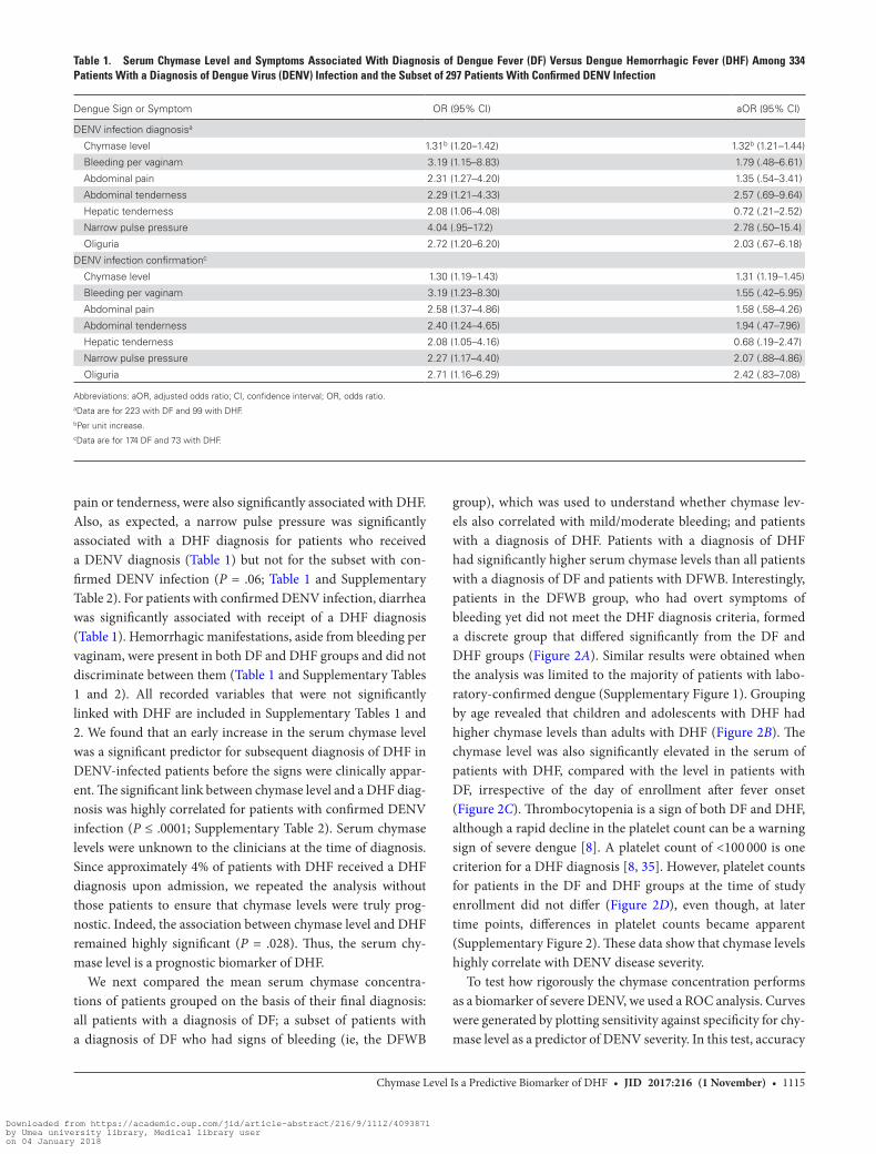
Independent variables associated with a diagnosis of DHF were investigated by multivariate logistic regression analysis (Table 1). Although chymase concentrations were measured in the first blood sample, collected at the time of enrollment, data on the remaining variables were recorded if they were recorded at any time between enrollment and study completion. Of the variables considered, serum chymase concentration at the time of presentation was a highly predictive biomarker for a DHF diagnosis for all patients with a diagnosis of DENV infection and for those with confirmed DENV infection (Table 1). Even after adjustment for the other variables, the odds of having DHF was 1.32 times more likely than the odds of having DF (adjusted odds ratio, 1.32; 95% confidence interval, 1.21–1.44) with every unit increase in serum chymase level (Table 1). As expected, many warning signs associated with subsequent development of severe dengue, such as hepatic and abdominal
20
50
40
30
20
10
0
80
Age
(yea
rs) A
ge (years)
MaleA
B C
Female
53.6% of patients 43.4% of patients
Total DF
DHF/DSS
60
40
20
0
60
40
Freq
uenc
y
20
0
80
60
40
20
0
15 10 5 0Frequency
Freq
uenc
y50
40
30
20
10
00 2 4
Days of Fever
6 8
Freq
uenc
y
5 10 15 20
0 2 4
Days of Fever
6 8
Figure 1. Demographic characteristics and fever duration at the time of recruitment among patients with dengue. A, Age distribution of male and female patients enrolled in this study, with a dominance of working-age males. B, Histogram depicting fever duration at the time of patient enrollment. C, There was no significant difference between the day of recruitment for patients who received a diagnosis of dengue fever (DF) versus those who received a diagnosis of dengue hemorrhagic fever (DHF) or dengue shock syndrome (DSS).
Downloaded from https://academic.oup.com/jid/article-abstract/216/9/1112/4093871by Umea university library, Medical library useron 04 January 2018
Chymase Level Is a Predictive Biomarker of DHF • JID 2017:216 (1 November) • 1115
pain or tenderness, were also significantly associated with DHF. Also, as expected, a narrow pulse pressure was significantly associated with a DHF diagnosis for patients who received a DENV diagnosis (Table 1) but not for the subset with con-firmed DENV infection (P = .06; Table 1 and Supplementary Table 2). For patients with confirmed DENV infection, diarrhea was significantly associated with receipt of a DHF diagnosis (Table 1). Hemorrhagic manifestations, aside from bleeding per vaginam, were present in both DF and DHF groups and did not discriminate between them (Table 1 and Supplementary Tables 1 and 2). All recorded variables that were not significantly linked with DHF are included in Supplementary Tables 1 and 2. We found that an early increase in the serum chymase level was a significant predictor for subsequent diagnosis of DHF in DENV-infected patients before the signs were clinically appar-ent. The significant link between chymase level and a DHF diag-nosis was highly correlated for patients with confirmed DENV infection (P ≤ .0001; Supplementary Table 2). Serum chymase levels were unknown to the clinicians at the time of diagnosis. Since approximately 4% of patients with DHF received a DHF diagnosis upon admission, we repeated the analysis without those patients to ensure that chymase levels were truly prog-nostic. Indeed, the association between chymase level and DHF remained highly significant (P = .028). Thus, the serum chy-mase level is a prognostic biomarker of DHF.
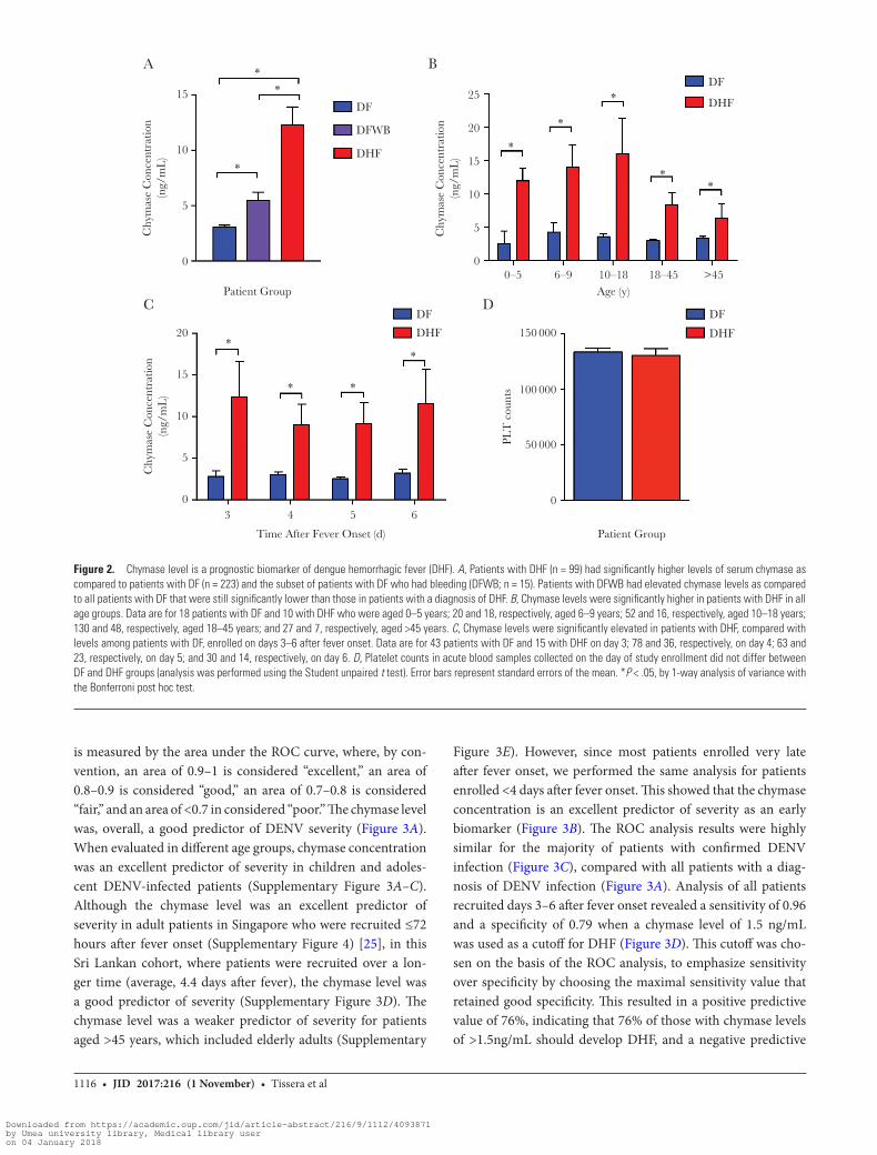
We next compared the mean serum chymase concentra-tions of patients grouped on the basis of their final diagnosis: all patients with a diagnosis of DF; a subset of patients with a diagnosis of DF who had signs of bleeding (ie, the DFWB
group), which was used to understand whether chymase lev-els also correlated with mild/moderate bleeding; and patients with a diagnosis of DHF. Patients with a diagnosis of DHF had significantly higher serum chymase levels than all patients with a diagnosis of DF and patients with DFWB. Interestingly, patients in the DFWB group, who had overt symptoms of bleeding yet did not meet the DHF diagnosis criteria, formed a discrete group that differed significantly from the DF and DHF groups (Figure 2A). Similar results were obtained when the analysis was limited to the majority of patients with labo-ratory-confirmed dengue (Supplementary Figure 1). Grouping by age revealed that children and adolescents with DHF had higher chymase levels than adults with DHF (Figure 2B). The chymase level was also significantly elevated in the serum of patients with DHF, compared with the level in patients with DF, irrespective of the day of enrollment after fever onset (Figure 2C). Thrombocytopenia is a sign of both DF and DHF, although a rapid decline in the platelet count can be a warning sign of severe dengue [8]. A platelet count of <100 000 is one criterion for a DHF diagnosis [8, 35]. However, platelet counts for patients in the DF and DHF groups at the time of study enrollment did not differ (Figure 2D), even though, at later time points, differences in platelet counts became apparent (Supplementary Figure 2). These data show that chymase levels highly correlate with DENV disease severity.
To test how rigorously the chymase concentration performs as a biomarker of severe DENV, we used a ROC analysis. Curves were generated by plotting sensitivity against specificity for chy-mase level as a predictor of DENV severity. In this test, accuracy
Table 1. Serum Chymase Level and Symptoms Associated With Diagnosis of Dengue Fever (DF) Versus Dengue Hemorrhagic Fever (DHF) Among 334 Patients With a Diagnosis of Dengue Virus (DENV) Infection and the Subset of 297 Patients With Confirmed DENV Infection
Dengue Sign or Symptom OR (95% CI) aOR (95% CI)
DENV infection diagnosisa
Chymase level 1.31b (1.20–1.42) 1.32b (1.21–1.44)
Bleeding per vaginam 3.19 (1.15–8.83) 1.79 (.48–6.61)
Abdominal pain 2.31 (1.27–4.20) 1.35 (.54–3.41)
Abdominal tenderness 2.29 (1.21–4.33) 2.57 (.69–9.64)
Hepatic tenderness 2.08 (1.06–4.08) 0.72 (.21–2.52)
Narrow pulse pressure 4.04 (.95–17.2) 2.78 (.50–15.4)
Oliguria 2.72 (1.20–6.20) 2.03 (.67–6.18)
DENV infection confirmationc
Chymase level 1.30 (1.19–1.43) 1.31 (1.19–1.45)
Bleeding per vaginam 3.19 (1.23–8.30) 1.55 (.42–5.95)
Abdominal pain 2.58 (1.37–4.86) 1.58 (.58–4.26)
Abdominal tenderness 2.40 (1.24–4.65) 1.94 (.47–7.96)
Hepatic tenderness 2.08 (1.05–4.16) 0.68 (.19–2.47)
Narrow pulse pressure 2.27 (1.17–4.40) 2.07 (.88–4.86)
Oliguria 2.71 (1.16–6.29) 2.42 (.83–7.08)
Abbreviations: aOR, adjusted odds ratio; CI, confidence interval; OR, odds ratio.aData are for 223 with DF and 99 with DHF.bPer unit increase.cData are for 174 DF and 73 with DHF.
Downloaded from https://academic.oup.com/jid/article-abstract/216/9/1112/4093871by Umea university library, Medical library useron 04 January 2018
1116 • JID 2017:216 (1 November) • Tissera et al
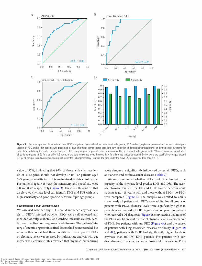
is measured by the area under the ROC curve, where, by con-vention, an area of 0.9–1 is considered “excellent,” an area of 0.8–0.9 is considered “good,” an area of 0.7–0.8 is considered “fair,” and an area of <0.7 in considered “poor.” The chymase level was, overall, a good predictor of DENV severity (Figure 3A). When evaluated in different age groups, chymase concentration was an excellent predictor of severity in children and adoles-cent DENV-infected patients (Supplementary Figure 3A–C). Although the chymase level was an excellent predictor of severity in adult patients in Singapore who were recruited ≤72 hours after fever onset (Supplementary Figure 4) [25], in this Sri Lankan cohort, where patients were recruited over a lon-ger time (average, 4.4 days after fever), the chymase level was a good predictor of severity (Supplementary Figure 3D). The chymase level was a weaker predictor of severity for patients aged >45 years, which included elderly adults (Supplementary
Figure 3E). However, since most patients enrolled very late after fever onset, we performed the same analysis for patients enrolled <4 days after fever onset. This showed that the chymase concentration is an excellent predictor of severity as an early biomarker (Figure 3B). The ROC analysis results were highly similar for the majority of patients with confirmed DENV infection (Figure 3C), compared with all patients with a diag-nosis of DENV infection (Figure 3A). Analysis of all patients recruited days 3–6 after fever onset revealed a sensitivity of 0.96 and a specificity of 0.79 when a chymase level of 1.5 ng/mL was used as a cutoff for DHF (Figure 3D). This cutoff was cho-sen on the basis of the ROC analysis, to emphasize sensitivity over specificity by choosing the maximal sensitivity value that retained good specificity. This resulted in a positive predictive value of 76%, indicating that 76% of those with chymase levels of >1.5ng/mL should develop DHF, and a negative predictive
15DF
A B
C D
DF
DHF
DF
DHF
DF
DHF 150 000
100 000
50 000PLT
cou
nts
0
DFWB
25
*
*
*
**
20
15
10
Chy
mas
e C
once
ntra
tion
(ng/
mL
)
5
0
20
15
10
Chy
mas
e C
once
ntra
tion
(ng/
mL
)
5
0
0–5
3 4 5
Time After Fever Onset (d) Patient Group
6
6–9 10–18 18–45 >45
DHF10
**
*
*
* *
*
Chy
mas
e C
once
ntra
tion
(ng/
mL
)
5
0
Age (y)Patient Group
Figure 2. Chymase level is a prognostic biomarker of dengue hemorrhagic fever (DHF). A, Patients with DHF (n = 99) had significantly higher levels of serum chymase as compared to patients with DF (n = 223) and the subset of patients with DF who had bleeding (DFWB; n = 15). Patients with DFWB had elevated chymase levels as compared to all patients with DF that were still significantly lower than those in patients with a diagnosis of DHF. B, Chymase levels were significantly higher in patients with DHF in all age groups. Data are for 18 patients with DF and 10 with DHF who were aged 0–5 years; 20 and 18, respectively, aged 6–9 years; 52 and 16, respectively, aged 10–18 years; 130 and 48, respectively, aged 18–45 years; and 27 and 7, respectively, aged >45 years. C, Chymase levels were significantly elevated in patients with DHF, compared with levels among patients with DF, enrolled on days 3–6 after fever onset. Data are for 43 patients with DF and 15 with DHF on day 3; 78 and 36, respectively, on day 4; 63 and 23, respectively, on day 5; and 30 and 14, respectively, on day 6. D, Platelet counts in acute blood samples collected on the day of study enrollment did not differ between DF and DHF groups (analysis was performed using the Student unpaired t test). Error bars represent standard errors of the mean. *P < .05, by 1-way analysis of variance with the Bonferroni post hoc test.
Downloaded from https://academic.oup.com/jid/article-abstract/216/9/1112/4093871by Umea university library, Medical library useron 04 January 2018
Chymase Level Is a Predictive Biomarker of DHF • JID 2017:216 (1 November) • 1117
value of 97%, indicating that 97% of those with chymase lev-els of <1.5ng/mL should not develop DHF. For patients aged 0–5 years, a sensitivity of 1 is maintained at this cutoff value. For patients aged >45 year, the sensitivity and specificity were 1.0 and 0.92, respectively (Figure 3). These results confirm that an elevated chymase level can identify DHF and DSS with very high sensitivity and good specificity for multiple age groups.
PECs Influence Serum Chymase Levels
We assessed whether any PECs could influence chymase lev-els in DENV-infected patients. PECs were self-reported and included obesity, diabetes, and cardiac, musculoskeletal, cere-brovascular, liver, or lung-associated diseases. The patients’ his-tory of anemia or gastrointestinal disease had been recorded, but none in this cohort had these conditions. The impact of PECs on chymase levels was assessed by multivariate analysis with age in years as a covariate. This revealed that chymase levels during
acute dengue are significantly influenced by certain PECs, such as diabetes and cardiovascular diseases (Table 2).
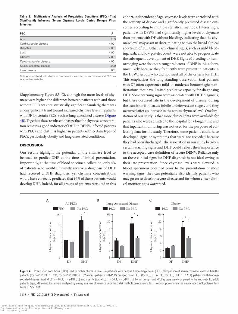
We next questioned whether PECs could interfere with the capacity of the chymase level predict DHF and DSS. The aver-age chymase levels in the DF and DHF groups between adult patients (age, >18 years) with and those without PECs (no-PEC) were compared (Figure 4). The analysis was limited to adults since nearly all patients with PECs were adults. For all groups of patients with PECs, chymase levels were significantly higher in patients who received a DHF diagnosis as compared to patients who received a DF diagnosis (Figure 4), emphasizing that none of the PECs would prevent the use of chymase level as a biomarker of DHF. For patients with any PEC (Figure 4A) and the subset of patients with lung-associated diseases or obesity (Figure 4B and 4C), patients with DHF had significantly higher levels of chymase than no-PEC DHF patients. For patients with car-diac diseases, diabetes, or musculoskeletal diseases as PECs
All PatientsA B
C D
Fever Duration <4 d1.0
0.8
Sens
itivi
ty 0.6
0.4
0.2
0.00.0 0.2 0.4 0.6 0.8
AUC = 0.80
1.0
Confirmed DENV Infection1.0
0.8
Sens
itivi
ty 0.6
0.4
0.2
0.0
1.00.9
0.7
0.5
0.3
0.1
0.8Se
nsiti
vity
1-Specificity
0.6
0.4
0.2
0.0
1.00.9
0.7
0.5
0.3
0.1
0.8
0.6
0.4
0.2
0.0
0.0 0.2 0.4
1-Specificity
0.6 0.8
AUC = 0.80
1.0All P
atien
ts0–
5 6–9
10–1
818
–45
>45
1.0
0.8
Sens
itivi
ty 0.6
0.4
0.2
0.00.0 0.2 0.4
1-Specificity1-Specificity
SpecificitySensitivity
0.6 0.8
AUC = 0.90
1.0
Age (y)
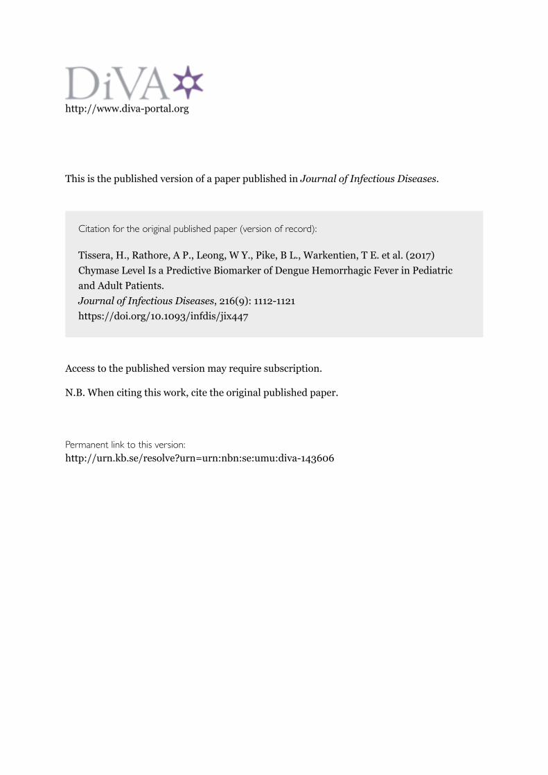
Figure 3. Receiver operator characteristic curve (ROC) analysis of chymase level for patients with dengue. A, ROC analysis graphs are presented for the total patient pop-ulation. B, ROC analysis for patients who presented <4 days after fever demonstrates excellent early detection of dengue hemorrhagic fever or dengue shock syndrome for patients tested during the acute phase of disease. C, ROC analysis graph of patients who were confirmed to be positive for dengue virus (DENV) infection is similar to that of all patients in panel A. D, For a cutoff of 1.5 ng/mL in the serum chymase level, the sensitivity for all groups ranged between 0.9–1.0, while the specificity averaged around 0.8 for all groups, including various age groups presented in Supplementary Figure 3. The area under the curve (AUC) is provided for panels A–C.
Downloaded from https://academic.oup.com/jid/article-abstract/216/9/1112/4093871by Umea university library, Medical library useron 04 January 2018
1118 • JID 2017:216 (1 November) • Tissera et al
(Supplementary Figure 5A–C), although the mean levels of chy-mase were higher, the difference between patients with and those without PECs was not statistically significant. Similarly, there was a nonsignificant trend toward increased chymase levels in patients with DF for certain PECs, such as lung-associated diseases (Figure 4B). Together, these results emphasize that the chymase concentra-tion remains a good indicator of DHF in DENV-infected patients with PECs and that it is higher in patients with certain types of PECs, particularly obesity and lung-associated conditions.
DISCUSSION
Our results highlight the potential of the chymase level to be used to predict DHF at the time of initial presentation. Importantly, at the time of blood specimen collection, only 4% of patients who would ultimately receive a diagnosis of DHF had received a DHF diagnosis; yet chymase concentrations would have correctly predicted that 96% of those patients would develop DHF. Indeed, for all groups of patients recruited in this
cohort, independent of age, chymase levels were correlated with the severity of disease and significantly predicted disease out-comes according to multiple statistical methods. Interestingly, patients with DFWB had significantly higher levels of chymase than patients with DF without bleeding, indicating that the chy-mase level may assist in discriminating within the broad clinical spectrum of DF. Other early clinical signs, such as mild bleed-ing, rash, and low platelet count, were not able to prognosticate the subsequent development of DHF. Signs of bleeding or hem-orrhaging were also not strong predictors of DHF in this cohort, most likely because they frequently were present in patients in the DFWB group, who did not meet all of the criteria for DHF. This emphasizes the long-standing observation that patients with DF often experience mild-to-moderate hemorrhagic man-ifestations that have limited predictive capacity for diagnosing DHF. Some warning signs were associated with DHF diagnosis, but these occurred late in the development of disease, during the transition from acute febrile to defervescent stages, and they occurred after an increase in the serum chymase level. One lim-itation of our study is that more clinical data were available for patients who were admitted to the hospital for a longer time and that inpatient monitoring was not used for the purposes of col-lecting data for the study. Therefore, some patients could have developed signs or symptoms that were not recorded because they had been discharged. The association in our study between certain warning signs and DHF could reflect their importance to the accepted case definition of severe DENV. Reliance only on these clinical signs for DHF diagnosis is not ideal owing to their late presentation. Since chymase levels were elevated in blood specimens obtained prior to the presentation of most warning signs, they can potentially also identify patients who may go on to develop severe disease and for whom closer clini-cal monitoring is warranted.
Table 2. Multivariate Analysis of Preexisting Conditions (PECs) That Significantly Influence Serum Chymase Levels During Dengue Virus Infection
PEC P
Any .289
Cardiovascular disease <.001
Diabetes <.001
Lung <.001
Obesity <.001
Cerebrovascular disease <.001
Musculoskeletal disease .999
Liver disease <.001
Data were analyzed with chymase concentration as a dependent variable and PECs as independent variables.
A B C
20
PEC
All PECs Lung-Associated Disease Obesity
No PEC PEC No PEC PEC No PEC
10
Chy
mas
e C
once
ntra
tion
(ng/
mL
)
64
32
16
8
4
2
DF DHF DF DHF DF DHF1
Chy
mas
e C
once
ntra
tion
(ng/
mL
)
64*
**
32
16
8
4
2
1
Chy
mas
e C
once
ntra
tion
(ng/
mL
)
1
Figure 4. Preexisting conditions (PECs) lead to higher chymase levels in patients with dengue hemorrhagic fever (DHF). Comparison of serum chymase levels in healthy patients (for no-PEC, DF: n = 191, for no-PEC, DHF: n = 82) versus patients with PECs grouped by all PECs (for PEC, DF: n = 33, for PEC, DHF: n = 17; A), patients with lung-as-sociated diseases (with-PEC: n = 6-DF, n = 2-DHF; B), and obesity (with-PEC: n = 5-DF, n = 5-DHF; C). For all groups, with-PEC groups were compared to the without-PEC adult patients (age, >18 years). Data were analyzed by 2-way analysis of variance with the Sidak multiple comparisons test. Post-hoc power analyses are included in Supplementary Table 3. *P < .001.
Downloaded from https://academic.oup.com/jid/article-abstract/216/9/1112/4093871by Umea university library, Medical library useron 04 January 2018
Chymase Level Is a Predictive Biomarker of DHF • JID 2017:216 (1 November) • 1119
Our data indicate that the chymase level is a stronger bio-marker for children and adolescents than for adults. A lower chymase level in adults with DHF could reflect their increased likelihood of having other comorbidities that could exacerbate disease or influence the diagnosis of severity, potentially inde-pendent of vascular complications. Elderly patients frequently have atypical dengue presentations and often experience coin-fections [36]. Older adults may also present later with a febrile response after inoculation with virus, which could alter the detection parameters and the study time points since they are anchored here to the onset of fever. Studies have not identified any influence of age on the DENV intrinsic incubation period; however, these studies were performed in young adults [37]. Even though the oldest adults in this study had lower levels of chymase during DHF, a conservative cutoff of 1.5 ng/mL was still able to predict DHF in this population with a sensitivity approaching 1 and a specificity of >0.95. Further studies are needed to determine whether these cutoffs are valid for addi-tional patient cohorts, including those in other dengue-en-demic regions.
Previously, we reported that, in a cohort of adult Singaporean patients, chymase levels were significantly correlated with dis-ease severity. Those data had a sensitivity of 1.0, a false-positive rate of 0.83, and an area under the ROC curve of 0.99 for adults recruited up to 72 hours after fever onset (Supplementary Figure 4) [25]. In that study, measurement of the chymase concentra-tion in sequential serum samples showed that levels decreased over time as acute disease resolved [25]. In this cohort, patients were recruited at longer times after fever onset, but we did not observe significant differences between the levels of chymase, based on the day of study enrollment. Longitudinal serum monitoring of individual patients, which was not performed here, might also demonstrate that chymase levels are higher at earlier time points. Going forward, it will be important to assess whether early measurement of the chymase level and interven-tion can improve the outcomes of DENV-infected patients and promote allocation of healthcare resources to those most likely to develop severe DENV.
Our results show that PECs influence the levels of chymase during DHF. In particular, the link between high chymase levels and DHF in patients with lung-associated PECs is interesting because, although the specific conditions were not specified, some of these patients presumably had asthma, which has a known MC-promoted etiology. In the 1981 Cuban epidemic, asthma was also frequently a PEC of patients with DHF [38]. Higher chymase levels indicate increased activation of MCs in patients with DHF with PECs. Although we previously observed that febrile control patients without dengue (most of whom had respiratory virus infections) did not have elevated chymase levels [25], some other infections activate MCs. One patient with DHF in this study had a chymase concentration
that was approximately 100 times the levels of other patients with DHF and received a diagnosis of bacterial coinfection. This raises the point that chymase induction is not necessarily DENV specific and can be detected in other conditions that promote MC activation, including bacterial infections and noninfectious inflammatory conditions, such as asthma or anaphylaxis. Thus, measurement of the serum chymase level alone should not serve as a diagnostic test for DENV and, for DENV infection progno-sis, must be interpreted in the context of results of other tests performed to confirm infection or in the context of a strong sus-picion of DENV. Furthermore, since PECs influence chymase concentrations in DENV-infected patients, additional studies should determine whether differing cutoffs for patients with PECs would improve the prognostic value of the chymase level.
Our data demonstrate that the chymase concentration is a strong predictor of DHF in febrile patients with acute-phase DENV infection, with the potential to identify those at risk for complications more robustly and at earlier time points than many clinical warning signs. Since chymase is an angioten-sin-converting enzyme and can influence vasoconstriction [28], further studies are needed to determine its contributions to the pathophysiology of DENV disease.
Supplementary Data
Supplementary materials are available at The Journal of Infectious Diseases online. Consisting of data provided by the authors to benefit the reader, the posted materials are not copyedited and are the sole responsibility of the authors, so questions or com-ments should be addressed to the corresponding author.
Notes
Acknowledgment. We thank October Sessions for provid-ing information on DENV-confirmed cases from the study by Tisserra et al [3].
Disclaimer. B. L. P. and T. W. are US military service mem-bers, and this work was prepared as part of their official duties. However, the opinions and assertions contained herein are those of the authors and are not to be construed as official or reflect-ing the views of the Department of the Navy, the Department of Defense, the US government.
Financial support. This work was supported by the US Naval Medical Research Center–Asia (awards Duke/Duke-NUS/RECA[PILOT]/2013/0008 and Duke/Duke-NUS/RECA[Matching Fund]/2015/0016) and the Seventh Framework Programme of the European Community (grant 282589).
Potential conflicts of interest. All authors: No reported con-flicts of interest. All authors have submitted the ICMJE Form for Disclosure of Potential Conflicts of Interest. Conflicts that the editors consider relevant to the content of the manuscript have been disclosed.
Downloaded from https://academic.oup.com/jid/article-abstract/216/9/1112/4093871by Umea university library, Medical library useron 04 January 2018
1120 • JID 2017:216 (1 November) • Tissera et al
References
1. Gubler DJ. Dengue, urbanization and globalization: the unholy trinity of the 21(st) century. Trop Med Health 2011; 39:3–11.
2. Bhatt S, Gething PW, Brady OJ, et al. The global distribution and burden of dengue. Nature 2013; 496:504–7.
3. Tissera H, Amarasinghe A, Gunasena S, et al. Laboratory-enhanced dengue sentinel surveillance in Colombo District, Sri Lanka: 2012–2014. PLoS Negl Trop Dis 2016; 10:e0004477.
4. Snow GE, Haaland B, Ooi EE, Gubler DJ. Review article: Research on dengue during World War II revisited. Am J Trop Med Hyg 2014; 91:1203–17.
5. SABIN AB. Research on dengue during World War II. Am J Trop Med Hyg 1952; 1:30–50.
6. Simmons CP, Farrar JJ, Nguyen vV, Wills B. Dengue. N Engl J Med 2012; 366:1423–32.
7. St John AL, Abraham SN, Gubler DJ. Barriers to preclinical investigations of anti-dengue immunity and dengue patho-genesis. Nat Rev Microbiol 2013; 11:420–6.
8. Dengue: Guidelines for Diagnosis, Treatment, Prevention and Control: New Edition. Geneva, 2009.
9. Vaughn DW, Green S, Kalayanarooj S, et al. Dengue viremia titer, antibody response pattern, and virus serotype corre-late with disease severity. J Infect Dis 2000; 181:2–9.
10. Libraty DH, Endy TP, Houng HS, et al. Differing influences of virus burden and immune activation on disease severity in secondary dengue-3 virus infections. J Infect Dis 2002; 185:1213–21.
11. Singla M, Kar M, Sethi T, et al. Correction: immune response to dengue virus infection in pediatric patients in New Delhi, India-association of viremia, inflammatory mediators and monocytes with disease severity. PLoS Negl Trop Dis 2016; 10:e0004642.
12. Chen RF, Liu JW, Yeh WT, et al. Altered T helper 1 reaction but not increase of virus load in patients with dengue hemor-rhagic fever. FEMS Immunol Med Microbiol 2005; 44:43–50.
13. Simmons CP, Chau TN, Thuy TT, et al. Maternal antibody and viral factors in the pathogenesis of dengue virus in infants. J Infect Dis 2007; 196:416–24.
14. Fox A, Le NM, Simmons CP, et al. Immunological and viral determinants of dengue severity in hospitalized adults in Ha Noi, Viet Nam. PLoS Negl Trop Dis 2011; 5:e967.
15. Sung C, Wei Y, Watanabe S, et al. Extended evaluation of virological, immunological and pharmacokinetic endpoints of CELADEN: a randomized, placebo-controlled trial of celgosivir in dengue fever patients. PLoS Negl Trop Dis 2016; 10:e0004851.
16. Namvongsa V, Sirivichayakul C, Songsithichok S, Chanthavanich P, Chokejindachai W, Sitcharungsi R. Differences in clinical features between children and adults with dengue hemorrhagic fever/dengue shock syndrome. Southeast Asian J Trop Med Public Health 2013; 44:772–9.
17. Kittigul L, Pitakarnjanakul P, Sujirarat D, Siripanichgon K. The differences of clinical manifestations and laboratory findings in children and adults with dengue virus infection. J Clin Virol 2007; 39:76–81.
18. Alera MT, Srikiatkhachorn A, Velasco JM, et al. Incidence of dengue virus infection in adults and children in a pro-spective longitudinal cohort in the philippines. PLoS Negl Trop Dis 2016; 10:e0004337.
19. Gamble J, Bethell D, Day NP, et al. Age-related changes in microvascular permeability: a significant factor in the susceptibility of children to shock? Clin Sci (Lond) 2000; 98:211–6.
20. Thein TL, Leo YS, Fisher DA, et al. Risk factors for fatality among confirmed adult dengue inpatients in Singapore: a matched case-control study. PLoS One 2013; 8:e81060.
21. Low JG, Ooi EE. Dengue–old disease, new challenges in an ageing population. Ann Acad Med Singapore 2013; 42:373–5.
22. Sam SS, Omar SF, Teoh BT, Abd-Jamil J, AbuBakar S. Review of Dengue hemorrhagic fever fatal cases seen among adults: a retrospective study. PLoS Negl Trop Dis 2013; 7:e2194.
23. Pichainarong N, Mongkalangoon N, Kalayanarooj S, Chaveepojnkamjorn W. Relationship between body size and severity of dengue hemorrhagic fever among children aged 0-14 years. Southeast Asian J Trop Med Public Health 2006; 37:283–8.
24. Hadinegoro SR, Arredondo-García JL, Capeding MR, et al.; CYD-TDV Dengue Vaccine Working Group. Efficacy and long-term safety of a dengue vaccine in regions of endemic disease. N Engl J Med 2015; 373:1195–206.
25. St John AL, Rathore AP, Raghavan B, Ng ML, Abraham SN. Contributions of mast cells and vasoactive products, leu-kotrienes and chymase, to dengue virus-induced vascular leakage. Elife 2013; 2:e00481.
26. St John AL, Rathore AP, Yap H, et al. Immune surveillance by mast cells during dengue infection promotes natural killer (NK) and NKT-cell recruitment and viral clearance. Proc Natl Acad Sci U S A 2011; 108:9190–5.
27. St John AL. Influence of mast cells on dengue protective immunity and immune pathology. PLoS Pathog 2013; 9:e1003783.
28. Kunder CA, St John AL, Abraham SN. Mast cell modulation of the vascular and lymphatic endothelium. Blood 2011; 118:5383–93.
29. Wernersson S, Pejler G. Mast cell secretory granules: armed for battle. Nat Rev Immunol 2014; 14:478–94.
30. Reilly CF, Tewksbury DA, Schechter NM, Travis J. Rapid conversion of angiotensin I to angiotensin II by neutrophil and mast cell proteinases. J Biol Chem 1982; 257:8619–22.
31. Caughey GH. Mast cell proteases as protective and inflam-matory mediators. Adv Exp Med Biol 2011; 716:212–34.
32. Syenina A, Jagaraj CJ, Aman SA, Sridharan A, St John AL. Dengue vascular leakage is augmented by mast cell
Downloaded from https://academic.oup.com/jid/article-abstract/216/9/1112/4093871by Umea university library, Medical library useron 04 January 2018
Chymase Level Is a Predictive Biomarker of DHF • JID 2017:216 (1 November) • 1121
degranulation mediated by immunoglobulin Fcgamma receptors. Elife 2015; 4.
33. Wilder-Smith A, Renhorn KE, Tissera H, et al. DengueTools: innovative tools and strategies for the surveillance and con-trol of dengue. Glob Health Action 2012; 5.
34. WHO. Dengue hemorrhagic fever: diagnosis, treatment, pre-vention and control. Geneva: World Health Organization. 1997.
35. Deen JL, Harris E, Wills B, et al. The WHO dengue classifi-cation and case definitions: time for a reassessment. Lancet 2006; 368:170–3.
36. Rowe EK, Leo YS, Wong JG, et al. Challenges in dengue fever in the elderly: atypical presentation and risk of severe dengue and hospital-acquired infection [corrected]. PLoS Negl Trop Dis 2014; 8:e2777.
37. Nishiura H, Halstead SB. Natural history of dengue virus (DENV)-1 and DENV-4 infections: reanalysis of classic studies. J Infect Dis 2007; 195:1007–13.
38. Guzman MG, Kouri G, Soler M, et al. Dengue 2 virus enhancement in asthmatic and non asthmatic individual. Mem Inst Oswaldo Cruz 1992; 87:559–64.
Downloaded from https://academic.oup.com/jid/article-abstract/216/9/1112/4093871by Umea university library, Medical library useron 04 January 2018