Embed Size (px)
Citation preview
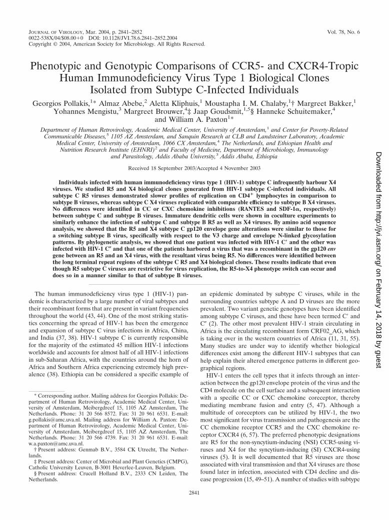
JOURNAL OF VIROLOGY, Mar. 2004, p. 2841–2852 Vol. 78, No. 60022-538X/04/$08.00�0 DOI: 10.1128/JVI.78.6.2841–2852.2004Copyright © 2004, American Society for Microbiology. All Rights Reserved.
Phenotypic and Genotypic Comparisons of CCR5- and CXCR4-TropicHuman Immunodeficiency Virus Type 1 Biological Clones
Isolated from Subtype C-Infected IndividualsGeorgios Pollakis,1* Almaz Abebe,2 Aletta Kliphuis,1 Moustapha I. M. Chalaby,1† Margreet Bakker,1
Yohannes Mengistu,3 Margreet Brouwer,4‡ Jaap Goudsmit,1,5§ Hanneke Schuitemaker,4and William A. Paxton1*
Department of Human Retrovirology, Academic Medical Center, University of Amsterdam,1 and Center for Poverty-RelatedCommunicable Diseases,5 1105 AZ Amsterdam, and Sanquin Research at CLB and Landsteiner Laboratory, Academic
Medical Center, University of Amsterdam, 1066 CX Amsterdam,4 The Netherlands, and Ethiopian Health andNutrition Research Institute (EHNRI)2 and Faculty of Medicine, Department of Microbiology, Immunology
and Parasitology, Addis Ababa University,3 Addis Ababa, Ethiopia
Received 18 September 2003/Accepted 4 November 2003
Individuals infected with human immunodeficiency virus type 1 (HIV-1) subtype C infrequently harbour X4viruses. We studied R5 and X4 biological clones generated from HIV-1 subtype C-infected individuals. Allsubtype C R5 viruses demonstrated slower profiles of replication on CD4� lymphocytes in comparison tosubtype B viruses, whereas subtype C X4 viruses replicated with comparable efficiency to subtype B X4 viruses.No differences were identified in CC or CXC chemokine inhibitions (RANTES and SDF-1�, respectively)between subtype C and subtype B viruses. Immature dendritic cells were shown in coculture experiments tosimilarly enhance the infection of subtype C and subtype B R5 as well as X4 viruses. By amino acid sequenceanalysis, we showed that the R5 and X4 subtype C gp120 envelope gene alterations were similar to those fora switching subtype B virus, specifically with respect to the V3 charge and envelope N-linked glycosylationpatterns. By phylogenetic analysis, we showed that one patient was infected with HIV-1 C� and the other wasinfected with HIV-1 C� and that one of the patients harbored a virus that was a recombinant in the gp120 envgene between an R5 and an X4 virus, with the resultant virus being R5. No differences were identified betweenthe long terminal repeat regions of the subtype C R5 and X4 biological clones. These results indicate that eventhough R5 subtype C viruses are restrictive for virus replication, the R5-to-X4 phenotype switch can occur anddoes so in a manner similar to that of subtype B viruses.
The human immunodeficiency virus type 1 (HIV-1) pan-demic is characterized by a large number of viral subtypes andtheir recombinant forms that are present in variant frequenciesthroughout the world (43, 44). One of the most striking statis-tics concerning the spread of HIV-1 has been the emergenceand expansion of subtype C virus infections in Africa, China,and India (37, 38). HIV-1 subtype C is currently responsiblefor the majority of the estimated 45 million HIV-1 infectionsworldwide and accounts for almost half of all HIV-1 infectionsin sub-Saharan Africa, with the countries around the horn ofAfrica and Southern Africa experiencing extremely high prev-alence (38). Ethiopia can be considered a specific example of
an epidemic dominated by subtype C viruses, while in thesurrounding countries subtype A and D viruses are the moreprevalent. Two variant genetic genotypes have been identifiedamong subtype C viruses, and these have been termed C� andC� (2). The other most prevalent HIV-1 strain circulating inAfrica is the circulating recombinant form CRF02_AG, whichis taking over in the western countries of Africa (11, 31, 55).Many studies are under way to identify whether biologicaldifferences exist among the different HIV-1 subtypes that canhelp explain their altered emergence patterns in different geo-graphical regions.
HIV-1 enters the cell types that it infects through an inter-action between the gp120 envelope protein of the virus and theCD4 molecule on the cell surface and a subsequent interactionwith a specific CC or CXC chemokine coreceptor, therebymediating membrane fusion and entry (5, 47). Although amultitude of coreceptors can be utilized by HIV-1, the twomost significant for virus transmission and pathogenesis are theCC chemokine receptor CCR5 and the CXC chemokine re-ceptor CXCR4 (6, 57). The preferred phenotypic designationsare R5 for the non-syncytium-inducing (NSI) CCR5-using vi-ruses and X4 for the syncytium-inducing (SI) CXCR4-usingviruses (5). It is well documented that R5 viruses are thoseassociated with viral transmission and that X4 viruses are thosefound later in infection, associated with CD4 decline and dis-ease progression (15, 49–51). A number of studies with subtype
* Corresponding author. Mailing address for Georgios Pollakis: De-partment of Human Retrovirology, Academic Medical Center, Uni-versity of Amsterdam, Meibergdreef 15, 1105 AZ Amsterdam, TheNetherlands. Phone: 31 20 566 8572. Fax: 31 20 961 6531. E-mail:[email protected]. Mailing address for William A. Paxton: De-partment of Human Retrovirology, Academic Medical Center, Uni-versity of Amsterdam, Meibergdreef 15, 1105 AZ Amsterdam, TheNetherlands. Phone: 31 20 566 4739. Fax: 31 20 961 6531. E-mail:[email protected].
† Present address: Genmab B.V., 3584 CK Utrecht, The Nether-lands.
‡ Present address: Center of Microbial and Plant Genetics (CMPG),Catholic University Leuven, B-3001 Heverlee-Leuven, Belgium.
§ Present address: Crucell Holland B.V., 2333 CN Leiden, TheNetherlands.
2841
on February 14, 2018 by guest
http://jvi.asm.org/
Dow
nloaded from
B-infected individuals have determined that between 40 and50% of AIDS patients can harbor viruses of the SI, and pre-sumably the X4, phenotype (27, 28). Numerous studies haverevealed that the frequency of SI emergence among subtypeC-infected individuals is far lower than that identified for theother subtypes (1, 7, 12, 35, 39), although a number of recentstudies have found a higher frequency of the X4 phenotype(13, 26).
The molecular alterations associated with the R5-to-X4switch in vivo are not fully understood, although many of thefeatures of the gp120 envelope viral protein involved in core-ceptor usage have been revealed. The V3 region is highlyassociated with the coreceptor phenotype, with the overallamino acid charge being central to coreceptor usage: higherpositive charges are associated with the SI phenotype andutilization of the CXCR4 coreceptor (17, 18, 41, 48). TheV1V2 region has also been associated with coreceptor usage,especially in cooperation with the V3 region of the envelope(41). Cooperation between the V3 and the V1V2 regions ofgp120 have been shown to influence not only the receptorusage pattern but also its replication phenotype, with a majordeterminant being the N-linked glycosylation site downstreamof the first V3 cysteine (41). The N-linked glycosylation pat-terns of the gp120 protein also confer a significant effect on theHIV-1 antibody neutralization responses mounted, with alter-ations in N-linked glycosylation patterns being able to success-fully mask effective antibody neutralization responses (3, 56).The CC chemokines RANTES, macrophage inflammatoryprotein 1� (MIP-1�), and MIP-1�, the natural ligands for theCCR5 chemokine receptor, and stromal cell-derived factor 1�(SDF-1�), the natural ligand for the CXCR4 coreceptor, suc-cessfully block the replication of HIV-1 in vitro (9, 14). Fur-thermore, the association between a large array of geneticpolymorphisms within the chemokine and chemokine receptorgenes and disease progression rates indicates a strong associ-ation between the chemokine network and viral replication invivo (53). Alterations within the gp120 envelope are thereforethought to influence the extent to which HIV-1 can be con-trolled by antibody-mediated immune responses as well as hostchemokine and chemokine expression levels.
There is evidence that specific genetic alterations within thelong terminal repeat (LTR) regions of HIV-1 can be associatedwith the different virus subtypes. The LTR regions of HIV-1subtype C viruses have been shown to have an additionalNF-�B site inserted in comparison to the other subtypes (24,36). It has been speculated that an additional NF-�B site mayhelp confer a replication advantage to the subtype C virusesover the other subtypes, and a number of studies have de-scribed enhancements to tat-induced transcription as well asvirus replication through an additional NF-�B site (25, 33, 34,36), although another study has suggested that this additionalsite is redundant with respect to biological function (45).
Why individuals infected with one HIV-1 subtype shouldswitch their virus phenotype from R5 to X4 more frequentlythan those infected with other subtypes infers either a restric-tion in the host favoring the expansion of one subtype overanother or, alternatively, differences in the biological charac-teristics of these viruses. The observation that different sub-types can predominate in similar geographical locations andamong individuals with similar environmental settings suggests
a restriction at the level of the virus (55). In this study we havechosen to investigate the phenotype and genotype of HIV-1biological clones generated from two individuals who wereinfected with HIV-1 subtype C viruses and who had beenidentified as harboring primary isolates with the SI phenotype(1).
MATERIALS AND METHODS
Patient selection and generation of biologically cloned HIV-1. Two individualswere selected for study from a previously described Ethiopian cohort of HIV-1subtype C-infected individuals (1). Both individuals had CD4 cell counts of�150/mm3 and harbored SI viruses as determined by replication on the MT-2cell line (1). The cocirculating HIV-1 subtype C quasi-species were clonedbiologically from peripheral blood mononuclear cells (PBMC) by a previouslypublished method, and the individual biological clones were subsequently stud-ied (28, 54). In summary, graded numbers of patient PBMCs (range, 1 � 104 to2 � 104 cells/well) were cocultivated in 96-well plates with 1 � 105 phytohemag-glutinin (PHA)-stimulated PBMC from healthy blood donor volunteers. Theproportion (F) of infected cells was determined from the formula for the Poissondistribution, F ln (F0), where F0 is the fraction of negative cultures. Onlyvirus clones obtained from a dilution that gave rise to progeny virus in fewer than33% of parallel cultures were considered clonal. The homogeneity of the isolateswas tested by amplification of the V1V3 region and the subsequent ligation intothe TOPO-A plasmid (Invitrogen, Breda, The Netherlands). Between 10 and 20individual bacterial colonies were picked and subsequently DNA sequenced.Only homogeneous biological clones were used in these studies. Generatedbiologically cloned viruses were propagated on activated CD4� lymphocytes, andviral growth was monitored by p24 antigen production in the culture superna-tants by using a standard enzyme-linked immunosorbent assay (ELISA).
Virus replication on U87.CD4 coreceptor cells, CD4� lymphocytes, and mac-rophages. The virus coreceptor utilization phenotype was determined by mea-suring viral replication on the U87.CD4 cell line expressing, independently, anarray of variant chemokine receptors (CCR1, CCR2b, CCR3, CCR5, andCXCR4), a gift from D. Littman, Skirball Institute, New York, N.Y. These cellswere maintained in Dulbecco minimal essential medium supplemented with 10%fetal calf serum (FCS) plus the antibiotics puromycin (1 �g/ml) and neomycin(300 �g/ml). Coreceptor utilization was determined by adding 200 �l of virusstock to 3.0 � 104 U87.CD4 cells (plated 20 to 24 h previously in a 96-wellflat-bottom culture plate) expressing the specific coreceptor under analysis. Thecells were infected for 18 h before being washed twice with phosphate-bufferedsaline and fed with 200 �l of fresh medium. On day 10 of culture, the cells werescored for syncytium formation and the p24 levels in the culture supernatantswere determined using a standard ELISA. Coreceptor usage was also monitoredby infecting CD4� lymphocytes isolated from an individual homozygous for awild-type CCR5 gene (CCR5wt/wt) or from an individual homozygous for the32-bp deletion in the CCR5 gene (CCR5�32/�32) and monitoring p24 productionon days 7 and 10 of culture.
CD4� lymphocytes were prepared by the following method. PBMCs wereisolated from fresh buffy coats (Sanquin, Amsterdam, The Netherlands) or freshblood draws from laboratory workers by standard Ficoll-Hypaque density cen-trifugation and frozen in multiple vials at high concentrations, thawed whenrequired, and activated with 5 �g of PHA (Sigma, Zwijndrecht, The Nether-lands) and cultured in RPMI 1640 medium containing 10% FCS, penicillin (100U/ml), and streptomycin (100 U/ml) with recombinant interleukin-2 (100 U/ml).On day 4 of culture, the cells underwent CD4� enrichment by removing CD8�
lymphocytes with CD8 immunomagnetic beads and a magnet (Dynal, Oslo,Norway), giving rise to cultures of 90% CD4� lymphocyte purity.
Macrophages were obtained from standard buffy coats that were prepared asdescribed above. After Ficoll-Hypaque density centrifugation, the cells wereresuspended in RPMI 1640 medium supplemented with 10% AB� human se-rum, containing 20% FCS, 2 mM glutamine, penicillin (100 U/ml) and strepto-mycin (100 U/ml), after which they were plated at 106 cells/cm2 for 5 days at 37°Cin six-well tissue culture plates. Nonadherent cells were removed by extensivewashing with RPMI–20% FCS medium, and the adherent cells were infectedwith each virus (input, between 200 and 1,000 50% tissue culture infective doses[TCID50]). Virus replication was monitored by assaying for p24 in the culturesupernatant on days 7, 10, 14, and 18 postinfection.
Replication kinetics of generated virus stocks. All virus stocks were generatedby the multiple passaging of each virus through CD4� lymphocytes. Viral stockswere assayed to determine their TCID50 on CD4� lymphocytes by using apreviously described method (41). The viruses were tested for their ability to
2842 POLLAKIS ET AL. J. VIROL.
on February 14, 2018 by guest
http://jvi.asm.org/
Dow
nloaded from
replicate on CD4� lymphocytes isolated from a CCR5wt/wt individual. The rep-lication kinetics of each virus was measured by infecting CD4� lymphocytes fromCCR5wt/wt individuals with 200 TCID50 of virus, and replication was monitoredby measuring p24 antigen levels in the culture supernatants on days 4, 7, 10, and14 of culture.
Chemokine inhibition assays. Chemokine inhibition assays were performed inwhich CD4� lymphocytes (2.0 � 105/well) were incubated with twofold limitingdilutions of the desired chemokine, RANTES or SDF-1� (ITK Diagnostics BV,Uithoorn, The Netherlands) and incubated for 30 min at 37°C. AMD3100 wasobtained through the National Institutes of Health repository. Each well wasinoculated with 100 TCID50 of virus and cultured for 14 days. On days 10 and 14,the amount of p24 was determined using a standard ELISA. Virus inhibition wasdetermined by calculating the percent p24 inhibition in the presence of chemo-kine and in comparison to a control infected well where no chemokine was addedand utilizing the data from the day where peak p24 levels were reached.
DC enhancement to viral replication. Immature dendritic cells (iDCs) werecultured from standard buffy coats (Sanquin) processed by standard Ficoll-Hypaque centrifugation. Monocytes were selected by adhesion in culture flasksfor 3 h and then cultured in the presence of 500 U of IL-4 per ml and 800 U ofgranulocyte-macrophage colony-stimulating factor (a gift from T. Geijtenbeek,Free University, Amsterdam, The Netherlands) per ml for 7 days. These cultureconditions have previously been shown to be suitable for cells expressing highlevels of DC-SIGN as measured by flow cytometry (21). These iDCs were utilizedin coculture experiments where suboptimal concentrations of virus were incu-bated with 2.0 � 104 iDCs and 2.0 � 105 CD4� lymphocytes isolated from anindividual with the the CCR5wt/wt genotype, and the amount of p24 in the culturesupernatant was determined on days 5 and 7 of culture. Chemokine inhibitionassays were also performed as described above in the presence of iDCs at thesame ratios described.
DNA sequencing and analysis of the envelope regions. PHA-stimulatedPBMCs from healthy blood donor volunteers were infected with each virus, andon day 5 the cellular DNA was extracted from 106 cells by using a silica-basedisolation method (10). The gp140 region of the envelope gene (between the KpnIand BamHI cloning sites) was amplified using the primers 5�-KpnENV (ACTTGTGGGTCACAGTCTATTATGGGGTACC) and 3�-BamENV (GCTCCGCAGGTCGTCCCAGGCAAGTGCTAAGGATCCG). The DNA was se-quenced using a set of primers spanning the length of the DNA, with an averagedistance of 200 nucleotides between primers. Both DNA strands were se-quenced, with the sequencing being performed on an automated sequencer withthe Thermo Sequenase BigDye Terminator cycle-sequencing kit (Applied Bio-systems, Foster City, Calif.) as specified by the manufacturer. The primer se-quence and conditions for PCR amplification of the U3 region of the LTR(U3LTR) have been described in detail previously (16).
The sequences were aligned manually based on the alignment of the LosAlamos database reference sequences for subtyping. Phylogenetic analysis of the
aligned sequences was performed using the neighbor-joining MEGA method(29) and confirmed by the DNADIST, NEIGHBOR, and DRAWTREE optionsof the PHYLIP software package (http://evolution.Genetics.Washington.edu/phylip.html) (20). The distance matrix was generated by Kimura’s two-param-eter estimation (29). Based on 100 replications, a bootstrap value equal to orgreater than 70% is considered significant (22, 23). Other sequences obtainedfrom the Los Alamos database were included as reference sequences. Theboot-scanning method as implemented in the SIMPLOT program was used todetect and analyze the recombinant viruses (46; S. C. Ray, 1999, http://www.med.jhu.edu/deptmed/scray/download/simplot). Our analysis was performed by cal-culating the distances for a sliding window of 200 nucleotides of the test se-quences, moving along the alignment of a panel of reference sequences byincrements of 20 bp. One hundred replications were generated by the bootstrapmethod for each window, and the percent bootstrap values were plotted againstthe nucleotide position of the sequence of the reference panel.
Nucleotide sequence accession numbers. The GenBank accession numbers forthe gp120 envelope sequences are AY452640 to AY452650, and those for theLTR sequences are AY452651 to AY452661.
RESULTS
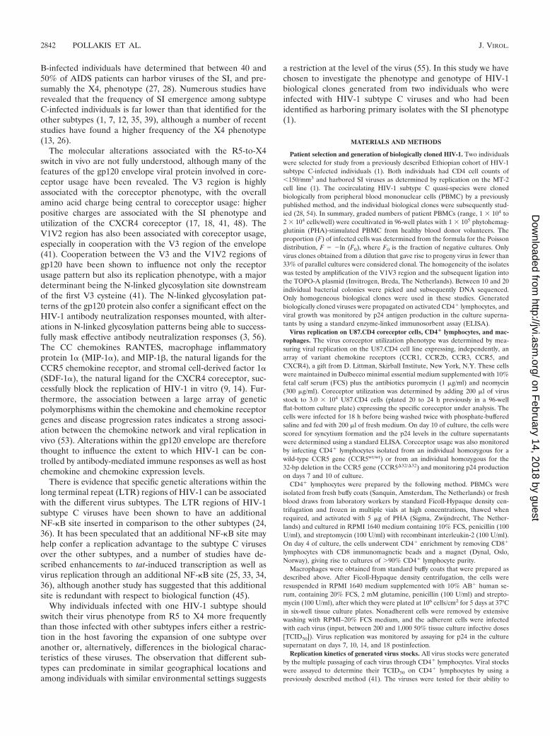
Generation of subtype C R5 and X4 biologically clonedviruses and their ability to replicate on variant cell types.From the study of 48 patients with CD4 counts of �150 cell/mm3, who had at least one AIDS-defining illness, 3 (6.3%)were identified as harboring viruses with the capacity to inducesyncytia on MT-2 cells (1). Biological clones were generatedfrom two of these individuals (PHD74 and PHD79), and theresultant viruses were studied for replication on various celltypes, namely, U87.CD4 cells expressing variant coreceptors,CD4� lymphocytes, and macrophages; a summary of the re-sults is shown in Table 1. Four virus clones were studied fromeach donor, with both R5 and X4 viruses being generated fromeach individual. One virus from donor PHD74 was of theR3/X4 phenotype (C4), while all the other viruses used a singlecoreceptor. Five other viruses were used in this study to allowfor further comparisons to be made: one X4 molecular clonedsubtype C virus (SE12808), two subtype B primary isolates(one R5 and one X4 [NSI-18 and SI-19, respectively]), and twosubtype B molecular cloned viruses (one R5 and one X4 [SF-
TABLE 1. Characteristics of HIV-1 subtype C biological clones
Donor Virus name Subtype Replication onMT-2 cells
Replication on CD4�a:Macrophageb Receptorc TCID50/mld
CCR5wt/wt CCR5�32/�32
PHD74 H4 C � 0 R5 103.09
PHD74 D3 C � 0 R5 102.22
PHD74 F3 C � � � 0 X4 102.56
PHD74 C4 C � � � 0 X4 105.72
PHD79 C1 C � 7,260 R5 103.72
PHD79 C12 C � 0 R5 104.15
PHD79 H8 C � � � 0 X4 103.97
PHD79 B8 C � � � 0 X4 104.85
None NSI-18 B � 9,440 R5 104.76
None SF-162 B � 10,000 R5 105.43
None SI-19 B � � � 0 X4 105.54
None SF-2 B � � � 0 X4 105.54
None SE12808 C � � � 0 X4 103.09
a Replication on CD4� lymphocytes isolated from an individual homozygous for a wild-type CCR5 (CCR5wt/wt) gene or homozygous for the 32-bp deletion in theCCR5 gene (CCR5�32/�32).
b Replication on macrophages [culture supernatant (picrograms/milliliter)].c Replication on U87.CD4 cells expressing the coreceptor of choice.d Determined on CCR5wt/wt CD4� lymphocytes.
VOL. 78, 2004 COMPARISON OF CCR5- AND CXCR4-TROPIC HIV-1 CLONES 2843
on February 14, 2018 by guest
http://jvi.asm.org/
Dow
nloaded from
162 and SF-2, respectively]). Replication of the subtype B andC viruses on MT-2 cells and on CD4� lymphocytes isolatedfrom either CCR5wt/wt or CCR5�32/�32 individuals correlatedwith the replication profile on U87.CD4 cells expressing eitherCCR5 or CXCR4. Only one of the R5 subtype C clones dem-onstrated replication on macrophages in comparison to boththe subtype B viruses, suggesting that the CCR5-utilizing sub-type C virus isolates replicate weakly on this cell type relativeto subtype B viruses.
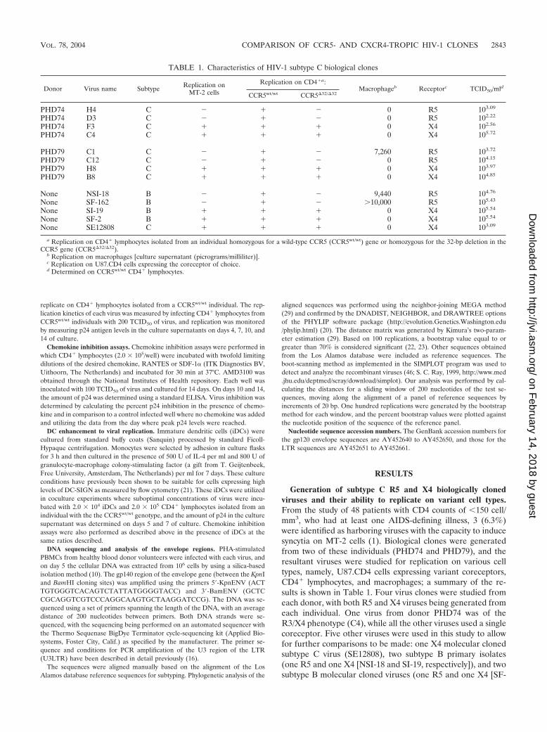
Reduced replication kinetics of subtype C R5 but not X4viruses. We sought to identify whether replication of the sub-type C viruses on PBMCs was comparable to what is commonlyfound for subtype B viruses. For this purpose, we analyzed thereplication of each subtype C biological clone on CD4� lym-phocytes isolated from three different individuals, wild-type forthe CCR5 gene, who demonstrated variation in the replicationprofile of the NSI-18 subtype B R5 virus (Fig. 1A). All infec-tions were performed with the same viral input (200 TCID50/ml) with the same activated batch of CD4� lymphocytes. We
observed low-level replication of the NSI-18 virus on CD4�
lymphocytes from donor 3, in comparison to the results forlymphocytes from the other two donors, which correlated withlow cell surface expression of the CCR5 coreceptor (data notshown). All the subtype C R5 viruses demonstrated the samepattern of restricted replication on CD4� lymphocytes fromdonor 3 in comparison to either donor 1 or donor 2, and thesubtype C R5 viruses demonstrated slower kinetics of replica-tion on lymphocytes from all three donors in comparison to thesubtype B control virus (Fig. 1A). When comparisons weremade between p24 values on day 7, the subtype B virus hadreached its peak p24 value on CD4� lymphocytes in all threedonors while the subtype C viruses were only at the beginningof the logarithmic phase of their replication cycle. The repli-cation curve we observed for the NSI-18 subtype B virus wasrepresentative of the curves for other subtype B viruses testedat the same TCID50 of infection (data not shown). In contrast,all the subtype C X4 viruses replicated to the same degree onthe same CD4�-enriched lymphocyte batches as the subtype B
FIG. 1. Replication kinetics of HIV-1 isolates on CD4� lymphocytes. (A) Replication of a single subtype B R5 virus and three subtype Cbiologically cloned R5 viruses generated from donors PHD74 and PHD79 were monitored on PHA-activated CD4� lymphocytes. (B) Replicationof a single subtype B X4 virus, a subtype C biologically cloned X4 virus from the AIDS repository, and three biologically cloned viruses from donorsPHD74 and PHD79 were monitored on PHA-activated CD4� enriched lymphocytes. In both panels, virus replication was monitored by measuringp24 in the culture supernatants on days 4, 7, 10, and 14 of culture.
2844 POLLAKIS ET AL. J. VIROL.
on February 14, 2018 by guest
http://jvi.asm.org/
Dow
nloaded from
viruses, with peak virus production in culture supernatantreached by day 7 (Fig. 1B). The similarity in replication of theX4 viruses on CD4� lymphocytes isolated from donor 3 indi-cates that these cells can replicate HIV-1 as efficiently as cellsfrom the other two donors, suggesting that the restriction ob-served with the CCR5-using viruses is an envelope-restrictedphenomenon.
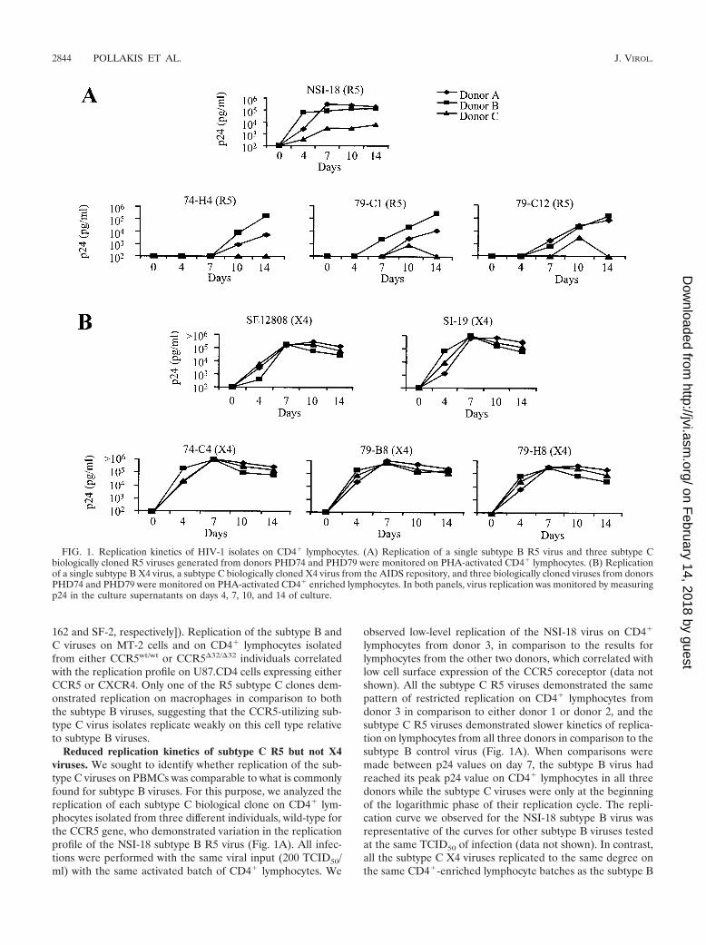
No differences between CC or CXC inhibition of the subtypeC or B viruses. To identify the sensitivities of our viruses to theinhibitory effects of either CC or CXC chemokines, we per-formed in vitro inhibition experiments with limiting dilutionsof either RANTES or SDF-1� for the R5 and X4 viruses,respectively (Fig. 2). The inhibitions were performed withCD4� lymphocytes from two donors (donors A and B), andsimilar concentrations of CC or CXC chemokine were re-quired to inhibit virus replication by 90% in lymphocytesfrom both donors (Fig. 2). We also observed a wide variationin the required concentration of CC or CXC chemokine re-
quired to inhibit replication, which was neither HIV subtypenor R5 or X4 phenotype restricted.
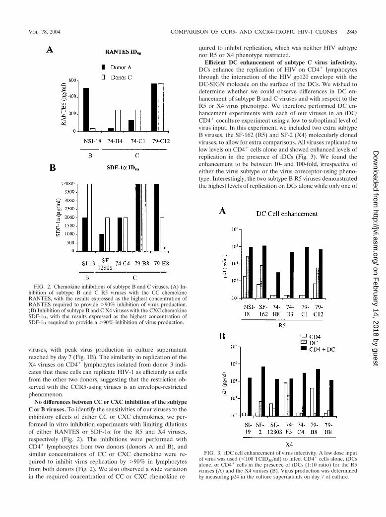
Efficient DC enhancement of subtype C virus infectivity.DCs enhance the replication of HIV on CD4� lymphocytesthrough the interaction of the HIV gp120 envelope with theDC-SIGN molecule on the surface of the DCs. We wished todetermine whether we could observe differences in DC en-hancement of subtype B and C viruses and with respect to theR5 or X4 virus phenotype. We therefore performed DC en-hancement experiments with each of our viruses in an iDC/CD4� coculture experiment using a low to suboptimal level ofvirus input. In this experiment, we included two extra subtypeB viruses, the SF-162 (R5) and SF-2 (X4) molecularly clonedviruses, to allow for extra comparisons. All viruses replicated tolow levels on CD4� cells alone and showed enhanced levels ofreplication in the presence of iDCs (Fig. 3). We found theenhancement to be between 10- and 100-fold, irrespective ofeither the virus subtype or the virus coreceptor-using pheno-type. Interestingly, the two subtype B R5 viruses demonstratedthe highest levels of replication on DCs alone while only one of
FIG. 2. Chemokine inhibitions of subtype B and C viruses. (A) In-hibition of subtype B and C R5 viruses with the CC chemokineRANTES, with the results expressed as the highest concentration ofRANTES required to provide 90% inhibition of virus production.(B) Inhibition of subtype B and C X4 viruses with the CXC chemokineSDF-1�, with the results expressed as the highest concentration ofSDF-1� required to provide a 90% inhibition of virus production.
FIG. 3. iDC cell enhancement of virus infectivity. A low dose inputof virus was used (�100 TCID50/ml) to infect CD4� cells alone, iDCsalone, or CD4� cells in the presence of iDCs (1:10 ratio) for the R5viruses (A) and the X4 viruses (B). Virus production was determinedby measuring p24 in the culture supernatants on day 7 of culture.
VOL. 78, 2004 COMPARISON OF CCR5- AND CXCR4-TROPIC HIV-1 CLONES 2845
on February 14, 2018 by guest
http://jvi.asm.org/
Dow
nloaded from
the four subtype C R5 viruses demonstrated such high levels,probably a reflection on the poor growth of subtype C R5viruses on CD4� lymphocytes in general (Fig. 3A). Althoughwe cannot rule out additional DC cytokine enhancement ofviral replication, previous studies have demonstrated the ne-cessity of the DC-SIGN interaction with the virus for increasedviral transfer via the ability of a DC-SIGN-specific monoclonalantibody to inhibit enhanced viral replication (21).
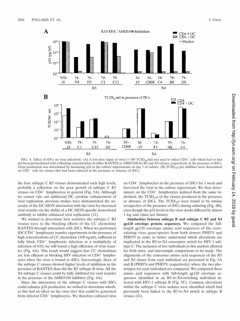
We wished to determine how sensitive the subtype C R5viruses were to the blocking effects of the CC chemokineRANTES through interaction with iDCs. When we performediDC/CD4� lymphocyte transfer experiments in the presence ofhigh concentrations of CC chemokine (100 ng/ml), sufficient tofully block CD4� lymphocyte infection at a multiplicity ofinfection of 0.03, we still found a high efficiency of virus trans-fer (Fig. 4A). This result would suggest that CC chemokinesare less efficient at blocking HIV infection of CD4� lympho-cytes when the virus is bound to iDCs. Interestingly, three ofthe subtype C viruses showed higher levels of inhibition in thepresence of RANTES than did the R5 subtype B virus. All theX4 subtype C viruses could be fully inhibited for viral transferin the presence of the AMD3100 inhibitor (Fig. 4A).
Since the interaction of the subtype C viruses with iDCscould enhance p24 production, we wished to determine wheth-er this had an effect on the virus titer that could be generatedfrom infected CD4� lymphocytes. We therefore cultured virus
on CD4� lymphocytes in the presence of iDCs for 1 week andharvested the virus in the culture supernatant. We then deter-mined, on the CD4� lymphocytes isolated from the same in-dividual, the TCID50s of the viruses produced in the presenceor absence of iDCs. The TCID50s were found to be similarirrespective of the presence of iDCs during culturing (Fig. 4B),even though the p24 levels in the virus stocks differed by almost1 log unit (data not shown).
Similarities between subtype B and subtype C R5 and X4gp120 envelope protein sequences. We compared the full-length gp120 envelope amino acid sequences of the cocir-culating virus quasi-species from both donors PHD74 andPHD79 in order to better understand which alterations areimplicated in the R5-to-X4 coreceptor switch for HIV-1 sub-type C. The inclusion of two individuals in this analysis allowedfor both intra- and intersample comparisons to be made. Thealignments of the consensus amino acid sequences of the R5and X4 clones from each individual are presented in Fig. 5Aand B (PHD74 and PHD79, respectively), where the two phe-notypes for each individual are compared. We compared theseamino acid sequences with full-length gp120 envelope se-quences identified in an R5-to-X4-switching individual in-fected with HIV-1 subtype B (Fig. 5C). Common alterationswithin the subtype C virus isolates were identified which hadpreviously been linked to the R5-to-X4 switch in subtype Bviruses (41).
FIG. 4. Effect of iDCs on virus infectivity. (A) A low-dose input of virus (�100 TCID50/ml) was used to infect CD4� cells which had or hadnot been preincubated with a blocking concentration of either RANTES or AMD3100 for R5 and X4 viruses, respectively, in the presence of iDCs.Virus production was determined by measuring p24 in the culture supernatants on day 7 of culture. (B) TCID50s per milliliter were determinedon CD4� cells for viruses that had been cultured in the presence or absence of iDCs.
2846 POLLAKIS ET AL. J. VIROL.
on February 14, 2018 by guest
http://jvi.asm.org/
Dow
nloaded from
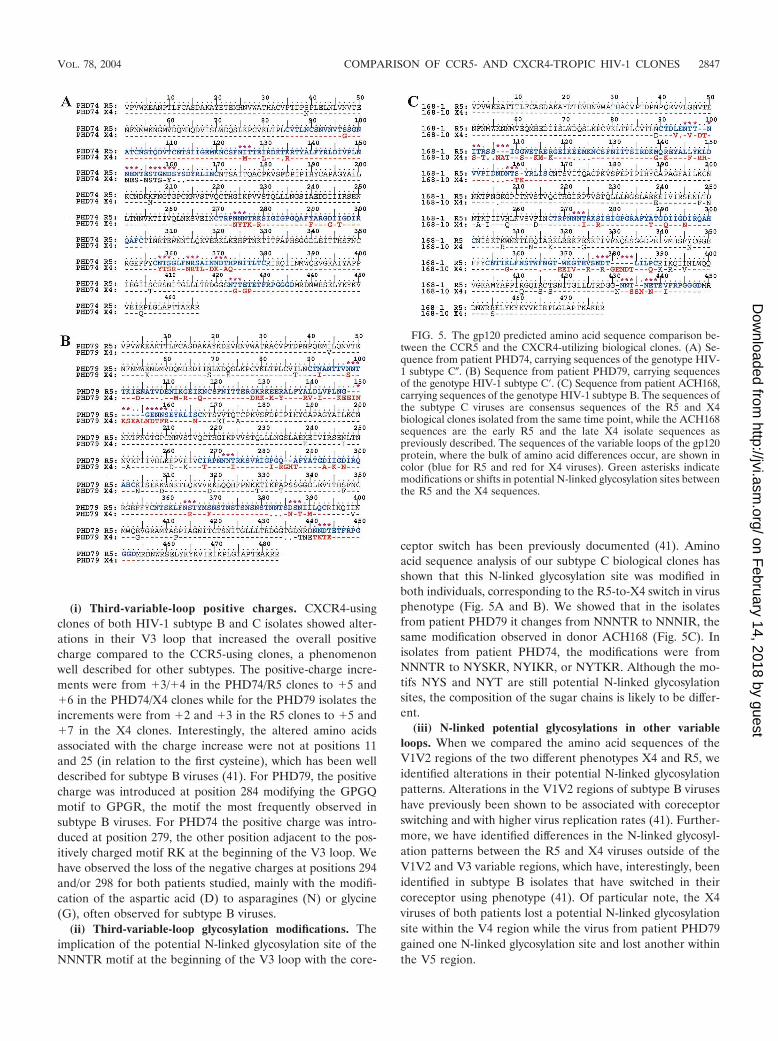
(i) Third-variable-loop positive charges. CXCR4-usingclones of both HIV-1 subtype B and C isolates showed alter-ations in their V3 loop that increased the overall positivecharge compared to the CCR5-using clones, a phenomenonwell described for other subtypes. The positive-charge incre-ments were from �3/�4 in the PHD74/R5 clones to �5 and�6 in the PHD74/X4 clones while for the PHD79 isolates theincrements were from �2 and �3 in the R5 clones to �5 and�7 in the X4 clones. Interestingly, the altered amino acidsassociated with the charge increase were not at positions 11and 25 (in relation to the first cysteine), which has been welldescribed for subtype B viruses (41). For PHD79, the positivecharge was introduced at position 284 modifying the GPGQmotif to GPGR, the motif the most frequently observed insubtype B viruses. For PHD74 the positive charge was intro-duced at position 279, the other position adjacent to the pos-itively charged motif RK at the beginning of the V3 loop. Wehave observed the loss of the negative charges at positions 294and/or 298 for both patients studied, mainly with the modifi-cation of the aspartic acid (D) to asparagines (N) or glycine(G), often observed for subtype B viruses.
(ii) Third-variable-loop glycosylation modifications. Theimplication of the potential N-linked glycosylation site of theNNNTR motif at the beginning of the V3 loop with the core-
ceptor switch has been previously documented (41). Aminoacid sequence analysis of our subtype C biological clones hasshown that this N-linked glycosylation site was modified inboth individuals, corresponding to the R5-to-X4 switch in virusphenotype (Fig. 5A and B). We showed that in the isolatesfrom patient PHD79 it changes from NNNTR to NNNIR, thesame modification observed in donor ACH168 (Fig. 5C). Inisolates from patient PHD74, the modifications were fromNNNTR to NYSKR, NYIKR, or NYTKR. Although the mo-tifs NYS and NYT are still potential N-linked glycosylationsites, the composition of the sugar chains is likely to be differ-ent.
(iii) N-linked potential glycosylations in other variableloops. When we compared the amino acid sequences of theV1V2 regions of the two different phenotypes X4 and R5, weidentified alterations in their potential N-linked glycosylationpatterns. Alterations in the V1V2 regions of subtype B viruseshave previously been shown to be associated with coreceptorswitching and with higher virus replication rates (41). Further-more, we have identified differences in the N-linked glycosyl-ation patterns between the R5 and X4 viruses outside of theV1V2 and V3 variable regions, which have, interestingly, beenidentified in subtype B isolates that have switched in theircoreceptor using phenotype (41). Of particular note, the X4viruses of both patients lost a potential N-linked glycosylationsite within the V4 region while the virus from patient PHD79gained one N-linked glycosylation site and lost another withinthe V5 region.
FIG. 5. The gp120 predicted amino acid sequence comparison be-tween the CCR5 and the CXCR4-utilizing biological clones. (A) Se-quence from patient PHD74, carrying sequences of the genotype HIV-1 subtype C�. (B) Sequence from patient PHD79, carrying sequencesof the genotype HIV-1 subtype C�. (C) Sequence from patient ACH168,carrying sequences of the genotype HIV-1 subtype B. The sequences ofthe subtype C viruses are consensus sequences of the R5 and X4biological clones isolated from the same time point, while the ACH168sequences are the early R5 and the late X4 isolate sequences aspreviously described. The sequences of the variable loops of the gp120protein, where the bulk of amino acid differences occur, are shown incolor (blue for R5 and red for X4 viruses). Green asterisks indicatemodifications or shifts in potential N-linked glycosylation sites betweenthe R5 and the X4 sequences.
VOL. 78, 2004 COMPARISON OF CCR5- AND CXCR4-TROPIC HIV-1 CLONES 2847
on February 14, 2018 by guest
http://jvi.asm.org/
Dow
nloaded from
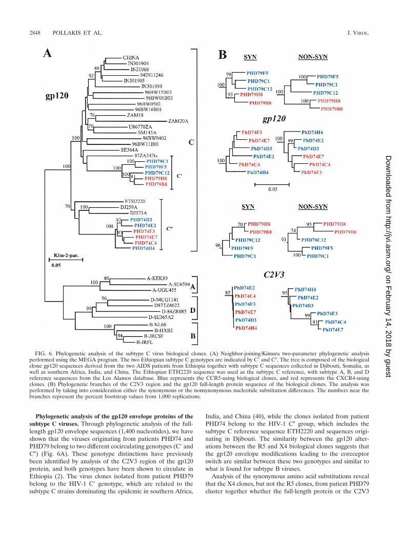
Phylogenetic analysis of the gp120 envelope proteins of thesubtype C viruses. Through phylogenetic analysis of the full-length gp120 envelope sequences (1,400 nucleotides), we haveshown that the viruses originating from patients PHD74 andPHD79 belong to two different cocirculating genotypes (C� andC�) (Fig. 6A). These genotype distinctions have previouslybeen identified by analysis of the C2V3 region of the gp120protein, and both genotypes have been shown to circulate inEthiopia (2). The virus clones isolated from patient PHD79belong to the HIV-1 C� genotype, which are related to thesubtype C strains dominating the epidemic in southern Africa,
India, and China (40), while the clones isolated from patientPHD74 belong to the HIV-1 C� group, which includes thesubtype C reference sequence ETH2220 and sequences origi-nating in Djibouti. The similarity between the gp120 alter-ations between the R5 and X4 biological clones suggests thatthe gp120 envelope modifications leading to the coreceptorswitch are similar between these two genotypes and similar towhat is found for subtype B viruses.
Analysis of the synonymous amino acid substitutions revealthat the X4 clones, but not the R5 clones, from patient PHD79cluster together whether the full-length protein or the C2V3
FIG. 6. Phylogenetic analysis of the subtype C virus biological clones. (A) Neighbor-joining/Kimura two-parameter phylogenetic analysisperformed using the MEGA program. The two Ethiopian subtype C genotypes are indicated by C� and C�. The tree is composed of the biologicalclone gp120 sequences derived from the two AIDS patients from Ethiopia together with subtype C sequences collected in Djibouti, Somalia, aswell as southern Africa, India, and China. The Ethiopian ETH2220 sequence was used as the subtype C reference, with subtype A, B, and Dreference sequences from the Los Alamos database. Blue represents the CCR5-using biological clones, and red represents the CXCR4-usingclones. (B) Phylogenetic branches of the C2V3 region and the gp120 full-length protein sequence of the biological clones. The analysis wasperformed by taking into consideration either the synonymous or the nonsynonymous nucleotide substitution differences. The numbers near thebranches represent the percent bootstrap values from 1,000 replications.
2848 POLLAKIS ET AL. J. VIROL.
on February 14, 2018 by guest
http://jvi.asm.org/
Dow
nloaded from
region of the gp120 protein are considered (Fig. 6B). Neitherthe X4 nor the R5 clones from patient PHD74 clustered to-gether by any significant bootstrap values, indicating that co-receptor usage does not necessarily determine the phyloge-netic lineage. In contrast, the nonsynonymous amino acidsubstitution analysis revealed that for both individuals therewas a marked segregation between the X4 and R5 clones,which was particularly apparent for the C2V3 region of thegp120 protein, demonstrating the strong involvement of thisregion in coreceptor utilization (Fig. 6B). In addition, the syn-onymous-versus-nonsynonymous amino acid substitution rates(ds/dn) for the gp120 region were 1.00 for PHD74 and 0.58 andPHD79 while those for the C2V3 region were 0.02 and 0.26,respectively, again demonstrating the strong positive selectiondriven by coreceptor utilization.
Clone PHD79C12 is of particular interest since phylogeneticanalysis (Fig. 6B) and boot-scanning analysis (data not shown)have identified it as a recombinant between the X4 and R5quasi-species cocirculating within patient PHD79. The virushas acquired the V1V2 envelope region from the X4 clones buthas remained a CCR5-using virus, indicating that for this virusthe V3 region is a strong determinant of coreceptor usage.Interestingly, this virus has the highest TCID50 of all the R5viruses (Table 1), demonstrating the highest resistance to theblocking effects of the CC chemokine RANTES on CD4�-lymphocytes (Fig. 2A).
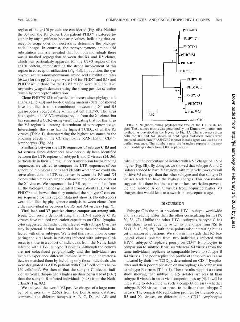
Similarity between the LTR sequences of subtype C R5 andX4 viruses. Since differences have previously been identifiedbetween the LTR regions of subtype B and C viruses (24, 36),particularly in their U3 regulatory transcription factor bindingsequences, we wished to compare the LTR sequences of ourgenerated biological clones and identify whether we could ob-serve alterations in LTR sequences between the R5 and X4clones, which may explain the enhanced replication capacity ofthe X4 viruses. We sequenced the U3R region amplified fromall the biological clones generated from patients PHD74 andPHD79 and showed that they matched the subtype C consen-sus sequence for this region (data not shown). No differenceswere identified by phylogenetic analysis between clones fromeither individual or between the R5 and X4 viruses (Fig. 7).
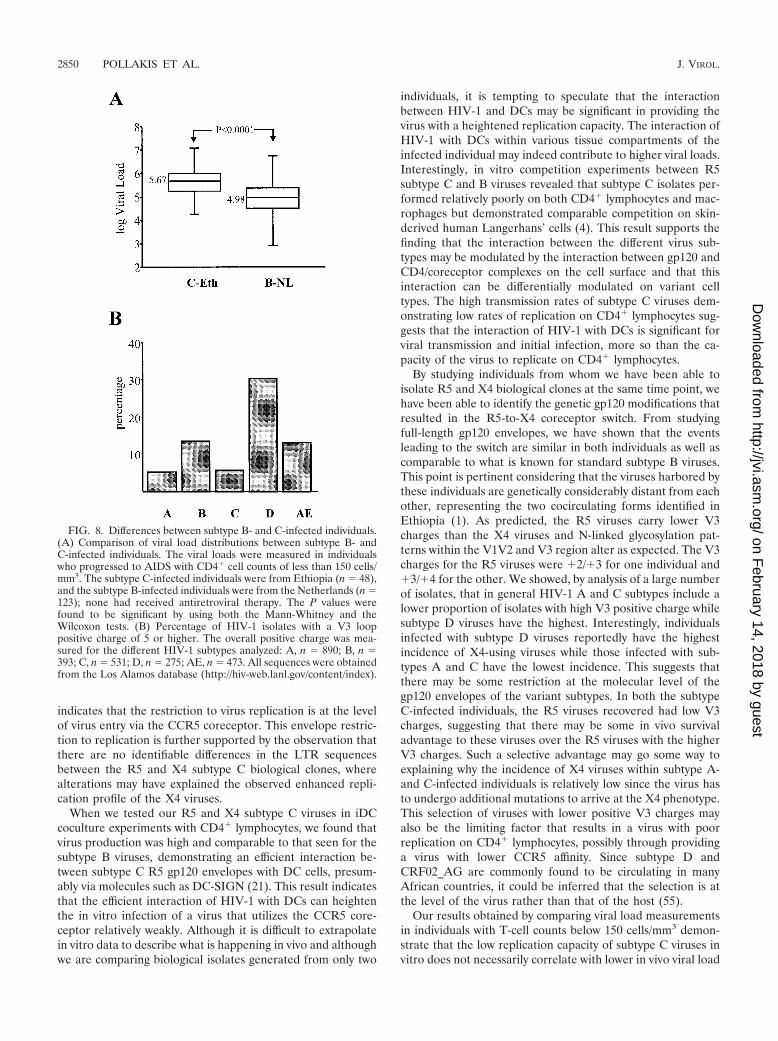
Viral load and V3 positive charge comparison among sub-types. Our results demonstrating that HIV-1 subtype C R5viruses have reduced replication capacities on CD4� lympho-cytes suggested that individuals infected with subtype C virusesmay in general harbor lower viral loads than individuals in-fected with other subtypes. We tested this assumption by com-paring the viral loads in patients infected with subtype C vi-ruses to those in a cohort of individuals from the Netherlandsinfected with HIV-1 subtype B isolates. Although the cohortsare not colocalized geographically and the individuals arelikely to experience different immune stimulation characteris-tics, we matched them by including only those individuals whowere designated as AIDS patients with CD4� cell counts below150 cells/mm3. We showed that the subtype C-infected indi-viduals from Ethiopia had a higher median log viral load (5.67)than the subtype B-infected individuals (4.98) from the Neth-erlands (Fig. 8A).
We analyzed the overall V3 positive charges of a large num-ber of viruses (n 2,562) from the Los Alamos database,compared the different subtypes A, B, C, D, and AE, and
calculated the percentage of isolates with a V3 charge of �5 orhigher (Fig. 8B). By doing so, we showed that subtype A and Cisolates tended to have V3 regions with relatively lower overallpositive V3 charges than the other subtypes and that subtype Dviruses tended to have the highest charges. This observationsuggests that there is either a virus or host restriction prevent-ing the subtype A or C viruses from acquiring higher V3charges that would lead to a switch in coreceptor usage.
DISCUSSION
Subtype C is the most prevalent HIV-1 subtype worldwideand is spreading faster than the other cocirculating forms (19,30, 38, 42). Unlike the other HIV-1 subtypes, subtype C hasbeen shown to infrequently switch its phenotype from NSI toSI (1, 8, 12, 35, 39). Both these points raise interesting but asyet unanswered questions. We show in this study that R5 bio-logical clones isolated from two individuals infected withHIV-1 subtype C replicate poorly on CD4� lymphocytes incomparison to subtype B viruses whereas X4 viruses from thesame individuals replicate to comparable levels to subtype BX4 viruses. The poor replication profile of these viruses is alsoindicated by their low TCID50s determined on CD4� lympho-cytes and their poor replication on macrophages in comparisonto subtype B viruses (Table 1). These results support a recentstudy showing that subtype C R5 isolates are less fit thansubtype B viruses in an ex vivo competition assay (4). It will beinteresting to determine in such a competition assay whethersubtype B X4 viruses also prove to be fitter than subtype Cviruses. The comparable replication profiles, for the subtype CR5 and X4 viruses, on different donor CD4� lymphocytes
FIG. 7. Neighbor-joining phylogenetic tree of the LTR/U3R re-gion. The distance matrix was generated by the Kimura two-parametermethod, as described in the legend to Fig. 1A. The sequences fromboth the R5 and X4 (shown in bold type) biological clones wereanalyzed, and isolate FR83HXB2 (shown in italic type) was used as theoutlier sequence. The numbers near the branches represent the per-cent bootstrap values from 1,000 replications.
VOL. 78, 2004 COMPARISON OF CCR5- AND CXCR4-TROPIC HIV-1 CLONES 2849
on February 14, 2018 by guest
http://jvi.asm.org/
Dow
nloaded from
indicates that the restriction to virus replication is at the levelof virus entry via the CCR5 coreceptor. This envelope restric-tion to replication is further supported by the observation thatthere are no identifiable differences in the LTR sequencesbetween the R5 and X4 subtype C biological clones, wherealterations may have explained the observed enhanced repli-cation profile of the X4 viruses.
When we tested our R5 and X4 subtype C viruses in iDCcoculture experiments with CD4� lymphocytes, we found thatvirus production was high and comparable to that seen for thesubtype B viruses, demonstrating an efficient interaction be-tween subtype C R5 gp120 envelopes with DC cells, presum-ably via molecules such as DC-SIGN (21). This result indicatesthat the efficient interaction of HIV-1 with DCs can heightenthe in vitro infection of a virus that utilizes the CCR5 core-ceptor relatively weakly. Although it is difficult to extrapolatein vitro data to describe what is happening in vivo and althoughwe are comparing biological isolates generated from only two
individuals, it is tempting to speculate that the interactionbetween HIV-1 and DCs may be significant in providing thevirus with a heightened replication capacity. The interaction ofHIV-1 with DCs within various tissue compartments of theinfected individual may indeed contribute to higher viral loads.Interestingly, in vitro competition experiments between R5subtype C and B viruses revealed that subtype C isolates per-formed relatively poorly on both CD4� lymphocytes and mac-rophages but demonstrated comparable competition on skin-derived human Langerhans’ cells (4). This result supports thefinding that the interaction between the different virus sub-types may be modulated by the interaction between gp120 andCD4/coreceptor complexes on the cell surface and that thisinteraction can be differentially modulated on variant celltypes. The high transmission rates of subtype C viruses dem-onstrating low rates of replication on CD4� lymphocytes sug-gests that the interaction of HIV-1 with DCs is significant forviral transmission and initial infection, more so than the ca-pacity of the virus to replicate on CD4� lymphocytes.
By studying individuals from whom we have been able toisolate R5 and X4 biological clones at the same time point, wehave been able to identify the genetic gp120 modifications thatresulted in the R5-to-X4 coreceptor switch. From studyingfull-length gp120 envelopes, we have shown that the eventsleading to the switch are similar in both individuals as well ascomparable to what is known for standard subtype B viruses.This point is pertinent considering that the viruses harbored bythese individuals are genetically considerably distant from eachother, representing the two cocirculating forms identified inEthiopia (1). As predicted, the R5 viruses carry lower V3charges than the X4 viruses and N-linked glycosylation pat-terns within the V1V2 and V3 region alter as expected. The V3charges for the R5 viruses were �2/�3 for one individual and�3/�4 for the other. We showed, by analysis of a large numberof isolates, that in general HIV-1 A and C subtypes include alower proportion of isolates with high V3 positive charge whilesubtype D viruses have the highest. Interestingly, individualsinfected with subtype D viruses reportedly have the highestincidence of X4-using viruses while those infected with sub-types A and C have the lowest incidence. This suggests thatthere may be some restriction at the molecular level of thegp120 envelopes of the variant subtypes. In both the subtypeC-infected individuals, the R5 viruses recovered had low V3charges, suggesting that there may be some in vivo survivaladvantage to these viruses over the R5 viruses with the higherV3 charges. Such a selective advantage may go some way toexplaining why the incidence of X4 viruses within subtype A-and C-infected individuals is relatively low since the virus hasto undergo additional mutations to arrive at the X4 phenotype.This selection of viruses with lower positive V3 charges mayalso be the limiting factor that results in a virus with poorreplication on CD4� lymphocytes, possibly through providinga virus with lower CCR5 affinity. Since subtype D andCRF02_AG are commonly found to be circulating in manyAfrican countries, it could be inferred that the selection is atthe level of the virus rather than that of the host (55).
Our results obtained by comparing viral load measurementsin individuals with T-cell counts below 150 cells/mm3 demon-strate that the low replication capacity of subtype C viruses invitro does not necessarily correlate with lower in vivo viral load
FIG. 8. Differences between subtype B- and C-infected individuals.(A) Comparison of viral load distributions between subtype B- andC-infected individuals. The viral loads were measured in individualswho progressed to AIDS with CD4� cell counts of less than 150 cells/mm3. The subtype C-infected individuals were from Ethiopia (n 48),and the subtype B-infected individuals were from the Netherlands (n 123); none had received antiretroviral therapy. The P values werefound to be significant by using both the Mann-Whitney and theWilcoxon tests. (B) Percentage of HIV-1 isolates with a V3 looppositive charge of 5 or higher. The overall positive charge was mea-sured for the different HIV-1 subtypes analyzed: A, n 890; B, n 393; C, n 531; D, n 275; AE, n 473. All sequences were obtainedfrom the Los Alamos database (http://hiv-web.lanl.gov/content/index).
2850 POLLAKIS ET AL. J. VIROL.
on February 14, 2018 by guest
http://jvi.asm.org/
Dow
nloaded from
measurements for this subtype (Fig. 8A). The results also dem-onstrate that subtype C-infected individuals can progress tolower T-cell counts and, presumably, to disease without aswitch in coreceptor usage phenotype. This finding has to beinterpreted in light of the observation that Ethiopian individ-uals, whether HIV-1 infected or not, tend to have lower CD4counts than other individuals (32). A number of factors, in-cluding cell type availability, coreceptor expression profiles,and HIV-1-specific immune responses, will obviously contrib-ute to the observation that despite the low in vitro replicationof the subtype C R5 viruses, these individual can develop highviral loads.
When we compared the sensitivities of the subtype C R5viruses to the blocking effects of the CC chemokine RANTES,we found a wide variation, probably reflecting differences inthe coreceptor binding domain of the gp120 envelope, whichmay alter the affinity for the CCR5 coreceptor. Notably, thevirus with the highest resistance to RANTES neutralization(PHD79C12) was identified by phylogenetic and boot-scanninganalysis to be a recombinant between an R5 virus and an X4virus from the same patient, with the virus possessing the V3region from an R5 virus and the V1V2 region from an X4virus. This is not the first time that recombination betweencocirculating R5 and X4 viruses has been described (52). Thisresult would indicate that the recombinant virus either has ahigher-affinity interaction with the CCR5 coreceptor or canbetter escape control by circulatory CC chemokines, therebyhelping explain its emergence in vivo.
It is still difficult to explain why subtype C viruses do notswitch coreceptors, considering that they require the sameenvelope alterations as subtype B viruses in order to do so. Thelow initial V3 charge seen in the V3 region may provide someexplanation, with more time required for the mutations in thegp120 envelope to accrue, allowing the switch to occur, espe-cially if the replication capacity of the viruses with the lower V3charge is restricted. It has been speculated that the low X4frequency with subtype C viruses is due to differences in cohortselection, time of monitoring, and coinfection profiles (13).Although these arguments may well be relevant, we think thatthey are unlikely to be the main explanations. In our Ethiopiancohort, we identified a very low incidence of X4 viruses amongAIDS patients, with only 3 of 48 patients in the cohort (6.25%)harboring X4 viruses, a similar observation to those in otherstudies (1, 8, 39). Coinfection with other pathogens and thegeneral immune activation profile of the host are likely to bemajor factors in directing viral coreceptor usage and switchingthrough alterations in such factors as coreceptor expressionlevels. However, individuals from countries with similar geo-graphical settings and infection profiles can have extremelydifferent outcomes with respect to the HIV-1 subtype beingpropagated and, more significantly, the virus phenotypes, whichis especially pertinent for subtype C and CRF02_AG viruses(55). Why subtype C viruses are spreading so fast is also amystery; again, the low V3 positive charges associated withsubtype C viruses may favor the presence within the infectedpopulation of viruses harboring a transmission advantage overviruses with the higher positive V3 charges and the X4 pheno-type.
ACKNOWLEDGMENTS
This work was supported by grants from the European Union(QLK2-CT-1999-01321 “EuroVac”), the American Foundation ofAIDS Research (grant 02721-28-RG), and the Ethio-NetherlandsAIDS Research Program (ENARP). Virus SE18208 was providedthrough the EU Programme EVA/MRC Centralized Facility for AIDSReagents, NIBSC, United Kingdom (grants QLK2-CT-1999-00609and GP828102). W.A.P. is the recipient of a research fellowship fromthe Royal Dutch Academy for Arts and Sciences.
We thank Patrizia Carotenuto for providing viruses NSI-18 andSI-19.
REFERENCES
1. Abebe, A., D. Demissie, J. Goudsmit, M. Brouwer, C. L. Kuiken, G. Pollakis,H. Schuitemaker, A. L. Fontanet, and T. F. Rinke de Wit. 1999. HIV-1subtype C syncytium- and non-syncytium-inducing phenotypes and corecep-tor usage among Ethiopian patients with AIDS. AIDS 13:1305–1311.
2. Abebe, A., G. Pollakis, A. L. Fontanet, B. Fisseha, B. Tegbaru, A. Kliphuis,G. Tesfaye, H. Negassa, M. Cornelissen, J. Goudsmit, and T. F. Renke deWit. 2000. Identification of a genetic subcluster of HIV type 1 subtype C (C�)widespread in Ethiopia. AIDS Res. Hum. Retrovir. 16:1909–1914.
3. Back, N. K., L. Smit, J. J. De Jong, W. Keulen, M. Schutten, J. Goudsmit,and M. Tersmette. 1994. An N-glycan within the human immunodeficiencyvirus type 1 gp120 V3 loop affects virus neutralization. Virology 199:431–438.
4. Ball, S. C., A. Abraha, K. R. Collins, A. J. Marozsan, H. Baird, M. E.Quinones-Mateu, A. Penn-Nicholson, M. Murray, N. Richard, M. Lobritz,P. A. Zimmerman, T. Kawamura, A. Blauvelt, and E. J. Arts. 2003. Com-paring the ex vivo fitness of CCR5-tropic human immunodeficiency virustype 1 isolates of subtypes B and C. J. Virol. 77:1021–1038.
5. Berger, E. A. 1997. HIV entry and tropism: the chemokine receptor connec-tion. AIDS 11(Suppl. A):S3–S16.
6. Berger, E. A., R. W. Doms, E. M. Fenyo, B. T. Korber, D. R. Littman, J. P.Moore, Q. J. Sattentau, H. Schuitemaker, J. Sodroski, and R. A. Weiss. 1998.A new classification for HIV-1. Nature 391:240.
7. Bjorndal, A., H. Deng, M. Jansson, J. R. Fiore, C. Colognesi, A. Karlsson, J.Albert, G. Scarlatti, D. R. Littman, and E. M. Fenyo. 1997. Coreceptor usageof primary human immunodeficiency virus type 1 isolates varies according tobiological phenotype. J. Virol. 71:7478–7487.
8. Bjorndal, A., A. Sonnerborg, C. Tscherning, J. Albert, and E. M. Fenyo.1999. Phenotypic characteristics of human immunodeficiency virus type 1subtype C isolates of Ethiopian AIDS patients. AIDS Res. Hum. Retrovir.15:647–653.
9. Bleul, C. C., M. Farzan, H. Choe, C. Parolin, I. Clark-Lewis, J. Sodroski, andT. A. Springer. 1996. The lymphocyte chemoattractant SDF-1 is a ligand forLESTR/fusin and blocks HIV-1 entry. Nature 382:829–833.
10. Boom, R., C. J. Sol, M. M. Salimans, C. L. Jansen, P. M. Wertheim-vanDillen, and J. van der Noordaa. 1990. Rapid and simple method for purifi-cation of nucleic acids. J. Clin. Microbiol. 28:495–503.
11. Carr, J. K., M. O. Salminen, J. Albert, E. Sanders-Buell, D. Gotte, D. L. Birx,and F. E. McCutchan. 1998. Full genome sequences of human immunode-ficiency virus type 1 subtypes G and A/G intersubtype recombinants. Virol-ogy 247:22–31.
12. Cecilia, D., S. S. Kulkarni, S. P. Tripathy, R. R. Gangakhedkar, R. S.Paranjape, and D. A. Gadkari. 2000. Absence of coreceptor switch withdisease progression in human immunodeficiency virus infections in India.Virology 271:253–258.
13. Cilliers, T., J. Nhlapo, M. Coetzer, D. Orlovic, T. Ketas, W. C. Olson, J. P.Moore, A. Trkola, and L. Morris. 2003. The CCR5 and CXCR4 coreceptorsare both used by human immunodeficiency virus type 1 primary isolates fromsubtype C. J. Virol. 77:4449–4456.
14. Cocchi, F., A. L. Devico, A. Garzino-Demo, S. K. Arya, R. C. Gallo, and P.Lusso. 1995. Identification of RANTES, MIP-1 alpha, and MIP-1 beta as themajor HIV-suppressive factors produced by CD8� T cells. Science 270:1811–1815.
15. Connor, R. I., K. E. Sheridan, D. Ceradini, S. Choe, and N. R. Landau. 1997.Change in coreceptor use coreceptor use correlates with disease progressionin HIV-1-infected individuals. J. Exp. Med. 185:621–628.
16. de Baar, M. P., A. M. van der Schoot, J. Goudsmit, F. Jacobs, R. Ehren,K. H. van der Horn, P. Oudshoorn, F. De Wolf, and A. De Ronde. 1999.Design and evaluation of a human immunodeficiency virus type 1 RNA assayusing nucleic acid sequence-based amplification technology able to quantifyboth group M and O viruses by using the long terminal repeat as target.J. Clin. Microbiol. 37:1813–1818.
17. de Jong, J. J., A. De Ronde, W. Keulen, M. Tersmette, and J. Goudsmit.1992. Minimal requirements for the human immunodeficiency virus type 1V3 domain to support the syncytium-inducing phenotype: analysis by singleamino acid substitution. J. Virol. 66:6777–6780.
18. de Jong, J. J., J. Goudsmit, W. Keulen, B. Klaver, W. Krone, M. Tersmette,and A. de Ronde. 1992. Human immunodeficiency virus type 1 clones chi-
VOL. 78, 2004 COMPARISON OF CCR5- AND CXCR4-TROPIC HIV-1 CLONES 2851
on February 14, 2018 by guest
http://jvi.asm.org/
Dow
nloaded from
meric for the envelope V3 domain differ in syncytium formation and repli-cation capacity. J. Virol. 66:757–765.
19. de Martinez, A. M., E. F. Barbosa, P. C. Ferreira, F. A. Cardoso, J. Silveira,G. Sassi, C. M. da Silva, V. Mendonca-Signorini, and C. M. Antunes. 2002.Molecular epidemiology of HIV-1 in Rio Grande, RS, Brazil. Rev. Soc. Bras.Med Trop. 35:471–476.
20. Felsenstein, J. 1996. PHYLIP: Phylogeny Inference Package, v. 3.52. Uni-versity of Washington, Seattle.
21. Geijtenbeek, T. B., D. S. Kwon, R. Torensma, S. J. van Vliet, G. C. vanDuijnhoven, J. Middel, I. L. Cornelissen, H. S. Nottet, V. N. KewalRamani,D. R. Littman, C. G. Figdor, and Y. van Kooyk. 2000. DC-SIGN, a dendriticcell-specific HIV-1-binding protein that enhances trans-infection of T cells.Cell 100:587–597.
22. Hillis, D. M. 1997. Phylogenetic analysis. Curr. Biol. 7:R129–R131.23. Hillis, D. M., and J. J. Bull. 1993. An empirical test for bootstraping as a
method for assessing confidence in phylogenetic analysis. Syst. Biol. 42:182–192.
24. Hunt, G., and C. T. Tiemessen. 2000. Occurrence of additional NF-kappaB-binding motifs in the long terminal repeat region of South African HIV type1 subtype C isolates. AIDS Res. Hum. Retrovir. 16:305–306.
25. Jeeninga, R. E., M. Hoogenkamp, M. Armand-Ugon, M. de Baar, K. Verhoef,and B. Berkhout. 2000. Functional differences between the long terminalrepeat transcriptional promoters of human immunodeficiency virus type 1subtypes A through G. J. Virol. 74:3740–3751.
26. Johnston, E. R., L. S. Zijenah, S. Mutetwa, R. Kantor, C. Kittinunvorakoon,and D. A. Katzenstein. 2003. High frequency of syncytium-inducing andCXCR4-tropic viruses among human immunodeficiency virus type 1 subtypeC-infected patients receiving antiretroviral treatment. J. Virol. 77:7682–7688.
27. Karlsson, A., K. Parsmyr, K. Aperia, E. Sandstrom, E. M. Fenyo, and J.Albert. 1994. MT-2 cell tropism of human immunodeficiency virus type 1isolates as a marker for response to treatment and development of drugresistance. J. Infect. Dis. 170:1367–1375.
28. Koot, M., A. B. van’t Wout, N. A. Kootstra, R. E. de Goede, M. Tersmette,and H. Schuitemaker. 1996. Relation between changes in cellular load,evolution of viral phenotype, and the clonal composition of virus populationsin the course of human immunodeficiency virus type 1 infection. J. Infect.Dis. 173:349–354.
29. Kumar, S., K. Tamura, and M. Nei. 1993. Molecular Evolutionary GeneticsAnalysis (MEGA), version 1.01. Institute of Molecular Evolutionary Genet-ics, Pennsylvania State University, University Park.
30. Long, E. M., H. L. Martin, Jr., J. K. Kreiss, S. M. Rainwater, L. Lavreys,D. J. Jackson, J. Rakwar, K. Mandaliya, and J. Overbaugh. 2000. Genderdifferences in HIV-1 diversity at time of infection. Nat. Med. 6:71–75.
31. McCutchan, F. E., J. K. Carr, M. Bajani, E. Sanders-Buell, T. O. Harry,T. C. Stoeckli, K. E. Robbins, W. Gashau, A. Nasidi, W. Janssens, and M. L.Kalish. 1999. Subtype G and multiple forms of A/G intersubtype recombi-nant human immunodeficiency virus type 1 in Nigeria. Virology 254:226–234.
32. Messele, T., M. Abdulkadir, A. L. Fontanet, B. Petros, D. Hamann, M. Koot,M. T. Roos, P. T. Schellekens, F. Miedema, and T. F. Rinke de Wit. 1999.Reduced naive and increased activated CD4 and CD8 cells in healthy adultEthiopians compared with their Dutch counterparts. Clin. Exp. Immunol.115:443–450.
33. Montano, M. A., C. P. Nixon, T. Ndung’u, H. Bussmann, V. A. Novitsky, D.Dickman, and M. Essex. 2000. Elevated tumor necrosis factor-alpha activa-tion of human immunodeficiency virus type 1 subtype C in Southern Africais associated with an NF-kappaB enhancer gain-of-function. J Infect. Dis.181:76–81.
34. Montano, M. A., V. A. Novitsky, J. T. Blackard, N. L. Cho, D. A. Katzenstein,and M. Essex. 1997. Divergent transcriptional regulation among expandinghuman immunodeficiency virus type 1 subtypes. J. Virol. 71:8657–8665.
35. Morris, L., T. Cilliers, H. Bredell, M. Phoswa, and D. J. Martin. 2001. CCR5is the major coreceptor used by HIV-1 subtype C isolates from patients withactive tuberculosis. AIDS Res. Hum. Retrovir. 17:697–701.
36. Novitsky, V. A., M. A. Montano, M. F. McLane, B. Renjifo, F. Vannberg,B. T. Foley, T. P. Ndung’u, M. Rahman, M. J. Makhema, R. Marlink, and M.Essex. 1999. Molecular cloning and phylogenetic analysis of human immu-nodeficiency virus type 1 subtype C: a set of 23 full-length clones fromBotswana. J. Virol. 73:4427–4432.
37. Osmanov, S., C. Pattou, N. Walker, B. Schwardlander, and J. Esparza. 2002.Estimated global distribution and regional spread of HIV-1 genetic subtypesin the year 2000. J. Acquir. Immune Defic. Syndr. 29:184–190.
38. Papathanasopoulos, M. A., G. M. Hunt, and C. T. Tiemessen. 2003. Evolu-tion and diversity of HIV-1 in Africa—a review. Virus Genes 26:151–163.
39. Peeters, M., R. Vincent, J. L. Perret, M. Lasky, D. Patrel, F. Liegeois, V.Courgnaud, R. Seng, T. Matton, S. Molinier, and E. Delaporte. 1999. Evi-dence for differences in MT2 cell tropism according to genetic subtypes of
HIV-1: syncytium-inducing variants seem rare among subtype C HIV-1 vi-ruses. J. Acquir. Immune Defic. Syndr. 20:115–121.
40. Pollakis, G., A. Abebe, A. Kliphuis, T. F. Rinke de Wit, B. Fisseha, B.Tegbaru, G. Tesfaye, H. Negassa, Y. Mengistou, A. L. Fontanet, I. L. Cor-nelissen, and J. Goudsmit. 2003. Recombination of HIV type 1C (C�/C�) inEthiopia: possible link of EthHIV-1C� to subtype C sequences from thehigh-prevalence epidemics in India and southern Africa. AIDS Res. Hum.Retrovir. 999–1008.
41. Pollakis, G., S. Kang, A. Kliphuis, M. I. Chalaby, J. Goudsmit, and W. A.Paxton. 2001. N-linked glycosylation of the HIV type-1 gp120 envelopeglycoprotein as a major determinant of CCR5 and CXCR4 coreceptor uti-lization. J. Biol. Chem. 276:13433–13441.
42. Robbins, K. E., C. I. Bandea, A. Levin, J. J. Goedert, W. A. Blattner, G.Brubaker, T. M. Brown, G. Schochetman, M. L. Kalish, J. Shao, and T. R.O’Brien. 1996. Genetic variability of human immunodeficiency virus type 1 inrural northwest Tanzania. AIDS Res. Hum. Retrovir. 12:1389–1391.
43. Robertson, D. L., J. P. Anderson, J. A. Bradac, J. K. Carr, B. Foley, R. K.Funkhouser, F. Gao, B. H. Hahn, M. L. Kalish, C. Kuiken, G. H. Learn, T.Leitner, F. McCutchan, S. Osmanov, M. Peeters, D. Pieniazek, M. Salminen,P. M. Sharp, S. Wolinsky, and B. Korber. 2000. HIV-1 nomenclature pro-posal. Science 288:55–56.
44. Robertson, D. L., B. H. Hahn, and P. M. Sharp. 1995. Recombination inAIDS viruses. J. Mol. Evol. 40:249–259.
45. Roof, P., M. Ricci, P. Genin, M. A. Montano, M. Essex, M. A. Wainberg, A.Gatignol, and J. Hiscott. 2002. Differential regulation of HIV-1 clade-spe-cific B, C, and E long terminal repeats by NF-kappaB and the Tat transac-tivator. Virology 296:77–83.
46. Salminen, M. O., J. K. Carr, D. S. Burke, and F. E. McCutchan. 1995.Identification of breakpoints in intergenotypic recombinants of HIV type 1by bootscanning. AIDS Res. Hum. Retrovir. 11:1423–1425.
47. Sattentau, Q. J., A. G. Dalgleish, R. A. Weiss, and P. C. Beverley. 1986.Epitopes of the CD4 antigen and HIV infection. Science 234:1120–1123.
48. Scarlatti, G., E. Tresoldi, A. Bjorndal, R. Fredriksson, C. Colognesi, H. K.Deng, M. S. Malnati, A. Plebani, A. G. Siccardi, D. R. Littman, E. M. Fenyo,and P. Lusso. 1997. In vivo evolution of HIV-1 co-receptor usage andsensitivity to chemokine-mediated suppression. Nat. Med. 3:1259–1265.
49. Schuitemaker, H., M. Koot, N. A. Kootstra, M. W. Dercksen, R. E. de Goede,R. P. van Steenwijk, J. M. Lange, J. K. Schattenkerk, F. Miedema, and M.Tersmette. 1992. Biological phenotype of human immunodeficiency virustype 1 clones at different stages of infection: progression of disease is asso-ciated with a shift from monocytotropic to T-cell-tropic virus population.J. Virol. 66:1354–1360.
50. Tersmette, M., R. E. de Goede, B. J. Al, I. N. Winkel, R. A. Gruters, H. T.Cuypers, H. G. Huisman, and F. Miedema. 1988. Differential syncytium-inducing capacity of human immunodeficiency virus isolates: frequent de-tection of syncytium-inducing isolates in patients with acquired immunode-ficiency syndrome (AIDS) and AIDS-related complex. J. Virol. 62:2026–2032.
51. Tersmette, M., R. A. Gruters, F. De Wolf, R. E. de Goede, J. M. Lange, P. T.Schellekens, J. Goudsmit, H. G. Huisman, and F. Miedema. 1989. Evidencefor a role of virulent human immunodeficiency virus (HIV) variants in thepathogenesis of acquired immunodeficiency syndrome: studies on sequentialHIV isolates. J. Virol. 63:2118–2125.
52. van Rij, R. P., M., Worobey, J. A. Visser, and H. Schuijtemaker. 2003.Evolution of R5 and X4 human immunodeficiency virus type 1 gag sequencesin vivo: evidence for recombination. Virology 314:451–459.
53. Van Rij, R. P., and H. Schuitemaker. 2002. Host genetic factors in theclinical course of HIV-1 infection: chemokines and chemokine receptors.Commun. Genet. 5:88–101.
54. van’t Wout, A. B., H. Blaak, L. J. Ran, M. Brouwer, C. Kuiken, and H.Schuitemaker. 1998. Evolution of syncytium-inducing and non-syncytium-inducing biological virus clones in relation to replication kinetics during thecourse of human immunodeficiency virus type 1 infection. J. Virol. 72:5099–5107.
55. Vergne, L., A. Bourgeois, E. Mpoudi-Ngole, R. Mougnutou, J. Mbuagbaw, F.Liegeois, C. Laurent, C. Butel, L. Zekeng, E. Delaporte, and M. Peeters.2003. Biological and genetic characteristics of HIV infections in Cameroonreveals dual group M and O infections and a correlation between SI-inducingphenotype of the predominant CRF02_AG variant and disease stage. Virol-ogy 310:254–266.
56. Wei, X., J. M. Decker, S. Wang, H. Hui, J. C. Kappes, X. Wu, J. F. Salazar-Gonzalez, M. G. Salazar, J. M. Kilby, M. S. Saag, N. L. Komarova, M. A.Nowak, B. H. Hahn, P. D. Kwong, and G. M. Shaw. 2003. Antibody neutral-ization and escape by HIV-1. Nature 422:307–312.
57. Zhang, Y. J., and J. P. Moore. 1999. Will multiple coreceptors need to betargeted by inhibitors of human immunodeficiency virus type 1 entry? J. Vi-rol. 73:3443–3448.
2852 POLLAKIS ET AL. J. VIROL.
on February 14, 2018 by guest
http://jvi.asm.org/
Dow
nloaded from


![Moderate Restriction of Macrophage-Tropic Human ......9], and this observation led to the identification of the CCR5 and CXCR4 chemokine receptors as HIV-1 co-receptors for viral fusion](https://img.pdfslide.net/doc/110x75/606645ddad14062d597e7589/moderate-restriction-of-macrophage-tropic-human-9-and-this-observation.jpg)