Embed Size (px)
Citation preview
1Andrew D. Perron, MD, FACEP
How to Read a Head CTHow to Read a Head CT
(or “How I learned to stop worrying and love computed tomography”)

Andrew D. Perron, MD, FACEPAndrew D. Perron, MD, FACEP
EM Residency Program DirectorEM Residency Program Director
Department of Emergency MedicineDepartment of Emergency MedicineMaine Medical CenterMaine Medical Center
Portland, MEPortland, ME
Andrew D. Perron, MD, FACEP2

3Andrew D. Perron, MD, FACEP
Head CTHead CT
• Has assumed a critical role in the daily practice of Emergency Medicine for evaluating intracranial emergencies. (e.g. Trauma, Stroke, SAH, ICH).
• Most practitioners have limited experience with interpretation.
• In many situations, the Emergency Physician must initially interpret and acton the CT without specialist assistance.
4Andrew D. Perron, MD, FACEP
Head CTHead CT
• Most EM training programs have no formalized training process to meet this need.
• Many Emergency Physicians are uncomfortable interpreting CTs.
• Studies have shown that EPs have a significant “miss rate” on cranial CT interpretation.
5Andrew D. Perron, MD, FACEP
Head CTHead CT
• In medical school, we are taught a systematic technique to interpret ECGs (rate, rhythm, axis, etc.) so that all aspects are reviewed, and no findings are missed.
6Andrew D. Perron, MD, FACEP
Head CTHead CT
• The intent of this session is to introduce a similar systematic method of cranial CT interpretation, based on the mnemonic…

7Andrew D. Perron, MD, FACEP
Head CTHead CT
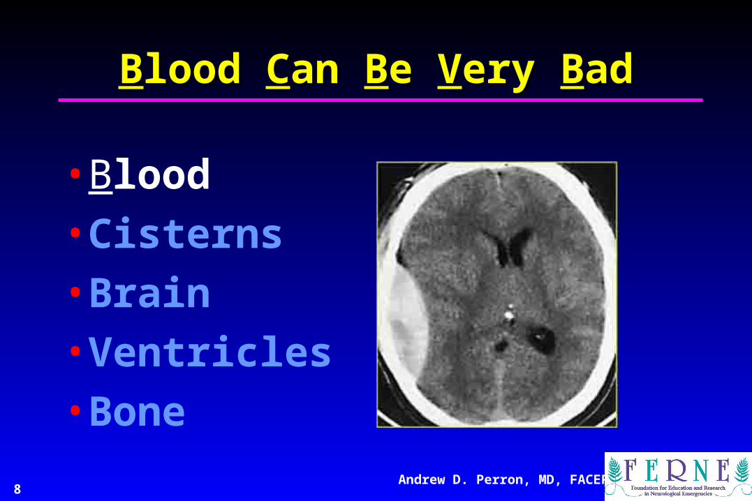
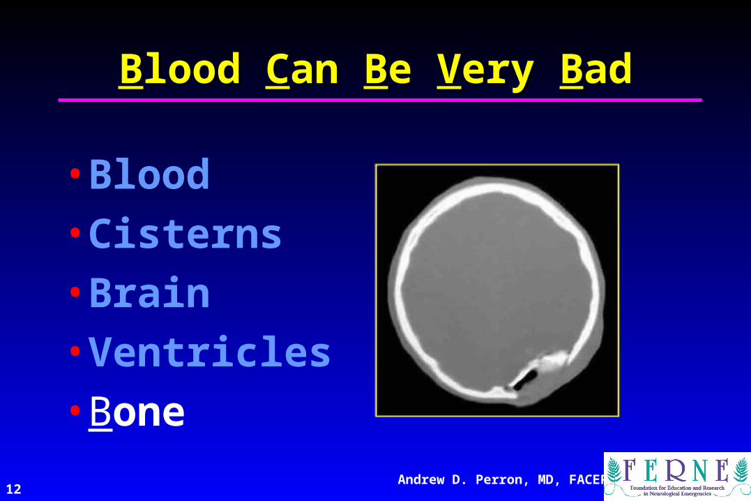
“Blood Can Be Very Bad”
8Andrew D. Perron, MD, FACEP
BBlood lood CCan an BBe e VVery ery BBadad
• Blood
• Cisterns
• Brain
• Ventricles
• Bone

9Andrew D. Perron, MD, FACEP
BBlood lood CCan an BBe e VVery ery BBadad
• Blood
• Cisterns
• Brain
• Ventricles
• Bone

10Andrew D. Perron, MD, FACEP
BBlood lood CCan an BBe e VVery ery BBadad
• Blood
• Cisterns
• Brain
• Ventricles
• Bone

11Andrew D. Perron, MD, FACEP
BBlood lood CCan an BBe e VVery ery BBadad
• Blood
• Cisterns
• Brain
• Ventricles
• Bone
12Andrew D. Perron, MD, FACEP
BBlood lood CCan an BBe e VVery ery BBadad
• Blood
• Cisterns
• Brain
• Ventricles
• Bone

13Andrew D. Perron, MD, FACEP
CT Scan BasicsCT Scan Basics• A CT image is a computer-generated
picture based on multiple x-ray exposures taken around the periphery of the subject.
• X-rays are passed through the subject, and a scanning device measures the transmitted radiation.
• The denser the object, the more the beam is attenuated, and hence fewer x-rays make it to the sensor.

14Andrew D. Perron, MD, FACEP
CT Scan BasicsCT Scan Basics• The denser the object, the whiter it is on CT
– Bone is most dense = + 1000 Hounsfield U.
– Air is the least dense = - 1000H Hounsfield U.
15Andrew D. Perron, MD, FACEP
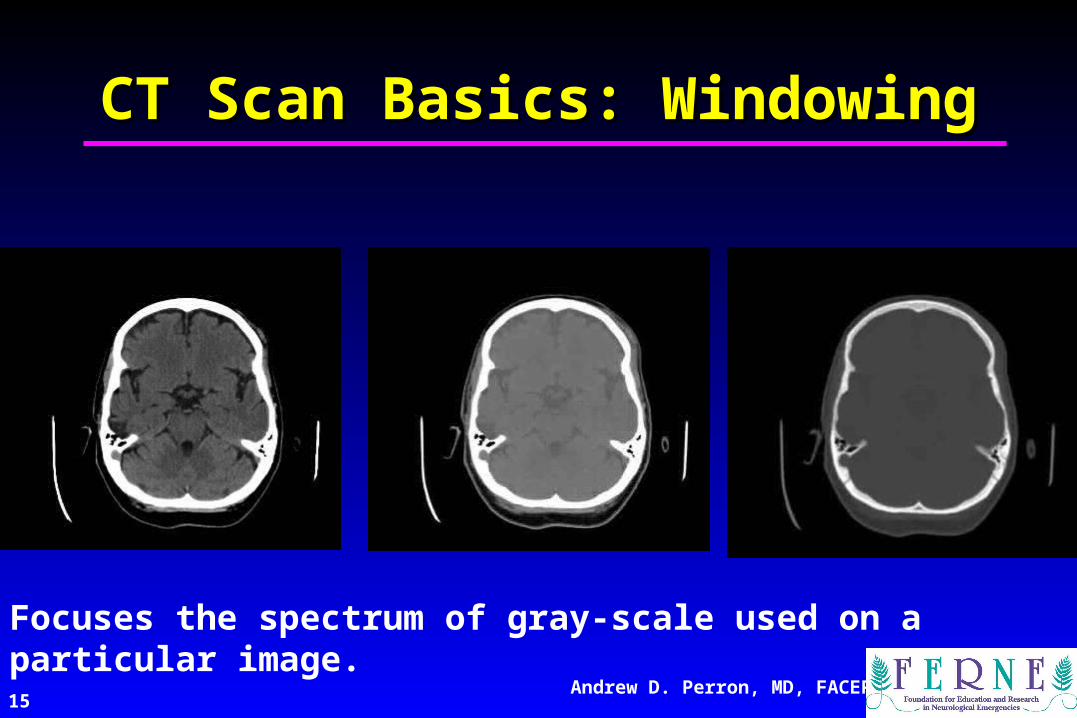
CT Scan Basics: WindowingCT Scan Basics: Windowing
Focuses the spectrum of gray-scale used on a particular image.
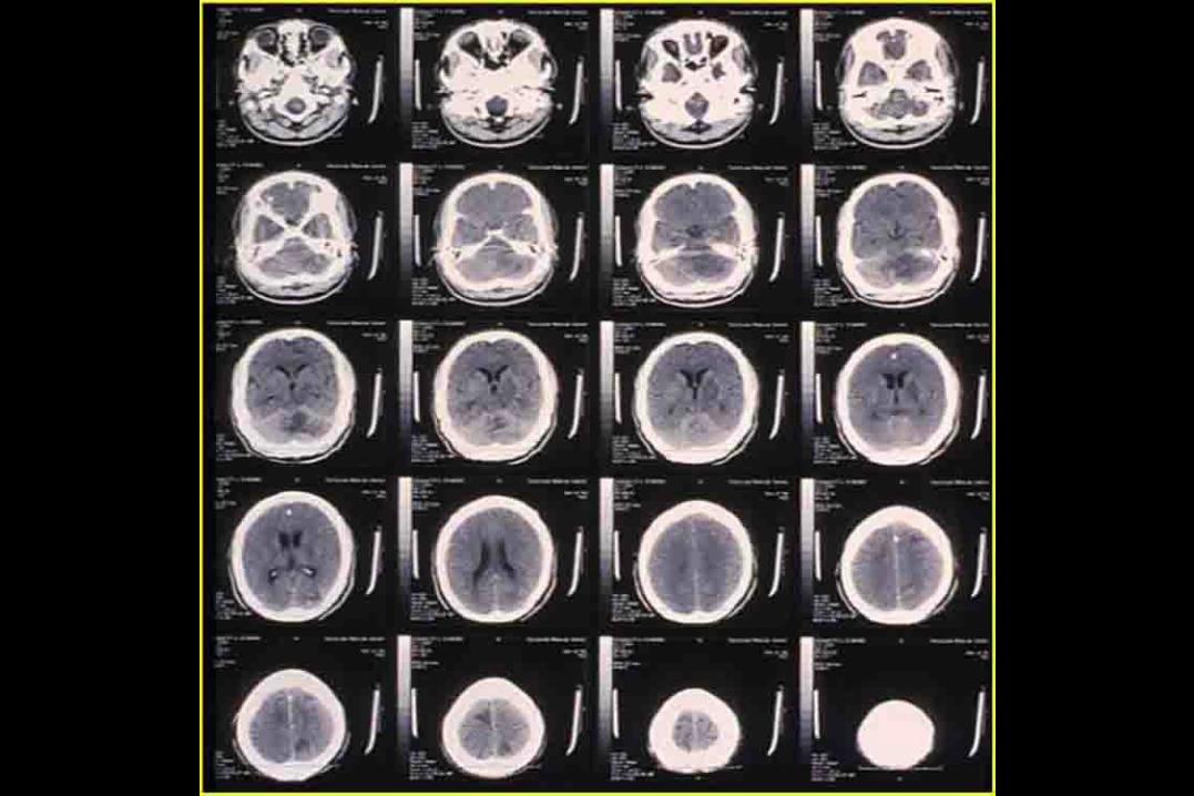
2 Sheet Head CT
17Andrew D. Perron, MD, FACEP
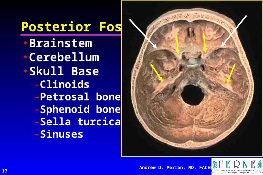
•Brainstem•Cerebellum•Skull Base
–Clinoids–Petrosal bone–Sphenoid bone–Sella turcica–Sinuses
Posterior FossaPosterior Fossa
18Andrew D. Perron, MD, FACEP
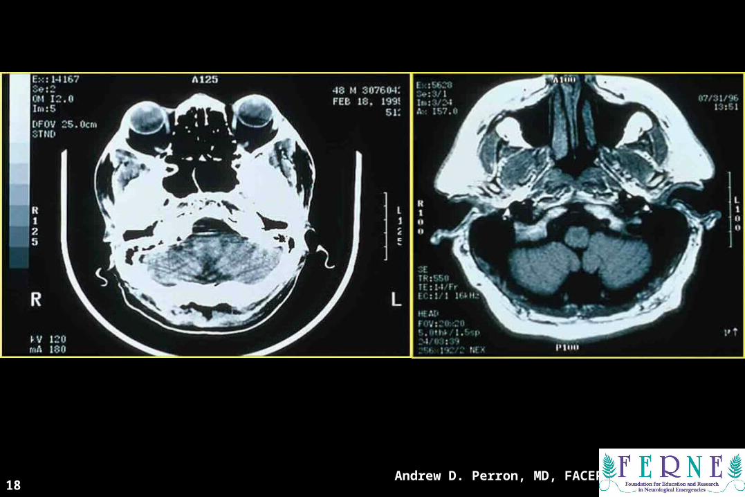
CT ScanCT Scan

CT ScanCT Scan
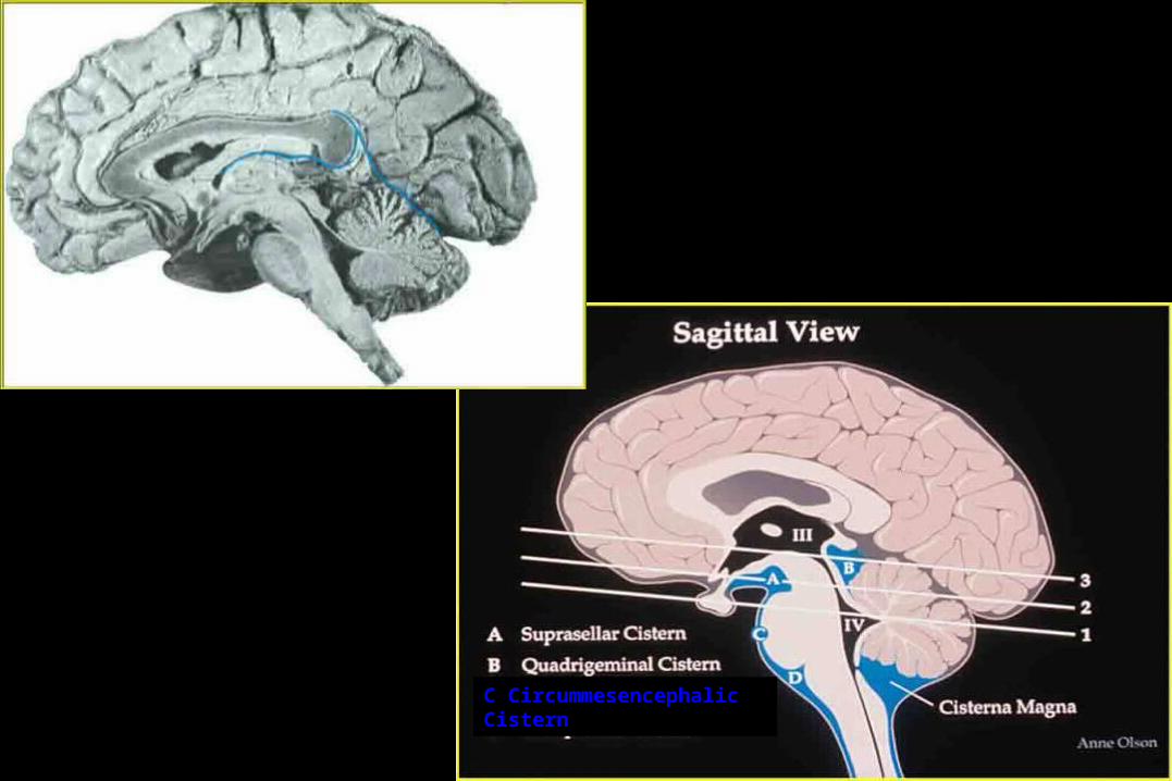
Sagittal ViewSagittal View
C Circummesencephalic Cistern

21Andrew D. Perron, MD, FACEP
CT DiagnosticsCT Diagnostics
Where is the most sensitive area to examine the CT for increased ICP?
A. Lateral Ventricles
B. IVth ventricle
C. Basilar Cisterns
D. Gyral pattern
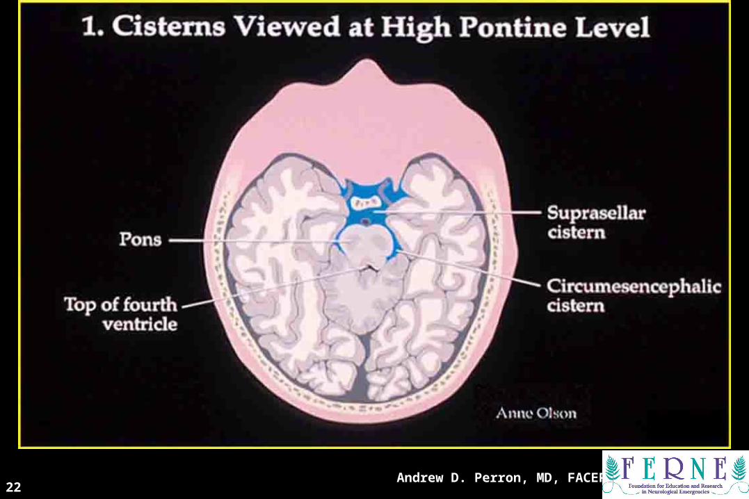
22Andrew D. Perron, MD, FACEP
CisternsCisterns
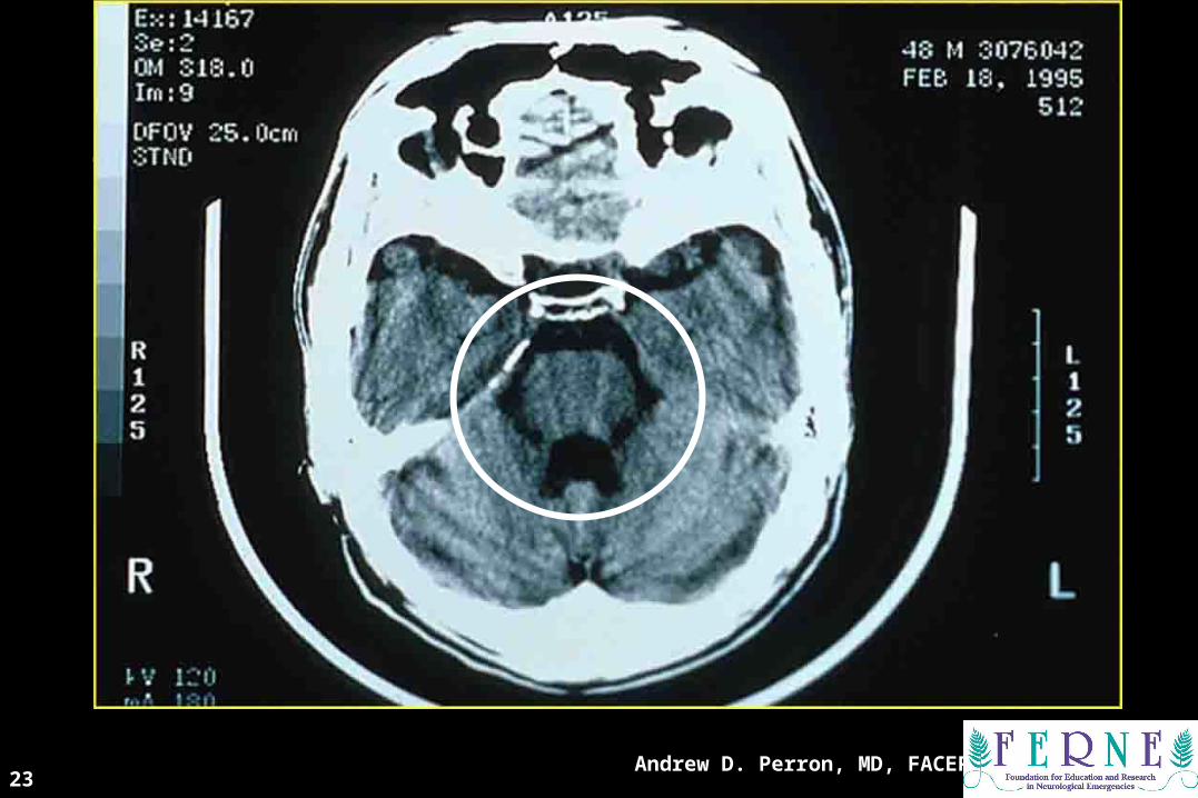
23Andrew D. Perron, MD, FACEP
CT ScanCT Scan
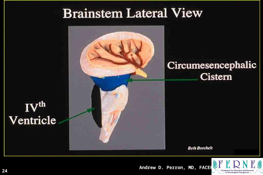
24Andrew D. Perron, MD, FACEP
Brainstem Lateral ViewBrainstem Lateral View

25Andrew D. Perron, MD, FACEP
2nd Key Level22ndnd Key Level Sagittal View Key Level Sagittal View
Circummesencephalic Cistern

26Andrew D. Perron, MD, FACEP
Cisterns at Cerebral Peduncles Cisterns at Cerebral Peduncles LevelLevel

27Andrew D. Perron, MD, FACEP
CT ScanCT Scan

28Andrew D. Perron, MD, FACEP
Suprasellar CisternSuprasellar Cistern
29Andrew D. Perron, MD, FACEP
CT DiagnosticsCT Diagnostics
Where is the most sensitive area to examine the CT for ventricular dilation?
A. IIIrd ventricle
B. IVth ventricle
C. Temporal horns of lateral ventricles

30Andrew D. Perron, MD, FACEP
CT ScanCT Scan
31Andrew D. Perron, MD, FACEP
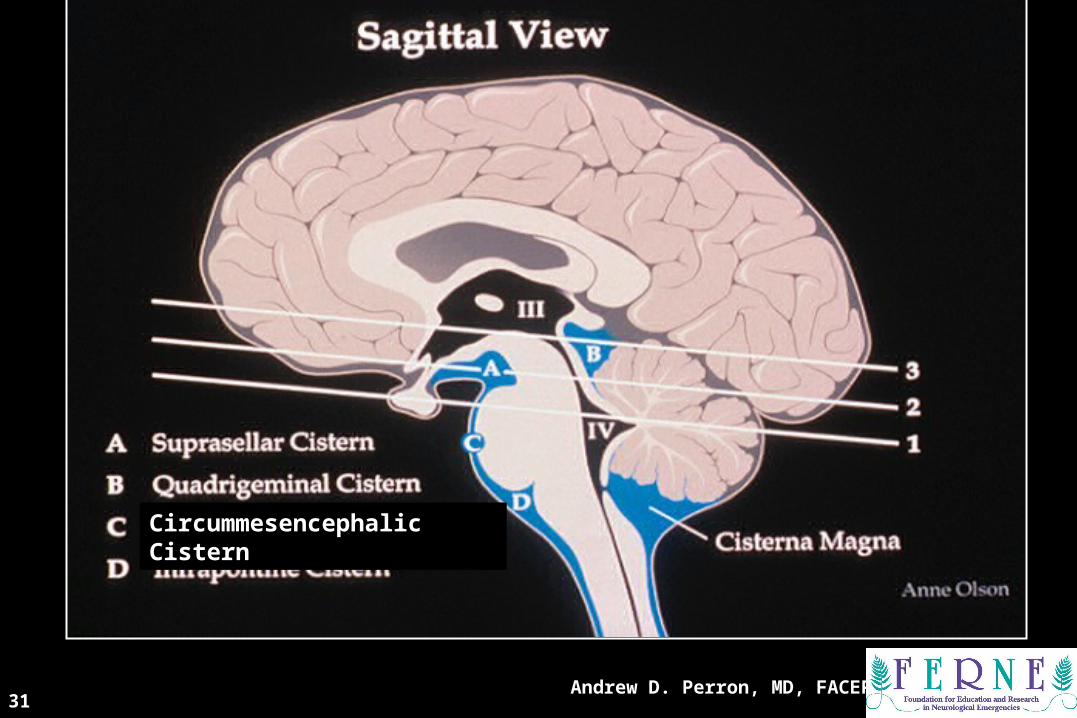
33rdrd Key Level Sagittal View Key Level Sagittal View
Circummesencephalic Cistern
32Andrew D. Perron, MD, FACEP
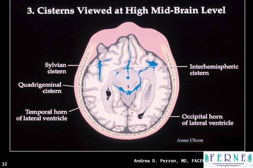
Cisterns at High Mid-Brain LevelCisterns at High Mid-Brain Level

CT ScanCT Scan

34Andrew D. Perron, MD, FACEP
VentriclesVentricles
35Andrew D. Perron, MD, FACEP
CSF ProductionCSF Production
• Produced in choroid plexus in the lateral ventricles Foramen of Monroe IIIrd Ventricle Acqueduct of Sylvius IVth Ventricle Lushka/Magendie
• 0.5-1 cc/min• Adult CSF volume is approx. 150 cc’s.• Adult CSF production is approx. 500-
700 cc’s per day.

CT ScansCT Scans
Andrew D. Perron, MD, FACEP37
38Andrew D. Perron, MD, FACEP
A Few Kid-Specific ThoughtsA Few Kid-Specific Thoughts

39Andrew D. Perron, MD, FACEP
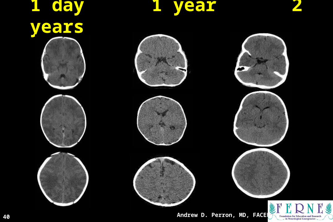
A Few Kid-Specific ThoughtsA Few Kid-Specific Thoughts• Premature Infants (30-34 weeks):
Larger sylvian, basilar (circummesencephalic) cisterns.Larger subarachnoid spacesThin cerebral cortex (Gray matter)Prominent white matter (with higher water content)Limited cortical gyral patternVentricles are variable: slit-like to well-developed
• Term Infant (36-41 weeks):Small, slit-like lateral ventriclesContinued white-matter prominenceMore prominent sulcal patternTemporal horns unlikely to be seen
• 1st & 2nd years of Life:Marked growth of all lobes of the brain (proportionally greatest in frontal lobes)Wide variation in lateral ventricle size (3rd and 4th fairly constant)Temporal horns unlikely to be seen.
Andrew D. Perron, MD, FACEP40
1 day 1 day 1 year 1 year 2 years 2 years
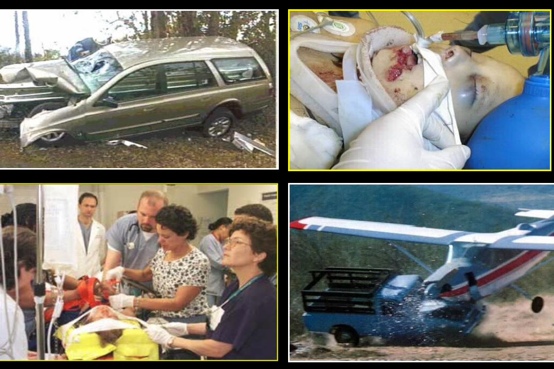
Trauma PicturesTrauma Pictures

42Andrew D. Perron, MD, FACEP
BB is for Blood is for Blood
• 1st decision: Is blood present?
• 2nd decision: If so, where is it?
• 3rd decision: If so, what effect is it having?

43Andrew D. Perron, MD, FACEP
CT DiagnosticsCT Diagnostics
At what point does blood become isodense with brain?
A. About 48 hours
B. About 1 week
C. About 2 weeks
D. After 1 month
44Andrew D. Perron, MD, FACEP
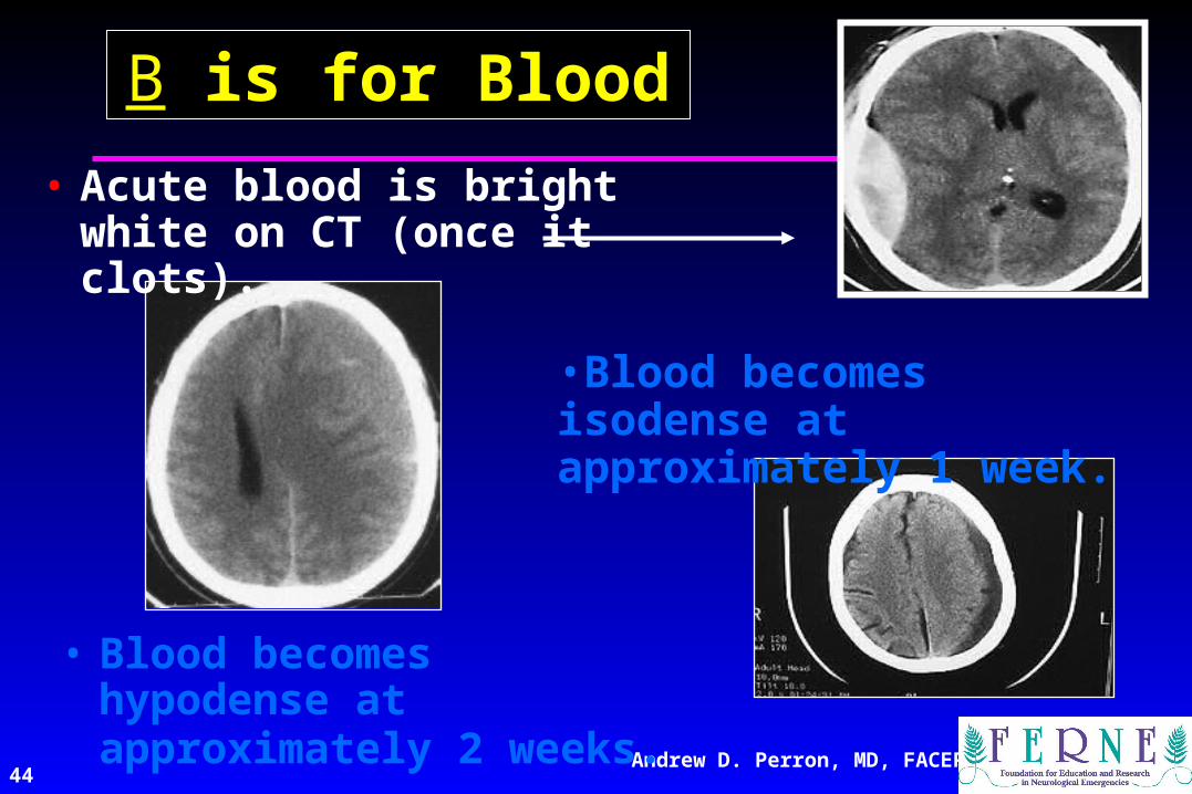
BB is for Blood is for Blood
• Blood becomes hypodense at approximately 2 weeks.
•Blood becomes isodense at approximately 1 week.
• Acute blood is bright white on CT (once it clots).

45Andrew D. Perron, MD, FACEP
BB is for Blood is for Blood
• Blood becomes hypodense at approximately 2 weeks.
• Blood becomes isodense at approximately 1 week.
• Acute blood is bright white on CT (once it clots).
46Andrew D. Perron, MD, FACEP
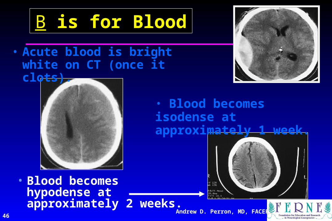
BB is for Blood is for Blood
• Blood becomes hypodense at approximately 2 weeks.
• Blood becomes isodense at approximately 1 week.
• Acute blood is bright white on CT (once it clots).

47Andrew D. Perron, MD, FACEP
Epidural HematomaEpidural Hematoma
• Lens shaped
• Does not cross sutures
• Classically described with injury to middle meningeal artery
• Low mortality if treated prior to unconsciousness
( < 20%)

48Andrew D. Perron, MD, FACEP
CT ScansCT Scans

49Andrew D. Perron, MD, FACEP
Subdural HematomaSubdural Hematoma
• Typically falx or sickle-shaped.
• Crosses sutures, but does not cross midline.
• Acute subdural is a marker for severe head injury. (Mortality approaches 80%)
• Chronic subdural usually slow venous bleed and well tolerated.

CT Scan CT Scan
Andrew D. Perron, MD, FACEP50

51Andrew D. Perron, MD, FACEP
Subarachnoid HemorrhageSubarachnoid Hemorrhage
52Andrew D. Perron, MD, FACEP
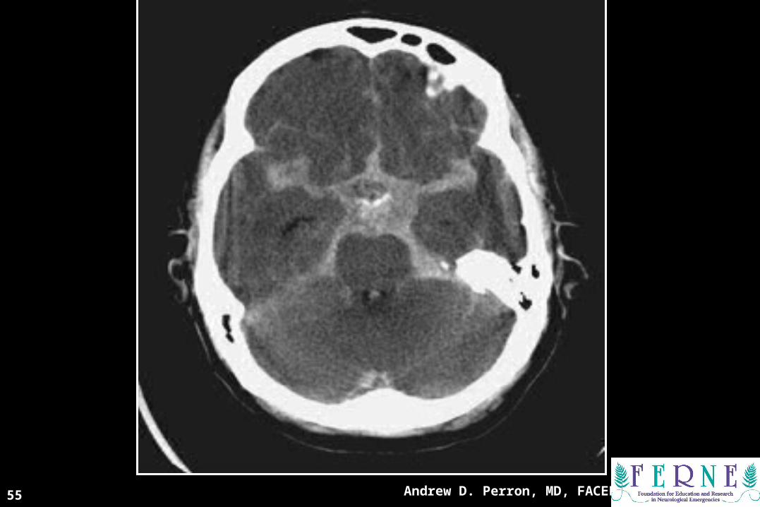
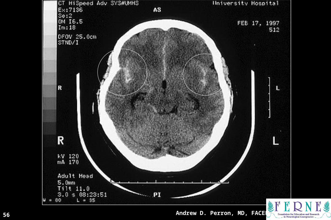
Subarachnoid HemorrhageSubarachnoid Hemorrhage• Blood in the cisterns/cortical gyral surface
– Aneurysms responsible for 75-80% of SAH– AVM’s responsible for 4-5%– Vasculitis accounts for small proportion (<1%)– No cause is found in 10-15%– 20% will have associated acute hydrocephalus

53Andrew D. Perron, MD, FACEP
CT DiagnosticsCT Diagnostics
What is the sensitivity of CT for SAH?
A. 100%
B. 95%
C. 80%
D. Depends…I need a lot more information to answer.

54Andrew D. Perron, MD, FACEP
• 98-99% at 0-12 hours
• 90-95% at 24 hours
• 80% at 3 days
• 50% at 1 week
• 30% at 2 weeks
Depends on generation of scanner and who is reading scan and how much blood there is.
CT Scan Sensitivity for SAHCT Scan Sensitivity for SAH
Andrew D. Perron, MD, FACEP
CT ScanCT Scan
55
CT ScanCT Scan
Andrew D. Perron, MD, FACEP56

57Andrew D. Perron, MD, FACEP
Intraventricular/Intraventricular/Intraparenchymal HemorrhageIntraparenchymal Hemorrhage
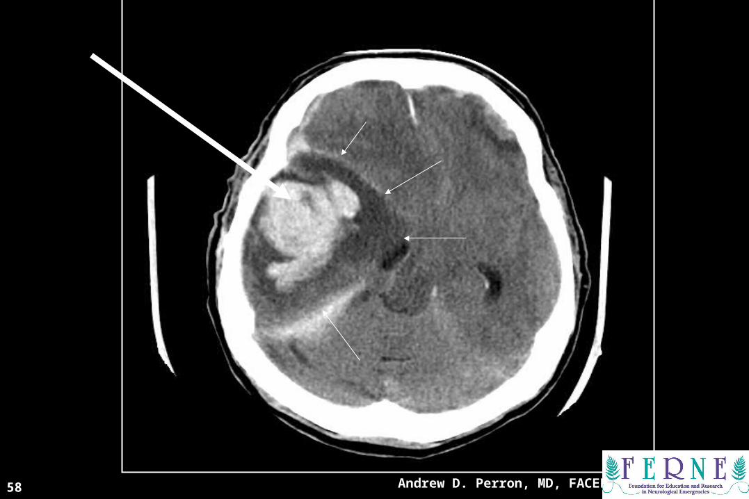
CT ScanCT Scan
Andrew D. Perron, MD, FACEP58

59Andrew D. Perron, MD, FACEP
CC is for CISTERNS is for CISTERNS
• 4 key cisterns– Circummesencephalic
– Suprasellar
– Quadrigeminal
– Sylvian
((BBlood lood CCan an BBe e VVery ery BBad)ad)
Circummesencephalic

60Andrew D. Perron, MD, FACEP
CisternsCisterns• 2 Key questions to answer regarding
cisterns:– Is there blood?
– Are the cisterns open?

Andrew D. Perron, MD, FACEP61
Andrew D. Perron, MD, FACEP62
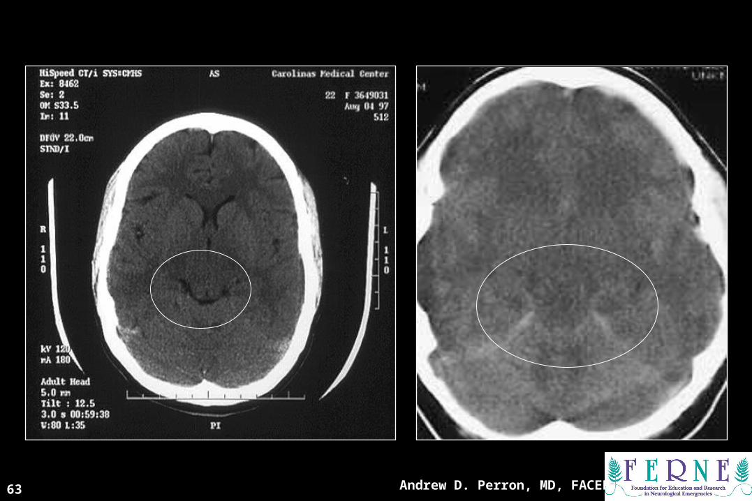
Andrew D. Perron, MD, FACEP63
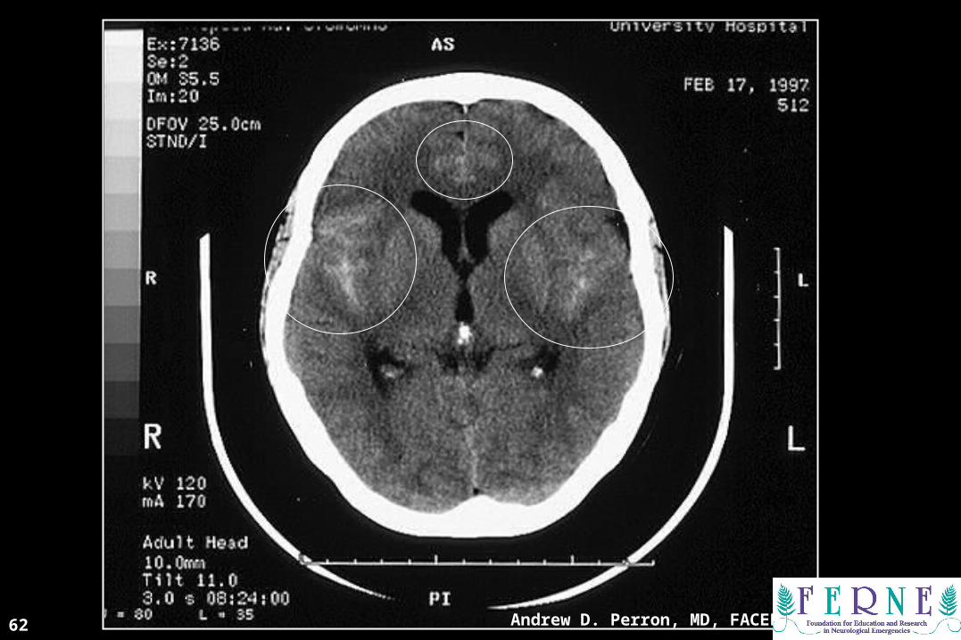
64Andrew D. Perron, MD, FACEP
BB is for is for BBRAINRAIN((BBlood lood CCan an BBe e VVery ery BBad)ad)
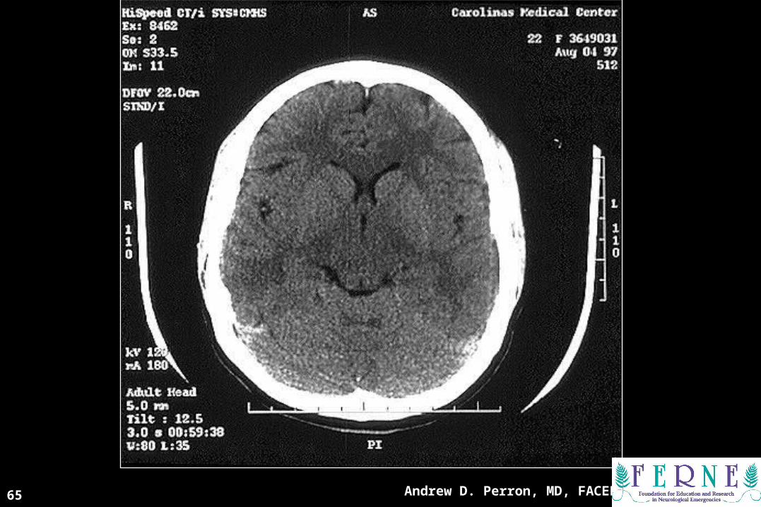
Andrew D. Perron, MD, FACEP65
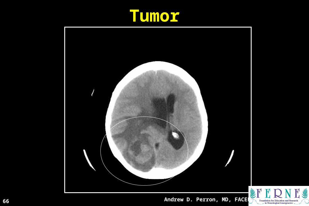
TumorTumor
Andrew D. Perron, MD, FACEP66
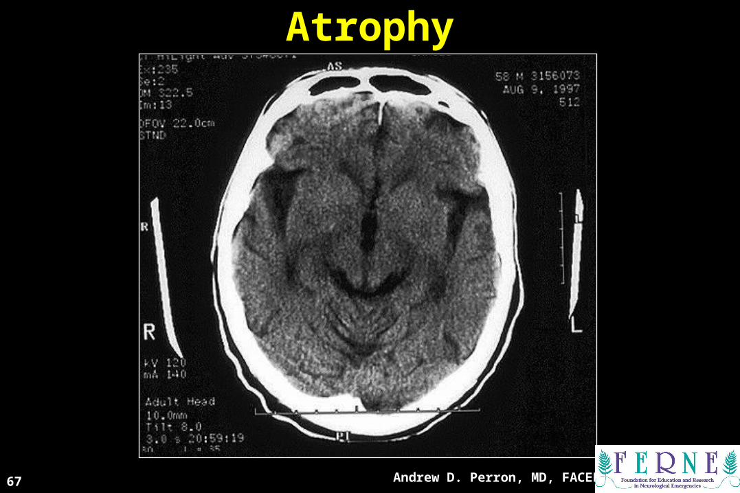
AtrophyAtrophy
Andrew D. Perron, MD, FACEP67
68Andrew D. Perron, MD, FACEP
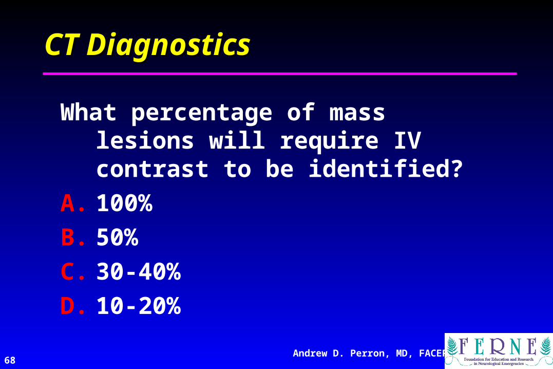
CT DiagnosticsCT Diagnostics
What percentage of mass lesions will require IV contrast to be identified?
A. 100%
B. 50%
C. 30-40%
D. 10-20%

AbscessAbscess
Andrew D. Perron, MD, FACEP69
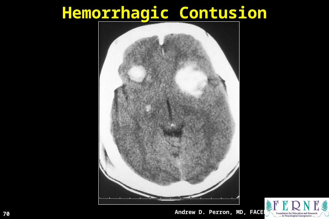
Hemorrhagic ContusionHemorrhagic Contusion
Andrew D. Perron, MD, FACEP70
Andrew D. Perron, MD, FACEP71
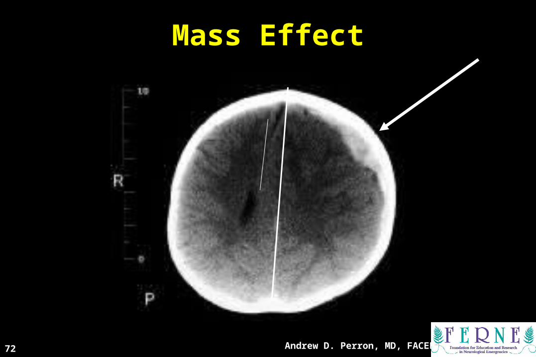
Mass EffectMass Effect
Andrew D. Perron, MD, FACEP72
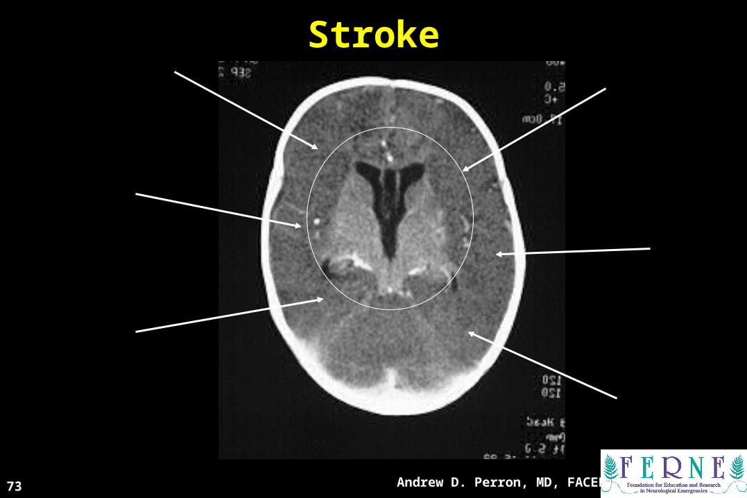
StrokeStroke
Andrew D. Perron, MD, FACEP73
74Andrew D. Perron, MD, FACEP
Intracranial AirIntracranial Air

Intracranial AirIntracranial Air
Andrew D. Perron, MD, FACEP75
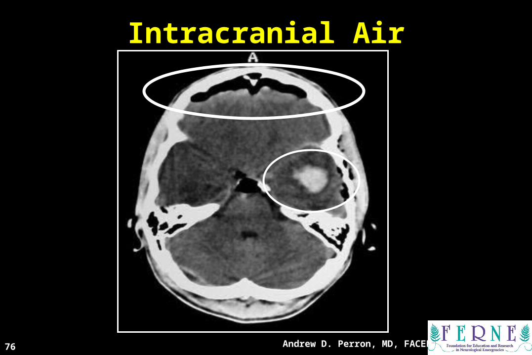
Intracranial AirIntracranial Air
Andrew D. Perron, MD, FACEP76

77Andrew D. Perron, MD, FACEP
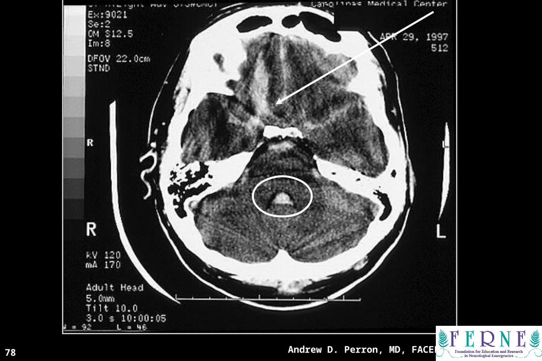
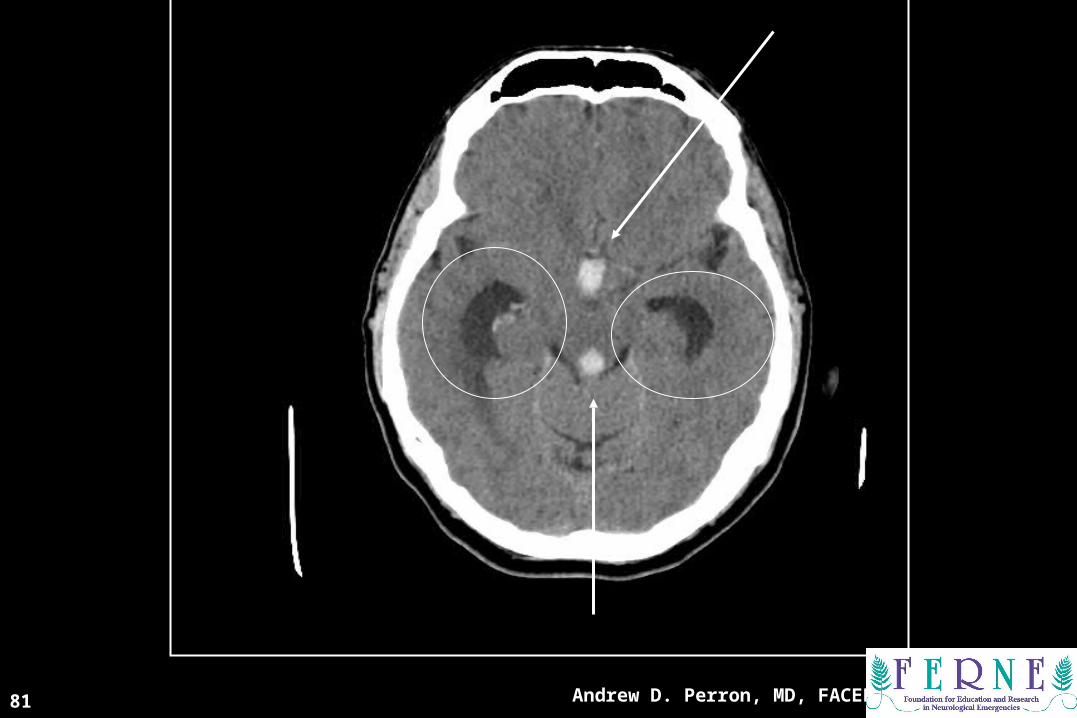
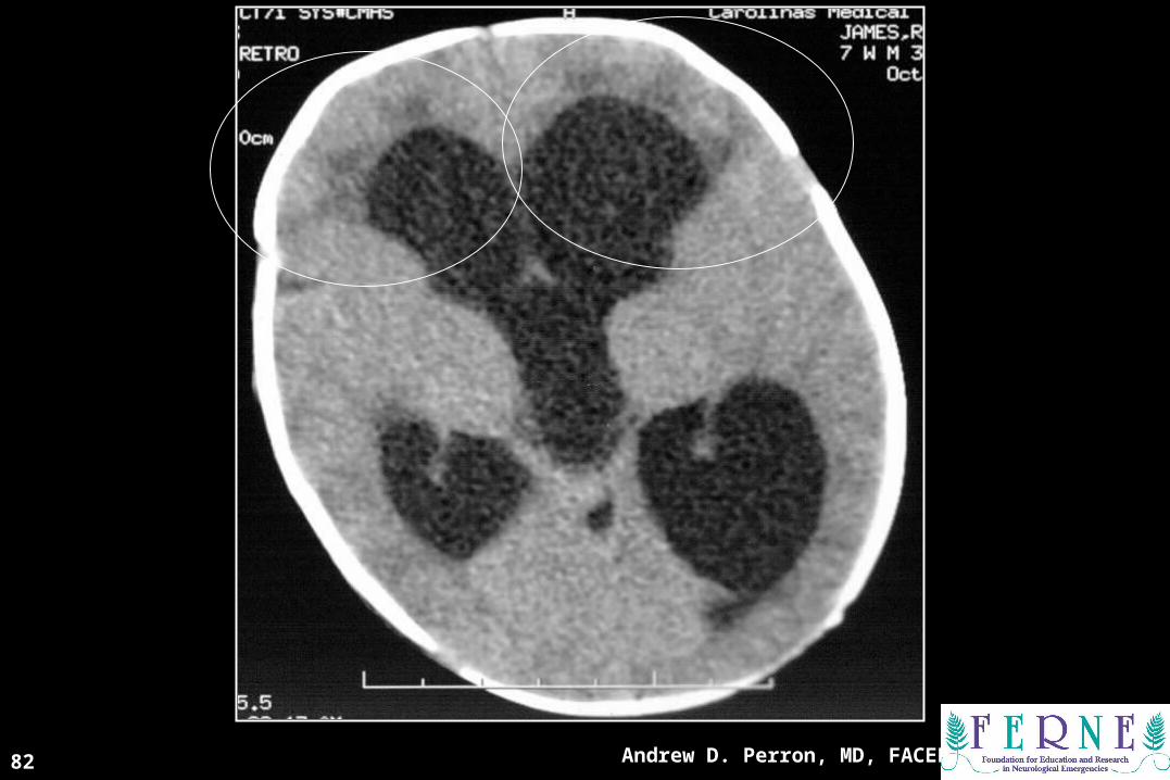
VV is for is for VVENTRICLESENTRICLES((BBlood lood CCan an BBe e VVery ery BBad)ad)
Andrew D. Perron, MD, FACEP78

Andrew D. Perron, MD, FACEP79

Ex-Vacuo PhenomenonEx-Vacuo Phenomenon
Andrew D. Perron, MD, FACEP80
Andrew D. Perron, MD, FACEP81
Andrew D. Perron, MD, FACEP82

Andrew D. Perron, MD, FACEP83
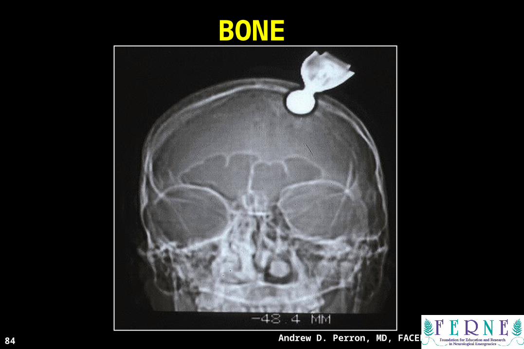
BONEBONE
Andrew D. Perron, MD, FACEP84

Andrew D. Perron, MD, FACEP85

Andrew D. Perron, MD, FACEP86
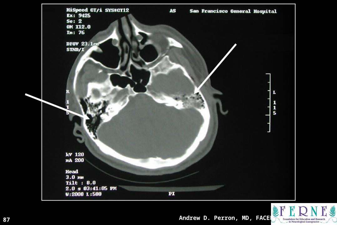
Andrew D. Perron, MD, FACEP87

Andrew D. Perron, MD, FACEP88
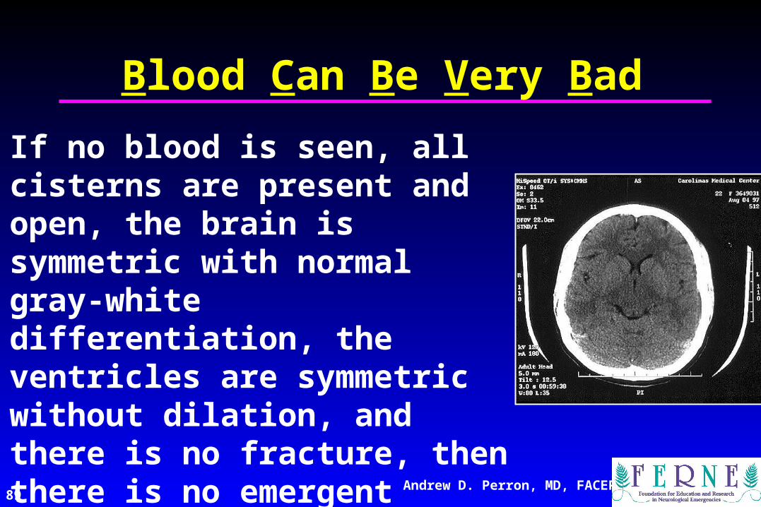
89Andrew D. Perron, MD, FACEP
If no blood is seen, all cisterns are present and open, the brain is symmetric with normal gray-white differentiation, the ventricles are symmetric without dilation, and there is no fracture, then there is no emergent diagnosis from the CT scan.
Blood Can Be Very Bad

RIPRIP
91Andrew D. Perron, MD, FACEP
QuestionsQuestions
ferne_acep_2005_peds_perron_ich_bcbvb_fshow.ppt 04/18/23 13:13
[email protected]@ferne.org
Andrew D. Perron, MD, FACEPAndrew D. Perron, MD, FACEP
[email protected](207) 662-7015207) 662-7015


![[Gokigenyou] One Shot Worrying](https://img.pdfslide.net/doc/110x75/577c7eb81a28abe054a22ff3/gokigenyou-one-shot-worrying.jpg)