Embed Size (px)
Citation preview

Androgen receptor-driven chromatinlooping in prostate cancerDayong Wu1*, Chunpeng Zhang1*, Yanping Shen1,2, Kenneth P. Nephew3 andQianben Wang1,2
1Department of Molecular and Cellular Biochemistry and the Comprehensive Cancer Center, The Ohio State University College ofMedicine, Columbus, OH 43210, USA2Ohio State Biochemistry Graduate Program, The Ohio State University, Columbus, OH 43210, USA3Medical Sciences Program, Department of Cellular and Integrative Physiology, Indiana University School of Medicine,Bloomington, IN 47405, USA
The androgen receptor (AR) is important for prostatecancer development and progression. Genome-widemapping of AR binding sites in prostate cancer has foundthat the majority of AR binding sites are located withinnon-promoter regions. These distal AR binding regionsregulate AR target genes (e.g. UBE2C) involved in pros-tate cancer growth through chromatin looping. In addi-tion to long-distance gene regulation, looping has beenshown to induce spatial proximity of two genes other-wise located far away along the genomic sequence andthe formation of double-strand DNA breaks, resulting inaberrant gene fusions (e.g. TMPRSS2–ERG) that alsocontribute to prostate tumorigenesis. Elucidating themechanisms of AR-driven chromatin looping will in-crease our understanding of prostate carcinogenesisand may lead to the identification of new therapeutictargets.
AR signaling in prostate cancerAlthough significant strides have been made in prostatecancer research and treatment, prostate cancer remainsone of the most commonly diagnosed cancers and the secondleading cause of cancer deaths in American men [1]. The‘male’ hormones or androgens, exerting their biological effectthrough androgen receptor (AR), play important roles inprostate cancer development and progression [2,3]. There-fore androgen ablation therapy, including surgical andchemical castration, is the first-line therapeutic approachfor advanced androgen-dependent prostate cancer (ADPC).Although this therapy is initially effective, ADPC ultimatelyprogresses into an incurable, castration-resistant stage ofthe disease (CRPC), involving the reactivation of AR signal-ing. The mechanisms for AR reactivation after castrationinclude AR amplification, increased androgen sensitivity,increased intracellular synthesis of androgen, a constitutive-ly active AR lacking the ligand binding domain (LBD),activation of growth factor pathways, and retinoblastoma(RB) loss-induced, E2F1-mediated AR overexpression [3–6].Thus, aberrantly active AR signaling is present in bothADPC and CRPC.
AR is a ligand-dependent transcription factor belongingto the nuclear hormone receptor (NR) superfamily [7]. To
understand how AR signaling contributes to ADPC andCRPC, recent studies have utilized genome-wide chroma-tin immunoprecipitation (ChIP) techniques to define ARbinding sites across the entire human genome in prostatecancer cells [8–13]. In these studies, AR ChIP-enrichedDNA was amplified and hybridized to tiling DNA micro-arrays (ChIP-on-chip), or subjected to massively parallelhigh-throughput sequencing (ChIP-seq) [14,15]. Thesestudies have greatly advanced understanding of AR bind-ing to chromatin, the functional interplay between AR andchromatin, and AR-mediated regulation of target genesinvolved in prostate tumorigenesis. In this review wediscuss how the majority of AR binding sites in the genomeare located within non-promoter distal regions andthe importance of this for transcriptional regulation. Atthese sites, several transcription factors act as positive ornegative regulators of AR function and affect AR-mediatedgene regulation. We then discuss two types of AR binding-driven chromatin looping in prostate cancer – the AR-driven chromatin looping leading to target gene expression(without genomic rearrangement) and AR-driven chroma-tin looping that results in gene fusion (with genomic rear-rangement). The elucidation of the mechanisms of ARsignaling in prostate cancer has translational implicationsin the development of new therapies for prostate cancer.
Features of the genome-wide AR binding atlas inprostate cancerDistal AR binding sites: their location and importanceIn the pre-genome-wide ChIP era, studies on the canonicalAR target gene PSA (prostate specific antigen) found thatAR primarily binds to the PSA enhancer rather than to thepromoter region [16]. Consistent with the findings from thePSA gene, genome-wide mapping of AR binding sites inprostate cancer cells revealed that most AR binding sitesare not within the promoter region of AR-regulated genes.Approximately 86–95% of AR binding sites identified inADPC cell models (LNCaP [12,13] and VCaP [13]) andCRPC cell models (LNCaP-abl [12] and C4-2B [11]) arelocated within non-promoter regions. Interestingly, simi-lar distal AR binding patterns are also observed in animmortalized normal human prostate epithelium cell line(HPr-1) [17], and non-prostate androgen-responsive cells(human primary skeletal muscle myoblasts [18]) or tissues
Review
Corresponding author: Wang, Q. ([email protected]).* These authors contributed equally to this work.
474 1043-2760/$ – see front matter ! 2011 Elsevier Ltd. All rights reserved. doi:10.1016/j.tem.2011.07.006 Trends in Endocrinology and Metabolism, December 2011, Vol. 22, No. 12
(mouse epididymis [19]). These studies strongly indicatethat distal binding may be a general rule for AR action.
Several lines of evidence suggest that at least part ofthese distal AR binding regions are important fortranscriptional regulation. First, some AR binding regions,when cloned upstream of minimal promoters, function asenhancers in reporter gene assays [8,10,11]. Second, tran-scriptional coactivators [e.g. the histone acetyltransferase-s(HAT), BRM-containing chromatin-remodeling complexand the Mediator complex] and RNA polymerase II(Pol II) bind to some distal AR binding sites [8,10,12,13,20,21], indicating that these sites are transcriptionallyactive. Third, some distal AR binding sites are associatedwith active histone modifications, including H3K4 mono-and di-methylation (H3K4me1 and H3K4me2) and H3acetylation (H3Ac) [10–13], which create a permissivechromatin environment that facilitates active transcrip-tion. Fourth, the nucleosomes present at some distal ARbinding regions contain the H2A.Z variant [22], which hasbeen reported to correlate with gene activation [23,24].Collectively, these observations suggest that some distalAR binding sites act as transcriptional enhancers.
Transcription factors that positively or negatively affectAR-mediated gene expressionDe novo motif searching (i.e. searching for common sequencepatterns) or known motif scanning within the AR bindingregions across the genome has identified that, in additionto AR binding motifs, many other transcription factormotifs (e.g. Forkhead, GATA, OCT, ETS, and NKX3-1)are significantly enriched within the AR binding regions,compared with genomic background. These observationssuggest that transcription factors recognizing these motifsmay be recruited to AR binding regions and play importantroles in AR-mediated gene expression in prostate cancer,either by enhancing or repressing AR action. Consistentwith this hypothesis, the pioneer factors FoxA1 and GATA2were reported to be recruited to a significant portion ofAR binding sites in ADPC and CRPC cells, facilitatingAR binding and enhancing AR-mediated transcription[8,11,13,22]. In addition, OCT1, a member of the POUdomain family of transcription factors, may facilitate PolII binding to AR-bound regions after short-term and pro-longed androgen stimulation [8,22]. By contrast, E26 trans-formation-specific (ETS) family member ERG binding sitesthat significantly overlap with AR binding sites in VCaPcells repress AR action and AR target gene expression [13].These studies suggest that AR-mediated gene expressionin prostate cancer involves combinatorial transcriptionalregulation.
AR-driven chromatin looping leads to target geneexpressionDistal AR binding regions communicate with target genepromoters through chromatin loopingBecause the majority of AR binding sites are located atdistal regions, how these distal binding sites interact withtheir target gene promoters and regulate target geneexpression remains an open question. The looping modelproposes that distal transcription factor binding sitesinteract with the proximal promoter regions, looping out
the intervening DNA [25,26]. Chromosome conformationcapture (3C) technology developed almost a decade ago [27]allows this proposed model to be tested. In this assay, thedistal and proximal genomic regions, which have beencross-linked, are digested by the same restriction enzymeand re-ligated under extensively diluted DNA concentra-tions. The re-ligation of distal/proximal regions should bedetected by PCR if the distal region is physically associatedwith the proximal region [27,28]. Using this assay and itsderived ChIP-3C (ChIP combined with 3C) assay, recentstudies have demonstrated that distal AR binding sitesregulate well-known AR target genes such as PSA [16] andTMPRSS2 [8,21] through chromatin looping. Chromatinlooping also drives the expression of crucial AR targetgenes directly involved in prostate cancer growth. Forexample, the CRPC-specific AR target gene UBE2C (ubi-quitin-conjugating enzyme E2C) has an essential role inpromoting CRPC cell growth [12,21] by inactivating the M-phase checkpoint [29] or by increasing the pool of activeanaphase-promoting complex/cyclosome (APC/C) [30]. Itwas recently demonstrated that two CRPC cell-specificAR-bound enhancers, located –32.8 kb and +41.6 kb fromthe transcription start site (TSS) of the UBE2C gene,interact with the UBE2C promoters through chromatinlooping in CRPC but not in ADPC cells, and that thislooping is required for UBE2C gene expression and CRPCgrowth [12,21].
Although the 3C assay is a powerful approach for study-ing enhancer/promoter interaction, it is not feasible toperform standard 3C for each AR binding site to identifyor validate its target gene because there are more than10 000 AR binding sites in the genome. The development ofhigh-throughput adaptation of 3C assays, 4C (circular 3Cor 3C-on-chip) [31], 5C (3C-carbon copy) [32], Hi-C [33] andCHIA-PET (chromatin interaction analysis by paired-endsequencing) [34], should allow for genome-wide assess-ment of AR-bound enhancer/promoter interactions in thenear future.
Mechanisms for AR-mediated chromatin looping inCRPCChromatin looping is established and/or maintained byprotein–protein interaction between enhancer-bound tran-scription factors and promoter-bound proteins [25,26,35].Although the distally bound transcription factors maydirectly interact with promoter-bound proteins, in manycases transcriptional coregulators or Pol II facilitate and/or mediate such interactions [26,36]. Indeed, recent studiesdemonstrated that both transcription factors [e.g. AR, es-trogen receptor (ER), FoxA1 and GATA-1] [8,16,21,34,37]and coactivators (e.g. Med12, SRC-1, p300/CBP and BRG1)[38–41] play essential roles in looping formation and/ormaintenance. Among these coactivators, Mediator, an evo-lutionally conserved multiprotein complex including !30subunits [42], is of particular interest. Although previousstudies imply that Mediator is important for chromatinlooping at several loci [41,43], a recent study found thatMediator may mediate genome-wide enhancer/promoterinteractions. The global Mediator-mediated looping is sta-bilized by cohensin, a protein which embraces the loop[39]. These studies suggest that Mediator is an essential
Review Trends in Endocrinology and Metabolism December 2011, Vol. 22, No. 12
475
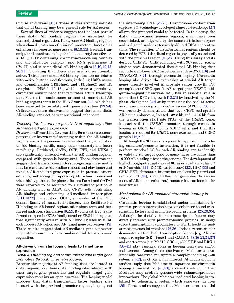
chromatin architectural factor. Consistent with this notion,a recent study demonstrated that silencing of the Mediatorsubunit MED1, an AR coactivator [16,44], significantlydecreased the interaction between the AR-bound UBE2Cenhancers and the UBE2C promoter in the CRPC cell modelLNCaP-abl, leading to decreased UBE2C gene expression[21]. Importantly, phosphorylation of MED1 at Thr1032 byphosphatidylinositol 3-kinase (PI3K)/AKT was proposed asa mechanism for MED1-mediated looping in CRPC cells byenhancing interactions between enhancer-bound FoxA1/ARand promoter-bound Pol II and TATA binding protein [21].Based on the findings from these studies, we propose alooping model for AR-mediated gene regulation in CRPC(Figure 1). Furthermore, it seems reasonable to hypothesizethat other looping-related coactivators such as SRC-1, p300/CBP [38], and cohesin [39] may also be involved in AR-mediated looping in CRPC (Figure 1). Further studies areneeded to test these hypotheses.
AR-driven chromatin looping results in gene fusionOverview of AR-regulated fusion genes in prostate cancerAlthough genomic rearrangements are important for phys-iological processes such as variable, diversity and joining[V(D)J] recombination and class switch recombination(CSR) in lymphocytes, aberrant genomic rearrangementscould lead to gene fusions in prostate cells contributing toprostate cancer initiation and progression [45,46]. Forexample, the TMPRSS2–ERG fusion gene, which isexpressed and functional in !50% of primary prostatecancer and !30% of CRPC patients [45,47,48], is generatedby the juxtaposition of the 50 untranslated region of the ARtarget gene TMPRSS2 (21q22.3) [8] and the 50 end of theoncogene ERG (21q22.2) [45,49]. The juxtaposition of
TMPRSS2 and ERG occurs either through balanced trans-locations with a ‘closed chain’ pattern (i.e. without loss ofgenetic material) or through interstitial deletions (Edel)[50–52].
The TMPRSS2–ERG fusion gene was first identified byChinnaiyan and colleagues using an integrative computa-tional and experimental approach [49]. An algorithmtermed COPA (cancer outlier profile analysis) was usedto identify ERG as an outlier gene (i.e. a gene overex-pressed in a subset of patients) from gene expressiondatasets in the Oncomine database [53]. Exon-walk PCRand 50-RNA-ligase-mediated rapid amplification of comple-mentary DNA ends (50-RLM-RACE) assays were thenperformed to identify the fusion between TMPRSS2 andERG [45,49]. Using the same approach, many other an-drogen-regulated genes fused with members of the ETSgene family (e.g. TMPRSS2–ETV1 [49], SLC45A3–ETV1[54], and CANT1–ETV4 [55,56]), have been identified.Recently, more androgen-regulated gene fusions includingnon-ETS fusions (e.g. NDRG1–ERG [57], SLC45A3–BRAF[58] and TMPRSS2–FKBP5–ERG [59]) have been identi-fied in prostate cancer by using the newly developedpaired-end RNA sequencing (PE RNA-seq) method [60].In this approach total RNA is fragmented and convertedinto double-stranded cDNA. The cDNA fragments gothrough adapter ligation and PCR amplification processes,and the final cDNA library is used for paired-end high-throughput sequencing. By integrating publically avail-able AR ChIP-seq data [13] and standard AR ChIP data[8,61,62] with gene expression and FISH (fluorescencein situ hybridization) analysis data, we have summarizedthe published AR-regulated fusion genes (Table S1 in thesupplementary material online).
AR
p160
p300
Active histonemodifications
p-MED1
Cohesin
TSS
FoxA1
TBPpolII
TRENDS in Endocrinology & Metabolism
AR
p160 p p
p300
Active histonemodification s
p-MED1
Cohesin
TSS
1FoxA1FoxA1
TBPpolII
Figure 1. A proposed looping model for AR-regulated gene expression in CRPC. Active histone modifications (e.g. H3K4me1 and H3K4me2) facilitate the binding of AR andits collaborating transcription factors (e.g. FoxA1) to distal nucleosome-depleted regions [12,22]. p-MED1 mediates chromatin looping by facilitating interactions betweendistal transcription factors and the basal transcriptional apparatus [21,39]. Other coactivators (e.g. p160 coactivators and p300 [38]) may also enhance chromatin looping.Cohension may stabilize chromatin looping by embracing the loop [39]. AR, androgen receptor; H3K4me1 and H3K4me2, H3K4 mono- and di-methylation; p-MED1, PI3K/AKT phosphorylated MED1.
Review Trends in Endocrinology and Metabolism December 2011, Vol. 22, No. 12
476

In addition to AR-regulated fusion genes, many non-ARregulated fusion genes have been reported. For example,ETV1 was found to be fused with the housekeeping geneHNRPA2B1 [54]. In addition, none of the most recentlyidentified 50 gene fusion partners (e.g. ALG5, PIGU, andTNPO1) by PE RNA-seq are androgen-regulated [59].These findings suggest that there are multiple mecha-nisms for the regulation of fusion gene expression inprostate cancer.
Function of AR-regulated fusion genesThe binding of liganded AR to the regulatory elements ofthe 50 partner of a fusion gene drives the overexpression ofthe 30 partner, which is often an oncogene (e.g. ERG), thuscontributing to prostate carcinogenesis. For example, ithas been reported that AR-driven expression of the ETSfamily members ETV1, ETV5 and ERG promotes invasionof benign prostate cells (e.g. RWPE, PrEC and BPH-1) andADPC cells (e.g. LNCaP and VCaP) by activating aninvasion-associated transcriptional program and thus in-ducing the plasminogen activator pathway [13,54,63–65].Although overexpression of ETV1 and ETV5 showed noeffect on cell proliferation [54,63], recent studies found thatERG overexpression also suppresses the AR-mediateddifferentiation program to promote LNCaP and VCaP cellgrowth [13,65]. Although ERG expression is necessary forprostate cancer cell invasion and growth, transgenic over-expression of ERG in mouse prostate is insufficient toinduce prostatic intraepithelial neoplasia (PIN) and pros-tate cancer [64,65], suggesting that ERG overexpressioncooperates with other genetic alterations in prostate tu-morigenesis. Consistent with this hypothesis, recent stud-ies using mouse models have indicated that loss of thetumor suppressor gene PTEN collaborates with ERG over-expression to develop PIN and prostate cancer [66,67]. Inaddition, assessment of human prostate samples identifiedan association of TMPRSS2–ERG with deletion of chromo-some 3p14, which includes the potential tumor suppressorgenes FoxP1, RYBP and SHQ1, suggesting a potential newcooperation in prostate tumorigenesis [68].
In addition to AR-regulated ETS fusion genes, AR-driven non-ETS fusion genes also have a crucial role inprostate cancer growth and invasion. For example, the ARtarget gene SLC45A3 was reported to be fused with BRAF ,a gene encoding a serine/threonine-specific protein kinaseinvolved in the mitogen-activated protein kinase (MAPK)signaling pathway, in less than 2% of prostate cancerpatients. Overexpression of SLC45A3–BRAF in RWPEcells increases cell proliferation and invasion, and leadsto the formation of small tumors in immunodeficient mice.Although ETS fusion genes are considered to be poortherapeutic targets, the function of the SLC45A3–BRAFfusion gene can be readily inhibited by RAF and mitogen-activated protein kinase kinase (MAP2K1) inhibitors, in-dicating that this fusion gene is a druggable target [58].
Mechanisms for AR-mediated gene fusionIn general, gene fusions require the spatial proximity oftwo genomic regions otherwise located far apart on thegenome, the formation of double-strand DNA breaks(DSBs), and error-prone DNA repair, which together might
lead to illegitimate DNA recombination [46,69]. In additionto the ability of AR to regulate the expression of fusiongenes directly, as described above, recent studies focusingon the TMPRSS2–ERG fusion have found that androgenand/or AR might contribute to the processes that drivegene fusion. For example, androgen stimulation increasesthe spatial association of TMPRSS2 and ERG. Inspired bythe finding that estrogen is able to induce chromatin loop-ing [38], two independent studies [61,70] found that dihy-drotestosterone (DHT) treatment of LNCaP cells inducescolocalization of TMPRSS2 and ERG. Further studiesfound that this androgen-induced chromatin looping ismediated by AR, which is recruited to the TMPRSS2and ERG breakpoints upon DHT treatment [61,62](Table S1). In agreement with the notion that breakpointsare associated with active histone modifications [46], theseAR binding regions at the TMPRSS2 and ERG breakpointsare enriched in histone H3K79 methylation and H4K16acetylation [61]. A recent study that combined prostatecancer whole-genome sequencing data with VCaP ChIP-seq data [13] further revealed a genome-wide association ofAR binding and active histone modifications (H3K4me3,H3K36me3 and H3Ac) near breakpoints in TMPRSS2–ERG positive patients [50]. These active histone modifica-tions could facilitate AR binding and gene fusions.
In addition, DHT-bound AR recruits enzymes capable ofinducing DSB. DHT stimulation has been found to induceAR-mediated recruitment of topoimerase II beta (TOP2B) tobreakpoints [62], an enzyme producing transient DSB thatare required for ER- [71,72] and AR-regulated transcription[62]. Of note is also the observation that, in the presence ofexogenous genotoxic stress (e.g. irradiation), DHT treat-ment markedly increases the mRNA and protein expressionof activation-induced cytidine deaminase (AID) [61], a lym-phoid-specific enzyme that changes cytosine to uracil andcould ultimately lead to DSB [73]. AR then recruits AID tochromatin via its coactivator Gadd45 [61]. Along these lines,genotoxic stress also increases the expression of long inter-spersed nuclear element-1 (LINE-1) retroelement encodedopen reading frame 2 (ORF2) endonuclease. Although ORF2is recruited to translocation regions in a DHT-dependentmanner, no physical interaction between AR and ORF2 hasbeen detected [61].
Finally, androgen stimulation increases recruitment ofproteins involved in non-homologous end joining (NHEJ).There are two main pathways for DSB repair, namely thehomologous recombination (HR) pathway and the NHEJpathway. The error-prone NHEJ is the major pathway forthe repair of DSB in eukaryotes [74]. It has been reportedthat, upon DHT stimulation and increased DSB, severalproteins involved in NHEJ including Ku70-Ku80 and atax-ia telangiectasia-mutated protein (ATM) are recruited tothe breakpoints, resulting in TMPRSS2–ERG fusion[61,62]. Significantly, pharmacologic inhibitors and smallinterfering RNAs (siRNAs) targeting NHEJ pathway com-ponents have been shown to reduce the formation ofde novo TMPRSS2–ERG gene fusions [62]. Based on thesefindings, a model for AR-mediated TMPRSS2–ERG fusionin prostate cancer is proposed (Figure 2). Evidence suggeststhat this model is generalizable to other AR-regulated fusiongenes such as SLC45A3–ETV1 [61].
Review Trends in Endocrinology and Metabolism December 2011, Vol. 22, No. 12
477

Concluding remarksAR expression and functionality have been well documen-ted in both ADPC and CRPC [2,3]. Recent studies havefound that the distal-binding AR transcription complex,including AR and associated transcription factors andcoactivators, regulates the expression of several AR targetgenes involved in prostate cancer growth through chroma-tin looping. By using a global 3C assay, future studiesshould address whether such a long-range combinatorialregulation can be generalized to include other AR-depen-dent genes in the genome. This would allow the identifica-tion of all AR direct target genes involved in prostatetumorigenesis and might lead to the development of newtherapies for the disease. In addition to the looping mech-anism, future studies should also examine whether othermechanisms for enhancer function (e.g. spreading and non-coding RNA [26]) participate in AR-mediated long-rangegene regulation.
With regard to AR-mediated gene fusion, significantprogress has been made in our understanding of the mech-anisms of AR-driven chromatin looping that leads to gene
fusions. The finding that inhibition of the NHEJ pathwayreduces TMPRSS2–ERG gene fusion [62] suggests thatfuture clinical trials may consider combining agents tar-geting both the AR and NHEJ pathway proteins. Despiteprogress on gene fusion mechanisms, it is still unclear onwhy gene fusions only occur in a limited number of genomicregions. It has been proposed that histone modifications,coactivators or noncoding RNAs may assist in the selectionof these regions [75]. Future work should test thesepossibilities. Finally, because gene fusion at the RNAlevel without genomic rearrangement has recently beenreported [59,76,77], future investigations should includewhether AR has a role in driving the formation of suchchimeric RNAs that also promote prostate carcinogenesis.
AcknowledgmentsStudies performed in the laboratory of Q.W. were supported by grantsfrom National Institute of Health (R00 CA126160, U54 CA113001), andthe Ohio State University Comprehensive Cancer Center. We thankDrs. Jindan Yu, Wei Li and Zhong Chen for critically reviewing of themanuscript. We apologize to the colleagues whose relevant work was notcited due to space limitation.
Active histonemodifications
AR AR AR
AIDORF2
AR
TOP2B
AID ORF2
DHT+genotoxic stress DHT
NHEJ(a)
(b)
(c)
TRENDS in Endocrinology & Metabolism
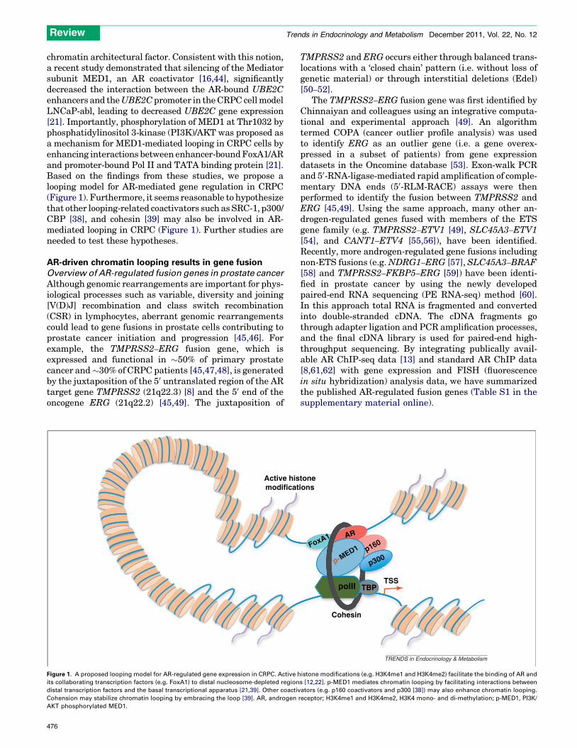
Figure 2. AR-mediated TMPRSS2-ERG gene fusion in prostate cancer cells. (a) In the presence of genotoxic stress, AR binds to TMPRSS2 and ERG breakpoints and recruitsAID upon DHT treatment. Exposure of prostate cancer cells under genotoxic stress to DHT also increases ORF2 recruitment to breakpoints [61]. DHT stimulation only leadsto AR–TOP2B complex loading on breakpoints [62]. (b) The recruitment of AID, ORF2 and TOP2B to breakpoints causes DSB [61,62]. (c) NHEJ machinery is recruited to thebreakpoints, leading to error-prone DSB repair and gene fusions such as TMPRSS2–ERG gene fusion [61,62]. AID, activation-induced cytidine deaminase; DHT,dihydrotestosterone; NHEJ, non-homologous end joining; ORF2, open reading frame 2; TOP2B, topoimerase II beta.
Review Trends in Endocrinology and Metabolism December 2011, Vol. 22, No. 12
478

Appendix A. Supplementary dataSupplementary data associated with this article can befound, in the online version, at doi:10.1016/j.tem.2011.07.006.
References1 Jemal, A. et al. (2010) Cancer statistics, 2010. CA Cancer J. Clin. 60,
277–3002 Heinlein, C.A. and Chang, C. (2004) Androgen receptor in prostate
cancer. Endocr. Rev. 25, 276–3083 Knudsen, K.E. and Penning, T.M. (2010) Partners in crime: deregulation
of AR activity and androgen synthesis in prostate cancer. TrendsEndocrinol. Metab. 21, 315–324
4 Feldman, B.J. and Feldman, D. (2001) The development of androgen-independent prostate cancer. Nat. Rev. Cancer 1, 34–45
5 Attard, G. et al. (2009) Steroid hormone receptors in prostate cancer: ahard habit to break? Cancer Cell 16, 458–462
6 Sharma, A. et al. (2010) The retinoblastoma tumor suppressor controlsandrogen signaling and human prostate cancer progression. J. Clin.Invest. 120, 4478–4492
7 Mangelsdorf, D.J. et al. (1995) The nuclear receptor superfamily: thesecond decade. Cell 83, 835–839
8 Wang, Q. et al. (2007) A hierarchical network of transcription factorsgoverns androgen receptor-dependent prostate cancer growth. Mol. Cell27, 380–392
9 Massie, C.E. et al. (2007) New androgen receptor genomic targetsshow an interaction with the ETS1 transcription factor. EMBO Rep.8, 871–878
10 Takayama,K.etal. (2007)Identificationofnovelandrogenresponsegenesin prostate cancer cells by coupling chromatin immunoprecipitationand genomic microarray analysis. Oncogene 26, 4453–4463
11 Jia, L. et al. (2008) Genomic androgen receptor-occupied regions withdifferent functions, defined by histone acetylation, coregulators andtranscriptional capacity. PLoS ONE 3, e3645
12 Wang, Q. et al. (2009) Androgen receptor regulates a distincttranscription program in androgen-independent prostate cancer.Cell 138, 245–256
13 Yu, J. et al. (2010) An integrated network of androgen receptor,polycomb, and TMPRSS2–ERG gene fusions in prostate cancerprogression. Cancer Cell 17, 443–454
14 Wang, Q. and Brown, M. (2009) Mapping the androgen receptorcistrome. In Androgen Action in Prostate Cancer (Tindall, D. andMohler, J., eds), pp. 663–680, Springer
15 Chen, Z. et al. (2010) Histone modifications and chromatin organizationin prostate cancer. Epigenomics 2, 551–560
16 Wang, Q. et al. (2005) Spatial and temporal recruitment of androgenreceptor and its coactivators involves chromosomal looping andpolymerase tracking. Mol. Cell 19, 631–642
17 Bolton, E.C. et al. (2007) Cell- and gene-specific regulation of primarytarget genes by the androgen receptor. Genes Dev. 21, 2005–2017
18 Wyce, A. et al. (2010) Research resource: the androgen receptormodulates expression of genes with critical roles in muscledevelopment and function. Mol. Endocrinol. 24, 1665–1674
19 Hu, S. et al. (2010) Research resource: genome-wide mapping of in vivoandrogen receptor binding sites in mouse epididymis. Mol. Endocrinol.24, 2392–2405
20 Makkonen, H. et al. (2009) Long-range activation of FKBP51transcription by the androgen receptor via distal intronic enhancers.Nucleic Acids Res. 37, 4135–4148
21 Chen, Z. et al. (2011) Phospho-MED1-enhanced UBE2C locus loopingdrives castration-resistant prostate cancer growth. EMBO J. 30,2405–2419
22 He, H.H. et al. (2010) Nucleosome dynamics define transcriptionalenhancers. Nat. Genet. 42, 343–347
23 Barski, A. et al. (2007) High-resolution profiling of histonemethylations in the human genome. Cell 129, 823–837
24 Wang, Z. et al. (2008) Combinatorial patterns of histone acetylationsand methylations in the human genome. Nat. Genet. 40, 897–903
25 Dekker, J. (2008) Gene regulation in the third dimension. Science 319,1793–1794
26 Bulger, M. and Groudine, M. (2011) Functional and mechanisticdiversity of distal transcription enhancers. Cell 144, 327–339
27 Dekker, J. et al. (2002) Capturing chromosome conformation. Science295, 1306–1311
28 Hagege, H. et al. (2007) Quantitative analysis of chromosomeconformation capture assays (3C-qPCR). Nat. Protoc. 2, 1722–1733
29 Reddy, S.K. et al. (2007) Ubiquitination by the anaphase-promotingcomplex drives spindle checkpoint inactivation. Nature 446, 921–925
30 van Ree, J.H. et al. (2010) Overexpression of the E2 ubiquitin-conjugating enzyme UbcH10 causes chromosome missegregationand tumor formation. J. Cell Biol. 188, 83–100
31 Zhao, Z. et al. (2006) Circular chromosome conformation capture (4C)uncovers extensive networks of epigenetically regulated intra- andinterchromosomal interactions. Nat. Genet. 38, 1341–1347
32 Dostie, J. et al. (2006) Chromosome Conformation Capture CarbonCopy (5C): a massively parallel solution for mapping interactionsbetween genomic elements. Genome Res. 16, 1299–1309
33 Lieberman-Aiden, E. et al. (2009) Comprehensive mapping of long-range interactions reveals folding principles of the human genome.Science 326, 289–293
34 Fullwood, M.J. et al. (2009) An oestrogen-receptor-alpha-bound humanchromatin interactome. Nature 462, 58–64
35 Miele, A. and Dekker, J. (2008) Long-range chromosomal interactionsand gene regulation. Mol. Biosyst. 4, 1046–1057
36 Nolis, I.K. et al. (2009) Transcription factors mediate long-rangeenhancer-promoter interactions. Proc. Natl. Acad. Sci. U.S.A. 106,20222–20227
37 Vakoc, C.R. et al. (2005) Proximity among distant regulatory elementsat the beta-globin locus requires GATA-1 and FOG-1. Mol. Cell 17,453–462
38 Hu, Q. et al. (2008) Enhancing nuclear receptor-induced transcriptionrequires nuclear motor and LSD1-dependent gene networkingin interchromatin granules. Proc. Natl. Acad. Sci. U.S.A. 105,19199–19204
39 Kagey, M.H. et al. (2010) Mediator and cohesin connect gene expressionand chromatin architecture. Nature 467, 430–435
40 Kim, S.I. et al. (2009) BRG1 requirement for long-range interaction of alocus control region with a downstream promoter. Proc. Natl. Acad. Sci.U.S.A. 106, 2259–2264
41 Park, S.W. et al. (2005) Thyroid hormone-induced juxtaposition ofregulatory elements/factors and chromatin remodeling of Crabp1dependent on MED1/TRAP220. Mol. Cell 19, 643–653
42 Malik, S. and Roeder, R.G. (2010) The metazoan Mediator co-activatorcomplex as an integrative hub for transcriptional regulation. Nat. Rev.Genet. 11, 761–772
43 Degenhardt, T. et al. (2009) Population-level transcription cyclesderive from stochastic timing of single-cell transcription. Cell 138,489–501
44 Wang, Q. et al. (2002) A coregulatory role for the TRAP-mediatorcomplex in androgen receptor-mediated gene expression. J. Biol.Chem. 277, 42852–42858
45 Kumar-Sinha, C. et al. (2008) Recurrent gene fusions in prostatecancer. Nat. Rev. Cancer 8, 497–511
46 Mani, R.S. and Chinnaiyan, A.M. (2010) Triggers for genomicrearrangements: insights into genomic, cellular and environmentalinfluences. Nat. Rev. Genet. 11, 819–829
47 Cai, C. et al. (2009) Reactivation of androgen receptor-regulatedTMPRSS2:ERG gene expression in castration-resistant prostatecancer. Cancer Res. 69, 6027–6032
48 Attard, G. et al. (2009) Characterization of ERG, AR and PTEN genestatus in circulating tumor cells from patients with castration-resistant prostate cancer. Cancer Res. 69, 2912–2918
49 Tomlins, S.A. et al. (2005) Recurrent fusion of TMPRSS2 andETS transcription factor genes in prostate cancer. Science 310,644–648
50 Berger, M.F. et al. (2011) The genomic complexity of primary humanprostate cancer. Nature 470, 214–220
51 Mehra, R. et al. (2008) Characterization of TMPRSS2–ETS geneaberrations in androgen-independent metastatic prostate cancer.Cancer Res. 68, 3584–3590
52 Mehra, R. et al. (2007) Heterogeneity of TMPRSS2 gene rearrangementsin multifocal prostate adenocarcinoma: molecular evidence for anindependent group of diseases. Cancer Res. 67, 7991–7995
53 Rhodes, D.R. et al. (2004) ONCOMINE: a cancer microarray databaseand integrated data-mining platform. Neoplasia 6, 1–6
Review Trends in Endocrinology and Metabolism December 2011, Vol. 22, No. 12
479

54 Tomlins, S.A. et al. (2007) Distinct classes of chromosomalrearrangements create oncogenic ETS gene fusions in prostate cancer.Nature 448, 595–599
55 Han, B. et al. (2008) A fluorescence in situ hybridization screen for E26transformation-specific aberrations: identification of DDX5–ETV4fusion protein in prostate cancer. Cancer Res. 68, 7629–7637
56 Hermans, K.G. et al. (2008) Two unique novel prostate-specific andandrogen-regulated fusion partners of ETV4 in prostate cancer. CancerRes. 68, 3094–3098
57 Pflueger, D. et al. (2009) N-myc downstream regulated gene 1 (NDRG1)is fused to ERG in prostate cancer. Neoplasia 11, 804–811
58 Palanisamy, N. et al. (2010) Rearrangements of the RAF kinase pathwayin prostate cancer, gastric cancer and melanoma. Nat. Med. 16, 793–798
59 Pflueger, D. et al. (2011) Discovery of non-ETS gene fusions in humanprostate cancer using next-generation RNA sequencing. Genome Res.21, 56–67
60 Maher, C.A. et al. (2009) Chimeric transcript discovery by paired-endtranscriptome sequencing. Proc. Natl. Acad. Sci. U.S.A. 106, 12353–12358
61 Lin, C. et al. (2009) Nuclear receptor-induced chromosomal proximityand DNA breaks underlie specific translocations in cancer. Cell 139,1069–1083
62 Haffner, M.C. et al. (2010) Androgen-induced TOP2B-mediated double-strand breaks and prostate cancer gene rearrangements. Nat. Genet.42, 668–675
63 Helgeson, B.E. et al. (2008) Characterization of TMPRSS2:ETV5 andSLC45A3:ETV5 gene fusions in prostate cancer. Cancer Res. 68, 73–80
64 Klezovitch, O. et al. (2008) A causal role for ERG in neoplastictransformation of prostate epithelium. Proc. Natl. Acad. Sci. U.S.A.105, 2105–2110
65 Tomlins, S.A. et al. (2008) Role of the TMPRSS2-ERG gene fusion inprostate cancer. Neoplasia 10, 177–188
66 Carver, B.S. et al. (2009) Aberrant ERG expression cooperates with lossof PTEN to promote cancer progression in the prostate. Nat. Genet. 41,619–624
67 King, J.C. et al. (2009) Cooperativity of TMPRSS2–ERG with PI3-kinase pathway activation in prostate oncogenesis. Nat. Genet. 41,524–526
68 Taylor, B.S. et al. (2010) Integrative genomic profiling of humanprostate cancer. Cancer Cell 18, 11–22
69 Nussenzweig, A. and Nussenzweig, M.C. (2010) Origin of chromosomaltranslocations in lymphoid cancer. Cell 141, 27–38
70 Mani, R.S. et al. (2009) Induced chromosomal proximity and genefusions in prostate cancer. Science 326, 1230
71 Ju, B.G. et al. (2006) A topoisomerase IIbeta-mediated dsDNA breakrequired for regulated transcription. Science 312, 1798–1802
72 Perillo, B. et al. (2008) DNA oxidation as triggered by H3K9me2demethylation drives estrogen-induced gene expression. Science 319,202–206
73 Nambiar, M. and Raghavan, S.C. (2011) How does DNA break duringchromosomal translocations? Nucleic Acids Res. 39, 5813–5825
74 Lieber, M.R. (2008) The mechanism of human nonhomologous DNAend joining. J. Biol. Chem. 283, 1–5
75 Mathas, S. and Misteli, T. (2009) The dangers of transcription. Cell 139,1047–1049
76 Maher, C.A. et al. (2009) Transcriptome sequencing to detect genefusions in cancer. Nature 458, 97–101
77 Kannan, K. et al. (2011) Recurrent chimeric RNAs enriched in humanprostate cancer identified by deep sequencing. Proc. Natl. Acad. Sci.U.S.A. 108, 9172–9177
Review Trends in Endocrinology and Metabolism December 2011, Vol. 22, No. 12
480