Embed Size (px)
Citation preview
Córdoba is a city located near the geographical center of Argentina, in the foothills of the Sierras Chicas on the Suquía River, about 700 km northwest of Buenos Aires. It is the capital of Córdoba Province. Córdoba is the second-largest city in Argentina after the federal capital Buenos Aires, with about 1.5 million inhabitants. The National University of Córdoba, (UNC), is the oldest university in Argentina, and one of the oldest in the Americas. Since the early 20th century it has been the second largest university in the country (after the University of Buenos Aires) in terms of the number of students, faculty, and academic programs. As the location of the first university founded in the land that is now Argentina, Córdoba has earned the nickname La Docta(roughly translated, "The Wise").
MY
HEART
São Paulo is the largest city in Brazil, the largest city in the western and southern hemispheres, and the world's eighth largest city by population. The metropolis is anchor to the São Paulo metropolitan area, ranked as the second-most populous metropolitan area in the Americas and among the five-largest metropolitan areas on the planet. São Paulo is the capital of the state of São Paulo, the most populous Brazilian state and exerts strong regional influence in commerce and finance as well as arts and entertainment. São Paulo maintains strong international influence and is considered an Alpha – World City. The name of the city honors Saint Paul.
ANDRÉS RICARDO PÉREZ-RIERA, M.D. Ph.D.Chief of Electro-Vectorcardiography Sector – Cardiology Discipline -
ABC Faculty of Medicine - ABC Foundation - Santo André – São Paulo – Brazil

CAUCASIAN MAN WITH MULTIPLE RISK FACTORSAND ACUTE CORONARY SYNDROME
HOMBRE BLANCO CON MÚLTIPLES FACTORES DE RIESGO Y SINDROME CORONARIO AGUDO
The following ECG/VCGs were obtained from 53 years-old male sedentary, with coronary artery disease, diabetes mellitus type 2, centripetal obesity grade II (waist circumference 106cm and BMI =36), high blood pressure and familial dysbetalipoproteinemia ("Broad beta disease".). He denies vices.The patient reffer that approximately an hour and a half he feel at rest an opressive retrosternal pain radiating to jaw, accompanied by cold sweating. The pain had approximately 20 minutes in duration. Inexpressive physical examWhat is the ECG/VCG diagnosis? Which is the appropriate approach?-----------------------------------------------------------------------------------------------------------------------------------Los siguientes ECG / VCG se obtuvieron de un hombre sedentário, de 53 años de edad con enfermedad arterial coronaria, diabetes mellitus tipo 2, obesidad centrípeta grado II( IMC=36 circunferencia abdominal de 106cm), hipertensión arterial sistémica y disbetalipoproteinemia familiar(enfermedad beta ancha). Niega vicios.El paciente refiere que aproximadamente una hora y media atrás sitió un dolor opresivo retroesternal en reposo irradiado a mandíbula y acompañado de sudoración fría. El dolor duró aproximadamente 20 minutos. Examen físico NDN.¿Cuál es el diagnóstico de ECG / VCG? ¿ Cual es el abordaje adecuado?

ECG/VCG CORRELATION FRONTAL PLANE
aVRaVL
IIIII
aVF
Y
XITP

ECG/VCG CORRELATION FRONTAL PLANE
V6
V1 V4
V5
V2 V3
X
Z
N/2
N/2

ECG/VCG CORRELATION RIGHT SAGITTAL PLANE
Y
aVF
Z V2T
P

Colleagues commentaries

Andres,You are being facetious when you wrote in the initial clinical history – “he denies vices”. He has two major vices – food and an aversion to exercise. Having said that, you did not indicate what medications he was on but with a description of classic ischemic chest pain occurring at rest and lasting for 20 minutes with his past history – it is go directly to the hospital and if available, the cathlab for possible PCI. If he was not having an MI, he had severe ischemia and with pain occurring at rest resulting in a diagnosis of unstable angina warranting aggressive intervention (pharmacologic or interventional).As to his ECG/VCG – the origin of his atrial complexes is probably from the sino-atrial node (sinus). What the rhythm is cannot be determined from isolated single complexes in each lead. HisECG is markedly splintered in the inferior leads and the mid-precordial leads. I would diagnose both an anterior wall MI as well as LVH. Further, he has LBBB making additional diagnoses difficult and I am not expert in reading the VCG. I suspect that there is more but I cannot say. I also cannot assess his ventricular function from this ECG but it unlikely to be normal and he cannot afford additional myocardial damage (no one can afford any myocardial damage which is why prevention is so important but that is no longer an option for this individual). Independent of what other problems he may have and even past problems with coronary artery disease, he has an acute problem at the moment and the level of the chest pain occurring at rest warrants aggressive intervention.Paul Paul A. Levine MD, FHRS, FACC, CCDS25876 The Old Road #14Stevenson Ranch, CA 91381Cell: 661 565-5589Fax: 661 253-2144Email: [email protected]

Andrés Você está sendo brincalhão quando escreve na história clínica "ele nega vícios“. Ele tem dois vícios principais Gula e uma aversão ao exercício Dito isto, você não indicou quais os medicamentos que ele estava tomando, mas com uma descrição de dor no peito clássica isquêmica ocorrendo em repouso e com duração de 20 minutos e com o seu passado histórico a conduta é ir diretamente para o hospital e, se disponível, ao laboratório de hemodinâmica para intervenção percutânea coronaria Se ele não estava tendo um infarto do miocárdio ele isquemia tinha grave dor por ocorrem em repouso, resultando em um diagnóstico de angina instável que requer intervenção agressiva (farmacológica ou intervencionistas).Quanto à o ECG / VCG – ao ritmo é provavelmente sinusal, mas não posso determinar a partir de um complexo único isolado.Seu ECG é marcadamente fragmentado nas derivações inferiores e nas precordiais médias.
Eu diagnosticaria um IM de parede anterior, bem como HVE. além disso, ele tem BRE. Fazer diagnósticos adicionais é difícil uma vez que eu não sou especialista em VCG. Suspeito que há mais mas não posso dizer.
Tampouco posso avaliar a sua função ventricular a partir deste ECG, mas é pouco provável que seja normal.
Independente dos outros problemas que ele pode ter passado e até mesmo problemas com doença arterial coronariana, ele tem um problema grave no momento: a dor torácica que nos assinala a necessidade de uma intervenção agressiva.

ECG Caucasiano, quadro clínico de dor de origem coronária há 1h e 30 minutos com 20 minutos de duração, típica, sudorese, autolimitada – não refere medicação – portador de DAC, dislipidemia (DLP), HAS, Obesidade e DM2 – muito alto risco. Assintomatico agora e com este ECG:Ritmo sinusal FC: 94 bpm Duração = P = 0,08 QRS = 0,12 PR = 0,16 Eixos: P = +60º QRS = +30º T = 120º Alterações morfológicas: QRS alargado c/ entalhes em DII, DII ve aVF e V4 Indice de Sokolow-Lyon(S V1 +R V5 ou V6) : 51 Mv Onda T (V4 a V6): Invertidas, de base larga, profunda e simétrica (asa de gaivota invertida)Vecto: Impressão de amputação de alça no plano frontal Plano horizontal com alça de ativação ventricular normal mas com alça de repolarização sentido de rotação horário o que sugere infartoLaudo:Bloqueio completo do Ramo esquerdo de 3º grauInfarto ínfero-lateralIsquemia aguda de parede lateral Conduta:Cateterismo coronárioDr. Adail - Bahia - Brasil

Ours final commentaries

ECG/VCG CORRELATION FRONTAL PLANE
aVRaVL
IIIII
aVF
Y
XITP
P axis +60º, PR interval: 160ms, QRS axis: near 0°, QRS duration: 130ms
Monophasic, broad R wave, recorded in the
left leads I and aVL
QRS complex of the QS type in aVR
CLBBB
CLBBB
QRS/ST-T angle near the 180ºCLBBB

ECG/VCG CORRELATION FRONTAL PLANE
V6
V1 V4
V5
V2 V3
X
Z
T
N/2
N/2
HR=94bpm
Broad R waveIn V5-6: CLBBB
QRS/ST-T angle near the 180º
Symmetricaland deep T waves
E POINT
CCW
J POINT
ST segment elevation

QRS duration 120ms R wave very tall R peak time 70ms, symmetrical T wave: LVH + atypical LBBB.
TYPICAL LBBB PATTERNIN LEFT LEADS

A Our present caseAtypical LBBB
Classical Uncomplicated LBBB
RAMO EFERENTEÁ DIREITA
Z
X
V1V2
V6
Á
ROTAÇÃO HORÁRIA
II
III
IV
I
RIGHT EFFERENTBRANCH
Z
X
V1V2
V6
CLOCKWISEROTATIONII
III
IV
I
LEFTAFFERENTBRANCH
TT
B
1. Broad QRS loop shape. 2. All QRS loop with CW rotation. As rule ,
CW of the T loop in HP is indicative of heart disease,
3. Only middle QRS-loop with conduction delay
4. E and J point not coincident: singnificative Injury vector ST segment elevation
5. Primary T loop change: round and large with clock wise rotation (CWR) and T-looplength-to-width ratio 1:1
1. Narrow and long QRS loop shape2. Initial portion of QRS loop with counter clock
wise rotation (CCW).3. Middle-final QRS-loop conduction delay4. T- Loop morphology elliptical, narrow “normal”
or linear with counter clock wise rotation and T-loop length-to-width ratio ≥ 3:1
J
E
Injury vectorCCW
cw

VECTOCARDIOGRAPHIC CRITERIA OF CLBBB NOT COMPLICATED IN THE HP
X
Z
V6
V1
V2
T
V3
V5
V4
MIDDLE FINAL DELAY
X
Z
V6
V1
V2
T
V3
V5
V4
AFFERENTBRANCH
EFFERENTBRANCH
- Narrow, long QRS loop, and with morphology usually in 8.
- Main portions of QRS loop of CLOCKWISE rotation. CCW rotation may indicate parietal CLBBB or complicated with lateral infarction or severe LVH.
- Maximal vector of QRS located in the left posterior quadrant (between –40º to -80º) and of increased magnitude (>2 mV).
- T loop of counterclockwise recording. The clockwise rotation of T wave in this plane suggests CLBBB complicated with infarction or LVH.
OF FAST RECORDING
OF SLOW RECORDING LOCATED TO THE LEFT
ST VECTOR LOCATED
IN THE RIGHT ANTERIOR QUADRANT
Vectocardiographic criteria of classification for CLBBB, highlighting clockwise rotation of QRS loop in the HP. The middle final delay is in the opposite location of repolarization (ST-T loop).

- Vector of initial 10 ms, to the left and inferior; rarely to the left and superior;
- QRS loop of counterclockwise rotation or in eight;-- QRS loop with characteristic middle final delay;
- Direction of maximal vector usually between +30º and –30º;
- Vectors of ST and T opposite to QRS (angle around 180º) and of counterclockwise rotation.
VECTOCARDIOGRAPHIC CRITERIA OF CLBBB NOT COMPLICATED IN THE FP
DI
DIIDIII
aVLaVR
aVF
Y
X DI
DIIDIII
aVLaVR
aVF
Y
X
PT
VCG characteristics in the frontal plane in CLBBB.

- Vector of initial 10 ms to the front and below (or to the back);
- QRS loop of clockwise rotation (RSP) or counterclockwise (LSP) rarely in 8;
- QRS loop with characteristic middle final delay;
- Direction of maximal vector of posterior orientation (between +1500 and -1750);
- T loop of location opposite to the QRS loop (anterior) and of clockwise (RSP) or counterclockwise (LSP) rotation.
VECTOCARDIOGRAPHIC CRITERIA OF CLBBB NOT COMPLICATED IN THE RSP
Y
Z
RAMO EFERENTE
RAMO AFERENTE
T
Y
Z
EFFERENT BRANCH
AFFERENT BRANCH
T
VCG characteristics in the right sagittal plane in CLBBB.

What are ECG's Points?Answer: Only twoThe J point: Point of convergence between the end of QRS complex and the onset of ST segment.The Ja Point : of junction between the end of the P wave and the onset of PR segment.
The Ja Point The J Point
P
PR segment (PRs)
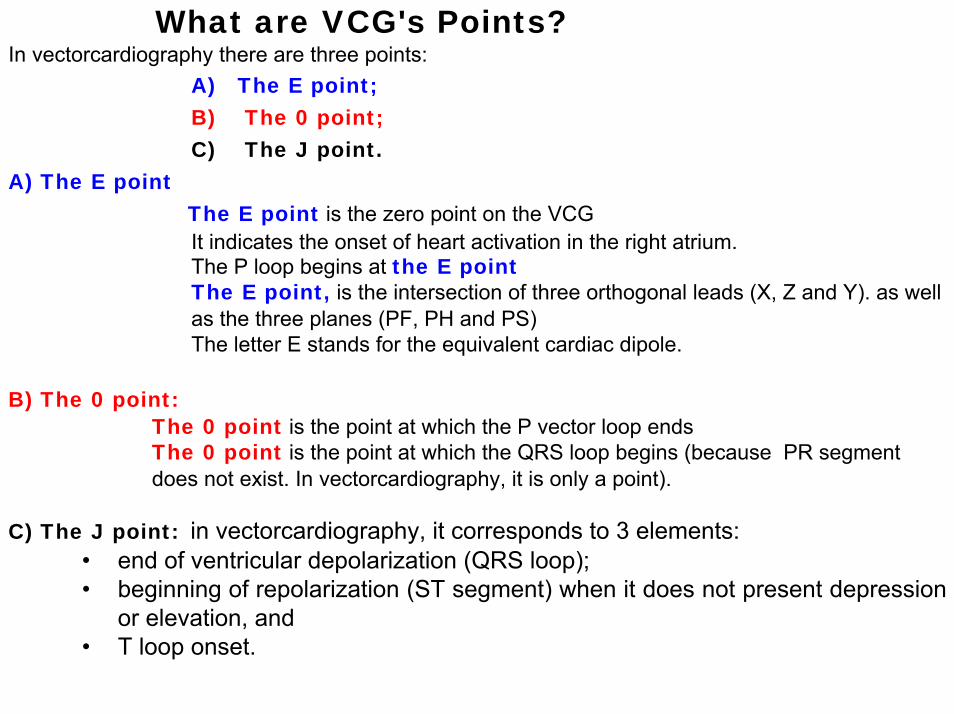
In vectorcardiography there are three points:A) The E point;B) The 0 point; C) The J point.
A) The E pointThe E point is the zero point on the VCGIt indicates the onset of heart activation in the right atrium. The P loop begins at the E pointThe E point, is the intersection of three orthogonal leads (X, Z and Y). as wellas the three planes (PF, PH and PS)The letter E stands for the equivalent cardiac dipole.
B) The 0 point:The 0 point is the point at which the P vector loop endsThe 0 point is the point at which the QRS loop begins (because PR segmentdoes not exist. In vectorcardiography, it is only a point).
C) The J point: in vectorcardiography, it corresponds to 3 elements: • end of ventricular depolarization (QRS loop); • beginning of repolarization (ST segment) when it does not present depression
or elevation, and • T loop onset.
What are VCG's Points?
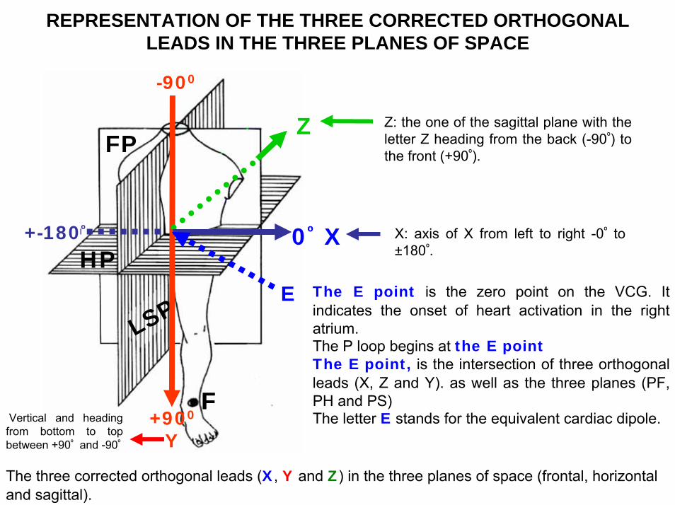
REPRESENTATION OF THE THREE CORRECTED ORTHOGONAL LEADS IN THE THREE PLANES OF SPACE
F
FP
LSP
0º X
Z
+900
Y
F
HPE
X: axis of X from left to right -0º to ±180º.
Z: the one of the sagittal plane with the letter Z heading from the back (-90º) to the front (+90º).
Vertical and heading from bottom to top between +90º and -90º
-900
+-180º
The E point is the zero point on the VCG. It indicates the onset of heart activation in the right atrium. The P loop begins at the E pointThe E point, is the intersection of three orthogonal leads (X, Z and Y). as well as the three planes (PF, PH and PS)The letter E stands for the equivalent cardiac dipole.
The three corrected orthogonal leads (X, Y and Z) in the three planes of space (frontal, horizontal and sagittal).

THE THREE POINTS OF VCG
T LOOP
E
RA
LA
0 POINTE POINT
RA
LA
0 POINTE POINT
J POINT
QRS LOOP P LOO
P
EFFERENT LIMB
AFFERENT LIMB
(fasterregistration)
E POINT
T LOOP
J & 0 POINTS
RA
10MS (is inscribed more slowly)
Normal T loop tends to be elliptical in shape

THE THREE BASIC POINTS IN VECTORCARDIOGRAPHY
In situations where there is depression or elevation of ST segment, the J point does not coincide with the 0 point, and the greater or lesser distance between both points indicatethe greater or lesser ST segment elevation or depression.
The phenomenon is observed in:1. Early Repolarization Pattern (ERP): ST elevation can be normal in young people and
some middle age people -- this requires no treatment is called a J point elevation or ERP. 2. Acute coronary syndrome with ST segment elevation: ST segment elevation myocardial
infarction (STEMI). 3. Prinzmetal angina, also known as variant angina or angina inverse.4. Pericarditis5. Brugada syndrome6. Idiopathic Ventricular Fibrillation7. Arrhytmogenic Right Ventricular Cardiomyopathy/Dysplasia, (ARVC/D).
The S-T vector is the vector joining the 0 and J points.The T loop begins at the J point and ends at the E point.