Embed Size (px)
Citation preview

Anemia
Dr. Kenneth Wurtz

Variables which Tend to Raise the Hematocrit
• Dehydration• Fingerstick (heelstick, earlobe) samples• Prolonged tourniquet stasis• Exposure to cold• Increased muscular activity• Upright position• Centrifugation techniques (especially with bizarre cell
shapes)Variables which Tend to Decrease the Hematocrit
• Volume overload• Supine position• Capillary tube leakage during centrifugation• Automated techniques

Representative Normal Values (Coulter S)
31.5-3631.5-36MCHC (g/dl RBC’s)
27-3227-32MCH (pg)
82-9882-98MCV (fl)
37-4742-54HCT (%)
12-1614-18HGB (g/dl blood)
Adult FemaleAdult Male

Peripheral Blood Smear

Help from the Peripheral Smear
• Normal RBC Morphology– Anemia of chronic inflammation (malignancy)– Chronic renal failure (most patients)– Hypoendocrine states (most patients)– Early iron deficiency– Aplastic anemia
• Abnormal RBC morphology– Combined iron and folate deficiency– Sideroblastic anemia– Leukoerythroblastosis– Renal failure (some patients)– Hypothyroidism (some patients)

Common Causes of Various RBC Abnormalities
•Liver disease•C hemoglobin (AC,CC,SC)•SS Disease•Postsplenectomy•Thalassemia•Artifact
Target Cells
•Marked iron deficiency•Megaloblastic anemia (severe)•Microangiopathic hemolysis•Leukoerythroblastosis•Hemoglobinopathies
Marked Anisocytsis and Poikilocytosis
•Liver disease (central targeting)•Megaloblastic anemia (macroovalocytes)•Reticulocytosis•Newborn•Preleukemia (mimics megaloblastic morphology)
Macrocytosis
•Iron Deficiency •Thalassemia•Sideroblastic anemia•Chronic inflammation
Hypochromia, Microcytosis

Common Causes of Various RBC Abnormalities Cont.
•Postsplenectomy•Sideroblastic anemia•Megaloblastic anemia•Alcohol•Marked hemolysis•Thalassemia
Pappenheimer Bodies
• Postsplenectomy•Megaloblastic anemia• Erythroleukemia
Howell-Jolly Bodies
•Leukoerythroblastosis•Megaloblastic anemias•Thalassemia
Tear Drop Cells
•Hereditary acanthocytosis•Liver disease (spur cells)•Renal disease (burr cells)•Post splenectomy•Hypothyroidism•Microangiopathic hemolysis
Spiculated RBC’s

Common Causes of Various RBC Abnormalities Cont.
•Hereditary ovalocytosis•Megaloblastic anemia•Iron deficiency•Thalassemia
Ovalocytes
•Heredity spherocytosis•Autoimmune hemolysis•Hemoglobin C disorders (CC,SC)•Severe burns
Spherocytes

Iron Deficient Anemia

Sickle Cell

B-12 and Folate Deficiency

Spherocyte

Hemolytic Anemia

Polychromasia

Howell Jolly

Anemia’s Associated with Various Clinical States
Bleeding, Hypersplenism, Folate deficiency, Ethanol depression of production, Sideroblastic anemia, Iron deficiency, Hemolysis
Alcoholic Liver Disease:
Microangiopathic hemolysis, Immune hemolysis, Decreased production
Malignancy:
Anemia of Inflammation, Microangiopathic Hemolysis, Oxidative Hemolysis (G-6-PD deficiency), Other Hemolytic Mechanisms
Bacterial Infection:
Immune Hemolysis, Decreased ProductionViral Infection:
HemoglobinopathiesFar East Origin:
G-6-PD Deficiency, ThalassemiaMediterranian Origin:
G-6-PD Deficiency, Hemoglobinopatheis, ThalassemiaBlacks:
Iron DeficiencyFemale:
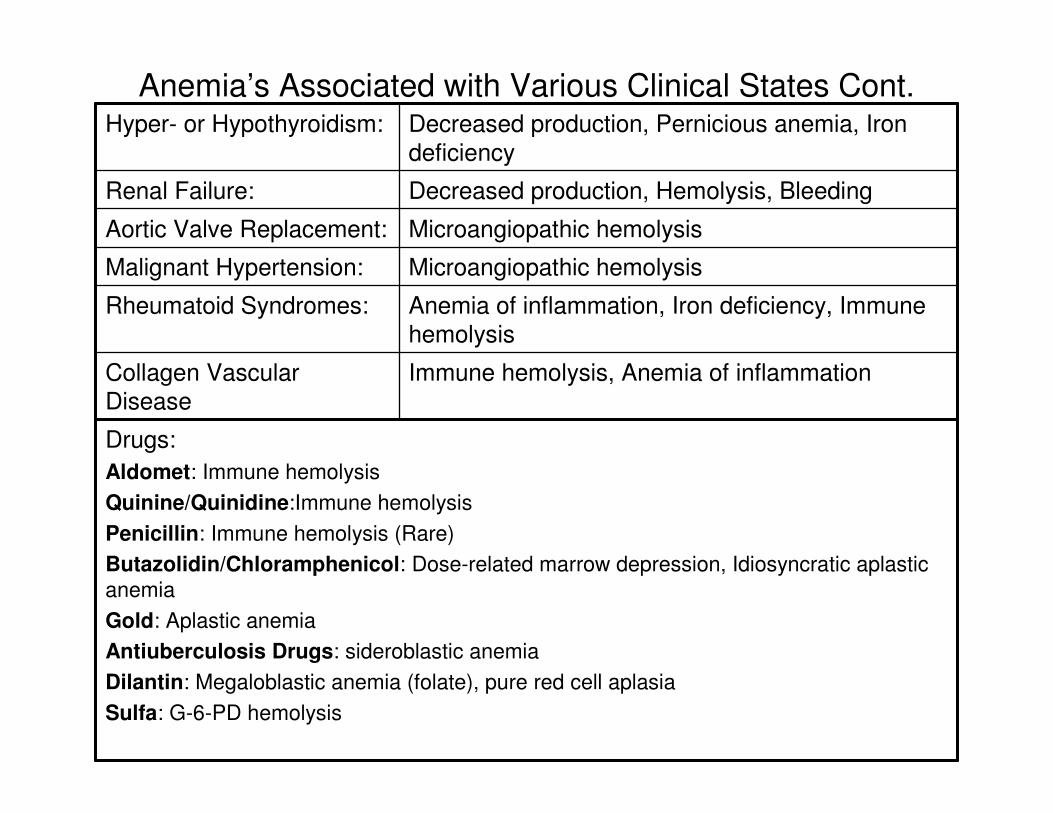
Anemia’s Associated with Various Clinical States Cont.
Immune hemolysis, Anemia of inflammationCollagen Vascular Disease
Drugs:Aldomet: Immune hemolysisQuinine/Quinidine:Immune hemolysisPenicillin: Immune hemolysis (Rare)Butazolidin/Chloramphenicol: Dose-related marrow depression, Idiosyncratic aplasticanemiaGold: Aplastic anemiaAntiuberculosis Drugs: sideroblastic anemiaDilantin: Megaloblastic anemia (folate), pure red cell aplasiaSulfa: G-6-PD hemolysis
Anemia of inflammation, Iron deficiency, Immune hemolysis
Rheumatoid Syndromes:
Microangiopathic hemolysisMalignant Hypertension:
Microangiopathic hemolysisAortic Valve Replacement:
Decreased production, Hemolysis, BleedingRenal Failure:
Decreased production, Pernicious anemia, Iron deficiency
Hyper- or Hypothyroidism:

Serum Ferritin
• The bulk of iron in the adult male body is in the hemoglobin of the circulating red cells (approx. 2600mg). Myoglobin (muscle...400mg) Ferritin and hemosiderin (approx. 800mg)(female approx. 200mg) scattered thoughout the body in the RES (monocyte-macropahge system).
• Ferritin is an iron storage protein. You can measure the ferritin.
***Sometimes we correct the anemia with iron, but not correct the iron deficiency. So, give iron for several months.

Transferrin/TIBC, Iron, Iron Saturation
• Transferrin is a transport protein in the plasma which binds the serum iron and then transports the iron to the developing red cells in the bone marrow.
• You can measure the serum iron and transferrin and calculate the % iron saturation.
• FE/transferrin (or TIBC) = %iron saturation • Iron Absorption Iron is in inorganic (ferric and ferrous)
forms and in organic (heme) forms. • Ferric is common dietary iron. • Heme iron is present in meats. Animal origin is best
absorbed. Phosphates and tannic acid (in tea) may inhibit absorption.
• Duodenum and jejunum are areas or iron absorption.• Achlorhydria or gastric removal or atrophic gastritis are
reasons for lack of iron absorption.

Representative Date Bases at Various Stages in the Slow Development of Severe Iron Deficient Anemia
m=mu
AbsentAbsentAbsentAbsentAbsent Bone Marrow Stores
4+ poikilocytosis4+ hypochromia
1+ poikilocytosis1+ hypochromia
NormalNormalNormalPeripheral Smear
1353060Serum Ferritin(10-200 m g/ml)
450400300300300TIBC250-375 m g/ml
2020356070SI(65-175 m g%)
2933333333MCHC(32-36g/dl)
6875818892MCV(82-98 fl)
1927354242HCT

Anemia-Approach to work-up
1. What is the patients MCV? Cell Size?2. Mechanism (production, bleeding,
hemolysis)3. Patient problem list.

ExampleHCT 10% reticulocyte count 5%
Reticulocyte index= 5% x 10/50 = 1%
-In this example (which uses for ease of calculation an arbitrary normal HCT of 50%) there is an inappropriately low reticulocyte index, indicating that bone marrow production is at least a contributing factor in the etiology of anemia. The elevated reticulocyte count of 5% is misleading. For this degree of anemia an appropriate reticulocyte count would be at least 15%:
reticulocyte index = 15% X 10/50= 3%

Low MCV
• Iron Deficiency• Thalassemia• Anemia of Chronic Disease• Sideroblastic Anemia

Treatment Tips (Dr. Wurtz)• FESO4 300mg= 60mg Elemental Iron• Avoid time-release and Enteric coated• Parenteral iron sometimes necessary if GI
intolerance exists or persists.• Indicated in patients with small and large bowel
inflammation, rapid transit GI problems, malabsorption, proven noncompliance, when time to respond is an urgent issue( third trimester of pregnancy, severe anemia). Side effects. Dosing for IM/IV iron.
• Vitamin C- not worth the cost– GI side effects

Administration of intravenous iron dextranMATERIEL• 2 50cc bags .9 NS • 1 500cc bag 0.9 NS • 1 IMED cassette tubing • 1 10cc syringe with needle • 1 1cc syringe with needle • Fe Dextrin in 2ml or 5ml ampoules • Alcohol wipes • 1 Buretrol
PROCEDURE1. Obtain MD orders and ensure her/his ability to be in attendance, to administer test dose,
and to be available during the infusion of full dose. Dosecalculation: Mg iron = [0.3 x wt (lbs) x 100 (14.8 - Hb)]/14.8 Note: in this calculation, 14.8 is the desired final hemoglobin value. The "Hb" in the equation
is the patient's current hemoglobin value. NOTE: Potential for anaphylactic reaction requires the administration of test dose.2. Prepare full dose of Fe Dextran as ordered in 10cc syringe - use single-dose ampoules. NOTE: Multi-dose vials of Fe dextran should not be used for IV administration due to
their phenol content.3. Transfer 25 mg of dose into 1cc syringe and inject into the 50cc bag of 0.9 NS (or as
ordered by M.D.)

Administration of intravenous iron dextrinNOTE: The use of 5% dextrose as diluent is associated with increased
incidence of local pain and phlebitis. 250 to 1000 ml of 0.9 NS diluent is recommended for the full dose of iron dextran.
4. Place IV access, hang bag 500cc 0.9 NS using an IMED cassettetubing and regulate IV to KVO.
5. With the M.D. present, hook the 50 ml bag with Fe dextran 25mg test dose into a side-port of the main IV tubing set, and run the test dose in over
10-15 minutes, carefully observing the patient's vital signs. NOTE: M.D. to stay in attendance.6. Monitor patient for 15-20 minutes for signs of adverse reaction. 7. Only after the completion of the IV test dose should the remainder of
the iron dextran be added to the 500 cc 0.9 NS bag. Administer full dose of
Fe dextrin via IMED pump over 2-6 hours as ordered by M.D. NOTE: M.D. should be available during the infusion, but not necessarily
at bedside. Administration rate should not exceed 500 mg/hr.

Administration of intravenous iron dextran
8. Flush medication through tubing, using 0.9 NS 50cc bag. 9. D/C IV per institutional procedure. 10. Document on appropriate patient forms. Large IV does of iron dextrin can produce delayed adverse reactions
(1-2 days) which usually subside within 3-4 days. These include: arthralgias,
myalgias, moderate to high fever, backache, chills, dizziness, headache, malaise, nausea and/or vomiting. Nonsteroidal anti-inflammatory agents
usually give good relief of these symptoms. To provide prophylaxis against these problems, physicians can
premedicate patients with 60 mg of methylprednisolone. Premedication is
particularly helpful to patients with inflammatory conditions, such as rheumatoid arthritis. These flairs may be caused by the inflammatory cytokines
from reticuloendothelial cells, which are "activated" when they engulf the circulating iron dextran complexes in the circulation
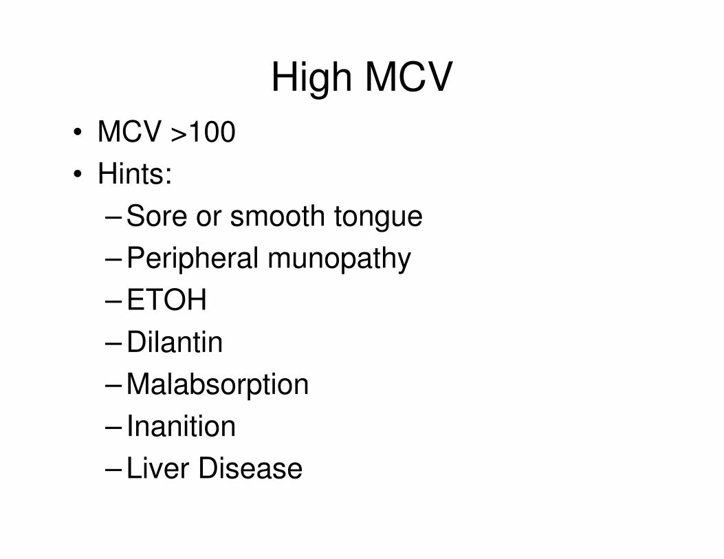
High MCV• MCV >100• Hints:
– Sore or smooth tongue– Peripheral munopathy– ETOH– Dilantin– Malabsorption– Inanition– Liver Disease

Differential Diagnosis of an MCV Greater than 100 fl
1. Spurious2. Reticulocytosis (marked)3. Liver Disease4. Alcoholism5. No Associated Disease6. Refractory Anemia (preleukemia, sideroblastic
anemia)7. Drugs8. Megaloblastic anemia’s

Causes of Vitamin B-12 and Folic Acid Megaloblastosis B-12• Pernicious anemia (acquired and congenital)• Gastrectomy• Illeal resection• Crohn’s disease• Fish tapeworm infestation• Blind loop syndrome• Nutritional deficiency (vegan’s diet, rare)• Familial selective malabsorption (Imerslund’s syndrome)• Dietary (old age, alcoholic, chronic disease)• Malabsorption (sprue)• Hemodialysis• Severe exfoliative skin disease (e.g. psoriasis)• Drugs:
– Interference with absorption or metabolism (dilantin, alcohol)– Dihydrofolate reductase inhibitors (methotrexate, trimethoprim)
Increased requirements: Pregnancy, Infancy, Hemolysis (e.g., Sickle Cell Anemia)

Serum B-12 Assay1. Spuriously low in some patients with folate deficiency.2. Spuriously low in some pregnant patients.3. May be elevated for weeks after one shot of B-12.4. Increased in myeloproliferative syndromes.
RBC Folate Assay1. Reflects chronic folate deficiency.2. Falsely low in some patients with B-12 deficiency.3. Falsely high in reticulocytes.
Serum Folate Assay1. A measure of recent dietary intake of folate.2. Usually normal or elevated in B-12 deficiency.

• Pernicious anemia is thought to be the true and most common cause of B12 deficiency. However, dietary B12 is rare. B12 stores are usually plentiful and take years to deplete. Shillings test no longer used to diagnose Pernicious Anemia. Measuring homocysteine and methylmalonic acid levels are used...and increased in B12 deficiency.

• Remember hyperhomocysteinemia may lead to atherosclerosis. If pernicious anemia is suspected, then measure parietal cell antibodies and intrinsic factor antibodies. With the ubiquitous use of gastric acid blocking agents (H2 and proton pump inhibitors ) B12 and folatelevels may be reduced.

Differentiating Between Folate and B-12 Megaloblastosis
All other combinations � Schilling test.
May confirm with schilling test
B-12 deficiencyDecreased Decreased (serum folate usually elevated)
Suggests B-12
NoneB-12 deficiencyDecreasedNl or elevatedSuggests B-12
Recheck B-12 after folate Rx for 1 week
Folate deficiency
Sl or decreased
Decreased Suggests Folate
NoneFolate deficiency
Nl or elevatedDecreasedSuggests Folate
Further Testing
InterpretationSerum B-12RBC FolateEtiology by History

Laboratory Features in Three Conditions Associated with an Elevated MCV
Normal or elevatedDecreased in B-12 deficiency; may be slightly decreased in folate deficiency
NormalSerum B-12
Normal or elevatedDecreased in folate deficiency; elevated in B-12 deficiency
Depends on dietSerum Folate
Frequently large and degranulated
NormalNormalPlatelet morphology
Abnormal mononuclear cells, no hyper segmentation
Hyper segmentedNormalWBC Morphology
CommonCommon Not commonNRB’s
Marked A and P, May mimic megaloblasticanemia
Marked A and P, Macro-ovalocytes, Tear drop cells
Targets No A and PRBC Morphology
Frequently decreasedFrequently decreasedVariablePlatelets
Frequently decreasedFrequently decreasedVariableWBC’s
Usually <110 flMaybe >110 flUsually < 110 flMCV
PreleukemiaMegaloblastic AnemiaLiver Disease

• Hemolysis and Bleeding Anemia with a normal or slightly elevated MCV and an appropriate Reticulocyte Index. Increase in reticulocyte count in absence of bleeding suggests HEMOLYSIS.
• Approach to suspected hemolysis :

Clinical States Associated with Intravascular Hemolysis
1. Acute hemolytic transfusion reactions.2. Severe and extensive burns.3. Physical trauma (e.g., March hemoglobinuria).4. Severe microangiopathic hemolysis (e.g.,
aortic valve prosthesis).5. Acute G-6-PD hemolysis.6. Paroxysmal nocturnal hemoglobinuria.7. Clostridial sepsis.

Common Clinical States Associated with Extravascular Hemolysis
1. Autoimmune hemolysis. 2. Delayed hemolytic transfusion
reactions.3. Hemoglobinopathies.4. Heredity spherocytosis.5. Hypersplenism.6. Hemolysis with liver disease.
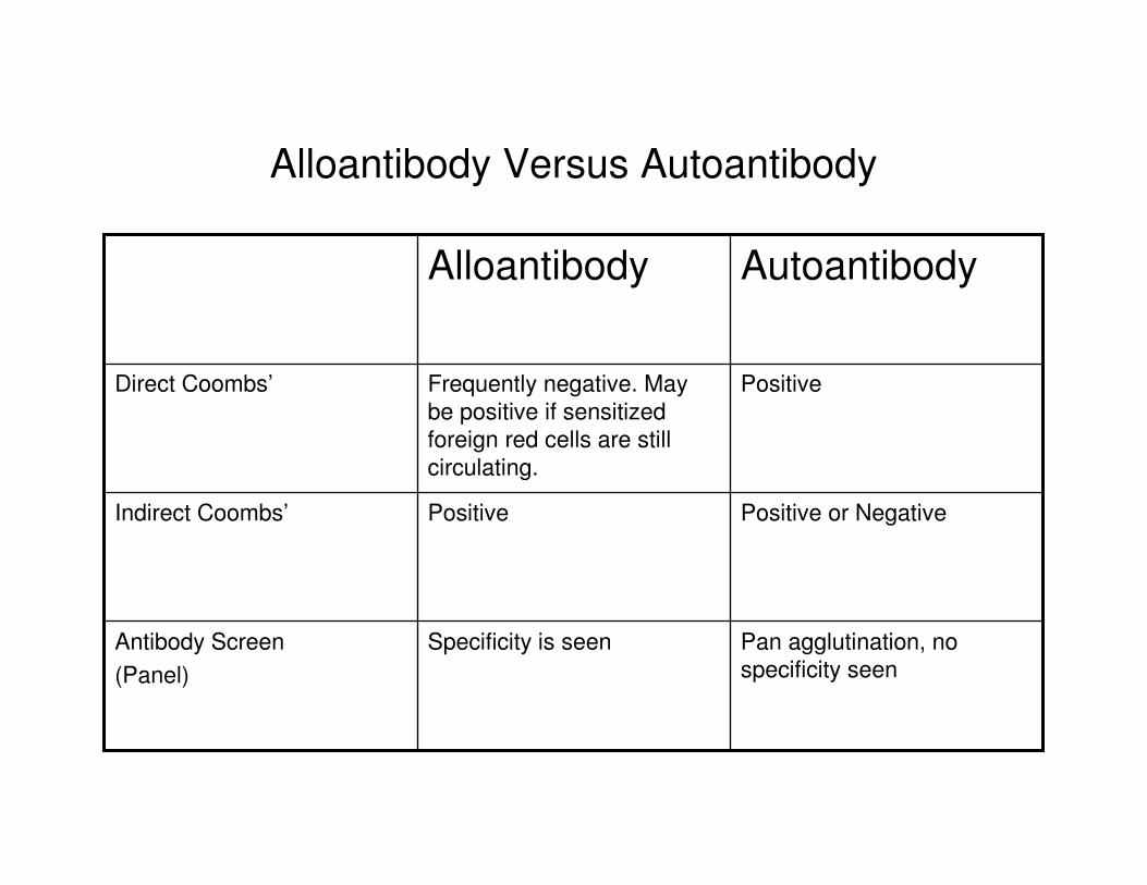
Alloantibody Versus Autoantibody
Pan agglutination, no specificity seen
Specificity is seenAntibody Screen(Panel)
Positive or NegativePositiveIndirect Coombs’
PositiveFrequently negative. May be positive if sensitized foreign red cells are still circulating.
Direct Coombs’
AutoantibodyAlloantibody

Anemia with a Normal MCV and Low Reticulocyte Index
Differential Diagnosis• Renal Failure• Anemia of Inflamatory Disease (anemia of malignancy)• Anemia of hypoendocrine states (hypothyroidism etc…)• Mild (early) Iron Deficiency• Combined iron deficiency and megaloblastic anemia• Sideroblastic Anemia• Bone Marrow inflatration (myelophthisis)• Bleeding or hemolysis plus one of the above

Inappropriately Normal or Elevated Serum Ferritin Levels
• Acute Liver Disease• Cirrhosis• Hodgkin’s Disease• Acute Leukemia• Solid Tumors (occasional)• Fever• Acute Inflammation• Renal Dialysis Patients• Recent Treatment with Iron

• Anemia of Renal Failure....decreased marrow erythropoiesis secondary to decreased erythropoietin (EPO). EPO and ESA are a whole 'nother topic !!! Interference by uremic toxins. Excess PTH (secondary PTH). Hemolytic component. Patients on hemodialysis...iron deficiency (bleeding into the coil, phlebotomy, GI bleed). Folate deficiency (folate is hemodialyzable). Measure GFR ! Don't always depend upon the serum creatinine. Measure ferritin. Inflammation elevates the ferritin level in iron deficient patients...so, a ferritin level less than 55 u g/l is highly suggestive of iron deficiency...whereas a level greater than 100 usually means iron stores are present.

• Anemia of Chronic Inflammation Very common ! Serum iron is low and TIBC is low, so % iron saturation may be low or normal, and serum ferritin is normal or elevated and bone marrow iron stores are normal or increased. Malignancies and acute and chronic infections. Not the best idea to measure iron parameters during acute febrile illness. Look at peripheral blood smear (PBS...I call it "the po man's bone marrow".

• Anemia of Hypoendocrine States Hypothyroidism, hypoadrenalism, hypopituitarism,androgenlack/deprivation. Don't forget andropause and testosterone. Lessened peripheral oxygen requirements. Mention BEPO and PEPO.
• Don't or Do Shotgun the Patient !!! New anemia w/u by shotgun...you might hit sumpin :CBC with diff, iron, transferrin, ferritin, % iron saturation, hgbelectrophoresis, reticulocyte count, LDH, SPEP, UPEP, B12, folate, T4, TSH, haptoglobin.
• What is left out ? • Myelodysplasia(MDS) and Myeloproliferative
Disorders(MPD) MDS...elevated MCV, other cell lines involved besides red blood cells. MPD...MCV variable and other cell lines involved.

Sideroblastic Anemia: Differential Diagnosis
• CongenitalAcquired
Primary IdiopathicSecondary
Drugs (alcohol, lead, antituberculosis drugs, chloramphenicol)Collagen vascular diseaseMultiple myelomaMarked hemolysisThalassemiaMegaloblastic anemiaPreleukemia and the non lymphocytic acute leukemia

Leukoerythroblastosis (Common Etiologies)
• Primary myelofibrosis (Agnogenic myeloid metaplasia)• End-stage polycythemia vera• Metastatic Cancer
– Breast– Prostate– Oat cell carcinoma of the lung
• Acute leukemia (occasionally)• Other hematologic malignancies on occasion (CLL,
multiple myeloma, lymphosarcoma)

Bone Marrow

Help from the Bone Marrow
• Bone Marrow NOT Helpful– Anemia of chronic disease– Chronic renal failure– Hypoendocrine states
• Bone Marrow Helpful– Myelophthisis (need a marrow biopsy)– Iron deficiency (serum ferritin is cheaper and less uncomfortable)– Combined iron deficiency and megaloblastic anemia– Sideroblastic anemia– Aplastic anemia– Primary marrow malignancy (leukemia, myeloma, etc.)

Why do a Bone Marrow?
• Q. Will a bone marrow (aspirate and biopsy) find something I did not already suspect by the patients' history, physical examination, lab, peripheral blood smear (PBS) and xrays ? A. Chances of finding something in the bone marrow that I did not already suspect are low.
• Q. What about the time involved, cost and discomfort of a bone marrow ? A. Discomfort is variable (sedation, anesthetic). The procedure usually takes less than 20 minutes (dependent on patient's body habitus).

Why do a Bone Marrow?
• Q. Costs? A. Interpretation of the bone marrow by the pathologist...maturity of the cells (red, white, platelet), number of cells vs. amount of fat , fibrosis, iron content. Blood (aspirate) from the bone marrow (sometimes also peripheral blood) can be sent for special studies like flow cytometry and cytogenetics. To make a diagnosis of 'refractory anemia' a bone marrow exam may be needed. 'Refractory' has variable interpretation...clinical or pathological ? When to give Erythrocyte Stimulating Agents (ESA) ? Empiric treatment for anemia ? Do you 'just give' iron, B12 or folate just to see if it will work ?

Anemia with Normal MCV and low Reticulocyte Index
• EPO (Erythropoietin)• RBC Morphology• Ferritin, serum iron, TIBC, Iron saturation• Hormone• Inflammation• Thyroid• Combined• Bone marrow failure• Peripheral blood smear