Embed Size (px)
Citation preview
Anesthetic Implications for the Physiological Changes in Pregnancy & Basic FHR Monitoring
J.E. Pellegrini, CRNA, PhD

Changes during the Puerperium
Changes to anatomy & physiology Most changes to physiology occur
during the 1st trimester Most changes to anatomy occur during
the 2nd and 3rd trimester Many of the changes are beneficial
As an anesthetist you must have a good understanding of these changes and so that you can determine if they will have an impact on your your anesthetic management
Physiological Changes of Pregnancy
Primarily we’ll discuss:Respiratory ChangesCardiovascular
ChangesGI/Hepatic/Renal
ChangesChanges in Neural
network (metabolism)
Factors influencing the Respiratory System
and endotracheal intubation
Weight gainBreast enlargementVascularity of the respiratory tract
mucosaPossible edema of the oropharynx,
nasopharyx, and vocal cords (**most prevalent in preeclampsia)
Progesterone-beneficial
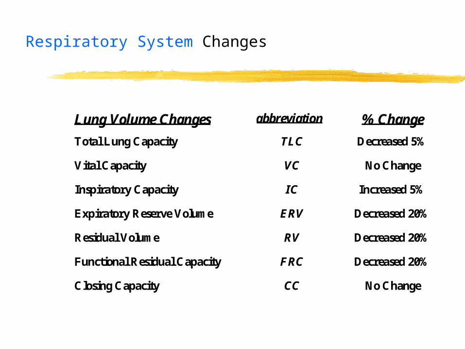
Respiratory System Changes
Lung Volume Changes abbreviation % Change
Total Lung Capacity TLC Decreased 5%
Vital Capacity VC No Change
Inspiratory Capacity IC Increased 5%
Expiratory Reserve Volume ERV Decreased 20%
Residual Volume RV Decreased 20%
Functional Residual Capacity FRC Decreased 20%
Closing Capacity CC No Change

Respiratory Changes with Pregnancy
Compensatory Respiratory System Changes
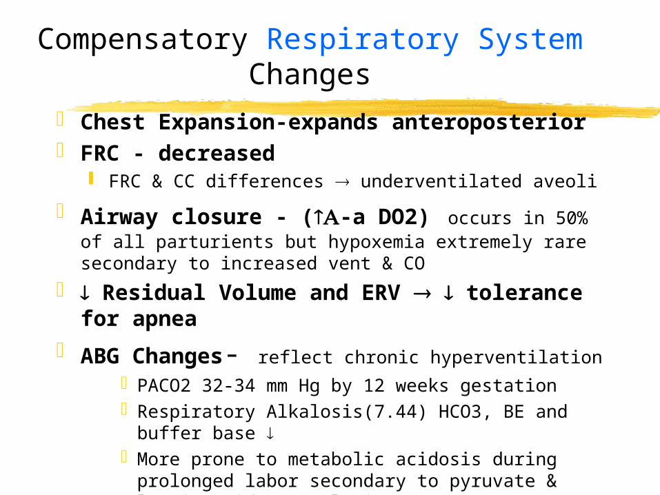
Chest Expansion-expands anteroposterior FRC - decreased
FRC & CC differences underventilated aveoli
Airway closure - (-a DO2) occurs in 50% of all parturients but hypoxemia extremely rare secondary to increased vent & CO
Residual Volume and ERV tolerance for apnea
ABG Changes- reflect chronic hyperventilationPACO2 32-34 mm Hg by 12 weeks gestationRespiratory Alkalosis(7.44) HCO3, BE and buffer
base More prone to metabolic acidosis during
prolonged labor secondary to pyruvate & lactic acid accumulation
Compensatory Respiratory System Changes
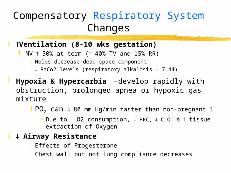
Ventilation (8-10 wks gestation) MV 50% at term ( 40% TV and 15% RR)
Helps decrease dead space component PaCo2 levels (respiratory alkalosis - 7.44)
Hypoxia & Hypercarbia -develop rapidly with obstruction, prolonged apnea or hypoxic gas mixture
PO2 can 80 mm Hg/min faster than non-pregnant
• Due to O2 consumption, FRC, C.O. & tissue extraction of Oxygen
Airway ResistanceEffects of ProgesteroneChest wall but not lung compliance decreases
Compensatory Respiratory System Changes
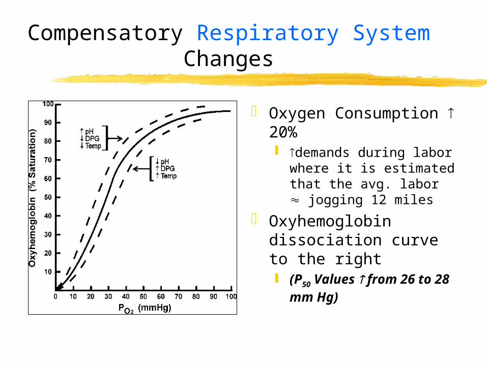
Oxygen Consumption 20% demands during labor
where it is estimated that the avg. labor jogging 12 miles
Oxyhemoglobin dissociation curve to the right (P50 Values from 26
to 28 mm Hg)
Clinical Implications of these Respiratory System Changes
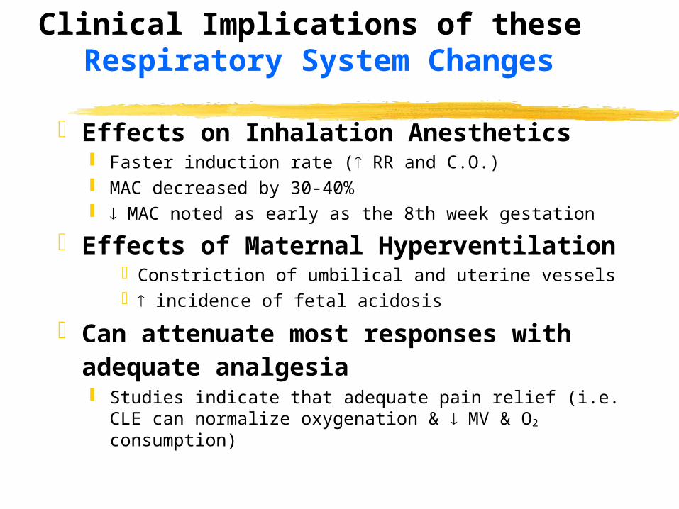
Effects on Inhalation Anesthetics Faster induction rate ( RR and C.O.) MAC decreased by 30-40% MAC noted as early as the 8th week gestation
Effects of Maternal Hyperventilation
Constriction of umbilical and uterine vessels incidence of fetal acidosis
Can attenuate most responses with adequate analgesia Studies indicate that adequate pain relief (i.e. CLE
can normalize oxygenation & MV & O2 consumption)
Cardiovascular System
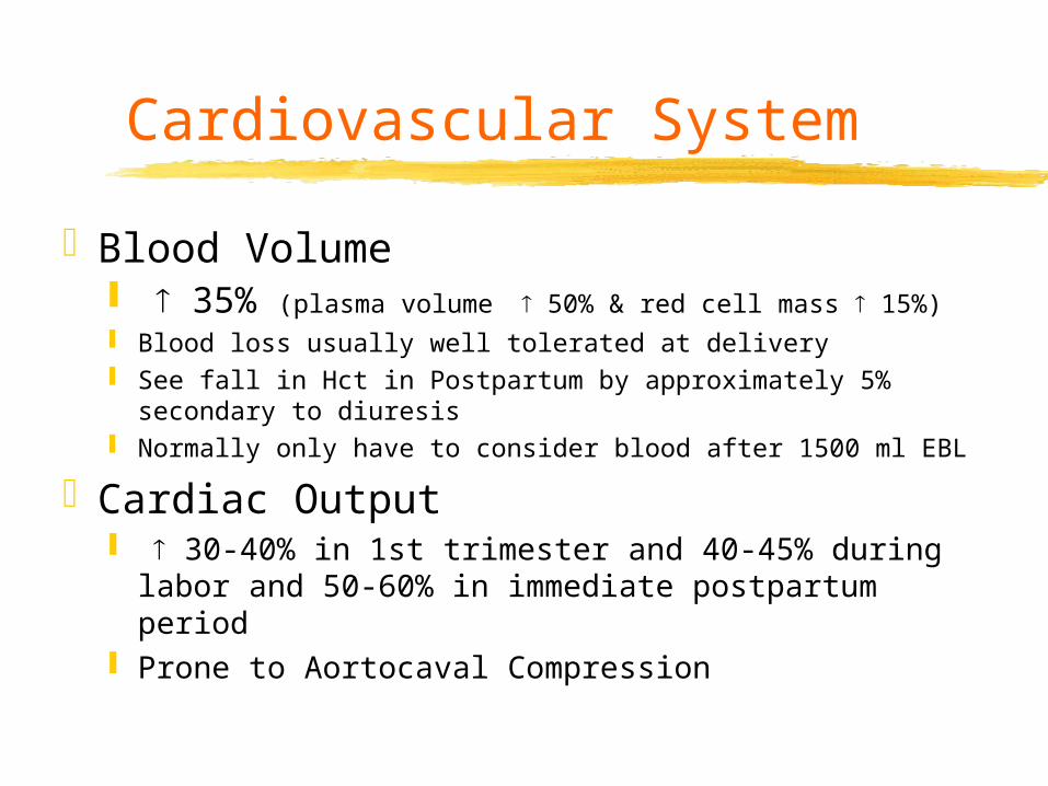
Blood Volume 35% (plasma volume 50% & red cell mass 15%) Blood loss usually well tolerated at delivery See fall in Hct in Postpartum by approximately 5%
secondary to diuresis Normally only have to consider blood after 1500 ml EBL
Cardiac Output 30-40% in 1st trimester and 40-45% during
labor and 50-60% in immediate postpartum period Prone to Aortocaval Compression
Changes in Cardiovascular System
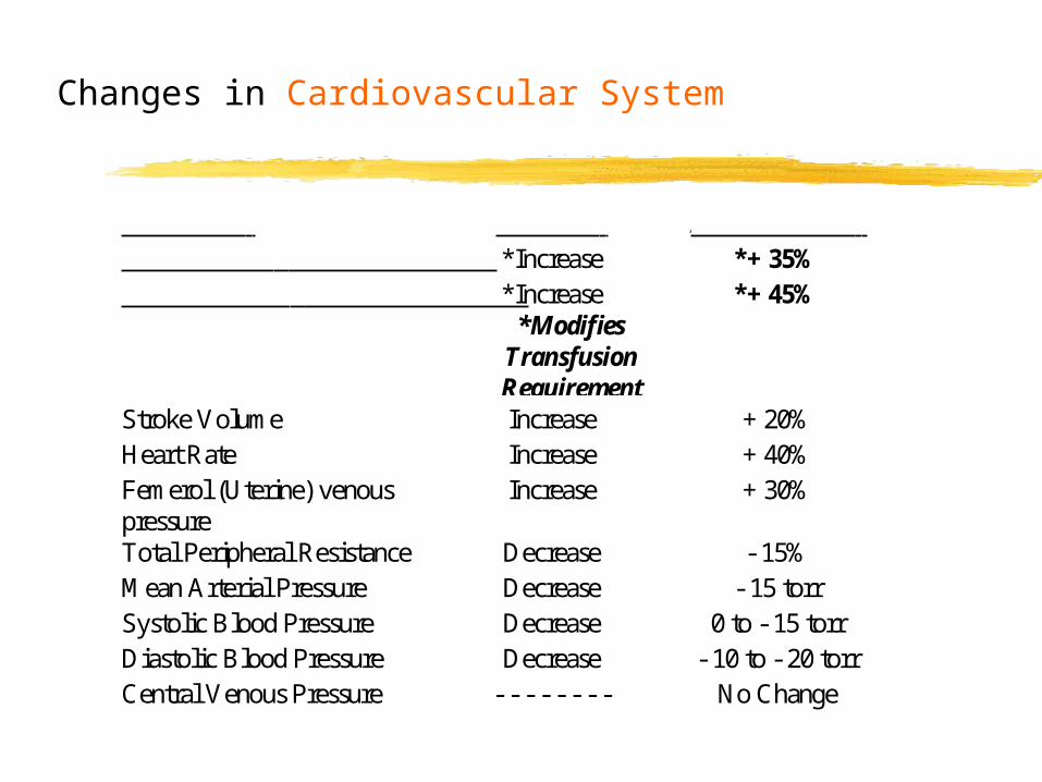
VARIABLE CHANGE AVG CHANGE Blood Volume *Increase *+ 35%Plasma Volume *Increase
*ModifiesTransfusionRequirement
*+ 45%
Stroke Volume Increase + 20%Heart Rate Increase + 40%Femerol (Uterine) venouspressure
Increase + 30%
Total Peripheral Resistance Decrease - 15%Mean Arterial Pressure Decrease - 15 torrSystolic Blood Pressure Decrease 0 to - 15 torrDiastolic Blood Pressure Decrease - 10 to - 20 torrCentral Venous Pressure - - - - - - - - No Change

Aorto-Caval SyndromeHypotension
20 weeks gestation Gravid Uterus Weight Can Decrease C.O. 30% Management Plan
Pre-induction hydrationLeft Uterine Displacement (or RUD)Ephedrine/Phenylephrine
Venal Caval Compression Distention of epidural venous
plexus Decrease LA dose 1/3 (>14
wks)
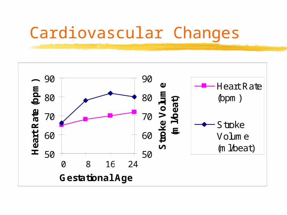
Cardiovascular Changes
50
60
70
80
90
Gestational Age
Hea
rt R
ate
(bp
m)
50
60
70
80
90
Str
oke
Vo
lum
e (m
l/b
eat)
Heart Rate(bpm)
StrokeVolume(ml/beat)
0 8 16 24

Anesthetic Significance of Cardiovascular Changes
Venodilation- increases accidental epidural vein puncture
Oxytocin with free H20 volume overload Hgb levels > 14 indicates low volume
status, HTN or diuresisC.O. high in 4 hrs postpartumB/P < 90 to 95 torr uterine blood flowHypotension occurs 75% with T4 level
Gastrointestinal Changes
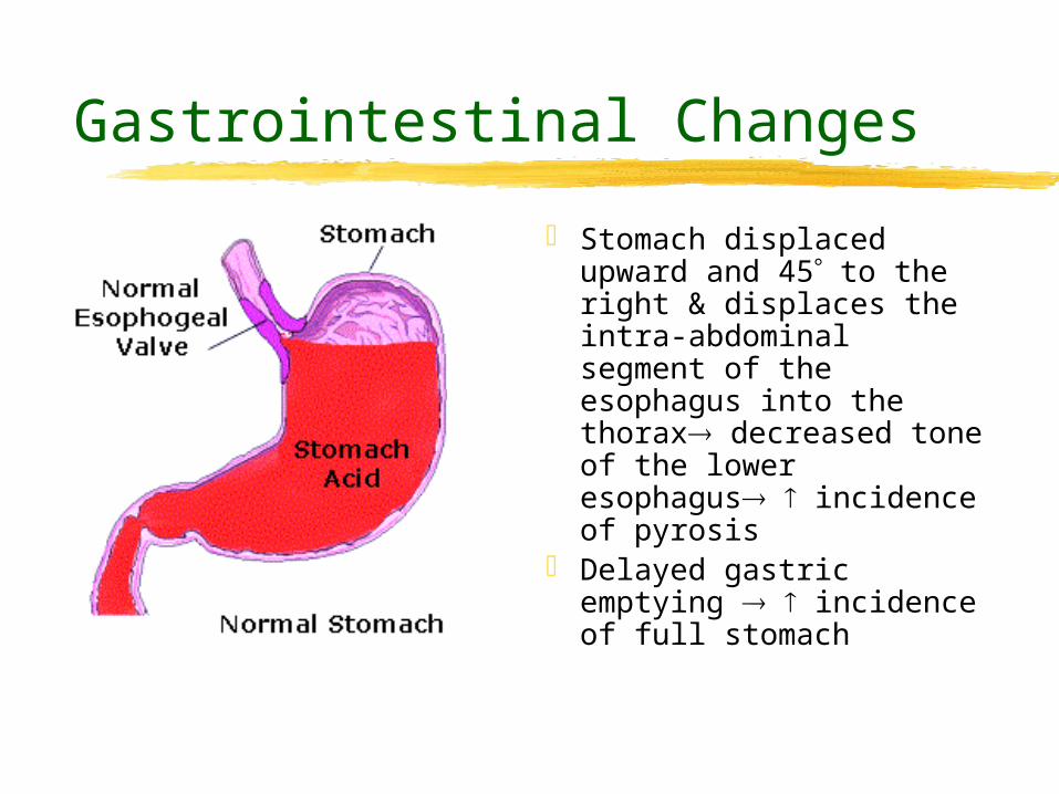
Stomach displaced upward and 45 to the right & displaces the intra-abdominal segment of the esophagus into the thorax decreased tone of the lower esophagus incidence of pyrosis
Delayed gastric emptying incidence of full stomach

Gastrointestinal Changes
Obesity - associated 2-20 fold in mortality (PIH, IDDM)
Progesterone Gastrointestinal motility & esophageal
sphincter tone
Parturients beyond 18th week of gestation more prone to vomiting and regurgitation Treat as full stomach at 12th week
*put it all together and this spells trouble
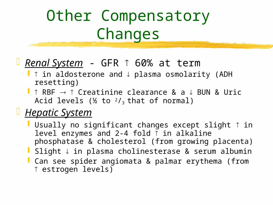
Other Compensatory Changes
Renal System - GFR 60% at term in aldosterone and plasma osmolarity (ADH
resetting) RBF Creatinine clearance & a BUN & Uric Acid
levels (½ to 2/3 that of normal)
Hepatic System Usually no significant changes except slight in level
enzymes and 2-4 fold in alkaline phosphatase & cholesterol (from growing placenta)
Slight in plasma cholinesterase & serum albumin Can see spider angiomata & palmar erythema (from
estrogen levels)
Neuromuscular Changes
EndorphinsMAC by 40%Sedative Effect from ProgesteroneChanges in SNS
See down-regulation Altered Response to Catecholamines
Altered Responses to Anesthesia
sensitivity of neural network Probably secondary to levels of
circulating progesteronePossible influence from circulating
endorphins
Applicable for both neuraxial and peripheral blockades
Applicable for parturients beyond 24th week gestationDecrease local anesthetic dose by as much
as 1/3
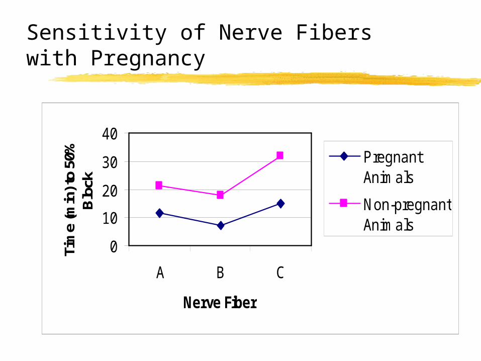
Sensitivity of Nerve Fibers with Pregnancy
0
10
20
30
40
A B C
Nerve Fiber
Tim
e (m
in) t
o 50
%
Blo
ck
Pregnant Animals
Non-pregnantAnimals
Summary
Multiple physiological changes in pregnancy have profound impact on your anesthetic management
The conservative approach is the best approach when dealing with the OB patient
Your principle patient is the parturient
Fetal Monitoring
No ideal way to assess fetal well-being
FHR one of the better methods FHR influenced by Para and sympathetic
outflow FHR responds to Baro & Chemo
receptors
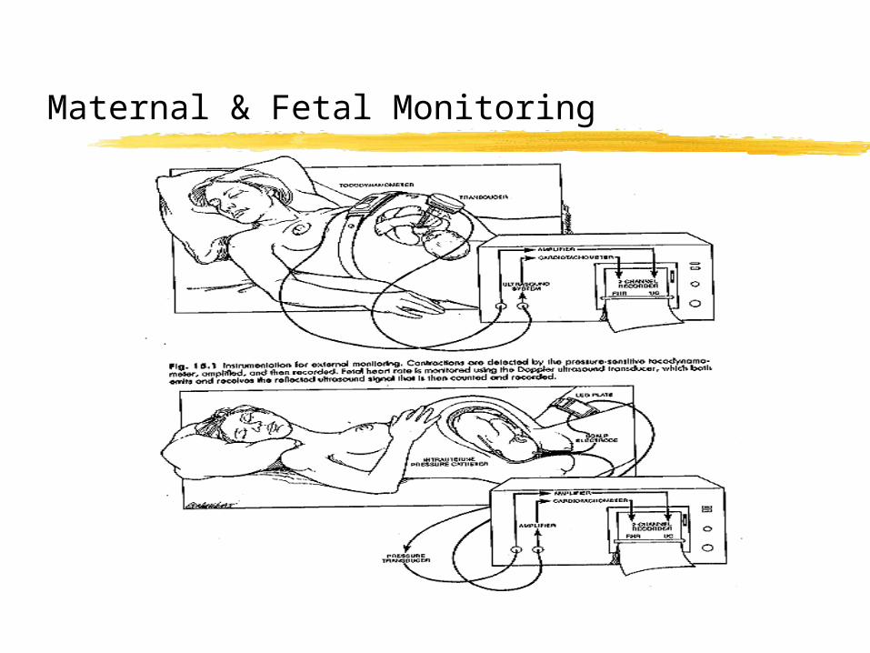
Maternal & Fetal Monitoring
Fetal Heart Rate
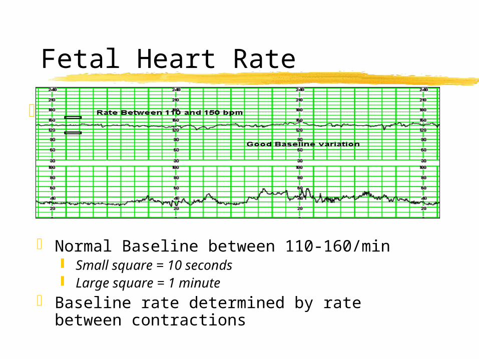
Normal Baseline between 110-160/min Small square = 10 seconds Large square = 1 minute
Baseline rate determined by rate between contractions

Three Primary Mechanisms that Uterine Contractions cause FHR Abnormalities
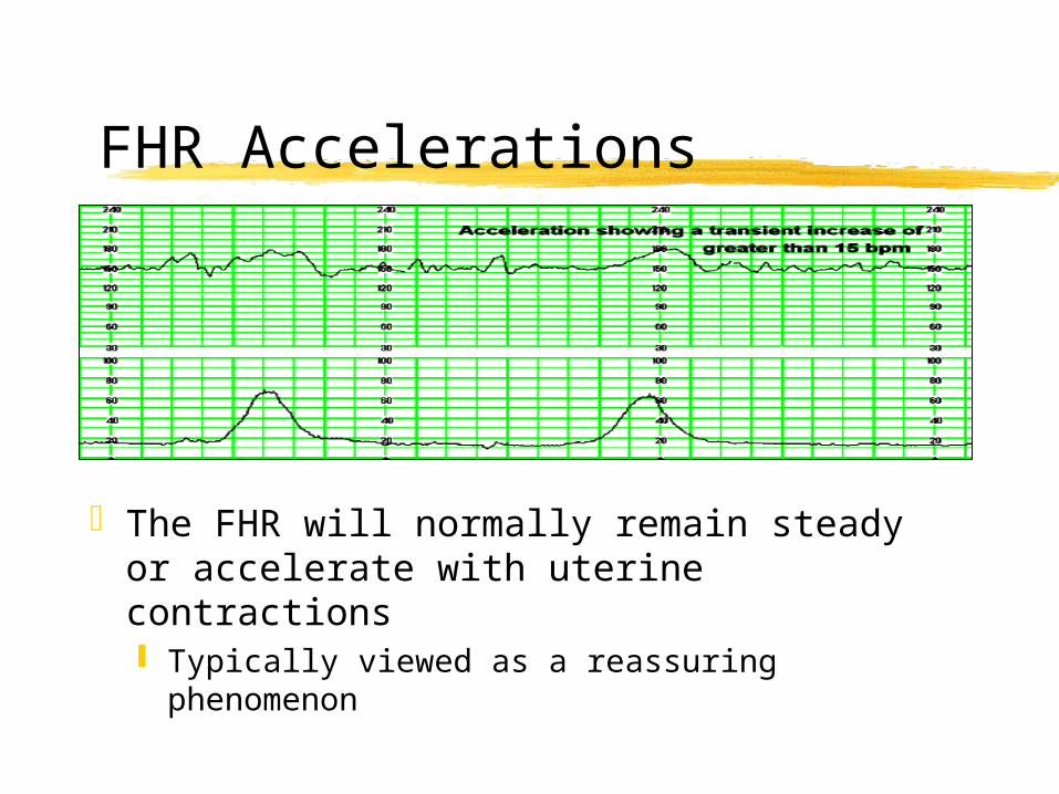
FHR Accelerations
The FHR will normally remain steady or accelerate with uterine contractions Typically viewed as a reassuring
phenomenon
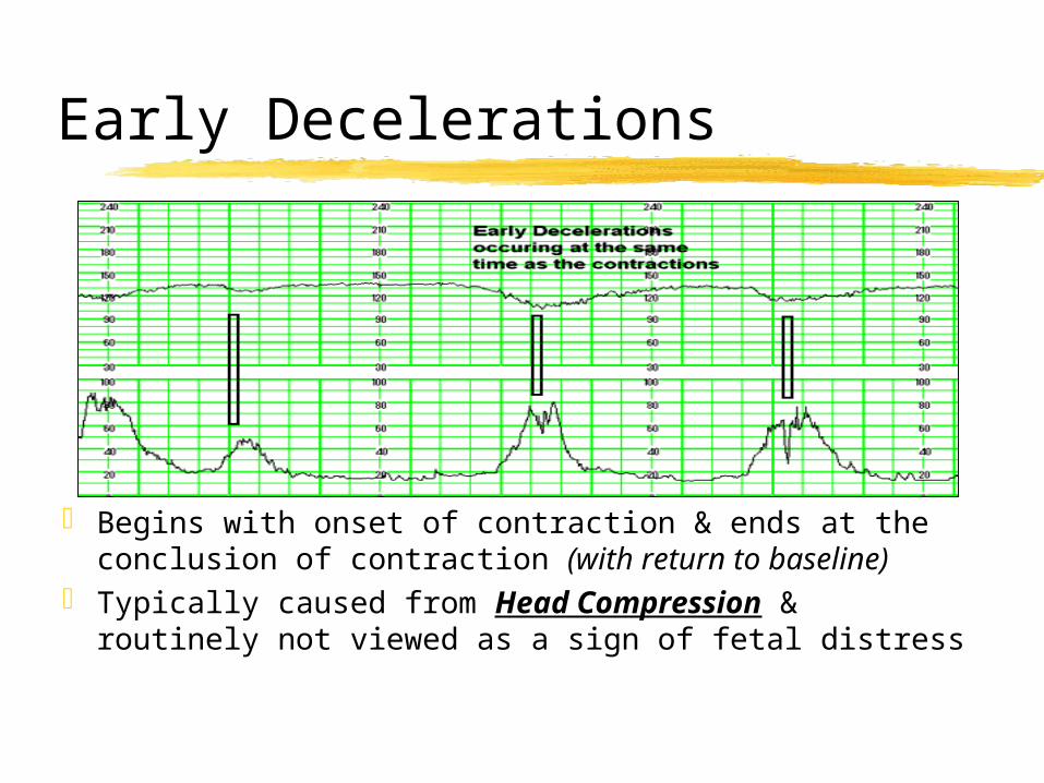
Early Decelerations
Begins with onset of contraction & ends at the conclusion of contraction (with return to baseline)
Typically caused from Head Compression & routinely not viewed as a sign of fetal distress
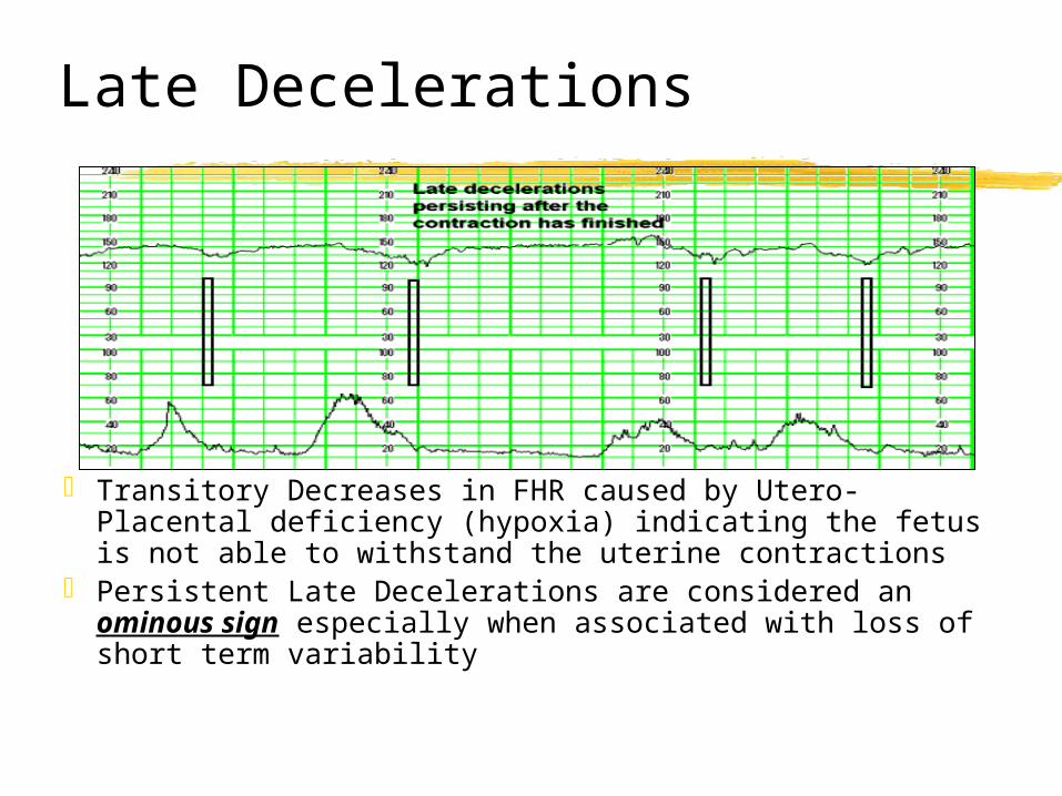
Late Decelerations
Transitory Decreases in FHR caused by Utero-Placental deficiency (hypoxia) indicating the fetus is not able to withstand the uterine contractions
Persistent Late Decelerations are considered an ominous sign especially when associated with loss of short term variability
Nonreassuring Patterns
Nonreassuring, or "warning," patterns suggest decreasing fetal capacity to cope with the stress of labor.
Nonreassuring Patterns (Warning Signs) Decrease in baseline variability Progressive tachycardia (>160bpm) Decrease in baseline FHR Intermittent late decelerations with good variability
Ominous patterns suggest possible fetal compromise.
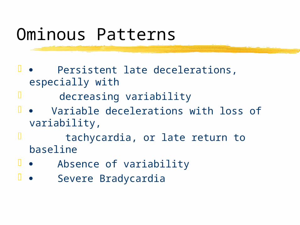
Ominous Patterns
Persistent late decelerations, especially with decreasing variability Variable decelerations with loss of variability, tachycardia, or late return to baseline Absence of variability Severe Bradycardia
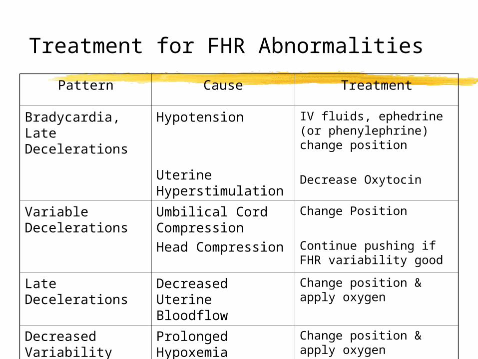
Treatment for FHR Abnormalities
Pattern Cause Treatment
Bradycardia, Late Decelerations
Hypotension
Uterine Hyperstimulation
IV fluids, ephedrine (or phenylephrine) change position
Decrease Oxytocin
Variable Decelerations
Umbilical Cord CompressionHead Compression
Change Position
Continue pushing if FHR variability good
Late Decelerations
Decreased Uterine Bloodflow
Change position & apply oxygen
Decreased Variability
Prolonged Hypoxemia
Change position & apply oxygen
So – In summary
If an ominous pattern appears to be present: Have the mother lie on her left side or in a
knee chest position immediately followed by:Increase IV fluid.Give her oxygen @ 10-12L to breathe by mask.Discontinue or decrease any CLE infusionNotify the obstetrical nursing staff & Obstetrician