Embed Size (px)
Citation preview
Angiopoietin-like 4 is a potent angiogenic factor and anovel therapeutic target for patients with proliferativediabetic retinopathySavalan Babapoor-Farrokhrana,1, Kathleen Jeea,1, Brooks Puchnera, Syed Junaid Hassana, Xiaoban Xina,Murilo Rodriguesa, Fabiana Kashiwabuchia, Tao Mab, Ke Hua,c, Monika Deshpandea, Yassine Daouda, Sharon Solomona,Adam Wenicka, Gerard A. Luttya, Gregg L. Semenzad, Silvia Montanerb, and Akrit Sodhia,2
aWilmer Eye Institute, Johns Hopkins University School of Medicine, Baltimore, MD 21287; bDepartment of Oncology and Diagnostic Sciences, School ofDentistry, and Department of Pathology, School of Medicine, Greenebaum Cancer Center, University of Maryland, Baltimore, MD 21201; dVascular Program,Institute for Cell Engineering, and Departments of Pediatrics, Medicine, Oncology, Radiation Oncology, Biological Chemistry, and Genetic Medicine, JohnsHopkins University School of Medicine, Baltimore, MD, 21205; and cDepartment of Ophthalmology, The First Affiliated Hospital of Chongqing MedicalUniversity, 400016 Chongqing, China
Edited by George D. Yancopoulos, Regeneron Pharmaceuticals, Inc., Tarrytown, NY, and approved May 1, 2015 (received for review December 15, 2014)
Diabetic eye disease is the most common cause of severe visionloss in the working-age population in the developed world, andproliferative diabetic retinopathy (PDR) is its most vision-threat-ening sequela. In PDR, retinal ischemia leads to the up-regulationof angiogenic factors that promote neovascularization. Therapiestargeting vascular endothelial growth factor (VEGF) delay thedevelopment of neovascularization in some, but not all, diabeticpatients, implicating additional factor(s) in PDR pathogenesis. Herewe demonstrate that the angiogenic potential of aqueous fluidfrom PDR patients is independent of VEGF concentration, providingan opportunity to evaluate the contribution of other angiogenicfactor(s) to PDR development. We identify angiopoietin-like 4(ANGPTL4) as a potent angiogenic factor whose expression is up-regulated in hypoxic retinal Müller cells in vitro and the ischemicretina in vivo. Expression of ANGPTL4 was increased in the aqueousand vitreous of PDR patients, independent of VEGF levels, correlatedwith the presence of diabetic eye disease, and localized to areasof retinal neovascularization. Inhibition of ANGPTL4 expression re-duced the angiogenic potential of hypoxic Müller cells; this effectwas additive with inhibition of VEGF expression. An ANGPTL4 neu-tralizing antibody inhibited the angiogenic effect of aqueous fluidfrom PDR patients, including samples from patients with low VEGFlevels or receiving anti-VEGF therapy. Collectively, our results sug-gest that targeting both ANGPTL4 and VEGF may be necessary foreffective treatment or prevention of PDR and provide the founda-tion for studies evaluating aqueous ANGPTL4 as a biomarker to helpguide individualized therapy for diabetic eye disease.
diabetes | neovascularization | hypoxia inducible factor-1 |angiopoietin-like 4 | vascular endothelial growth factor
Diabetic eye disease is the most common microvascular com-plication in the diabetic population and remains the leading
cause of blindness among working-age adults in the developedworld (1). Diabetic retinopathy (DR) is classified as either non-proliferative (NPDR) or proliferative (PDR). Although sustainedhyperglycemia is the major stimulus for the development ofNPDR (2), retinal ischemia is the prerequisite for PDR andresults in the up-regulation of angiogenic factors that promoteretinal neovascularization (3). If left untreated, neovascularizationcan lead to vitreous hemorrhage, retinal detachment, or glaucomaand result in profound and often irreversible loss of vision. For thelast 4 decades, the standard of care for PDR has been panretinalphotocoagulation (PRP), a process in which the peripheral is-chemic retina is killed (burned) with a laser to protect the patient’scentral vision (4). Although effective in reducing the risk of centralvision loss, PRP results in decreased peripheral and night vision intreated patients. Moreover, PDR can progress in patients despiteappropriate treatment. This emphasizes the importance of un-
derstanding the mechanism(s) promoting the development ofretinal neovascularization to identify targeted therapeutic ap-proaches for the prevention or treatment of PDR.In this regard, hypoxia-inducible factors (HIFs) activate the
transcription of multiple genes encoding angiogenic cytokinesthat promote retinal neovascularization in PDR (5). Among theangiogenic genes regulated by HIFs in PDR, considerable at-tention has focused on the contribution of vascular endothelialgrowth factor (VEGF). Several multicenter randomized con-trolled clinical trials have demonstrated the benefit of monthlyinjections with biologic molecules directed against VEGF totreat diabetic macular edema (DME) (6). These studies havedemonstrated a significant reduction in the progression to PDRin some—but not all—patients receiving anti-VEGF therapy (7,8), suggesting that other mediator(s) may participate in the de-velopment of PDR in many diabetic patients. Over the last 2decades, several angiogenic cytokines, growth factors, and in-flammatory mediators have been implicated in the pathogenesisof PDR (9). Despite these efforts, PRP treatment for PDR
Significance
In proliferative diabetic retinopathy (PDR), the most vision-threatening sequela of diabetic eye disease, retinal ischemialeads to increased expression of angiogenic factors that pro-mote neovascularization. Although therapies targeting thepotent angiogenic mediator vascular endothelial growth factorhave been remarkably successful for the treatment of diabeticmacular edema, this approach has not proven sufficient toprevent the development of retinal neovascularization, impli-cating additional angiogenic factor(s) in PDR pathogenesis. Wedemonstrate here that angiopoietin-like 4 is a potent angio-genic mediator with markedly increased expression in the eyesof PDR patients. Our studies identify a novel therapeutic targetfor the treatment of ocular neovascular disease and may havebroad implications for the treatment of other diseases de-pendent on pathologic angiogenesis.
Author contributions: S.M. and A.S. designed research; S.B.-F., K.J., B.P., S.J.H., X.X., M.R.,F.K., T.M., K.H., and M.D. performed research; Y.D., S.S., and A.W. contributed new re-agents/analytic tools; Y.D., S.S., and A.W. contributed patient samples; S.B.-F., K.J., B.P.,X.X., M.R., T.M., G.A.L., G.L.S., S.M., and A.S. analyzed data; and G.L.S., S.M., and A.S.wrote the paper.
The authors declare no conflict of interest.
This article is a PNAS Direct Submission.
Freely available online through the PNAS open access option.1S.B.-F. and K.J. contributed equally to this work.2To whom correspondence should be addressed. Email: [email protected].
This article contains supporting information online at www.pnas.org/lookup/suppl/doi:10.1073/pnas.1423765112/-/DCSupplemental.
E3030–E3039 | PNAS | Published online May 26, 2015 www.pnas.org/cgi/doi/10.1073/pnas.1423765112
remains unchanged. In an effort to identify an alternative thera-peutic approach for the treatment of retinal neovascularization,we set out to identify an ischemia-driven mediator that directlycontributes to the angiogenic phenotype in patients with PDR.
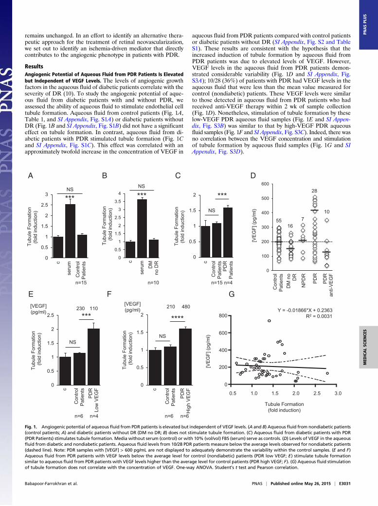
ResultsAngiogenic Potential of Aqueous Fluid from PDR Patients Is Elevatedbut Independent of VEGF Levels. The levels of angiogenic growthfactors in the aqueous fluid of diabetic patients correlate with theseverity of DR (10). To study the angiogenic potential of aque-ous fluid from diabetic patients with and without PDR, weassessed the ability of aqueous fluid to stimulate endothelial celltubule formation. Aqueous fluid from control patients (Fig. 1A,Table 1, and SI Appendix, Fig. S1A) or diabetic patients withoutDR (Fig. 1B and SI Appendix, Fig. S1B) did not have a significanteffect on tubule formation. In contrast, aqueous fluid from di-abetic patients with PDR stimulated tubule formation (Fig. 1Cand SI Appendix, Fig. S1C). This effect was correlated with anapproximately twofold increase in the concentration of VEGF in
aqueous fluid from PDR patients compared with control patientsor diabetic patients without DR (SI Appendix, Fig. S2 and TableS1). These results are consistent with the hypothesis that theincreased induction of tubule formation by aqueous fluid fromPDR patients was due to elevated levels of VEGF. However,VEGF levels in the aqueous fluid from PDR patients demon-strated considerable variability (Fig. 1D and SI Appendix, Fig.S3A); 10/28 (36%) of patients with PDR had VEGF levels in theaqueous fluid that were less than the mean value measured forcontrol (nondiabetic) patients. These VEGF levels were similarto those detected in aqueous fluid from PDR patients who hadreceived anti-VEGF therapy within 2 wk of sample collection(Fig. 1D). Nonetheless, stimulation of tubule formation by theselow-VEGF PDR aqueous fluid samples (Fig. 1E and SI Appen-dix, Fig. S3B) was similar to that by high-VEGF PDR aqueousfluid samples (Fig. 1F and SI Appendix, Fig. S3C). Indeed, there wasno correlation between the VEGF concentration and stimulationof tubule formation by aqueous fluid samples (Fig. 1G and SIAppendix, Fig. S3D).
A
0
0.5
1
1.5
2
2.5
3
Tubu
leFo
rmat
ion
(fold
indu
ctio
n)
c
seru
m
Con
trol
Pat
ient
s
***
n=15
NS
0
0.5
1
1.5
2
2.5
3
3.5
4
c
seru
m
DM
noD
R
Tubu
leFo
rmat
ion
(fold
indu
ctio
n)
***NS
n=10
B D
E
Tubu
leFo
rmat
ion
(fold
indu
ctio
n)
0
0.5
1
1.5
2
2.5
n=6
Con
trol
Pat
ient
sPD
RLo
wVE
GFc
n=4
***
NS
[VEGF](pg/ml)
110230
Tubu
leFo
rmat
ion
(fold
indu
ctio
n)
c
Con
trol
Pat
ient
s
PDR
Hig
h V
EGF
****
0
0.5
1
1.5
2
n=6 n=6
NS
[VEGF](pg/ml) 210 480
F G
[VEG
F](p
g/m
l)
0
200
400
600
800
0.5 1.5 2.0 2.5 3.01.0
Y = -0.01866*X + 0.2363R2 = 0.0031
Tubule Formation(fold induction)
Tubu
leFo
rmat
ion
(f old
indu
ctio
n)
c
Con
tr ol
Pat
ient
sPD
RP
atie
nts
***
n=15 n=4
NS
0
0.5
1
1.5
2
C
0
100
200
300
400
500
600
Con
trol
Pat
ient
sD
Mno DR
PDR
PDR
anti-
VEG
F
NPD
R
5516
28
107
[VEG
F](p
g/m
l)
Fig. 1. Angiogenic potential of aqueous fluid from PDR patients is elevated but independent of VEGF levels. (A and B) Aqueous fluid from nondiabetic patients(control patients; A) and diabetic patients without DR (DM no DR; B) does not stimulate tubule formation. (C) Aqueous fluid from diabetic patients with PDR(PDR Patients) stimulates tubule formation. Media without serum (control) or with 10% (vol/vol) FBS (serum) serve as controls. (D) Levels of VEGF in the aqueousfluid from diabetic and nondiabetic patients. Aqueous fluid levels from 10/28 PDR patients measure below the average levels observed for nondiabetic patients(dashed line). Note: PDR samples with [VEGF] > 600 pg/mL are not displayed to adequately demonstrate the variability within the control samples. (E and F)Aqueous fluid from PDR patients with VEGF levels below the average level for control (nondiabetic) patients (PDR low VEGF; E) stimulate tubule formationsimilar to aqueous fluid from PDR patients with VEGF levels higher than the average level for control patients (PDR high VEGF; F). (G) Aqueous fluid stimulationof tubule formation does not correlate with the concentration of VEGF. One-way ANOVA. Student’s t test and Pearson correlation.
Babapoor-Farrokhran et al. PNAS | Published online May 26, 2015 | E3031
MED
ICALSC
IENCE
SPN
ASPL
US

Angiogenic Potential of Aqueous Fluid from PDR Patients Is NotAffected by Anti-VEGF Therapy. Despite the variability in VEGFconcentrations in PDR aqueous fluid samples, the presence ofVEGF in these samples could still be responsible for the in-duction of tubule formation. To assess whether the angiogenicpotential of aqueous fluid from PDR patients was influenced by
anti-VEGF therapy, we tested the ability of aqueous fluid fromPDR patients (with high, average, or low levels of VEGF) tostimulate tubule formation in the presence of bevacizumab at a10-fold higher concentration than the dose which effectivelyneutralizes the highest levels of VEGF detected in the aqueousfluid of PDR patients (SI Appendix, Fig. S4 and Table 1). Treatment
Table 1. Patient samples for tubule formation assays
Patient Age, y Sex Phakic status* Prior vitrectomy DM type DM duration, y Prior PRP Anti-VEGF within 2–6 wk Anti-VEGF within 2 wk
Control1 70 F P No – – – – –
2 62 F P No – – – – –
3 83 M P No – – – – –
4 70 F P No – – – – –
5 75 M P No – – – – –
6 71 F P No – – – – –
7 64 F P No – – – – –
8 49 F P No – – – – –
9 50 M P No – – – – –
10 55 F P No – – – – –
11 65 F P No – – – – –
12 73 F P No – – – – –
13 55 F P No – – – – –
14 62 F P No – – – – –
15 59 M P No – – – – –
Diabetic (no DR)1 83 F P No II 9 – – –
2 70 M P No I 27 – – –
3 68 F P No II 12 – – –
4 76 F PP No II 5 – – –
5 65 M P No II 41 – – –
6 73 F P No II 4 – – –
7 91 M P No II 30 – – –
8 63 F P No I 36 – – –
9 68 F P No II 11 – – –
10 63 F P No II 9 – – –
PDR1 31 F P No II 19 Yes No No2 57 F P No II 25 No No No3 46 M P No II 20 Yes No No4 58 M P No II 20 No No No
High-VEGF PDR1 31 M P No I 25 No No No2 37 F P Yes I 27 Yes No No3 58 M P No II 20 No No No4 57 F P No II 25 No No No5 50 M P No II 4 No No No6 55 M P No II 2 Yes No No
Low-VEGF PDR1 55 M P Yes II 2 Yes No No2 58 M P Yes II 29 Yes No No3 52 F PP No I 26 Yes No No4 43 M P No I Unknown Yes No No
Anti-VEGF PDR1 50 M P No II 4 No No Yes2 35 M P No I Unknown Yes No Yes3 33 F P No I 23 No No Yes4 33 F P No I 23 Yes No Yes5 42 M P No II 12 Yes No Yes6 68 M P No II 15 No No Yes7 58 F P No II Unknown Yes No Yes8 57 M P No II Unknown Yes No Yes9 45 M P No II Unknown Yes No Yes
*At time of sample collection. DM, diabetes mellitus; DR, diabetic retinopathy; F, female; M, male; P, phakic; PDR, proliferative diabetic retinopathy; PP,pseudophakic; PRP, panretinal photocoagulation; VEGF, vascular endothelial growth factor.
E3032 | www.pnas.org/cgi/doi/10.1073/pnas.1423765112 Babapoor-Farrokhran et al.
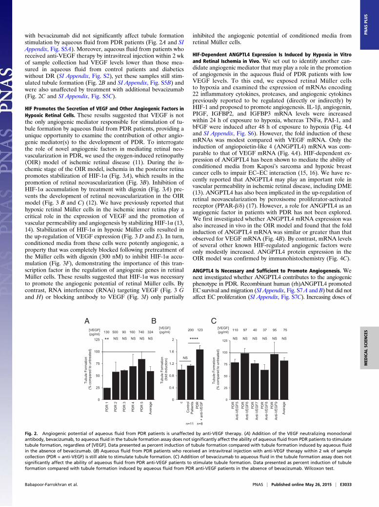
with bevacizumab did not significantly affect tubule formationstimulation by aqueous fluid from PDR patients (Fig. 2A and SIAppendix, Fig. S5A). Moreover, aqueous fluid from patients whoreceived anti-VEGF therapy by intravitreal injection within 2 wkof sample collection had VEGF levels lower than those mea-sured in aqueous fluid from control patients and diabeticswithout DR (SI Appendix, Fig. S2), yet these samples still stim-ulated tubule formation (Fig. 2B and SI Appendix, Fig. S5B) andwere also unaffected by treatment with additional bevacizumab(Fig. 2C and SI Appendix, Fig. S5C).
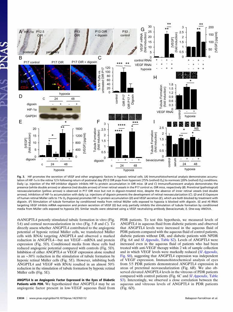
HIF Promotes the Secretion of VEGF and Other Angiogenic Factors inHypoxic Retinal Cells. These results suggested that VEGF is notthe only angiogenic mediator responsible for stimulation of tu-bule formation by aqueous fluid from PDR patients, providing aunique opportunity to examine the contribution of other angio-genic mediator(s) to the development of PDR. To interrogatethe role of novel angiogenic factors in mediating retinal neo-vascularization in PDR, we used the oxygen-induced retinopathy(OIR) model of ischemic retinal disease (11). During the is-chemic stage of the OIR model, ischemia in the posterior retinapromotes stabilization of HIF-1α (Fig. 3A), which results in thepromotion of retinal neovascularization (Fig. 3B). Inhibition ofHIF-1α accumulation by treatment with digoxin (Fig. 3A) pre-vents the development of retinal neovascularization in the OIRmodel (Fig. 3 B and C) (12). We have previously reported thathypoxic retinal Müller cells in the ischemic inner retina play acritical role in the expression of VEGF and the promotion ofvascular permeability and angiogenesis by stabilizing HIF-1α (13,14). Stabilization of HIF-1α in hypoxic Müller cells resulted inthe up-regulation of VEGF expression (Fig. 3 D and E). In turn,conditioned media from these cells were potently angiogenic, aproperty that was completely blocked following pretreatment ofthe Müller cells with digoxin (300 nM) to inhibit HIF-1α accu-mulation (Fig. 3F), demonstrating the importance of this tran-scription factor in the regulation of angiogenic genes in retinalMüller cells. These results suggested that HIF-1α was necessaryto promote the angiogenic potential of retinal Müller cells. Bycontrast, RNA interference (RNAi) targeting VEGF (Fig. 3 Gand H) or blocking antibody to VEGF (Fig. 3I) only partially
inhibited the angiogenic potential of conditioned media fromretinal Müller cells.
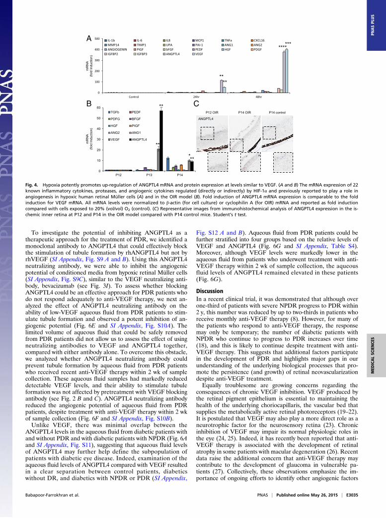
HIF-Dependent ANGPTL4 Expression Is Induced by Hypoxia in Vitroand Retinal Ischemia in Vivo. We set out to identify another can-didate angiogenic mediator that may play a role in the promotionof angiogenesis in the aqueous fluid of PDR patients with lowVEGF levels. To this end, we exposed retinal Müller cellsto hypoxia and examined the expression of mRNAs encoding22 inflammatory cytokines, proteases, and angiogenic cytokinespreviously reported to be regulated (directly or indirectly) byHIF-1 and proposed to promote angiogenesis. IL-1β, angiogenin,PIGF, IGFBP2, and IGFBP3 mRNA levels were increasedwithin 24 h of exposure to hypoxia, whereas TNFα, PAI-1, andbFGF were induced after 48 h of exposure to hypoxia (Fig. 4Aand SI Appendix, Fig. S6). However, the fold induction of thesemRNAs was modest compared with VEGF mRNA. Only theinduction of angiopoietin-like 4 (ANGPTL4) mRNA was com-parable to that of VEGF mRNA (Fig. 4A). HIF-dependent ex-pression of ANGPTL4 has been shown to mediate the ability ofconditioned media from Kaposi’s sarcoma and hypoxic breastcancer cells to impair EC–EC interaction (15, 16). We have re-cently reported that ANGPTL4 may play an important role invascular permeability in ischemic retinal disease, including DME(13). ANGPTL4 has also been implicated in the up-regulation ofretinal neovascularization by peroxisome proliferator-activatedreceptor (PPAR-β/δ) (17). However, a role for ANGPTL4 as anangiogenic factor in patients with PDR has not been explored.We first investigated whether ANGPTL4 mRNA expression wasalso increased in vivo in the OIR model and found that the foldinduction of ANGPTL4 mRNA was similar or greater than thatobserved for VEGF mRNA (Fig. 4B). By contrast, mRNA levelsof several other known HIF-regulated angiogenic factors wereonly modestly increased. ANGPTL4 protein expression in theOIR model was confirmed by immunohistochemistry (Fig. 4C).
ANGPTL4 Is Necessary and Sufficient to Promote Angiogenesis. Wenext investigated whether ANGPTL4 contributes to the angiogenicphenotype in PDR. Recombinant human (rh)ANGPTL4 promotedEC survival and migration (SI Appendix, Fig. S7A and B) but did notaffect EC proliferation (SI Appendix, Fig. S7C). Increasing doses of
A
T ubu
leFo
rmat
ion
(fold
indu
ctio
n)
c
Con
trol
P atie
nts
PD
R+
anti-
VEG
F
****
n=11 n=8
NS
0
0.4
0.8
1.2
1.6
2
[VEGF](pg/ml) 200 123
B C
T ubu
leFo
rmat
ion
(%co
mpa
red
toun
treat
ed)
PDR
1
PDR
2
PDR
3
PDR
4
PDR
5
Tubu
leFo
rmat
ion
(%co
mpa
red
toun
tr eat
ed)
[VEGF](pg/ml) 500 90130 160 740
** NS NS NS NS
Ave
rage
NS
324
0
25
50
75
100
125
[VEGF](pg/ml) 97 40 37 95110
NS NS NS NSNS
Aver
age
NS
75
0
25
50
75
100
125
PDR
Anti-
VEG
F5
PDR
Ant
i-VE G
F6PD
RA
nti-V
EG
F7PD
RAn
ti-VE
GF8
PDR
Anti-
VEG
F9
Fig. 2. Angiogenic potential of aqueous fluid from PDR patients is unaffected by anti-VEGF therapy. (A) Addition of the VEGF neutralizing monoclonalantibody, bevacizumab, to aqueous fluid in the tubule formation assay does not significantly affect the ability of aqueous fluid from PDR patients to stimulatetubule formation, regardless of [VEGF]. Data presented as percent induction of tubule formation compared with tubule formation induced by aqueous fluidin the absence of bevacizumab. (B) Aqueous fluid from PDR patients who received an intravitreal injection with anti-VEGF therapy within 2 wk of samplecollection (PDR + anti-VEGF) is still able to stimulate tubule formation. (C) Addition of bevacizumab to aqueous fluid in the tubule formation assay does notsignificantly affect the ability of aqueous fluid from PDR anti-VEGF patients to stimulate tubule formation. Data presented as percent induction of tubuleformation compared with tubule formation induced by aqueous fluid from PDR anti-VEGF patients in the absence of bevacizumab. Wilcoxon test.
Babapoor-Farrokhran et al. PNAS | Published online May 26, 2015 | E3033
MED
ICALSC
IENCE
SPN
ASPL
US
rhANGPTL4 potently stimulated tubule formation in vitro (Fig.5A) and corneal neovascularization in vivo (Fig. 5 B and C). Todirectly assess whether ANGPTL4 contributed to the angiogenicpotential of hypoxic retinal Müller cells, we transfected Müllercells with RNAi targeting ANGPTL4 and observed a markedreduction in ANGPTL4—but not VEGF—mRNA and proteinexpression (Fig. 5D). Conditioned media from these cells hadreduced angiogenic potential compared with controls (Fig. 5D).Inhibition of either ANGPTL4 or VEGF expression alone resultedin an ∼30% reduction in the stimulation of tubule formation byhypoxic retinal Müller cells (Fig. 5E). However, inhibiting bothANGPTL4 and VEGF with RNAi resulted in an almost 50%reduction in the stimulation of tubule formation by hypoxic retinalMüller cells (Fig. 5E).
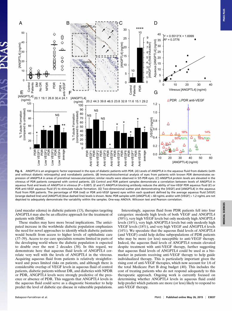
ANGPTL4 Is an Angiogenic Factor Expressed in the Eyes of DiabeticPatients with PDR. We hypothesized that ANGPTL4 may be anangiogenic factor present in low-VEGF aqueous fluid from
PDR patients. To test this hypothesis, we measured levels ofANGPTL4 in aqueous fluid from diabetic patients and observedthat ANGPTL4 levels were increased in the aqueous fluid ofPDR patients compared with the aqueous fluid of control patients,diabetic patients without DR, and diabetic patients with NPDR(Fig. 6A and SI Appendix, Table S2). Levels of ANGPTL4 wereincreased even in the aqueous fluid of patients who had beentreated with anti-VEGF therapy within 2 wk of sample collectionand in which VEGF levels were markedly reduced (SI Appendix,Fig. S8), suggesting that ANGPTL4 expression was independentof VEGF expression. Immunohistochemical analysis of eyesfrom 5/5 PDR patients demonstrated ANGPTL4 expression inareas of preretinal neovascularization (Fig. 6B). We also ob-served elevated ANGPTL4 levels in the vitreous of PDR patientscompared with control patients (Fig. 6C and SI Appendix, TableS3). Interestingly, we observed a close correlation between theaqueous and vitreous levels of ANGPTL4 in PDR patients(Fig. 6D).
% N
V Tu
fts v
s.un
treat
ed O
IR P
17
*** **
020406080
100120
1 2 3
cont
rol
OIR
P17
OIR
P17
+
Dig
oxin
CB
HIF-1α P12.5 OIR
P13 OIR
P13 control
posterior periphery
P13 OIR+ digoxin
A
D
HIF-1α
GAPDH
hypoxiaC 24
digoxin - +
48
- +
hypoxia
c 24
+
48
+
*
**
digoxin0
400
800
1200
1600
0
5
10
15
20
[VEG
F](fo
ld in
duct
ion)
[VEG
F] (p
g/m
l) *
*E F
G
VEG
F m
RN
A(fo
ld in
duct
ion)
control RNAiVEGF RNAi
hypoxia
00.30.60.91.21.51.8
Tubu
le F
orm
atio
n(fo
ld in
duct
ion)
**
- +VEGF RNAihypoxia
05
1015202530
+--
-+-
+-+
-++
**
0
50
100
150
200
00.5
11.5
22.5
3
[VEG
F]
(fold
indu
ctio
n)
******
[VEG
F] (p
g/m
l)
+ - + -- + +-- - + +
H
0
0.5
1
1.5
2
2.5
Tubu
le F
orm
atio
n(fo
ld in
duct
ion)
**
I
hypoxia
bevacizumab
* *****NS
seru
m
cont
rol
24
- +
48
- +
hypoxiac 24 48
digoxin
*
0
0.5
1
1.5
2
2.5
3
Tubu
le F
orm
atio
n(fo
ld in
duct
ion)
**** ***
cont
rol
- - - + +- - -
CD31
DAPI
P17 control P17 OIR P17 OIR + digoxin
Fig. 3. HIF promotes the secretion of VEGF and other angiogenic factors in hypoxic retinal cells. (A) Immunohistochemical analysis demonstrates accumu-lation of HIF-1α in the retina 12 h following return of postnatal day (P)12 OIR pups from hyperoxic [75% (vol/vol) O2] to normoxic [20% (vol/vol) O2] conditions.Daily i.p. injection of the HIF-inhibitor digoxin inhibits HIF-1α protein accumulation in OIR mice. (B and C) Immunofluorescent analysis demonstrates thepresence (white double arrows) or absence (red double arrows) of inner retinal vessels in the P17 control vs. OIR mice, respectively (B). Preretinal (pathological)neovascularization (yellow arrows) is observed in P17 OIR mice but not in digoxin-treated mice, despite the absence of inner retinal vessels (red doublearrows). Inhibition of HIF-1α accumulation with daily i.p. injections of digoxin prevents the development of retinal neovascularization (C). (D and E) Exposureof human retinal Müller cells to 1% O2 (hypoxia) promotes HIF-1α protein accumulation (D) and VEGF secretion (E), which are both blocked by treatment withdigoxin. (F) Stimulation of tubule formation by conditioned media from retinal Müller cells exposed to hypoxia is blocked with digoxin. (G and H) RNAitargeting VEGF inhibits mRNA expression and protein secretion of VEGF (G) but only partially inhibits the stimulation of tubule formation by conditionedmedia from Müller cells exposed to hypoxia (H). Similar results were obtained using a VEGF neutralizing antibody (bevacizumab; I). One-way ANOVA.
E3034 | www.pnas.org/cgi/doi/10.1073/pnas.1423765112 Babapoor-Farrokhran et al.
To investigate the potential of inhibiting ANGPTL4 as atherapeutic approach for the treatment of PDR, we identified amonoclonal antibody to ANGPTL4 that could effectively blockthe stimulation of tubule formation by rhANGPTL4 but not byrhVEGF (SI Appendix, Fig. S9 A and B). Using this ANGPTL4neutralizing antibody, we were able to inhibit the angiogenicpotential of conditioned media from hypoxic retinal Müller cells(SI Appendix, Fig. S9C), similar to the VEGF neutralizing anti-body, bevacizumab (see Fig. 3I). To assess whether blockingANGPTL4 could be an effective approach for PDR patients whodo not respond adequately to anti-VEGF therapy, we next an-alyzed the effect of ANGPTL4 neutralizing antibody on theability of low-VEGF aqueous fluid from PDR patients to stim-ulate tubule formation and observed a potent inhibition of an-giogenic potential (Fig. 6E and SI Appendix, Fig. S10A). Thelimited volume of aqueous fluid that could be safely removedfrom PDR patients did not allow us to assess the effect of usingneutralizing antibodies to VEGF and ANGPTL4 together,compared with either antibody alone. To overcome this obstacle,we analyzed whether ANGPTL4 neutralizing antibody couldprevent tubule formation by aqueous fluid from PDR patientswho received recent anti-VEGF therapy within 2 wk of samplecollection. These aqueous fluid samples had markedly reduceddetectable VEGF levels, and their ability to stimulate tubuleformation was not affected by pretreatment with VEGF blockingantibody (see Fig. 2 B and C). ANGPTL4 neutralizing antibodyreduced the angiogenic potential of aqueous fluid from PDRpatients, despite treatment with anti-VEGF therapy within 2 wkof sample collection (Fig. 6F and SI Appendix, Fig. S10B).Unlike VEGF, there was minimal overlap between the
ANGPTL4 levels in the aqueous fluid from diabetic patients withand without PDR and with diabetic patients with NPDR (Fig. 6Aand SI Appendix, Fig. S11), suggesting that aqueous fluid levelsof ANGPTL4 may further help define the subpopulation ofpatients with diabetic eye disease. Indeed, examination of theaqueous fluid levels of ANGPTL4 compared with VEGF resultedin a clear separation between control patients, diabeticswithout DR, and diabetics with NPDR or PDR (SI Appendix,
Fig. S12 A and B). Aqueous fluid from PDR patients could befurther stratified into four groups based on the relative levels ofVEGF and ANGPTL4 (Fig. 6G and SI Appendix, Table S4).Moreover, although VEGF levels were markedly lower in theaqueous fluid from patients who underwent treatment with anti-VEGF therapy within 2 wk of sample collection, the aqueousfluid levels of ANGPTL4 remained elevated in these patients(Fig. 6G).
DiscussionIn a recent clinical trial, it was demonstrated that although overone-third of patients with severe NPDR progress to PDR within2 y, this number was reduced by up to two-thirds in patients whoreceive monthly anti-VEGF therapy (8). However, for many ofthe patients who respond to anti-VEGF therapy, the responsemay only be temporary; the number of diabetic patients withNPDR who continue to progress to PDR increases over time(18), and this is likely to continue despite treatment with anti-VEGF therapy. This suggests that additional factors participatein the development of PDR and highlights major gaps in ourunderstanding of the underlying biological processes that pro-mote the persistence (and growth) of retinal neovascularizationdespite anti-VEGF treatment.Equally troublesome are growing concerns regarding the
consequences of chronic VEGF inhibition. VEGF produced bythe retinal pigment epithelium is essential to maintaining thehealth of the underlying choriocapillaris, the vascular bed thatsupplies the metabolically active retinal photoreceptors (19–22).It is postulated that VEGF may also play a more direct role as aneurotrophic factor for the neurosensory retina (23). Chronicinhibition of VEGF may impair its normal physiologic roles inthe eye (24, 25). Indeed, it has recently been reported that anti-VEGF therapy is associated with the development of retinalatrophy in some patients with macular degeneration (26). Recentdata raise the additional concern that anti-VEGF therapy maycontribute to the development of glaucoma in vulnerable pa-tients (27). Collectively, these observations emphasize the im-portance of ongoing efforts to identify other angiogenic factors
0
100
200
300
400
500
Control 24hr 48hr
IL-1b IL-6 IL8 MCP1 TNFa CXCL16MMP14 TIMP1 UPA PAI-1 ANG1 ANG2ANGIOGENIN PIGF bFGF PEDF HGF PDGFIGFBP2 IGFBP3 ANGPTL4 VEGF
B
A
0
10
20
30
40
50
60
P12 P13 P14
TGFb PEDF
PDFG BFGF
HGF PIGF
ANG2 ANG1
VEGF ANGPTL4
C
mR
NA
(fold
Indu
ctio
n)m
RN
A(fo
ldIn
d uct
ion)
**
****
**
***
**
**
***
ANGPTL4
P12 OIR P14 OIR P14 control
Fig. 4. Hypoxia potently promotes up-regulation of ANGPTL4 mRNA and protein expression at levels similar to VEGF. (A and B) The mRNA expression of 22known inflammatory cytokines, proteases, and angiogenic cytokines regulated (directly or indirectly) by HIF-1α and previously reported to play a role inangiogenesis in hypoxic human retinal Müller cells (A) and in the OIR model (B). Fold induction of ANGPTL4 mRNA expression is comparable to the foldinduction for VEGF mRNA. All mRNA levels were normalized to β-actin (for cell culture) or cyclophilin A (for OIR) mRNA and reported as fold inductioncompared with cells exposed to 20% (vol/vol) O2 (control). (C) Representative images from immunohistochemical analysis of ANGPTL4 expression in the is-chemic inner retina at P12 and P14 in the OIR model compared with P14 control mice. Student’s t test.
Babapoor-Farrokhran et al. PNAS | Published online May 26, 2015 | E3035
MED
ICALSC
IENCE
SPN
ASPL
US

that promote the development of diabetic eye disease and thatmay therefore serve as effective—and perhaps safer—targets forpatients with DR.In this regard, studies using animal models have demonstrated
that expression of a constitutively active HIF-1α mutant wassufficient to promote retinal neovascularization in vivo (28),whereas expression of VEGF alone was not sufficient to mediatethis effect (29–31). These results implicate additional HIF-regulatedangiogenic factor(s) in the promotion of retinal neovascularizationin PDR patients. Of interest, erythropoietin (EPO)—the first iden-tified HIF-regulated gene—has been implicated in the developmentof PDR (32, 33). However, PDR patients often have end-stagerenal disease and require parenteral injection of rhEPO on aregular basis. Additionally, in the eye, EPO has been found toreduce neuronal, glial, and vascular damage or dysfunction and toinhibit the breakdown of the blood–retinal barrier in a diabeticrodent model (34–36). EPO appears to play a complex and mul-tifaceted role in diabetic patients, as well as in diabetic eye disease,and modulation of EPO in this population should be approachedwith caution.The results of our study implicate an alternative angiogenic
factor, ANGPTL4, in the promotion of retinal neovascularization
in PDR patients. ANGPTL4 has been implicated in the up-regulation of retinal neovascularization by PPAR-β/δ (17). Weshow here that HIF-1 also plays an important role in regulatingexpression of ANGPTL4 in ischemic retinal disease. Expressionof ANGPTL4 was markedly increased in the aqueous and vitreousof PDR patients, and ANGPTL4 expression was localized toretinal neovascularization in PDR eyes. Inhibition of ANGPTL4reduced the angiogenic potential of hypoxic retinal cells; thiseffect was additive, with simultaneous inhibition of VEGF. Aneutralizing antibody for ANGPTL4 inhibited the angiogenicpotential of aqueous fluid from PDR patients, including aqueousfluid from PDR patients who had received anti-VEGF therapywithin 2 wk of sample collection. Although we cannot rule out acontribution from other angiogenic factors in the promotion ofretinal neovascularization in patients with PDR, our resultssuggest that targeting both ANGPTL4 and VEGF may be nec-essary for effective treatment or prevention of PDR. An essentialnext step will be the development of specific and effective anti-ANGPTL4 therapies. This will require the identification of therelevant endothelial cell receptor through which ANGPTL4 me-diates its pathological effects in the retina. Of note, as ANGPTL4has also been implicated in the promotion of vascular permeability
0
1
2
3
Tubu
le F
orm
atio
n(fo
ld in
duct
ion) ** ** **
rhANGPTL4 (ng/ml)
c 1 10 100
A
Ves
sels
per
mm
2
days
0
5
10
15
20
25
30
0 4 8 12 16
VEGFANGPTL4PBS
*** ** **
BPBS ANGPTL4 VEGF
D
Tubu
le F
orm
atio
n(fo
ld in
duct
ion)
C
control RNAiANGPTL4 RNAi
hypoxia
0
2
4
6
8 ** *
ANG
PTL4
mR
NA
(fold
indu
ctio
n)
[AN
GP
TL4]
(ng/
ml)
[AN
GP
TL4]
(fold
indu
ctio
n)
00.30.60.91.21.51.8
0
0.5
1
1.5
2
2.5 *** ***
0
100
200
300
400
0
10
20
30
40
[VE
GF]
(fol
d in
duct
ion)
[VE
GF]
(pg/
ml)
02468
10121416
VE
GF
mR
NA
(fol
d in
duct
ion) ***NS*** **
**
0
0.5
1
1.5
2
2.5 *
+--
-+-
+-+
-++
E
-+-+
--++
-+++
control RNAiANGPTL4 RNAi
VEGF RNAihypoxia
+---
+--+
-+-+
--++
-+++
0
10
20
30
40
50
Tubu
le F
orm
atio
n(%
inhi
bitio
n vs
. hyp
oxia
)
0
0.5
1
1.5
2
2.5
3
Tubu
le F
orm
atio
n(fo
ld in
duct
ion) ****
*****
NS ****
0
5
10
15
20
25
ANG
PTL4
mR
NA
(fold
Indu
ctio
n)
**
***
0
5
10
15
20
25
[AN
GP
TL4]
(fol
d in
duct
ion)
**
**
*
*****
[VE
GF]
(fol
d in
duct
ion)
0
5
10
15
20
25
30
VE
GF
mR
NA
(fol
d in
duct
ion) **
NS
**
*
0
5
10
15
20
25*
****
+---
+--+
-+-+
--++
-+++
+---
+--+
-+-+
--++
-+++
+---
+--+
-+-+
--++
-+++
+---
+--+
-+-+
--++
-+++
+--
-+-
+-+
-++
+--
-+-
+-+
-++
+--
-+-
+-+
-++
+--
-+-
+-+
-++
Fig. 5. ANGPTL4 is up-regulated by hypoxia and HIF in vitro and retinal ischemia in vivo and is necessary and sufficient to promote angiogenesis.(A) rhANGPTL4 potently stimulates tubule formation in vitro in a dose-dependent manner. (B and C) Representative photographs 1 wk following beadimplantation (B) and quantitation of vessels per mm2 (C) demonstrating that ANGPTL4 potently stimulates corneal neovascularization in vivo, similar to VEGF.(D) RNAi targeting ANGPTL4 causes a marked reduction in ANGPTL4—but not VEGF—mRNA and protein expression in human retinal Müller cells. This, inturn, results in reduced angiogenic potential of aqueous fluid from Müller cells pretreated with RNAi targeting ANGPTL4 compared with scrambled controls.(E) Inhibition of ANGPTL4 or VEGF mRNA and protein expression results in an ∼30% reduction in the stimulation of tubule formation by hypoxic retinalMüller cells. However, combined inhibition of both ANGPTL4 and VEGF mRNA and protein expression using RNAi in hypoxic retinal Müller cells results in analmost 50% reduction in the stimulation of tubule formation. One-way ANOVA.
E3036 | www.pnas.org/cgi/doi/10.1073/pnas.1423765112 Babapoor-Farrokhran et al.
(and macular edema) in diabetic patients (13), therapies targetingANGPTL4 may also be an effective approach for the treatment ofpatients with DME.These studies may have more broad implications. The antici-
pated increase in the worldwide diabetic population emphasizesthe need for novel approaches to identify which diabetic patientswould benefit from access to higher levels of ophthalmic care(37–39). Access to eye care specialists remains limited in parts ofthe developing world where the diabetic population is expectedto double over the next 2 decades (38). In this regard, wedemonstrate here that aqueous fluid levels of ANGPTL4 cor-relate very well with the levels of ANGPTL4 in the vitreous.Acquiring aqueous fluid from patients is relatively straightfor-ward and poses limited risks on patients, and although there isconsiderable overlap of VEGF levels in aqueous fluid of controlpatients, diabetic patients without DR, and diabetics with NPDRor PDR, ANGPTL4 levels were strongly predictive of the pres-ence or absence of PDR. This suggests that ANGPTL4 levels inthe aqueous fluid could serve as a diagnostic biomarker to helppredict the level of diabetic eye disease in vulnerable populations.
Interestingly, aqueous fluid from PDR patients fell into fourcategories: modestly high levels of both VEGF and ANGPTL4(50%), very high VEGF levels but only modestly high ANGPTL4levels (18%), very high ANGPTL4 levels but only modestly highVEGF levels (18%), and very high VEGF and ANGPTL4 levels(14%). We speculate that the aqueous fluid levels of ANGPTL4(and VEGF) could help define subpopulations of PDR patientswho may be more (or less) susceptible to anti-VEGF therapy.Indeed, the aqueous fluid levels of ANGPTL4 remain elevateddespite treatment with anti-VEGF therapy, further suggestingthat aqueous fluid levels of ANGPTL4 could be used as a bio-marker in patients receiving anti-VEGF therapy to help guideindividualized therapy. This is particularly important given therising cost of anti-VEGF therapies, which now account for 1/6 ofthe total Medicare Part B drug budget (40). This includes thecost of treating patients who do not respond adequately to thistherapeutic approach. Ongoing work is currently focused ondetermining whether ANGPTL4 levels in aqueous fluid couldhelp predict which patients are more (or less) likely to respond toanti-VEGF therapy.
%Tu
bule
Form
atio
n( c
ompa
red
toun
treat
ed)
PDR
Low
VEG
F1PD
RLo
wV E
GF2
PDR
Low
VEG
F3PD
RLo
wV E
GF4
*
NS
aver
age
[ANGPTL4](ng/ml) 26.8 30.419.120.1 24.1
***
*
0
20
40
60
80
100
120
F
0102030405060708090
100
%Tu
bule
Form
atio
n( c
ompa
red
toun
treat
ed)
PD
RA
nti-V
EG
F1PD
RAn
ti-V E
GF2
PDR
Anti-
VEG
F3PD
RAn
ti-VE
GF4
PDR
Anti -
V EG
F6PD
RA
nti-V
EG
F7PD
RA
nti -V
EGF8
PDR
Anti-
VEG
F5
aver
age
*
* *
*
*
[ANGPTL4](ng/ml) 39.8 46.5 11.6 15.1 29.232.823.820.2 44.3
NSNS
NS
*
G
0.0
0.2
0.4
0.6
0.8
1.0
1.2
10 20 30 40 50 60
[VEG
F](n
g/m
l)
[ANGPTL4] (ng/ml)
PDR PDR anti-VEGF
18% 14%
18%/50%50%/50%
B[A
NG
PTL4
](ng
/ml)
0
10
20
30
40
50C
ontro
lP
atie
nts
DM
No
DR
PD
R
PDR
+an
t i-VE
GF
NPD
R
A
E
0
5
10
15
20
25
30
35
40
0 10 20 30 40
Aque
ous
[AN
GPT
L4](
ng/m
l)
Vitreous [ANGPTL4] (ng/ml)
Y = 0.5513*X + 1.6999R2 = 0.3776
Con
trol
Pat
ient
s
0
5
10
15
20
25
30
[AN
GPT
L4](
ng/m
l)
10****
10
PDR
01
2
34
5
7
[AN
GP
TL4]
(fold
indu
ctio
n)
6
89
C DANGPTL4
control
Fig. 6. ANGPTL4 is an angiogenic factor expressed in the eyes of diabetic patients with PDR. (A) Levels of ANGPTL4 in the aqueous fluid from diabetic (withand without diabetic retinopathy) and nondiabetic patients. (B) Immunohistochemical analysis of eyes from patients with known PDR demonstrates ex-pression of ANGPTL4 in areas of preretinal neovascularization; similar results were observed in 5/5 PDR eyes. (C) ANGPTL4 protein levels are elevated in thevitreous of PDR patients compared with control patients. (D) Control and PDR patient samples demonstrate a correlation between levels of ANGPTL4 inaqueous fluid and levels of ANGPTL4 in vitreous (P = 0.007). (E and F) ANGPTL4 blocking antibody reduces the ability of low-VEGF PDR aqueous fluid (E) orPDR anti-VEGF aqueous fluid (F) to stimulate tubule formation. (G) Two-dimensional scatter plot demonstrating the [VEGF] and [ANGPTL4] in the aqueousfluid from PDR patients. The percentage of PDR (red) or PDR anti-VEGF (green) eyes within each quadrant defined by the average aqueous fluid [VEGF](orange dashed line) and [ANGPTL4] (blue dashed line) levels is shown. Note: PDR samples with [ANGPTL4] > 60 ng/mL and/or with [VEGF] > 1.2 ng/mL are notdepicted to adequately demonstrate the variability within the samples. One-way ANOVA. Wilcoxon test and Pearson correlation.
Babapoor-Farrokhran et al. PNAS | Published online May 26, 2015 | E3037
MED
ICALSC
IENCE
SPN
ASPL
US
As HIF-1 is a critical mediator of ocular neovascular disease,ANGPTL4 may play a key role in other vision-threatening dis-eases in which hypoxia (e.g., retinal ischemia) is a driving force,such as in retinal vein occlusion, sickle cell retinopathy, andretinopathy of prematurity. Our results suggest that ANGPTL4may contribute to retinal neovascularization in these patients.Ultimately, our observations provide the foundation for studiesto assess inhibition of ANGPTL4—alone or in combination withinhibition of VEGF—as a therapeutic approach for the treat-ment of PDR and other ocular neovascular diseases as well as forstudies investigating the use of aqueous fluid ANGPTL4 as adiagnostic and/or therapeutic biomarker for these diseases.
Materials and MethodsConstructs and Reagents. Recombinant human ANGPTL4, VEGF, and ANGPTL4(DuoSet) and VEGF (Quantikine) ELISA kits were purchased from R&D Sys-tems. Predesigned control (scrambled), ANGPTL4, and VEGF siRNA wereobtained from Santa Cruz. Lipofectamine RNAiMAX transfection reagentand Opti-MEM medium were obtained from Life Technologies. Hypoxiachambers were used to expose MIO-M1 cells (1% oxygen) and primarymurine Müller cells [3% (vol/vol) oxygen; exposure of primary murine Müllercells to lower oxygen concentrations resulted in cell death]. Digoxin anddesferrioxamine (DFO) were obtained from Sigma. Dimethyloxalylglycine(DMOG) was obtained from Cayman Pharmaceuticals.
Cell Culture. MIO-M1 cells were a generous gift from Astrid Limb (UniversityCollege London Institute of Ophthalmology) and cultured with DMEM(Invitrogen) containing 1 g/L glucose with 10% (vol/vol) FBS (Invitrogen) and1% penicillin/streptomycin (Cellgro). Immortalized human dermal micro-vascular endothelial cells (HMEC1) were obtained from the CDC and culturedwith DMEM containing 4.5 g/L glucose with 10% (vol/vol) FBS and 1% penicillin/streptomycin. Before treatments, the growth media were replaced with serumstarvation media containing 1% FBS.
siRNA Transfection. Cells were seeded at 60–80% confluence at transfection.Lipofectamine RNAiMAX reagent was diluted in Opti-MEM medium. Thirtypicomoles of siRNA from stock of 10 μM was diluted in Opti-MEM medium.Diluted siRNA was added to diluted Lipofectamine RNAiMAX reagent (1:1ratio) and incubated for 5 min at room temperature. siRNA–lipid complexwas added to cells and incubated at 37 °C for 24 h. The media were thenwashed out, and cells were ready for experiments.
Mice. Timed pregnant C57BL/6 mice (E14) (Charles River Laboratories) weretreated in accordance with the Association for Research in Vision andOphthalmology Statement for the Use of Animals in Ophthalmic and VisionResearch and the guidelines of the Johns Hopkins University Animal Care andUse Committee. OIR experiments were performed as previously described(11). Briefly, 7-d-old (P7) C57BL/6 mice and their mothers were exposed to75% (vol/vol) oxygen for 5 d. The mice were returned to room air at P12. Themice were killed and the eyes were collected at P12 (2 h after return tonormoxia), P13, P14, and/or P17. A subset of mice was given daily i.p. in-jection of vehicle or 2 mg/kg digoxin.
Western Blot. Cells in culture dishes were washed with PBS and lysed usingRIPA buffer (Sigma) with 10% (vol/vol) protease inhibitor mixture (Sigma).Cell lysates were then solubilized in LDS-sample buffer (Life Technologies)and incubated for 5 min at 95 °C. Lysates were subjected to 4–15% (wt/vol)gradient SDS/PAGE (Invitrogen). After blocking the membrane with 5% (wt/vol)milk (Bio-Rad), the membrane was then incubated with mouse anti-HIF-1α(BD, 610959) or rabbit anti-HIF-1α (Abcam, 2185) or with mouse anti-GAPDH monoclonal antibody (Fitzgerald) overnight at 4 °C. After washing,
the membrane was incubated with HRP-conjugated anti-mouse or anti-rabbitIgG (Cell Signaling) for 1 h and then visualized with ECL Super Signal West Femto(Thermo). Western blot scans are representative of at least three independentexperiments.
Quantitative RT-PCR.mRNAwas isolated from cultured cells or isolated retinaswith RNeasy Mini Kit (Qiagen), and cDNA was prepared with MuLV ReverseTranscriptase (Applied Biosystems). Quantitative real-time PCR was per-formed with Power SYBR Green PCR Master Mix (Applied Biosystems) andMyiQ Real-Time PCR Detection System (Bio-Rad). Cyclophilin A and β-actinwere used for normalization of mouse tissue and cell lines or for human celllines, respectively. Primers are listed in SI Appendix, Tables S5 and S6.
Immunohistochemistry and Immunofluorescence. Immunohistochemical de-tection of HIF-1α (Abcam, 2185) and ANGPTL4 (Abcam, 115798) was per-formed in paraffin-embedded human tissue and cryopreserved mouse tissuesections using ABC system (Dako) as previously described (13). CD31 anti-body was obtained from BD (550274). Images were captured using a con-focal microscope LSM 710 META (Carl Zeiss).
Tubule Formation Assay. Tubule formation assay was performed using growthfactor-reduced Matrigel (BD Biosciences; 356231). Sixty to eighty microlitersof Matrigel was added into a prechilled 96-well plate and placed in a 37 °CCO2 incubator for 30 min. HMECs were then counted and plated at 2 × 10⁴
cells per well on the Matrigel in a 96-well plate. Eighteen hours later, imageswere captured and analyzed using ImageJ software. Tubule formation assaywith patient samples was performed with an addition of 5 μL/well of aqueousfluid to the cell suspension before adding into the Matrigel-coated wells.VEGF neutralization was performed using 100 μg/mL of bevacizumab (JohnsHopkins University Pharmacy). ANGPTL4 neutralization was performed using10 μg/mL of an ANGPTL4 monoclonal antibody (Enzo, 804–732-C100).
Patient Samples. Institutional Review Board approval from the Johns HopkinsUniversity School of Medicine was obtained for all patient samples used inthis study. An a priori power analysis using a Cohen’s d effect size of 0.8based on results from in vivo studies and preliminary results, power of 0.8,and ⍺ of 0.05 yielded an estimated sample size of 26 control and PDR pa-tients. Aqueous and vitreous samples were collected from consenting pa-tients at the Wilmer Eye Institute undergoing cataract and/or vitrectomysurgery. Vitreous samples were immediately centrifuged at 16,000 × g for5 min at 4 °C, and supernatant was removed. Aqueous and vitreous sampleswere then aliquoted and stored at −80 °C before analysis.
ELISA. Aqueous diluted 1:10 and undiluted vitreous were analyzed forANGPTL4 and VEGF with ELISAs performed according to the manufacturer’sprotocols (R&D Systems).
Statistical Analysis. Results from cell culture and animal models are shown asmean ± SEM from at least three independent experiments. Results from clinicalsamples are shown as mean ± SD. Statistical differences between groups weredetermined by Student’s t test, Wilcoxon signed-rank test, and one-way ANOVAwhen indicated. Correlation was tested using Pearson’s method. Statisticalanalysis was performed using Microsoft Office and Prism 6.0 software (Graph-Pad). *P < 0.05; **P < 0.01; ***P < 0.001; ****P < 0.0001; NS, nonsignificant.
ACKNOWLEDGMENTS. This work was supported by the National Eye InstituteGrant K08-EY021189 (to A.S.), the Microscopy and Imaging Core Module andthe Animal Core Module of the Wilmer Core Grant EY001765, and an Unre-stricted Grant from Research to Prevent Blindness (to A.S.). A.S. gratefullyacknowledges the support he receives from the William and Ella Owens Med-ical Research Foundation, as a Career Development Award recipient from theResearch to Prevent Blindness Foundation.
1. Cheung N, Mitchell P, Wong TY (2010) Diabetic retinopathy. Lancet 376(9735):124–136.
2. Stitt AW (2010) AGEs and diabetic retinopathy. Invest Ophthalmol Vis Sci 51(10):4867–4874.
3. Durham JT, Herman IM (2011) Microvascular modifications in diabetic retinopathy.Curr Diab Rep 11(4):253–264.
4. Bressler NM, Beck RW, Ferris FL, 3rd (2011) Panretinal photocoagulation for pro-liferative diabetic retinopathy. N Engl J Med 365(16):1520–1526.
5. Kurihara T, Westenskow PD, Friedlander M (2014) Hypoxia-inducible factor (HIF)/vascularendothelial growth factor (VEGF) signaling in the retina. Adv Exp Med Biol 801:275–281.
6. Krispel C, Rodrigues M, Xin X, Sodhi A (2013) Ranibizumab in diabetic macular edema.World J Diabetes 4(6):310–318.
7. Bressler SB, et al. (2013) Exploratory analysis of the effect of intravitreal ranibizumabor triamcinolone on worsening of diabetic retinopathy in a randomized clinical trial.JAMA Ophthalmol 131(8):1033–1040.
8. Ip MS, Domalpally A, Hopkins JJ, Wong P, Ehrlich JS (2012) Long-term effects of ranibizu-mab on diabetic retinopathy severity and progression. Arch Ophthalmol 130(9):1145–1152.
9. Wang S, Park JK, Duh EJ (2012) Novel targets against retinal angiogenesis in diabeticretinopathy. Curr Diab Rep 12(4):355–363.
10. Funatsu H, et al. (2005) Aqueous humor levels of cytokines are related to vitreouslevels and progression of diabetic retinopathy in diabetic patients. Albrecht VonGraefes Arch Klin Exp Ophthalmol 243(1):3–8.
11. Smith LE, et al. (1994) Oxygen-induced retinopathy in the mouse. Invest OphthalmolVis Sci 35(1):101–111.
E3038 | www.pnas.org/cgi/doi/10.1073/pnas.1423765112 Babapoor-Farrokhran et al.
12. Yoshida T, et al. (2010) Digoxin inhibits retinal ischemia-induced HIF-1alpha expres-sion and ocular neovascularization. FASEB J 24(6):1759–1767.
13. Xin X, et al. (2013) Hypoxic retinal Muller cells promote vascular permeability by HIF-1-dependent up-regulation of angiopoietin-like 4. Proc Natl Acad Sci USA 110(36):E3425–E3434.
14. Rodrigues M, et al. (2013) VEGF secreted by hypoxic Müller cells induces MMP-2 ex-pression and activity in endothelial cells to promote retinal neovascularization inproliferative diabetic retinopathy. Diabetes 62(11):3863–3873.
15. Zhang H, et al. (2012) HIF-1-dependent expression of angiopoietin-like 4 and L1CAMmediates vascular metastasis of hypoxic breast cancer cells to the lungs. Oncogene31(14):1757–1770.
16. Ma T, et al. (2010) Viral G protein-coupled receptor up-regulates Angiopoietin-like 4promoting angiogenesis and vascular permeability in Kaposi’s sarcoma. Proc NatlAcad Sci USA 107(32):14363–14368.
17. Capozzi ME, McCollum GW, Savage SR, Penn JS (2013) Peroxisome proliferator-acti-vated receptor-β/δ regulates angiogenic cell behaviors and oxygen-induced retinop-athy. Invest Ophthalmol Vis Sci 54(6):4197–4207.
18. Fong DS, et al. (2004) Retinopathy in diabetes. Diabetes Care 27(Suppl 1):S84–S87.19. Blaauwgeers HG, et al. (1999) Polarized vascular endothelial growth factor secretion
by human retinal pigment epithelium and localization of vascular endothelial growthfactor receptors on the inner choriocapillaris. Evidence for a trophic paracrine re-lation. Am J Pathol 155(2):421–428.
20. Marneros AG, et al. (2005) Vascular endothelial growth factor expression in the ret-inal pigment epithelium is essential for choriocapillaris development and visualfunction. Am J Pathol 167(5):1451–1459.
21. Saint-Geniez M, Maldonado AE, D’Amore PA (2006) VEGF expression and receptoractivation in the choroid during development and in the adult. Invest Ophthalmol VisSci 47(7):3135–3142.
22. Saint-Geniez M, Kurihara T, Sekiyama E, Maldonado AE, D’Amore PA (2009) An es-sential role for RPE-derived soluble VEGF in the maintenance of the choriocapillaris.Proc Natl Acad Sci USA 106(44):18751–18756.
23. Zhang X, Bao S, Hambly BD, Gillies MC (2009) Vascular endothelial growth factor-A:A multifunctional molecular player in diabetic retinopathy. Int J Biochem Cell Biol41(12):2368–2371.
24. McLeod DS, et al. (2009) Relationship between RPE and choriocapillaris in age-relatedmacular degeneration. Invest Ophthalmol Vis Sci 50(10):4982–4991.
25. Kurihara T, Westenskow PD, Bravo S, Aguilar E, Friedlander M (2012) Targeted de-letion of Vegfa in adult mice induces vision loss. J Clin Invest 122(11):4213–4217.
26. Grunwald JE, et al. (2014) Risk of geographic atrophy in the comparison of age-related macular degeneration treatments trials. Ophthalmology 121(1):150–161.
27. Bakri SJ, et al. (2014) Intraocular pressure in eyes receiving monthly ranibizumab in2 pivotal age-related macular degeneration clinical trials. Ophthalmology 121(5):1102–1108.
28. Kelly BD, et al. (2003) Cell type-specific regulation of angiogenic growth factor geneexpression and induction of angiogenesis in nonischemic tissue by a constitutivelyactive form of hypoxia-inducible factor 1. Circ Res 93(11):1074–1081.
29. Tolentino MJ, et al. (1996) Intravitreous injections of vascular endothelial growthfactor produce retinal ischemia and microangiopathy in an adult primate. Ophthal-mology 103(11):1820–1828.
30. Ozaki H, et al. (1997) Intravitreal sustained release of VEGF causes retinal neo-vascularization in rabbits and breakdown of the blood-retinal barrier in rabbits andprimates. Exp Eye Res 64(4):505–517.
31. Ohno-Matsui K, et al. (2002) Inducible expression of vascular endothelial growthfactor in adult mice causes severe proliferative retinopathy and retinal detachment.Am J Pathol 160(2):711–719.
32. Watanabe D, et al. (2005) Erythropoietin as a retinal angiogenic factor in proliferativediabetic retinopathy. N Engl J Med 353(8):782–792.
33. Katsura Y, et al. (2005) Erythropoietin is highly elevated in vitreous fluid of patientswith proliferative diabetic retinopathy. Diabetes Care 28(9):2252–2254.
34. Zhang J, et al. (2008) Intravitreal injection of erythropoietin protects both retinalvascular and neuronal cells in early diabetes. Invest Ophthalmol Vis Sci 49(2):732–742.
35. Wang Q, et al. (2011) Long-term treatment with suberythropoietic Epo is vaso- andneuroprotective in experimental diabetic retinopathy. Cell Physiol Biochem 27(6):769–782.
36. McVicar CM, et al. (2011) Intervention with an erythropoietin-derived peptide pro-tects against neuroglial and vascular degeneration during diabetic retinopathy. Di-abetes 60(11):2995–3005.
37. Burgess PI, Msukwa G, Beare NA (2013) Diabetic retinopathy in sub-Saharan Africa:Meeting the challenges of an emerging epidemic. BMC Med 11:157.
38. Zheng Y, He M, Congdon N (2012) The worldwide epidemic of diabetic retinopathy.Indian J Ophthalmol 60(5):428–431.
39. Sivaprasad S, Gupta B, Crosby-Nwaobi R, Evans J (2012) Prevalence of diabetic reti-nopathy in various ethnic groups: A worldwide perspective. Surv Ophthalmol 57(4):347–370.
40. Hutton D, Newman-Casey PA, Tavag M, Zacks D, Stein J (2014) Switching to less ex-pensive blindness drug could save medicare part B $18 billion over a ten-year period.Health Aff (Millwood) 33(6):931–939.
Babapoor-Farrokhran et al. PNAS | Published online May 26, 2015 | E3039
MED
ICALSC
IENCE
SPN
ASPL
US