Embed Size (px)
Citation preview

Animal DevelopmentAnimal Development
•Nancy G. MorrisNancy G. Morris•Volunteer State Community CollegeVolunteer State Community College

Stages of Embryonic Development
Two early views of how animals developed from an egg competed for supporters until modern techniques were developed.
Preformation – suggests that the embryo contained all of its descendents as a series of successively smaller embryos within embryos. This was popular until about the 18th century. (Figure 47.1)

Stages of Embryonic Development
The second of two early views of how animals developed from an egg competed for supporters until modern techniques were developed:
Epigenesis – from egg to organism, an animal’s form develops gradually.Proposed by AristotleGained support in the 19th century as improved microscopy permitted scientists to observe embryos as they developed.
Fertilization…Fertilization…
activates the egg and brings nuclei of sperm and egg together
restores diploidy (from haploid sets of chromosomes from two individuals)
triggers onset of embryonic development

The Acrosomal ReactionThe Acrosomal Reaction
The acrosomal reaction is the discharge of hydrolytic enzymes from a vesicle in the acrosome of a sperm cell.
Sperm contact egg’s jelly coatAcrosome releases hydrolytic enzymesPenetration (Fig. 47.2)Proteins attach to specific receptors on egg’s vitelline layer = species specificityThe plasma membranes of the egg and sperm fuse
The Cortical ReactionThe Cortical Reaction
The fusion of egg & sperm membranes stimulates a series of changes in the egg’s cortex known as a cortical reaction.
Chemical reactions change the egg’s cortical granules.Granules fuse with the plasma membrane releasing enzymes separating the vitelline layer from the plasma membrane.Swelling “lifts” the vitelline membrane forming the fertilization membrane. Prevents penetration by other sperm
Activation of the EggActivation of the Egg
Chemical change (increase in Ca2+) results in metabolic changes that activate the egg cell. Figure 47.3
Cellular respiration & protein synthesis rates increase.Syngamy - sperm nucleus within the egg swells & merges with the egg nucleus to form the zygote.DNA replication begins & first division occurs about 90 minutes after syngamy. Figure 47.5

Fertilization in AnimalsFertilization in Animals
Capacitation (enhanced sperm function) results from secretion in the female’s reproductive tract.
Certain molecules on sperm’s surface are altered increasing motility.Capacitated sperm must reach the zona pellucida, the extra cellular matrix of the egg, containing a 3-D network of glycoprotein filaments.Microvilli from the egg pull the whole sperm cell into the egg cell.

Basic Developmental Basic Developmental VocabularyVocabulary
Fertilization – activates egg & brings together the nuclei of the egg and sperm
Cleavage partitions the zygote into many smaller cells.
Gastrulation rearranges the blastula to form a three-layered embryo with a primitive gut, the archenteron.
Organogenesis is the process by which the organs in the animal body form from the three embryonic germ layers.
Basic Developmental Basic Developmental VocabularyVocabulary
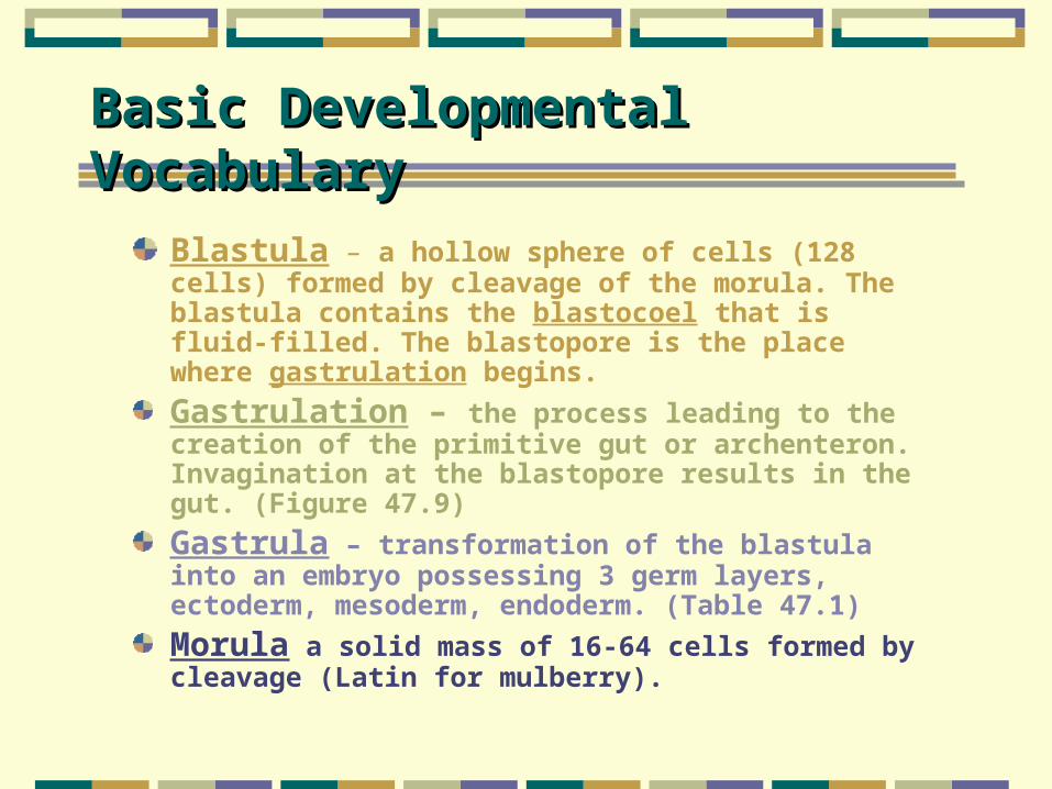
Blastula – a hollow sphere of cells (128 cells) formed by cleavage of the morula. The blastula contains the blastocoel that is fluid-filled. The blastopore is the place where gastrulation begins.
Gastrulation – the process leading to the creation of the primitive gut or archenteron. Invagination at the blastopore results in the gut. (Figure 47.9)
Gastrula – transformation of the blastula into an embryo possessing 3 germ layers, ectoderm, mesoderm, endoderm. (Table 47.1)
Morula a solid mass of 16-64 cells formed by cleavage (Latin for mulberry).

Figure 47.10
Gastrulation in the Frog

DDevelopment of evelopment of EExtraembryonic xtraembryonic
MMembranesembranes
YOLK SAC – develop blood vessels to carry nutrients into the embryo
AMINON – encloses the embryo in a fluid-filled sac, protecting from desiccation & absorbing shock
CHORION – cushions the embryo against mechanical shock
ALLANTOIS – disposal sac for uric acid

Figure 47.14
Development of extra- embryonic membranes in a chick
Mammalian DevelopmentMammalian Development
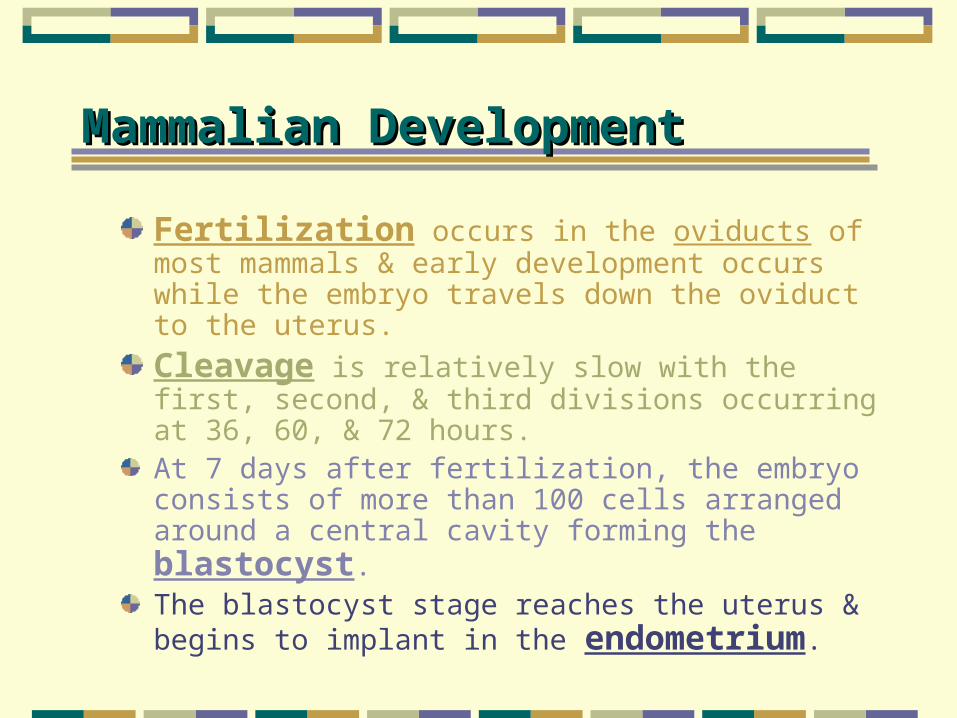
Fertilization occurs in the oviducts of most mammals & early development occurs while the embryo travels down the oviduct to the uterus.
Cleavage is relatively slow with the first, second, & third divisions occurring at 36, 60, & 72 hours.At 7 days after fertilization, the embryo consists of more than 100 cells arranged around a central cavity forming the blastocyst. The blastocyst stage reaches the uterus & begins to implant in the endometrium.
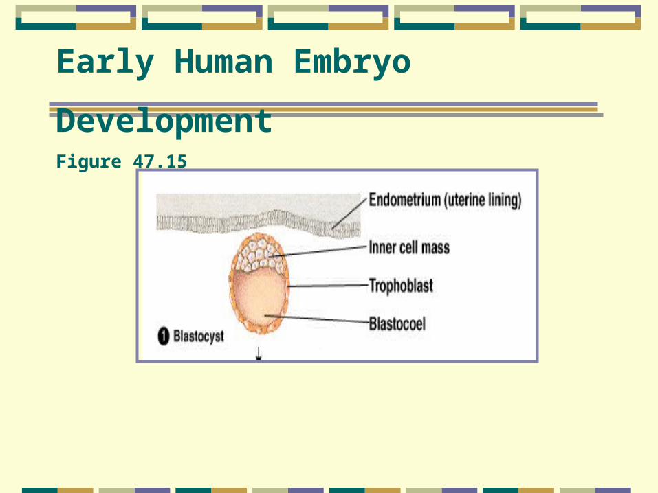
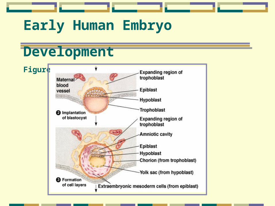
Early Human Embryo
DevelopmentFigure 47.15
DDevelopment of in Human Embryoevelopment of in Human Embryo
Trophoblast forms the chorion & fetal portion of the placenta (along with mesodermal tissue)
Epiblast cell layer forms the three germ layers & the amnion
Hypoblast forms the yolk sac
Early Human Embryo
DevelopmentFigure 47.15

Early Human Embryo
DevelopmentFigure 47.15

Figure 47.14
Convergent extension of a sheet of cells
A signal causes cells to elongate and crawl between each other. This results in the extension of the cell sheet in a direction perpendicular to the convergence.

Figure 47.16
Change in shape during morpho-genesis