Embed Size (px)
Citation preview
fgene-12-726474 October 6, 2021 Time: 16:53 # 1
REVIEWpublished: 12 October 2021
doi: 10.3389/fgene.2021.726474
Edited by:Gaurav K. Varshney,
Oklahoma Medical ResearchFoundation, United States
Reviewed by:Magdalena Sandu,
Spitalul Clinic de Copii Doctor VictorGomoiu, Romania
Olivia J. Veatch,University of Kansas Medical Center,
United States
*Correspondence:Delfien Syx
Specialty section:This article was submitted to
Genetics of Common and RareDiseases,
a section of the journalFrontiers in Genetics
Received: 16 June 2021Accepted: 10 August 2021
Published: 12 October 2021
Citation:Vroman R, Malfait A-M, Miller RE,Malfait F and Syx D (2021) Animal
Models of Ehlers–Danlos Syndromes:Phenotype, Pathogenesis,and Translational Potential.Front. Genet. 12:726474.
doi: 10.3389/fgene.2021.726474
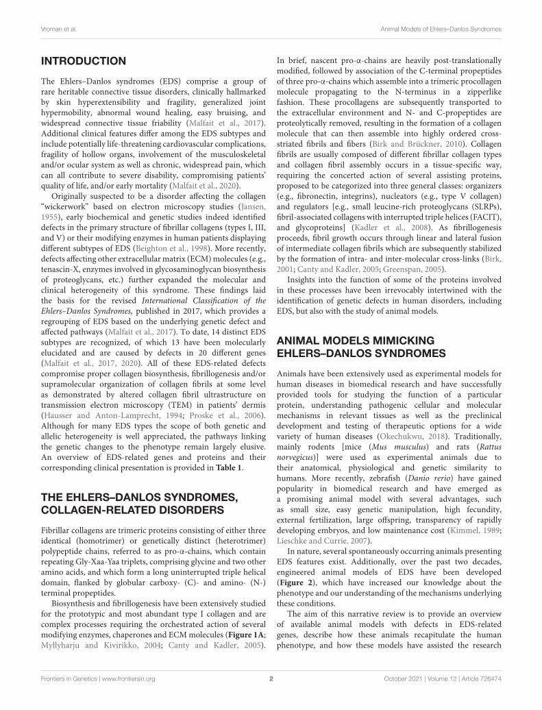
Animal Models of Ehlers–DanlosSyndromes: Phenotype,Pathogenesis, and TranslationalPotentialRobin Vroman1, Anne-Marie Malfait2, Rachel E. Miller2, Fransiska Malfait1 andDelfien Syx1*
1 Center for Medical Genetics, Department of Biomolecular Medicine, Ghent University, Ghent, Belgium, 2 Divisionof Rheumatology, Rush University Medical Center, Chicago, IL, United States
The Ehlers–Danlos syndromes (EDS) are a group of heritable connective tissuesdisorders mainly characterized by skin hyperextensibility, joint hypermobility andgeneralized tissue fragility. Currently, 14 EDS subtypes each with particular phenotypicfeatures are recognized and are caused by genetic defects in 20 different genes.All of these genes are involved in the biosynthesis and/or fibrillogenesis of collagensat some level. Although great progress has been made in elucidating the molecularbasis of different EDS subtypes, the pathogenic mechanisms underlying the observedphenotypes remain poorly understood, and consequentially, adequate treatment andmanagement options for these conditions remain scarce. To date, several animalmodels, mainly mice and zebrafish, have been described with defects in 14 of the20 hitherto known EDS-associated genes. These models have been instrumental indiscerning the functions and roles of the corresponding proteins during development,maturation and repair and in portraying their roles during collagen biosynthesis and/orfibrillogenesis, for some even before their contribution to an EDS phenotype waselucidated. Additionally, extensive phenotypical characterization of these models hasshown that they largely phenocopy their human counterparts, with recapitulation ofseveral clinical hallmarks of the corresponding EDS subtype, including dermatological,cardiovascular, musculoskeletal and ocular features, as well as biomechanical andultrastructural similarities in tissues. In this narrative review, we provide a comprehensiveoverview of animal models manifesting phenotypes that mimic EDS with a focus onengineered mouse and zebrafish models, and their relevance in past and future EDSresearch. Additionally, we briefly discuss domestic animals with naturally occurring EDSphenotypes. Collectively, these animal models have only started to reveal glimpses intothe pathophysiological aspects associated with EDS and will undoubtably continue toplay critical roles in EDS research due to their tremendous potential for pinpointing(common) signaling pathways, unveiling possible therapeutic targets and providingopportunities for preclinical therapeutic interventions.
Keywords: Ehlers–Danlos syndromes, EDS, animal models, mouse, zebrafish
Frontiers in Genetics | www.frontiersin.org 1 October 2021 | Volume 12 | Article 726474
fgene-12-726474 October 6, 2021 Time: 16:53 # 2
Vroman et al. Animal Models of Ehlers–Danlos Syndromes
INTRODUCTION
The Ehlers–Danlos syndromes (EDS) comprise a group ofrare heritable connective tissue disorders, clinically hallmarkedby skin hyperextensibility and fragility, generalized jointhypermobility, abnormal wound healing, easy bruising, andwidespread connective tissue friability (Malfait et al., 2017).Additional clinical features differ among the EDS subtypes andinclude potentially life-threatening cardiovascular complications,fragility of hollow organs, involvement of the musculoskeletaland/or ocular system as well as chronic, widespread pain, whichcan all contribute to severe disability, compromising patients’quality of life, and/or early mortality (Malfait et al., 2020).
Originally suspected to be a disorder affecting the collagen“wickerwork” based on electron microscopy studies (Jansen,1955), early biochemical and genetic studies indeed identifieddefects in the primary structure of fibrillar collagens (types I, III,and V) or their modifying enzymes in human patients displayingdifferent subtypes of EDS (Beighton et al., 1998). More recently,defects affecting other extracellular matrix (ECM) molecules (e.g.,tenascin-X, enzymes involved in glycosaminoglycan biosynthesisof proteoglycans, etc.) further expanded the molecular andclinical heterogeneity of this syndrome. These findings laidthe basis for the revised International Classification of theEhlers–Danlos Syndromes, published in 2017, which provides aregrouping of EDS based on the underlying genetic defect andaffected pathways (Malfait et al., 2017). To date, 14 distinct EDSsubtypes are recognized, of which 13 have been molecularlyelucidated and are caused by defects in 20 different genes(Malfait et al., 2017, 2020). All of these EDS-related defectscompromise proper collagen biosynthesis, fibrillogenesis and/orsupramolecular organization of collagen fibrils at some levelas demonstrated by altered collagen fibril ultrastructure ontransmission electron microscopy (TEM) in patients’ dermis(Hausser and Anton-Lamprecht, 1994; Proske et al., 2006).Although for many EDS types the scope of both genetic andallelic heterogeneity is well appreciated, the pathways linkingthe genetic changes to the phenotype remain largely elusive.An overview of EDS-related genes and proteins and theircorresponding clinical presentation is provided in Table 1.
THE EHLERS–DANLOS SYNDROMES,COLLAGEN-RELATED DISORDERS
Fibrillar collagens are trimeric proteins consisting of either threeidentical (homotrimer) or genetically distinct (heterotrimer)polypeptide chains, referred to as pro-α-chains, which containrepeating Gly-Xaa-Yaa triplets, comprising glycine and two otheramino acids, and which form a long uninterrupted triple helicaldomain, flanked by globular carboxy- (C)- and amino- (N-)terminal propeptides.
Biosynthesis and fibrillogenesis have been extensively studiedfor the prototypic and most abundant type I collagen and arecomplex processes requiring the orchestrated action of severalmodifying enzymes, chaperones and ECM molecules (Figure 1A;Myllyharju and Kivirikko, 2004; Canty and Kadler, 2005).
In brief, nascent pro-α-chains are heavily post-translationallymodified, followed by association of the C-terminal propeptidesof three pro-α-chains which assemble into a trimeric procollagenmolecule propagating to the N-terminus in a zipperlikefashion. These procollagens are subsequently transported tothe extracellular environment and N- and C-propeptides areproteolytically removed, resulting in the formation of a collagenmolecule that can then assemble into highly ordered cross-striated fibrils and fibers (Birk and Brückner, 2010). Collagenfibrils are usually composed of different fibrillar collagen typesand collagen fibril assembly occurs in a tissue-specific way,requiring the concerted action of several assisting proteins,proposed to be categorized into three general classes: organizers(e.g., fibronectin, integrins), nucleators (e.g., type V collagen)and regulators [e.g., small leucine-rich proteoglycans (SLRPs),fibril-associated collagens with interrupted triple helices (FACIT),and glycoproteins] (Kadler et al., 2008). As fibrillogenesisproceeds, fibril growth occurs through linear and lateral fusionof intermediate collagen fibrils which are subsequently stabilizedby the formation of intra- and inter-molecular cross-links (Birk,2001; Canty and Kadler, 2005; Greenspan, 2005).
Insights into the function of some of the proteins involvedin these processes have been irrevocably intertwined with theidentification of genetic defects in human disorders, includingEDS, but also with the study of animal models.
ANIMAL MODELS MIMICKINGEHLERS–DANLOS SYNDROMES
Animals have been extensively used as experimental models forhuman diseases in biomedical research and have successfullyprovided tools for studying the function of a particularprotein, understanding pathogenic cellular and molecularmechanisms in relevant tissues as well as the preclinicaldevelopment and testing of therapeutic options for a widevariety of human diseases (Okechukwu, 2018). Traditionally,mainly rodents [mice (Mus musculus) and rats (Rattusnorvegicus)] were used as experimental animals due totheir anatomical, physiological and genetic similarity tohumans. More recently, zebrafish (Danio rerio) have gainedpopularity in biomedical research and have emerged asa promising animal model with several advantages, suchas small size, easy genetic manipulation, high fecundity,external fertilization, large offspring, transparency of rapidlydeveloping embryos, and low maintenance cost (Kimmel, 1989;Lieschke and Currie, 2007).
In nature, several spontaneously occurring animals presentingEDS features exist. Additionally, over the past two decades,engineered animal models of EDS have been developed(Figure 2), which have increased our knowledge about thephenotype and our understanding of the mechanisms underlyingthese conditions.
The aim of this narrative review is to provide an overviewof available animal models with defects in EDS-relatedgenes, describe how these animals recapitulate the humanphenotype, and how these models have assisted the research
Frontiers in Genetics | www.frontiersin.org 2 October 2021 | Volume 12 | Article 726474
fgene-12-726474O
ctober6,2021Tim
e:16:53#
3
Vroman
etal.A
nimalM
odelsofE
hlers–Danlos
Syndrom
es
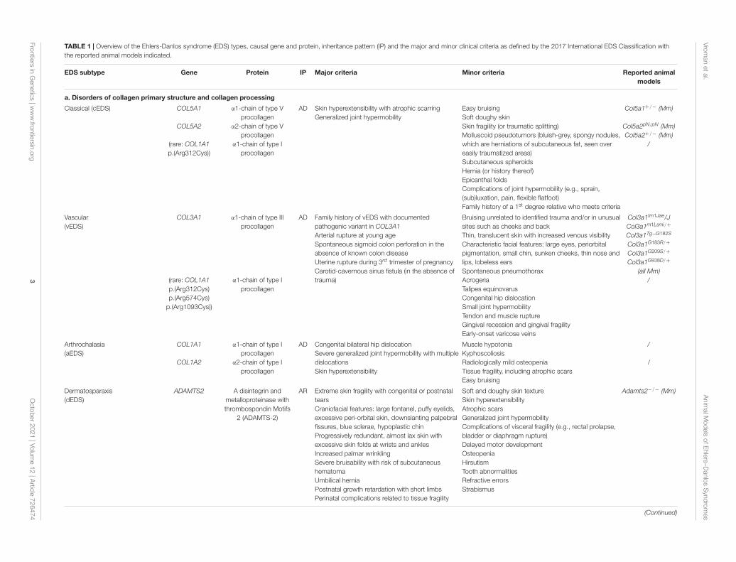
TABLE 1 | Overview of the Ehlers-Danlos syndrome (EDS) types, causal gene and protein, inheritance pattern (IP) and the major and minor clinical criteria as defined by the 2017 International EDS Classification withthe reported animal models indicated.
EDS subtype Gene Protein IP Major criteria Minor criteria Reported animalmodels
a. Disorders of collagen primary structure and collagen processing
Classical (cEDS) COL5A1
COL5A2
(rare: COL1A1p.(Arg312Cys))
α1-chain of type Vprocollagen
α2-chain of type Vprocollagen
α1-chain of type Iprocollagen
AD Skin hyperextensibility with atrophic scarringGeneralized joint hypermobility
Easy bruisingSoft doughy skinSkin fragility (or traumatic splitting)Molluscoid pseudotumors (bluish-grey, spongy nodules,which are herniations of subcutaneous fat, seen overeasily traumatized areas)Subcutaneous spheroidsHernia (or history thereof)Epicanthal foldsComplications of joint hypermobility (e.g., sprain,(sub)luxation, pain, flexible flatfoot)Family history of a 1st degree relative who meets criteria
Col5a1+/− (Mm)
Col5a2pN/pN (Mm)Col5a2+/− (Mm)
/
Vascular(vEDS)
COL3A1
(rare: COL1A1p.(Arg312Cys)p.(Arg574Cys)
p.(Arg1093Cys))
α1-chain of type IIIprocollagen
α1-chain of type Iprocollagen
AD Family history of vEDS with documentedpathogenic variant in COL3A1Arterial rupture at young ageSpontaneous sigmoid colon perforation in theabsence of known colon diseaseUterine rupture during 3rd trimester of pregnancyCarotid-cavernous sinus fistula (in the absence oftrauma)
Bruising unrelated to identified trauma and/or in unusualsites such as cheeks and backThin, translucent skin with increased venous visibilityCharacteristic facial features: large eyes, periorbitalpigmentation, small chin, sunken cheeks, thin nose andlips, lobeless earsSpontaneous pneumothoraxAcrogeriaTalipes equinovarusCongenital hip dislocationSmall joint hypermobilityTendon and muscle ruptureGingival recession and gingival fragilityEarly-onset varicose veins
Col3a1tm1Jae/JCol3a1m1Lsmi/+
Col3a1Tg−G182S
Col3a1G183R/+
Col3a1G209S/+
Col3a1G938D/+
(all Mm)/
Arthrochalasia(aEDS)
COL1A1
COL1A2
α1-chain of type Iprocollagen
α2-chain of type Iprocollagen
AD Congenital bilateral hip dislocationSevere generalized joint hypermobility with multipledislocationsSkin hyperextensibility
Muscle hypotoniaKyphoscoliosisRadiologically mild osteopeniaTissue fragility, including atrophic scarsEasy bruising
/
/
Dermatosparaxis(dEDS)
ADAMTS2 A disintegrin andmetalloproteinase withthrombospondin Motifs
2 (ADAMTS-2)
AR Extreme skin fragility with congenital or postnataltearsCraniofacial features: large fontanel, puffy eyelids,excessive peri-orbital skin, downslanting palpebralfissures, blue sclerae, hypoplastic chinProgressively redundant, almost lax skin withexcessive skin folds at wrists and anklesIncreased palmar wrinklingSevere bruisability with risk of subcutaneoushematomaUmbilical herniaPostnatal growth retardation with short limbsPerinatal complications related to tissue fragility
Soft and doughy skin textureSkin hyperextensibilityAtrophic scarsGeneralized joint hypermobilityComplications of visceral fragility (e.g., rectal prolapse,bladder or diaphragm rupture)Delayed motor developmentOsteopeniaHirsutismTooth abnormalitiesRefractive errorsStrabismus
Adamts2−/− (Mm)
(Continued)
Frontiersin
Genetics
|ww
w.frontiersin.org
3O
ctober2021
|Volume
12|A
rticle726474
fgene-12-726474O
ctober6,2021Tim
e:16:53#
4
Vroman
etal.A
nimalM
odelsofE
hlers–Danlos
Syndrom
es
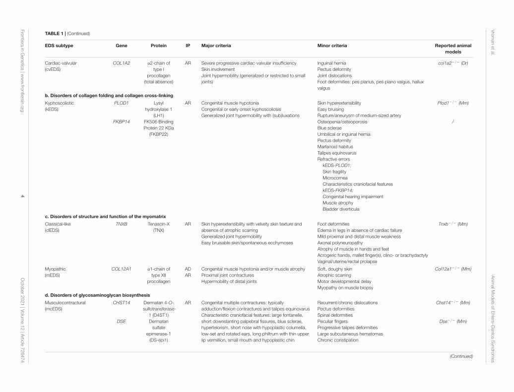
TABLE 1 | (Continued)
EDS subtype Gene Protein IP Major criteria Minor criteria Reported animalmodels
Cardiac-valvular(cvEDS)
COL1A2 α2-chain oftype I
procollagen(total absence)
AR Severe progressive cardiac-valvular insufficiencySkin involvementJoint hypermobility (generalized or restricted to smalljoints)
Inguinal herniaPectus deformityJoint dislocationsFoot deformities: pes planus, pes plano valgus, halluxvalgus
col1a2−/− (Dr)
b. Disorders of collagen folding and collagen cross-linking
Kyphoscoliotic(kEDS)
PLOD1
FKBP14
Lysylhydroxylase 1
(LH1)FK506 BindingProtein 22 KDa
(FKBP22)
AR Congenital muscle hypotoniaCongenital or early onset kyphoscoliosisGeneralized joint hypermobility with (sub)luxations
Skin hyperextensibilityEasy bruisingRupture/aneurysm of medium-sized arteryOsteopenia/osteoporosisBlue scleraeUmbilical or inguinal herniaPectus deformityMarfanoid habitusTalipes equinovarusRefractive errors
kEDS-PLOD1:Skin fragilityMicrocorneaCharacteristics craniofacial featureskEDS-FKBP14:Congenital hearing impairmentMuscle atrophyBladder diverticula
Plod1−/− (Mm)
/
c. Disorders of structure and function of the myomatrix
Classical-like(clEDS)
TNXB Tenascin-X(TNX)
AR Skin hyperextensibility with velvety skin texture andabsence of atrophic scarringGeneralized joint hypermobilityEasy bruisable skin/spontaneous ecchymoses
Foot deformitiesEdema in legs in absence of cardiac failureMild proximal and distal muscle weaknessAxonal polyneuropathyAtrophy of muscle in hands and feetAcrogeric hands, mallet finger(s), clino- or brachydactylyVaginal/uterine/rectal prolapse
Tnxb−/− (Mm)
Myopathic(mEDS)
COL12A1 α1-chain oftype XII
procollagen
ADAR
Congenital muscle hypotonia and/or muscle atrophyProximal joint contracturesHypermobility of distal joints
Soft, doughy skinAtrophic scarringMotor developmental delayMyopathy on muscle biopsy
Col12a1−/− (Mm)
d. Disorders of glycosaminoglycan biosynthesis
Musculocontractural(mcEDS)
CHST14
DSE
Dermatan 4-O-sulfotransferase-
1 (D4ST1)Dermatan
sulfateepimerase-1
(DS-epi1)
AR Congenital multiple contractures: typicallyadduction/flexion contractures and talipes equinovarusCharacteristic craniofacial features: large fontanelle,short downslanting palpebral fissures, blue sclerae,hypertelorism, short nose with hypoplastic columella,low-set and rotated ears, long philtrum with thin upperlip vermillion, small mouth and hypoplastic chin
Recurrent/chronic dislocationsPectus deformitiesSpinal deformitiesPeculiar fingersProgressive talipes deformitiesLarge subcutaneous hematomasChronic constipation
Chst14−/− (Mm)
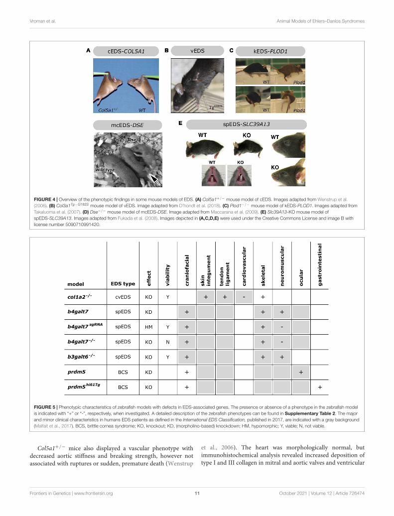
Dse−/− (Mm)
(Continued)
Frontiersin
Genetics
|ww
w.frontiersin.org
4O
ctober2021
|Volume
12|A
rticle726474
fgene-12-726474O
ctober6,2021Tim
e:16:53#
5
Vroman
etal.A
nimalM
odelsofE
hlers–Danlos
Syndrom
es
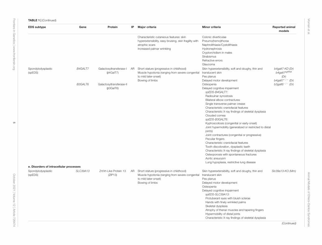
TABLE 1 | (Continued)
EDS subtype Gene Protein IP Major criteria Minor criteria Reported animalmodels
Characteristic cutaneous features: skinhyperextensibility, easy bruising, skin fragility withatrophic scarsIncreased palmar wrinkling
Colonic diverticulaePneumo(hemo)thoraxNephrolithiasis/CystolithiasisHydronephrosisCryptorchidism in malesStrabismusRefractive errorsGlaucoma
Spondylodysplastic(spEDS)
B4GALT7
B3GALT6
Galactosyltransferase-I(β4GalT7)
Galactosyltransferase-II(β3GalT6)
AR Short stature (progressive in childhood)Muscle hypotonia (ranging from severe congenitalto mild later-onset)Bowing of limbs
Skin hyperextensibility, soft and doughy, thin andtranslucent skinPes planusDelayed motor developmentOsteopeniaDelayed cognitive impairment
spEDS-B4GALT7:Radioulnar synostosisBilateral elbow contracturesSingle transverse palmar creaseCharacteristic craniofacial featuresCharacteristic X-ray findings of skeletal dysplasiaClouded corneaspEDS-B3GALT6:Kyphoscoliosis (congenital or early-onset)Joint hypermobility (generalized or restricted to distaljoints)Joint contractures (congenital or progressive)Peculiar fingersCharacteristic craniofacial featuresTooth discoloration, dysplastic teethCharacteristic X-ray findings of skeletal dysplasiaOsteoporosis with spontaneous fracturesAortic aneurysmLung hypoplasia, restrictive lung disease
b4galt7-KD (Dr)b4galt7sgRNA
(Dr)b4galt7−/− (Dr)b3galt6−/− (Dr)
e. Disorders of intracellular processes
Spondylodysplastic(spEDS)
SLC39A13 Zrt/Irt-Like Protein 13(ZIP13)
AR Short stature (progressive in childhood)Muscle hypotonia (ranging from severe congenitalto mild later-onset)Bowing of limbs
Skin hyperextensibility, soft and doughy, thin andtranslucent skinPes planusDelayed motor developmentOsteopeniaDelayed cognitive impairment
spEDS-SLC39A13:Protuberant eyes with bluish scleraeHands with finely wrinkled palmsSkeletal dysplasiaAtrophy of thenar muscles and tapering fingersHypermobility of distal jointsCharacteristic X-ray findings of skeletal dysplasia
Slc39a13-KO (Mm)
(Continued)
Frontiersin
Genetics
|ww
w.frontiersin.org
5O
ctober2021
|Volume
12|A
rticle726474
fgene-12-726474O
ctober6,2021Tim
e:16:53#
6
Vroman
etal.A
nimalM
odelsofE
hlers–Danlos
Syndrom
es
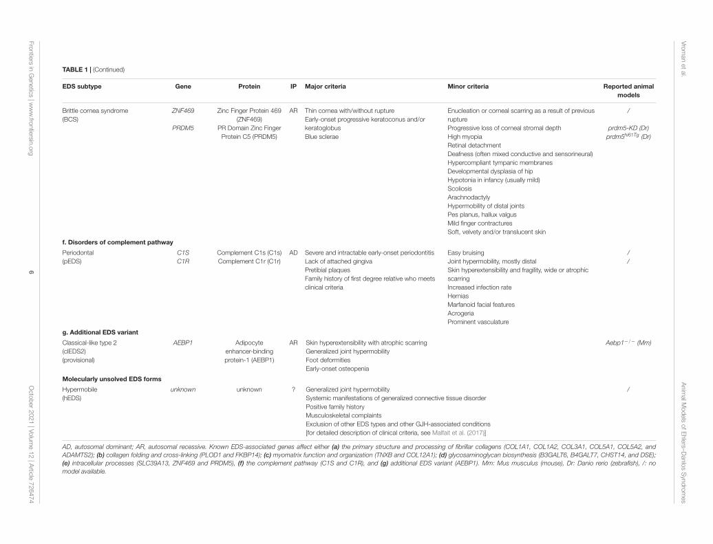
TABLE 1 | (Continued)
EDS subtype Gene Protein IP Major criteria Minor criteria Reported animalmodels
Brittle cornea syndrome(BCS)
ZNF469
PRDM5
Zinc Finger Protein 469(ZNF469)
PR Domain Zinc FingerProtein C5 (PRDM5)
AR Thin cornea with/without ruptureEarly-onset progressive keratoconus and/orkeratoglobusBlue sclerae
Enucleation or corneal scarring as a result of previousruptureProgressive loss of corneal stromal depthHigh myopiaRetinal detachmentDeafness (often mixed conductive and sensorineural)Hypercompliant tympanic membranesDevelopmental dysplasia of hipHypotonia in infancy (usually mild)ScoliosisArachnodactylyHypermobility of distal jointsPes planus, hallux valgusMild finger contracturesSoft, velvety and/or translucent skin
/
prdm5-KD (Dr)prdm5hi61Tg (Dr)
f. Disorders of complement pathway
Periodontal(pEDS)
C1SC1R
Complement C1s (C1s)Complement C1r (C1r)
AD Severe and intractable early-onset periodontitisLack of attached gingivaPretibial plaquesFamily history of first degree relative who meetsclinical criteria
Easy bruisingJoint hypermobility, mostly distalSkin hyperextensibility and fragility, wide or atrophicscarringIncreased infection rateHerniasMarfanoid facial featuresAcrogeriaProminent vasculature
//
g. Additional EDS variant
Classical-like type 2(clEDS2)(provisional)
AEBP1 Adipocyteenhancer-bindingprotein-1 (AEBP1)
AR Skin hyperextensibility with atrophic scarringGeneralized joint hypermobilityFoot deformitiesEarly-onset osteopenia
Aebp1−/− (Mm)
Molecularly unsolved EDS forms
Hypermobile(hEDS)
unknown unknown ? Generalized joint hypermobilitySystemic manifestations of generalized connective tissue disorderPositive family historyMusculoskeletal complaintsExclusion of other EDS types and other GJH-associated conditions[for detailed description of clinical criteria, see Malfait et al. (2017)]
/
AD, autosomal dominant; AR, autosomal recessive. Known EDS-associated genes affect either (a) the primary structure and processing of fibrillar collagens (COL1A1, COL1A2, COL3A1, COL5A1, COL5A2, andADAMTS2); (b) collagen folding and cross-linking (PLOD1 and FKBP14); (c) myomatrix function and organization (TNXB and COL12A1); (d) glycosaminoglycan biosynthesis (B3GALT6, B4GALT7, CHST14, and DSE);(e) intracellular processes (SLC39A13, ZNF469 and PRDM5), (f) the complement pathway (C1S and C1R), and (g) additional EDS variant (AEBP1). Mm: Mus musculus (mouse), Dr: Danio rerio (zebrafish), /: nomodel available.
Frontiersin
Genetics
|ww
w.frontiersin.org
6O
ctober2021
|Volume
12|A
rticle726474

fgene-12-726474 October 6, 2021 Time: 16:53 # 7
Vroman et al. Animal Models of Ehlers–Danlos Syndromes
FIGURE 1 | Schematic overview of collagen and glycosaminoglycan (GAG) biosynthesis and collagen fibrillogenesis. Molecules defective in Ehlers-Danlossyndromes (EDS) are highlighted in bold. (A) Fibrillar collagen biosynthesis starts with transcription and translation of pro-α-chains (step 1). Nascent pro-α-chains areheavily post-translationally modified by several proline and lysine hydroxylases and galactosyltransferases (step 2). The association of the C-terminal propeptides ofthree pro-α-chains, initiates triple helix formation which propagates to the N-terminus in a zipperlike fashion and is assisted by several molecular chaperones (step 3).The trimeric procollagen molecules aggregate laterally, are transported in secretory vesicles and are eventually directed to the extracellular environment (step 4).Removal of the N- and C-propeptides, by ADAMTS-2 and BMP-1/mTLD, respectively, results in the formation of a collagen molecule (step 5) that can then assembleinto highly ordered striated fibrils. The tissue-specific assembly of collagen fibrils requires the concerted action of several assisting proteins, categorized asorganizers, nucleators and regulators (step 6). At the plasma membrane, fibronectin and integrins serve as organizers of fibril assembly. Some collagens, such astype V collagen, function as nucleators, which initiate immature fibril assembly at the cell surface. Type V collagen co-assembles with type I collagen into heterotypicfibrils with the entire triple helical domain of type V collagen embedded within the fibril, whereas its partially processed N-propeptide domain protrudes to the fibrilsurface and controls fibrillogenesis by sterically hindering the addition of collagen monomers. The intermediate fibrils are then deposited into the extracellular matrix(ECM). Stabilization of these fibrils is provided by interactions with regulators such as the small leucine-rich proteoglycan (SLRP) decorin, tenascin-X and type XIIcollagen, which influence the rate of assembly, size and structure of the collagen fibrils. As fibrillogenesis proceeds, fibril growth occurs through linear and lateralfusion of intermediate collagen fibrils which are subsequently stabilized by the formation of covalent intra- and inter-molecular cross-links. (B) GAG biosynthesisstarts with the synthesis of a proteoglycan core protein which is subsequently modified by several Golgi-resident enzymes. Initially, a common linker regioncontaining four monosaccharides is formed. Biosynthesis of this tetrasaccharide linker region starts with the stepwise addition of a xylose (Xyl) residue to a specificserine residue of the core protein, catalyzed by xylosyltransferase-I and II (XylT-I/-II). Subsequently, two galactose (Gal) residues are added by galactosyltransferase-I(GalT-I or β4GalT7) and galactosyltransferase-II (GalT-II or β3GalT6). Finally, the addition of a glucuronic acid (GlcA), catalyzed by glucuronosyltransferase-I (GlcAT-I)
(Continued)
Frontiers in Genetics | www.frontiersin.org 7 October 2021 | Volume 12 | Article 726474
fgene-12-726474 October 6, 2021 Time: 16:53 # 8
Vroman et al. Animal Models of Ehlers–Danlos Syndromes
FIGURE 1 | (Continued)completes the formation of the linker region. The alternating addition of either N-acetyl-glucosamine (GlcNAc) or N-galactosyl-glucosamine (GalNAc) and GlcAdefines the composition of the GAG-chain and subdivides proteoglycans into heparan sulfate (HS) proteoglycans and chondroitin sulfate (CS)/dermatan sulfate (DS)proteoglycans. The GAG-chains are then further modified by epimerization and sulfation. DS synthesis necessitates the epimerization of GlcA towards iduronic acid(IdoA), which is catalyzed by DS epimerases–1 and -2 (DS-epi1 and DS-epi2). Subsequently, dermatan 4-O-sulfotransferase-1 (D4ST1) is able to catalyze4-O-sulfation of GalNAc, thereby preventing back-epimerization of the adjacent IdoA.
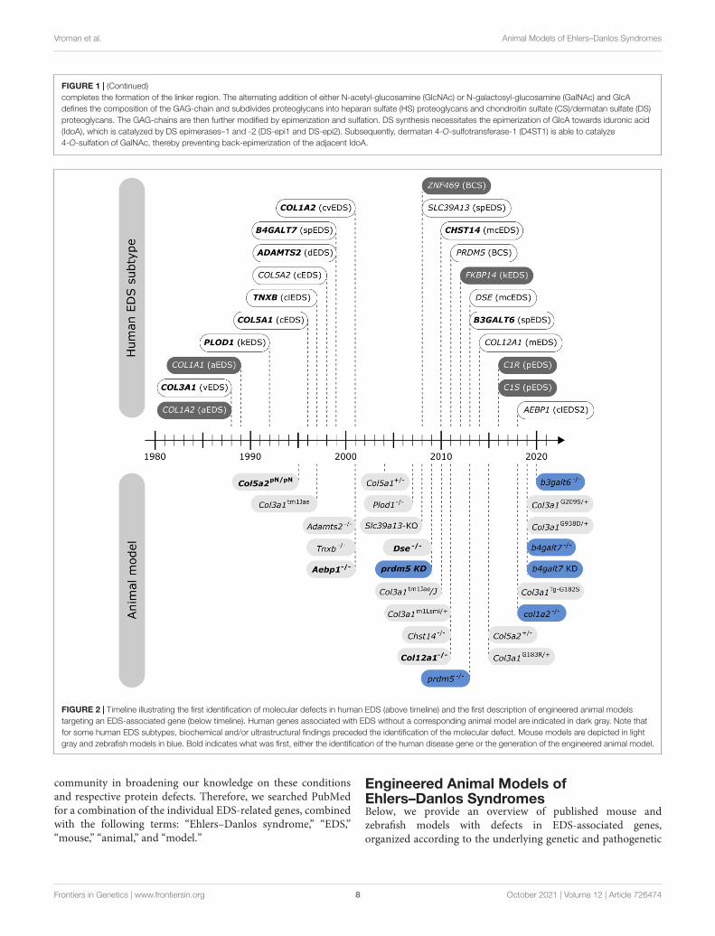
FIGURE 2 | Timeline illustrating the first identification of molecular defects in human EDS (above timeline) and the first description of engineered animal modelstargeting an EDS-associated gene (below timeline). Human genes associated with EDS without a corresponding animal model are indicated in dark gray. Note thatfor some human EDS subtypes, biochemical and/or ultrastructural findings preceded the identification of the molecular defect. Mouse models are depicted in lightgray and zebrafish models in blue. Bold indicates what was first, either the identification of the human disease gene or the generation of the engineered animal model.
community in broadening our knowledge on these conditionsand respective protein defects. Therefore, we searched PubMedfor a combination of the individual EDS-related genes, combinedwith the following terms: “Ehlers–Danlos syndrome,” “EDS,”“mouse,” “animal,” and “model.”
Engineered Animal Models ofEhlers–Danlos SyndromesBelow, we provide an overview of published mouse andzebrafish models with defects in EDS-associated genes,organized according to the underlying genetic and pathogenetic
Frontiers in Genetics | www.frontiersin.org 8 October 2021 | Volume 12 | Article 726474
fgene-12-726474 October 6, 2021 Time: 16:53 # 9
Vroman et al. Animal Models of Ehlers–Danlos Syndromes
mechanisms as defined in the 2017 EDS classification (Malfaitet al., 2017). A detailed summary of the available models ispresented in Figures 3, 4 and Supplementary Table 1 formouse models and Figures 5, 6 and Supplementary Table 2for zebrafish models. Although other (often conditional) mousemodels affecting an EDS-related gene have been reported, anextensive description of these models is beyond the scope of thisreview (for a brief summary, see Supplementary Information).
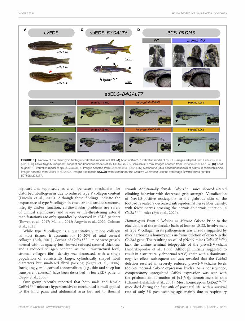
Models Affecting the Primary Structure andProcessing of Fibrillar CollagensModels of Classical Ehlers–Danlos Syndrome—Defects inType V CollagenClassical EDS (cEDS) is the most common, molecularlydefined EDS subtype. It is characterized by generalizedjoint hypermobility, skin hyperextensibility and skin fragility,manifesting as easy splitting of the skin upon minor trauma,which, in combination with delayed wound healing, resultsin the formation of widened atrophic scarring (Bowen et al.,2017). It is inherited in an autosomal dominant fashion andabout 90% of cEDS patients harbor a heterozygous mutationin either the COL5A1 or COL5A2 gene, encoding the pro-α1- and pro-α2-chain of type V collagen, respectively. Themajority of these mutations result in a non-functional COL5A1allele and give rise to COL5A1 haploinsufficiency with reducedtype V collagen protein levels, while mutations in COL5A1or COL5A2 that lead to a structural defect (e.g., glycinesubstitution or in-frame exon skip) have a dominant negativeeffect (Symoens et al., 2012; Colman et al., 2021). The mostcommon isoform of type V collagen is the [α1(V)]2α2(V)heterotrimer found in skin, tendon, ligaments, cornea, and bone,where it forms heterotypic fibrils with type I collagen. Otherisoforms, including the embryonic [α1(V)]3 homotrimer andthe α1(V)α2(V)α3(V) heterotrimer mainly found in placenta,are less abundant. Although type V collagen is quantitatively oflow abundance (about 2–5% of total collagen in bone, tendon,and dermis), it plays a key role during collagen fibrillogenesisby initiating fibril assembly and regulating heterotypic type I/Vcollagen fibril diameter through its partially retained α1(V)-N-propeptide that protrudes beyond the fibril surface (Birk, 2001;Wenstrup et al., 2004).
Several murine models have been generated to study thefunctional and regulatory roles of type V collagen during collagenfibrillogenesis, and as such these models also provide insights intocEDS pathogenesis.
Col5a1 Haploinsufficient Mice. Complete absence of the pro-α1(V)-chains in homozygous Col5a1 knockout (Col5a1−/−)mice was shown to result in lethality around embryonic day10 due to cardiovascular failure. Ultrastructural evaluation ofCol5a1−/− embryos using TEM revealed the virtual absenceof predermal mesenchymal fibril formation despite the presenceof normal amounts of type I collagen. These findings revealed acritical regulatory role of type V collagen in the initiation of earlyfibril formation and nucleation of type I collagen fibril assemblyduring early murine embryogenesis (Wenstrup et al., 2004).
Complete loss of the pro-α1(V)-chains has not been reported inhumans, most probably because it is lethal.
Heterozygous Col5a1 knockout (Col5a1+/−) mice are viableand mimic the most common molecular defect associated withcEDS, i.e., COL5A1 haploinsufficiency, leading to approximately50% reduction in type V collagen content (Wenstrup et al.,2006). Similar to cEDS patients, the skin of Col5a1+/− micewas hyperextensible with decreased tensile strength (Figure 4A).Ultrastructural analysis showed decreased fibril density in thesubscapular dermis, correlating with the decreased collagencontent, and consistent with dysfunctional regulation of collagenfibril nucleation when type V collagen is limited. Additionally,two different fibril subpopulations were observed: relativelynormal symmetrical fibrils with slightly larger diameters, andvery large, structurally aberrant fibrils with irregular contours, socalled “cauliflower” fibrils, representing an unregulated assemblyof type I collagen which virtually lacks type V collagen anddisrupted lateral fibril growth (Wenstrup et al., 2004, 2006).These ultrastructural abnormalities are highly reminiscent ofthe alterations in dermis of cEDS patients (Vogel et al.,1979). Most Col5a1+/− mice older than 6 months developedspontaneous, non-healing wounds indicative of skin fragility(DeNigris et al., 2015). Wound repair studies demonstratedslower in vivo closure of a subscapular skin wound in Col5a1+/−
mice. Subsequent in vitro studies on dermal fibroblasts fromCol5a1+/− mice showed that this impaired wound healing islikely attributed to an interplay of decreases in proliferationrate, attachment properties to components of the wound ECM(e.g., types I and III collagen and fibronectin) and migrationcapacity of Col5a1+/− fibroblasts (DeNigris et al., 2015).The latter finding is consistent with in vitro scratch assaysshowing delayed migration of dermal fibroblasts from cEDSpatients with COL5A1 haploinsufficiency (Viglio et al., 2008;DeNigris et al., 2015).
Although no overt joint hypermobility, another major featureof human cEDS, was described in Col5a1+/− mice (Wenstrupet al., 2006), biomechanical analysis of the flexor digitorumlongus (FDL) and patellar tendons showed reduced tensilestrength, suggesting increased elasticity. Ultrastructural analysesof FDL tendon showed unexpectedly mild abnormalities inCol5a1+/− mice with less regular cross-sectional profiles andsmaller diameter of collagen fibrils (Wenstrup et al., 2011).TEM of patellar tendon showed two distinct subpopulationsof smaller and larger diameter fibrils with mostly normalcircular fibril cross-section profiles but with larger diameterfibrils in Col5a1+/− mice (Johnston et al., 2017). Functionaland structural tendon pathology has also been described inthe patellar tendon of cEDS patients with low tendon stiffness,and abnormal ultrastructural findings with various amounts oflarge and irregular collagen fibrils combined with apparentlynormal fibrils (Nielsen et al., 2014). Tendons from Col5a1+/−
mice showed diminished mechanical recovery potential followingbilateral patellar tendon injury with smaller fibril sizes 6 weekspost-injury, indicating further failure in the healing responsein Col5a1+/− tendon. These findings point to a role fortype V collagen in healing and recovery following injury(Johnston et al., 2017).
Frontiers in Genetics | www.frontiersin.org 9 October 2021 | Volume 12 | Article 726474
fgene-12-726474 October 6, 2021 Time: 16:53 # 10
Vroman et al. Animal Models of Ehlers–Danlos Syndromes
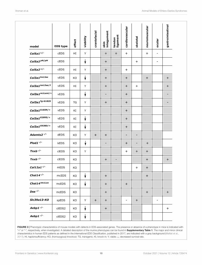
FIGURE 3 | Phenotypic characteristics of mouse models with defects in EDS-associated genes. The presence or absence of a phenotype in mice is indicated with“+” or “-”, respectively, when investigated. A detailed description of the murine phenotypes can be found in Supplementary Table 1. The major and minor clinicalcharacteristics in human EDS patients as defined in the International EDS Classification, published in 2017, are indicated with a gray background (Malfait et al.,2017). HI, haploinsufficiency; KO, (homozygous) knockout; TG, transgenic; KI, knock-in; Y, viable; ↓, decreased survival rate.
Frontiers in Genetics | www.frontiersin.org 10 October 2021 | Volume 12 | Article 726474
fgene-12-726474 October 6, 2021 Time: 16:53 # 11
Vroman et al. Animal Models of Ehlers–Danlos Syndromes
FIGURE 4 | Overview of the phenotypic findings in some mouse models of EDS. (A) Col5a1+/− mouse model of cEDS. Images adapted from Wenstrup et al.(2006). (B) Col3a1Tg−G182S mouse model of vEDS. Image adapted from D’hondt et al. (2018). (C) Plod1−/− mouse model of kEDS-PLOD1. Images adapted fromTakaluoma et al. (2007). (D) Dse−/− mouse model of mcEDS-DSE. Image adapted from Maccarana et al. (2009). (E) Slc39A13-KO mouse model ofspEDS-SLC39A13. Images adapted from Fukada et al. (2008). Images depicted in (A,C,D,E) were used under the Creative Commons License and image B withlicense number 5090710991420.
FIGURE 5 | Phenotypic characteristics of zebrafish models with defects in EDS-associated genes. The presence or absence of a phenotype in the zebrafish modelis indicated with “+” or “-”, respectively, when investigated. A detailed description of the zebrafish phenotypes can be found in Supplementary Table 2. The majorand minor clinical characteristics in humans EDS patients as defined in the International EDS Classification, published in 2017, are indicated with a gray background(Malfait et al., 2017). BCS, brittle cornea syndrome; KO, knockout; KD, (morpholino-based) knockdown; HM, hypomorphic; Y, viable; N, not viable.
Col5a1+/− mice also displayed a vascular phenotype withdecreased aortic stiffness and breaking strength, however notassociated with ruptures or sudden, premature death (Wenstrup
et al., 2006). The heart was morphologically normal, butimmunohistochemical analysis revealed increased deposition oftype I and III collagen in mitral and aortic valves and ventricular
Frontiers in Genetics | www.frontiersin.org 11 October 2021 | Volume 12 | Article 726474
fgene-12-726474 October 6, 2021 Time: 16:53 # 12
Vroman et al. Animal Models of Ehlers–Danlos Syndromes
FIGURE 6 | Overview of the phenotypic findings in zebrafish models of EDS. (A) Adult col1a2−/− zebrafish model of cvEDS. Images adapted from Gistelinck et al.(2018). (B) Larval b4galt7 morphant, crispant and knockout models of spEDS-B4GALT7. Scale bars: 1 mm. Images adapted from Delbaere et al. (2019a). (C) Adultb3galt6−/− zebrafish model of spEDS-B3GALT6. Images adapted from Delbaere et al. (2020). (D) Morpholino (MO)-based knockdown of prdm5 in zebrafish larvae.Images adapted from Meani et al. (2009). Images depicted in (A,C,D) were used under the Creative Commons License and image B with license number5078981221357.
myocardium, supposedly as a compensatory mechanism fordisturbed fibrillogenesis due to reduced type V collagen content(Lincoln et al., 2006). Although these findings indicate theimportance of type V collagen in vascular and cardiac structure,integrity and/or function, cardiovalvular problems are rarelyof clinical significance and severe or life-threatening arterialmanifestations are only sporadically observed in cEDS patients(Bowen et al., 2017; Malfait, 2018; Angwin et al., 2020; Colmanet al., 2021).
While type V collagen is a quantitatively minor collagenin most tissues, it accounts for 10–20% of total cornealcollagen (Birk, 2001). Cornea of Col5a1+/− mice were grosslynormal without opacity but showed reduced stromal thicknessand a reduced collagen content. At the ultrastructural level,stromal collagen fibril density was decreased, with a singlepopulation of consistently larger, cylindrically shaped fibrildiameters but unaltered fibril packing (Segev et al., 2006).Intriguingly, mild corneal abnormalities, (e.g., thin and steep buttransparent corneas) have been described in few cEDS patients(Segev et al., 2006).
Our group recently reported that both male and femaleCol5a1+/− mice are hypersensitive to mechanical stimuli appliedto the hind paws and abdominal area but not to thermal
stimuli. Additionally, female Col5a1+/− mice showed alteredclimbing behavior with decreased grip strength. Visualizationof NaV1.8-positive nociceptors in the glabrous skin of thefootpad revealed a decreased intraepidermal nerve fiber density,with fewer nerves crossing the dermis-epidermis junction inCol5a1+/− mice (Syx et al., 2020).
Homozygous Exon 6 Deletion in Murine Col5a2. Prior to theelucidation of the molecular basis of human cEDS, involvementof type V collagen in its pathogenesis was already suggested bymice harboring a homozygous in-frame deletion of exon 6 in theCol5a2 gene. The resulting so-called pN/pN mice (Col5a2pN/pN)lack the amino-terminal telopeptide of the pro-α2(V)-chain(Andrikopoulos et al., 1995). Although initially suggested toresult in a structurally abnormal α2(V)-chain with a dominant-negative effect, subsequent analyses revealed that the Col5a2deletion resulted in severely reduced pro-α2(V)-chains levels(despite normal Col5a2 expression levels). As a consequence,compensatory upregulated Col5a1 expression was seen withthe predominant formation of [α1(V)]3 homotrimers in skin(Chanut-Delalande et al., 2004). Most homozygous Col5a2pN/pN
mice died during the first 48h of postnatal life, with a survivalrate of only 5% past weaning age, mainly due to respiratory
Frontiers in Genetics | www.frontiersin.org 12 October 2021 | Volume 12 | Article 726474
fgene-12-726474 October 6, 2021 Time: 16:53 # 13
Vroman et al. Animal Models of Ehlers–Danlos Syndromes
problems triggered by varying degrees of lordosis and kyphosis.Surviving Col5a2pN/pN mice weighed approximately half of thewild-type littermates at 3 weeks of age, attributed to reducedmobility due to the progressive spinal deformities prohibitingnourishment. They displayed severe skin fragility (with multiplescars and bleeding lacerations) and increased stretchability ofthe skin, reminiscent of EDS. Histological analyses of the skinof Col5a2pN/pN mice revealed reduced dermal and increasedhypodermal thickness, with unusually localized hair folliclesin the latter, while ultrastructural analyses demonstrated thatdermal collagen fibrils were more disorganized, less tightlypacked and heterogeneous in size with areas devoid of bandedfibrils (Andrikopoulos et al., 1995; Chanut-Delalande et al., 2004).Dermal fibroblasts derived from Col5a2pN/pN mice produced asparse network of disorganized, thin fibrils in vitro and alsoshowed increased apoptosis (Chanut-Delalande et al., 2004).These observations underscore the importance of type V collagenand the [α1(V)]2α2(V) heterotrimer for proper cell-matrixinteractions during skin development. Additionally, pronouncedultrastructural disorganization of collagen fibrils with an overallincreased diameter was also seen in the corneal stroma, whichseemed thinner in Col5a2pN/pN mice (Andrikopoulos et al., 1995;Chanut-Delalande et al., 2004).
Col5a2 Haploinsufficient Mice. In 2015, a constitutive Col5a2knockout (Col5a2−/−) mouse model was created with completeabsence of pro-α2(V)-chains (Park et al., 2015). In contrastto Col5a2pN/pN mice, homozygous Col5a2−/− mice wereembryonic lethal around 12 days post conception (dpc) dueto cardiovascular insufficiency. In contrast to the absence offibrils in Col5a1−/− embryos, mesenchymal collagen fibrilswith abnormally large diameters and abnormal configurationswere observed in Col5a2−/− embryos, suggesting that [α1(V)]3homotrimers can at least partially compensate for the loss of[α1(V)]2α2(V) heterotrimers in the initiation of early embryoniccollagen fibril formation.
Whereas heterozygous Col5a2pN/+ mice did not display anovert phenotype, heterozygous Col5a2+/− mice representingCol5a2 haploinsufficiency did survive and showed increasedskin extensibility with decreased tensile strength, althoughless pronounced compared to Col5a1+/− mice. Ultrastructuralanalyses showed only mild collagen fibril abnormalities in thesubscapular dermis of Col5a2+/− mice with irregular collagenfibril contours mainly seen on longitudinal sections, but withoutthe pathognomonic “cauliflower”-shaped dermal collagen fibrilsfound in cross-sections of cEDS patients and Col5a1+/− mice.Reduced tensile strength and stiffness pointing to increasedfragility and elasticity, respectively, were also noted in theaorta of Col5a2+/− mice (Park et al., 2015). Additionally,experimentally increasing blood pressure by angiotensin IIadministration showed an increased incidence, diameter andseverity of abdominal aortic aneurysms with more than half ofCol5a2+/− mice dying of aortic arch dissection and rupture(Park et al., 2017). To date, no cEDS patients with COL5A2haploinsufficiency have been reported.
Taken together, Col5a1+/− mice mimic human cEDS the bestsince they represent the major molecular defect, i.e., COL5A1
haploinsufficiency, and faithfully recapitulate many of the clinical(including pain), biomechanical, morphological and biochemicalfeatures seen in cEDS patients (Wenstrup et al., 2006). Todate, no animal models harboring structural defects such asglycine substitutions or in-frame exon skips in the triple helicaldomain of the pro-α1- or pro-α2-chains of type V collagenhave been reported.
Models of Vascular Ehlers–Danlos Syndrome—Defects inType III CollagenVascular EDS (vEDS) is an autosomal dominant conditionmainly characterized by life-threatening complications, includingarterial aneurysms, dissections and ruptures, but also bowelperforations/ruptures and ruptures of the gravid uterus, resultingin a reduced life span with a median survival age of 51 years(Pepin et al., 2014; Byers et al., 2017). vEDS patients oftenhave thin, translucent skin, excessive bruising and present witha characteristic facial appearance. To date, the only evidence-based treatment strategy for vEDS that decreases the incidence ofarterial rupture is celiprolol, a long-acting β1-receptor antagonistwith partial β2-receptor agonist properties used for treatment ofhypertension and reducing arterial wall stress, without changinghemodynamic parameters (Ong et al., 2010). Nevertheless,caution is warranted due to some study limitations (Backer andBacker, 2019). The vast majority of vEDS patients harbors aheterozygous defect in the COL3A1 gene, encoding the pro-α1-chain of type III collagen. Most genetic defects are missenseor splice site mutations, introducing a glycine substitution orcreating an in-frame exon skip within the triple helical domain,respectively. Mutations leading to COL3A1 haploinsufficiencywere reported in a small proportion (less than 5%) of vEDSpatients and are associated with a delayed onset of complicationsby almost two decades (Pepin et al., 2014). Type III collagen is ahomotrimer consisting of three α1(III)-chains, found abundantlyin the wall of arteries, gastrointestinal tract, uterus and skin,where it often co-localizes with type I collagen and is thought toregulate fibril diameter (Epstein and Munderloh, 1975).
Several murine models have been generated to study the roleof (defective) type III collagen and vEDS pathogenesis.
Homozygous Col3a1 Knockout Mice. The first proposed vEDSmodel was a homozygous Col3a1 knockout (Col3a1tm1Jae) mouse,which had a 5% survival rate at weaning age with most deathsoccurring within 48 h after birth of unknown etiology. Survivinghomozygous Col3a1tm1Jae mice appeared normal but were 15%smaller than wild-type mice and had a reduced lifespan (6months), mainly caused by rupture of large blood vessels andoccasionally by intestinal rupture. About 60% of survivinghomozygous knockout mice displayed spontaneous skin lesions,including large open wounds. TEM analyses of aorta, skin,intestine, liver, heart, and lung showed a reduced number ofcollagen fibrils in mutant mice with variable fibril diameters ofalmost twice the size of those in wild-type mice, hinting at acritical role for type III collagen during collagen fibrillogenesis(Liu et al., 1997). The high mortality of this model precludespreclinical studies. Additionally, complete loss of type III collagenin human patients is rare and not associated with a typical vEDSphenotype. Biallelic COL3A1 mutations result in connective
Frontiers in Genetics | www.frontiersin.org 13 October 2021 | Volume 12 | Article 726474
fgene-12-726474 October 6, 2021 Time: 16:53 # 14
Vroman et al. Animal Models of Ehlers–Danlos Syndromes
tissue abnormalities with some phenotypic resemblance to vEDScombined with structural brain anomalies. (Plancke et al.,2009; Jørgensen et al., 2014; Horn et al., 2017; Vandervoreet al., 2017). In accordance with this human phenotype, thebrain of homozygous Col3a1tm1Jae mice showed cobblestone-likecortical malformation with pial basement membrane defects andneuronal overmigration, thereby suggesting a role for type IIIcollagen during brain development (Jeong et al., 2012).
Col3a1 Haploinsufficient Mice. Heterozygous Col3a1 knockout(Col3a1tm1Jae/J) mice, which model Col3a1 haploinsufficiency,were initially reported to be phenotypically normal withoutspontaneous life-threatening vascular or gastrointestinalcomplications or premature death up to 2 years of age (Liuet al., 1997). However, more detailed analysis revealed somelate-onset histological lesions in the aorta with fragmentationof the internal elastic lamina, which aggravated with age (from9 to 21 months) and were more pronounced in male (88%)versus female (47%) Col3a1tm1Jae/J mice (Cooper et al., 2010).Additionally, the abdominal aorta had decreased wall strengthand stiffness likely due to reduced thickness and collagencontent with increased matrix metalloproteinase (MMP) activity,particularly MMP-9 (Cooper et al., 2010; Tae et al., 2012;Goudot et al., 2018). In line with the vascular integrity deficits,experimentally increasing blood pressure by angiotensin IIadministration increased the susceptibility for thoracic aorticdissection in Col3a1tm1Jae/J mice (Faugeroux et al., 2013).Long-term treatment of Col3a1tm1Jae/J mice with doxycycline, atetracycline antibiotic and nonspecific MMP inhibitor, aimed atstrengthening the arterial walls by decreasing ECM degradationby MMPs, attenuated the decrease in aortic collagen content andprevented the development of spontaneous and stress-inducedaortic lesions (Briest and Talan, 2011; Briest et al., 2011; Taeet al., 2012). Colons of Col3a1tm1Jae/J mice showed reducedstrength and increased compliance, despite normal histology(Cooper et al., 2010). Bladders of Col3a1tm1Jae/J mice alsoshowed increased compliance, less densely packed collagenfibrils with a broader diameter distribution on ultrastructureof the lamina propria and decreased neurotransmitter function(Stevenson et al., 2006). Col3a1tm1Jae/J mice did not present witha clear skin phenotype, but faster experimental wound closurewith increased wound contracture was seen in a collagen gelcontraction assay with embryonic dermal fibroblasts in vitroand in vivo in adult mice (>1 year), accompanied by increasedmyofibroblast differentiation and increased scar tissue formation(Volk et al., 2011). Analysis of the skeleton revealed reducedtrabecular bone quantity but no craniofacial abnormalities inCol3a1tm1Jae/J mice (Volk et al., 2014) and a bilateral tibialfracture model showed impaired bone formation and alteredremodeling during fracture healing in Col3a1tm1Jae/J mice,without alterations in mechanical bone function (Miedelet al., 2015). These findings suggest a role for type III collagenin both cutaneous and skeletal development and repair.Taken together, haploinsufficient Col3a1tm1Jae/J mice modelonly a small subset of human vEDS patients (<5%) with amilder phenotype and although this model shows subclinical,age-dependent evidence for vascular and gastrointestinal
fragility, preclinical studies are largely limited by the lack ofovert disease.
Mice Harboring a Heterozygous In-frame Deletion inCol3a1. Another mouse model for vEDS was identifiedserendipitously during a gene-targeting study. Although thismodel was initially reported to harbor a 185 kb deletionaffecting the promotor region and exons 1–39 of Col3a1 therebyintroducing a premature termination codon (Smith et al., 2011),subsequent molecular characterization revealed an in-framedeletion of exons 33–39 (Dubacher et al., 2019). Whereashomozygous Col3a1m1Lsmi/m1Lsmi mice were embryonic lethal,heterozygous Col3a1m1Lsmi/+ mice displayed sudden death dueto acute aortic dissection, mostly between 4 and 10 weeks ofage, with incomplete penetrance (28%) and two times morefrequent in males versus females. Prior to aortic dissection,heterozygous Col3a1m1Lsmi/+ mice were indistinguishablefrom wild-type mice, without hypertension, aneurysms ordilatation of the ascending aorta in vivo and little evidence ofcardiovascular dysfunction. However, collagen content in themedia of the thoracic aorta was reduced in Col3a1m1Lsmi/+
mice and ultrastructural examination of the thoracic aortarevealed inconsistencies in the elastic laminae structure, tearingof the smooth muscle layer, and a reduced number of collagenfibrils with more variable and larger diameters in the adventitia,similar to findings in the skin (Smith et al., 2011; Dubacheret al., 2019). The thoracic aorta of Col3a1m1Lsmi/+ mice had alower maximal tensile strength compared to wild-type mice.This biomechanical integrity test of the thoracic aorta was usedas a readout for therapeutic intervention and was amelioratedfollowing treatment with the β-blocker celiprolol and MMPinhibitor doxycycline, but not with the angiotensin II receptortype 1 antagonist losartan or β-blocker bisoprolol (Dubacheret al., 2019; Gorosabel et al., 2019). Overall, this model appears tomimic only the vascular features of vEDS, which are more severecompared to Col3a1tm1Jae/J haploinsufficient mice, without overtevidence of skin fragility, gastrointestinal or uterus rupture(Smith et al., 2011).
Transgenic Col3a1 Mice. Since glycine substitutions in the pro-α1(III) triple helical domain are the main molecular defectsfound in the vast majority of vEDS patients (Pepin et al.,2014), our group developed a transgenic Col3a1 (Col3a1Tg−G182S)mouse model overexpressing a typical glycine substitution,p.(Gly182Ser) (D’hondt et al., 2018), corresponding to the mostfrequently reported human missense mutation [p.(Gly183Ser)] intype III collagen (Dalgleish, 1998). The skin of Col3a1Tg−G182S
mice was thin and easily torn when handled. At 13–14 weeks of age all male (but not female) Col3a1Tg−G182S
mice developed spontaneous transdermal wounds, requiringeuthanasia before any major vascular manifestations occurred(Figure 4B; D’hondt et al., 2018). These wounds were reminiscentof the dermal phenotype seen in surviving homozygousCol3a1−/− mice (Liu et al., 1997). Total collagen contentand tensile strength of 12-week-old Col3a1Tg−G182S mice wereseverely reduced in the abdominal skin and the thoraco-abdominal aorta with reduced thickness of the adventitiaof the aorta, indicative of cutaneous and vascular fragility
Frontiers in Genetics | www.frontiersin.org 14 October 2021 | Volume 12 | Article 726474
fgene-12-726474 October 6, 2021 Time: 16:53 # 15
Vroman et al. Animal Models of Ehlers–Danlos Syndromes
as seen in vEDS patients. Ultrastructural analysis revealeda reduced amount of collagen fibrils in the dermis andhighly variable fibril diameters with a tendency to thickerfibrils in the dermis and the adventitia of the thoracic aortaof Col3a1Tg−G182S mice. The latter also showed abnormaldistribution and morphology of smooth muscle cells andreduced contact with the elastic lamina. These clinical andultrastructural abnormalities were not detected in a controlline overexpressing wild-type type III collagen (Col3a1Tg−WT).Overall, these findings confirmed a key role for type III collagenduring collagen fibrillogenesis in the dermis and vasculature(D’hondt et al., 2018).
Heterozygous Col3a1 Knock-in Mice. Two groups independentlycreated heterozygous Col3a1 knock-in mouse models harboringdifferent glycine substitutions. The Col3a1G183R/+ mouse modelshows spontaneous thoracic aortic rupture resulting in a 50%mortality rate at 24 weeks of age which was lower in female(25%) compared to male (60%) mice (Fontaine et al., 2015).The observed arterial fragility was not preceded by dilatationof the aorta, but reduced abdominal aortic stiffening wasobserved (Fontaine et al., 2015; Goudot et al., 2018). TEManalysis of the aorta of Col3a1G183R/+ mice showed a lowerdensity of collagen fibrils with heterogeneous diameters anddilated endoplasmic reticulum in adventitial fibroblasts (Fontaineet al., 2015). Recently, two additional murine Col3a1 knock-in models were created, Col3a1G209S/+ and Col3a1G938D/+,harboring helical glycine substitutions previously identifiedin vEDS patients (Bowen et al., 2019). Both models sufferfrom sudden premature death due to spontaneous aorticrupture occasionally accompanied by dissection of the proximaldescending thoracic aorta but without aneurysms. As expectedfrom the location of the substituted glycine and the assemblyof type III procollagen from the C- to N-terminus (Mizunoet al., 2013), Col3a1G938D/+ mice showed a more severephenotype, including smaller body size, smaller aortas withdecreased collagen content and a median survival of 45 dayscompared to 400 days for Col3a1G209S/+ mice. Histologyof the aortic wall revealed occasional elastic fiber breaksand TEM of the proximal descending thoracic aorta showeddisrupted elastic lamellar units with thickened elastic fiberswith a moth-eaten appearance, disarray of vascular smoothmuscle cells (VSMCs) and paucity of collagen fibrils in thespace between VSMCs and elastic fibers in both models.Collagen fibrils in the aortic media of Col3a1G938D/+ micehad a wide variation in diameter and were generally smallerwhereas adventitial fibroblasts showed a dilated endoplasmicreticulum. Pharmacological blood pressure reduction usinglosartan, propranolol, atenolol and amlodipine had no impact onsurvival rate of the severe Col3a1G938D/+ model. Surprisingly,and in contrast to the ameliorated biomechanical findings inCol3a1m1Lsmi/+ aortas (Dubacher et al., 2019) and decreasedarterial rupture incidence in vEDS patients (Ong et al.,2010), celiprolol accelerated death from aortic dissection inboth Col3a1G209S/+ and Col3a1G938D/+ mice. Transcriptomeprofiling of the descending thoracic aorta revealed that thevascular rupture risk is mediated by excessive PLC/IP3/PKC/ERK
signaling and pharmacological attenuation of this pathwayby inhibition of IP3 (hydralazine), PKCβ (ruboxistaurin),or MEK/ERK (cobimetinib) increased survival in the severeCol3a1G938D/+ model. Furthermore, the increased risk for life-threatening vascular events associated with pregnancy andpuberty, also seen in vEDS patients (Murray et al., 2014; Byerset al., 2017), was shown to be rescued by inhibiting oxytocinsignaling (by removal of pups or oxytocin receptor antagonist),MEK (trametinib) or IP3 (hydralazine) in mild Col3a1G209S/+
female mice and androgen signaling (with androgen receptorantagonists bicalutamide or spironolactone) in Col3a1G938D/+
mice, respectively.In summary, vEDS is currently the only EDS subtype for
which mice are available with Col3a1 haploinsufficiency, agenomic multi-exon deletion or a glycine substitution (knock-in). Strikingly, sexual dimorphism is seen in almost all ofthe described vEDS models, with greater phenotypic severityin male mice compared to female mice, reflecting what isseen in vEDS patients (Pepin et al., 2014). Furthermore,vEDS mouse models are the only preclinical models in whichpharmacological studies have been performed (summarized inSupplementary Table 3).
Models of Rare Ehlers–Danlos Syndrome Subtypes—Defectsin Type I CollagenAlthough the majority of heterozygous mutations in COL1A1or COL1A2, encoding the pro-α1- and pro-α2-chain of typeI collagen, respectively, result in the brittle bone disorderosteogenesis imperfecta (OI), specific type I collagen defects canalso give rise to rare EDS subtypes.
Biallelic COL1A2 mutations resulting in complete loss ofpro-α2(I)-chains are associated with the cardiac-valvular EDSsubtype (cvEDS). The clinical hallmarks of cvEDS includesevere and progressive cardiac-valvular problems, combined withvariable skin hyperextensibility, atrophic scarring, and jointhypermobility (Brady et al., 2017). A recent study from our teamfocusing on type I collagen defects in zebrafish, described a col1a2knockout (col1a2−/−) zebrafish lacking pro-α2(I)-chains as thefirst zebrafish mutant for EDS (Figure 6A; Gistelinck et al., 2018).The skin of col1a2−/− zebrafish showed a disturbance of thetypical stripe pattern, increased fragility and 50% reduction indermal thickness. Consistent with these findings, biomechanicaltesting revealed decreased strength of the soft connective tissues,which is in line with the human phenotype. col1a2−/− zebrafishalso displayed marked kyphosis, most likely as a consequence oflocal distortion and dislocation of the intervertebral ligament inparts of the vertebral column. Additionally, a mild reduction ofbone thickness and mineralization in the vertebral column wasobserved in col1a2−/− zebrafish. These findings are consistentwith the presence of joint dislocations and the lack of a severeskeletal phenotype in cvEDS patients (Malfait et al., 2006).Despite the prominent propensity for cardiac-valvular disease inhuman patients, initial histological analysis of the adult heartfrom col1a2−/− mutants did not show overt morphologicalabnormalities of the cardiac valves and cardiac function andblood flow was normal in col1a2−/− larvae. More in-depth
Frontiers in Genetics | www.frontiersin.org 15 October 2021 | Volume 12 | Article 726474
fgene-12-726474 October 6, 2021 Time: 16:53 # 16
Vroman et al. Animal Models of Ehlers–Danlos Syndromes
studies are necessary to study the effect of absent pro-α2(I)-chainson the zebrafish cardiac morphology and functioning.
Models of Dermatosparaxis Ehlers–DanlosSyndrome—Defects in the Procollagen N-ProteinaseDermatosparaxis was first described in cattle showing severefragility of the skin and was the first collagen disorder thatwas characterized at the ultrastructural and biochemical levelin the animal world (Lapière et al., 1971; Hanset and Lapiere,1974). Human dermatosparaxis EDS (dEDS) is an autosomalrecessive condition clinically hallmarked by extreme skin fragilitywith excessive skin folds at the wrists and ankles, severebruisability and growth retardation. Patients have a typicalfacial gestalt with prominent and protuberant eyes with puffy,edematous eyelids, excessive periorbital skin, large fontanelsand/or wide cranial sutures, a hypoplastic chin and bluishor grayish sclerae. Animal dermatosparaxis and human dEDSare caused by biallelic mutations in ADAMTS2, encoding adisintegrin and metalloproteinase with thrombospondin-motifsType 1 Motif-2 (ADAMTS-2) (Malfait et al., 2017). ADAMTS-2 is the most prominent type I procollagen N-proteinasebut can also cleave the N-propeptide of types II, III, andV procollagen chains and possibly also other substrates(Colige et al., 2005).
A murine homozygous knockout model of Adamts2(Adamts2−/−) was generated. At birth, Adamts2−/− mice wereindistinguishable from their wild-type littermates. Around theage of two months, Adamts2−/− mice developed triangularfacies with a shorter snout, had less dense hair with thinnerhair follicles, but lacked craniofacial abnormalities or growthretardation, unlike dEDS patients (Li et al., 2001; Goff et al.,2006). Additionally, the skin of Adamts2−/− mice felt thinnerand softer and was extremely fragile resulting in easy tearingupon handling. While TEM analysis of the skin of 2-day-old Adamts2−/− mice was unremarkable, 2-month-oldAdamts2−/− mice showed collagen fibrils with an unusuallycurled morphology, a finding consistent with the “hieroglyphic”appearance of cross-sectional collagen fibrils in human dEDSpatients (Colige et al., 2004). Although the cutaneous features ofAdamts2−/− mice largely mimic findings in human dEDS skin,age-dependent changes have not been reported in dEDS patientsor animal dermatosparaxis. Histological and/or ultrastructuralevaluation of cartilage, skeleton and aorta of Adamts2−/− micewere unremarkable (Li et al., 2001; Goff et al., 2006). Only milddental changes were noted in Adamts2−/− mice, with normalincisors but subtle loss of the surface contour of the molarteeth, which contrasts the rather severe secondary dentitionabnormalities (e.g., micro- and hypodontia) seen in dEDSpatients (Goff et al., 2006; Brady et al., 2017). Adamts2−/− micehad abnormal lungs, characterized by decreased parenchymaldensity, with a pseudo-emphysematous appearance, but withoutinflammatory exudate or obvious fibrosis. Collagen fibrils in thelung appeared unaffected on TEM (Goff et al., 2006). FemaleAdamts2−/− mice were fertile, but male mice were sterile,pointing to an unexpected role for ADAMTS-2 in maturationof spermatogonia (Li et al., 2001). To date, no pregnancies have
been reported in affected individuals and no information onreproduction is available (Brady et al., 2017).
Notably, not all type I collagen-rich tissues of Adamts2−/−
mice (and human dEDS) are affected to the same degree.Although pro-α(I)-chains with a retained N-propeptide wereseen in skin extracts of Adamts2−/− mice as expected, fullyprocessed mature α-chains were also observed. Similar findingswere obtained for type II (pro)collagen in cartilage (Li et al.,2001), and types I and III (pro)collagen in aorta and lung(Goff et al., 2006) of Adamts2−/− mice, thereby indicating thatother enzymes (e.g., ADAMTS-3 and ADAMTS-14) can, at leastpartially, compensate for the loss of ADAMTS-2, in a tissue-specific way (Goff et al., 2006).
Of interest, two additional murine Adamts2 knockout models,Adamts2128 and Adamts21245, harboring a 28 and 245 bpgenomic deletion, respectively, were recently established toinvestigate the proteolytic inactivation of the ECM glycoprotein,reelin, by Adamts-2 in the adult brain and did not focus onthe associated EDS phenotype, but both models showed fragileskin and dull fur (Yamakage et al., 2019). This is the first reporthighlighting a neuronal function for ADAMTS-2 in addition toits role in collagen biosynthesis and connective tissue integrity.
Models Affecting Collagen Folding and Cross-LinkingModels of Kyphoscoliotic Ehlers–Danlos Syndrome—Defectsin Lysyl Hydroxylase 1Individuals with kyphoscoliotic EDS (kEDS) mainly sufferfrom congenital muscle hypotonia, progressive kyphoscoliosisand generalized joint hypermobility. Patients also present withabnormal scarring and easy bruising and have an increased riskof fatal arterial ruptures and, in some patients, ocular fragility.The majority of kEDS patients harbor biallelic mutations in thePLOD1 gene, encoding lysyl hydroxylase 1 (LH1), which catalyzeshydroxylation of specific helical lysine residues within the pro-αcollagen chains (Brady et al., 2017). As a result of LH1 deficiency,lysyl residues are underhydroxylated and hydroxylysyl residuesunderglycosylated, leading to impaired cross-link formation andmechanical instability of affected tissues (Rohrbach et al., 2011).kEDS-PLOD1 was the first EDS type to be characterized at thebiochemical level (Krane et al., 1972).
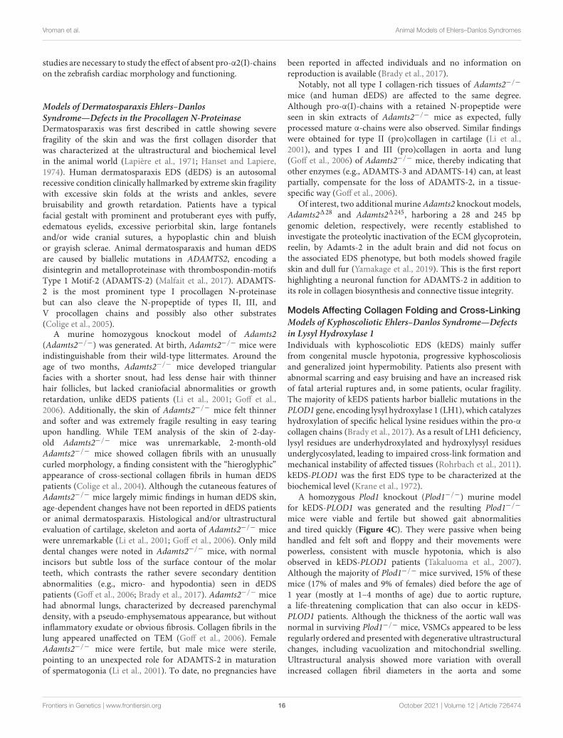
A homozygous Plod1 knockout (Plod1−/−) murine modelfor kEDS-PLOD1 was generated and the resulting Plod1−/−
mice were viable and fertile but showed gait abnormalitiesand tired quickly (Figure 4C). They were passive when beinghandled and felt soft and floppy and their movements werepowerless, consistent with muscle hypotonia, which is alsoobserved in kEDS-PLOD1 patients (Takaluoma et al., 2007).Although the majority of Plod1−/− mice survived, 15% of thesemice (17% of males and 9% of females) died before the age of1 year (mostly at 1–4 months of age) due to aortic rupture,a life-threatening complication that can also occur in kEDS-PLOD1 patients. Although the thickness of the aortic wall wasnormal in surviving Plod1−/− mice, VSMCs appeared to be lessregularly ordered and presented with degenerative ultrastructuralchanges, including vacuolization and mitochondrial swelling.Ultrastructural analysis showed more variation with overallincreased collagen fibril diameters in the aorta and some
Frontiers in Genetics | www.frontiersin.org 16 October 2021 | Volume 12 | Article 726474
fgene-12-726474 October 6, 2021 Time: 16:53 # 17
Vroman et al. Animal Models of Ehlers–Danlos Syndromes
fibrils had irregular contours. Similar ultrastructural alterationswere seen in the skin but skin hyperextensibility or fragilitywere lacking in Plod1−/− mice. In contrast to the humanphenotype, no kyphoscoliosis was observed in these mice. Inline with defective LH1 function, tissue-specific decreases intotal hydroxylysine residues were apparent in Plod1−/− mice,ranging from 22% in skin to 86% in lung. These findings suggestpartial compensation by the two other isoenzymes, LH2 and LH3(Takaluoma et al., 2007).
Overall, Plod1−/− mice seem to only partially capture thehuman kEDS-PLOD1 phenotype with the presence of musclehypotonia and aortic ruptures but without kyphoscoliosis ora clear skin phenotype. The latter might reflect differences inremaining hydroxylysine content between mice (22%) and inhuman (5%) (Steinmann et al., 2003; Takaluoma et al., 2007).
Models Affecting Myomatrix Function andOrganizationModels of Classical-Like Ehlers–Danlos Syndrome—Defectsin Tenascin-XClassical-like EDS (clEDS) is a rare autosomal recessivecondition, characterized by generalized joint hypermobility andskin hyperextensibility with easy bruising but without atrophicscarring. Patients harbor biallelic mutations in the TNXB gene,resulting in complete absence of the corresponding tenascin-X(TNX) protein (Brady et al., 2017; Green et al., 2020). TNX is aglycoprotein with an architectural function in the ECM and hasbeen shown to localize with collagen fibrils in tendon and dermis(Valcourt et al., 2015).
Tenascin-X was the first structural protein beyond fibrillarcollagens or their modifying enzymes associated with EDSand to investigate its hitherto unknown functions, two groupsindependently created homozygous Tnxb knockout (Tnxb−/−)mice (Matsumoto et al., 2001; Mao et al., 2002). AlthoughTnxb−/− mice appeared morphologically normal at birth theyshowed progressive skin hyperextensibility, similar to clEDSpatients, accompanied by a decreased ultrastructural densityof fibrils with normal size and shape, resulting in a reduceddermal collagen content. Despite near-normal collagen synthesisin vitro, Tnxb−/− skin fibroblasts showed defective depositionof type I collagen. Together with the ability of TNX toregulate the expression of several ECM molecules, these findingspointed to a regulatory role in collagen fibril deposition (Maoet al., 2002; Minamitani et al., 2004a). Nevertheless, conflictingresults have been reported for Tnxb−/− skin with normaldermal collagen content (Egging et al., 2007) and increasedcollagen fibril diameters with normal fibril density (Minamitaniet al., 2004b), which might be influenced by murine age,sample site and genetic background. Of note, opposed to theelastin fragmentation in clEDS patients, Tnxb−/− mice showedincreased dermal elastin density, which is probably less stable(Egging et al., 2006).
In contrast to cEDS, clEDS patients do not present withatrophic scarring or delayed wound healing. Tnxb−/− micemimicked the macroscopically normal wound closure of thedorsal skin in vivo, associated with reduced breaking strength(Egging et al., 2007). Subsequent in vitro experiments using
Tnxb−/− mouse embryonic fibroblasts suggested that the near-normal wound healing may be due to accelerated local matrixcontraction and tissue remodeling caused by increased activationof MMPs, upregulation of TGF-β1 expression and upregulatedcollagen synthesis followed by the promotion of cell proliferationand migration (Hashimoto et al., 2018). Together, these findingspointed to a role for TNX during the later phase of woundhealing when remodeling and maturation of the ECM occurs(Egging et al., 2007). Additionally, subcutaneous adipose tissueof Tnxb−/− mice was thicker and contains an increased amountof triglycerides and an altered fatty acid composition, suggestinga role during lipogenesis (Matsumoto et al., 2004).
Unlike clEDS patients, Tnxb−/− mice did not show overtsigns of joint hypermobility or ligamentous laxity (Egginget al., 2006), although tail and Achilles tendon displayedultrastructural changes in collagen fibril density similar toobservations in the skin (Mao et al., 2002). Tnxb−/− mice didpresent with mild muscle weakness, histological evidence ofmyopathy and increased turnover of the ECM in quadricepsmuscle (Voermans et al., 2011) as well as altered myofascialforce transmission (Huijing et al., 2010), consistent with themild to moderate muscle weakness observed in clEDS patients(Brady et al., 2017). Histology and TEM studies of the sciaticnerve revealed mildly reduced diameters of myelinated fibersand reduced collagen fibril density in the endoneurium inolder, but not young, Tnxb−/− mice, which may correspondto axonal polyneuropathy seen in clEDS patients (Matsumotoet al., 2002; Voermans et al., 2009, 2011; Sakai et al., 2017).Additionally, blood vessel formation (but not morphology) wasaltered in the peripheral nervous system of Tnxb−/− mice, witha decreased density of blood vessels with increased diameters(Sakai et al., 2017). Interestingly, Tnxb−/− mice were reportedto have mechanical allodynia but not thermal hypersensitivity.A chemical pain stimulus with formalin injection in the hindpaw evoked an increased pain response during the first (acute)phase (0–5 min) and early during the second (inflammatory)phase (16–30 min) in Tnxb−/− mice. Transcutaneous sinewave stimuli elicited a hypersensitive response of myelinated Aδ
and Aβ fibers, but not of unmyelinated C fibers. Additionally,Tnxb−/− mice showed molecular alterations in the dorsal hornof the spinal cord suggestive of spinal central sensitization(Okuda-Ashitaka et al., 2020).
Additional studies in Tnxb−/− mice revealed the presenceof reduced femoral bone mass with enhanced osteoclastdifferentiation and bone-resorbing ability (Kajitani et al., 2019),very mild genito-urinary complications (rectal prolapse in <1%)in female mice compared to uterine and vaginal prolapseseen in clEDS patients (Egging et al., 2008), attenuatedstromal neovascularization following cauterization (Sumiokaet al., 2018) and gastric dysfunction associated with abnormalgastric sensory function (Aktar et al., 2019). Additionally,Tnxb−/− mice showed increased anxiety-like behavior as wellas superior sensorimotor coordination and emotional learningand memory, without differences in locomotor or home cageactivity (Kawakami and Matsumoto, 2011; Voermans et al.,2011). Cognitive development was not studied in clEDS patients(Brady et al., 2017).
Frontiers in Genetics | www.frontiersin.org 17 October 2021 | Volume 12 | Article 726474
fgene-12-726474 October 6, 2021 Time: 16:53 # 18
Vroman et al. Animal Models of Ehlers–Danlos Syndromes
In summary, Tnxb−/− mice recapitulate the dermatological,mild muscular findings, and some neurological (including pain)findings of human clEDS but lack the articular aspects. Thesemice have been instrumental in demonstrating a regulatory rolefor TNX in a broad range of tissues.
Models of Myopathic Ehlers–Danlos Syndrome—Defects inType XII CollagenMyopathic EDS (mEDS) is a rare EDS subtype clinicallycharacterized by congenital muscle hypotonia and weaknesswith delayed motor development, proximal joint contractures incombination with distal joint hypermobility, scoliosis or kyphosisand abnormal scarring (Malfait et al., 2017; Delbaere et al.,2019b). mEDS is caused by mutations in COL12A1 encodingthe pro-α1-chain of type XII collagen, a homotrimer that is thelargest member of the FACIT family. Type XII collagen interactswith several ECM molecules (e.g., decorin and tenascin-X) andregulates the organization and mechanical properties of collagenfibrils (Chiquet et al., 2014). The majority of defects identifiedin COL12A1 give rise to heterozygous substitutions or in-frameexon skips, while a single biallelic loss-of-function COL12A1mutation was identified that results in a more severe phenotype(Hicks et al., 2014; Zou et al., 2014).
Before disease-causing mutations in COL12A1 wereidentified, type XII collagen-deficient (Col12a1−/−) micewere generated. Col12a1−/− mice were smaller and exhibitedskeletal abnormalities with shorter, more slender and fragile longbones as well as aberrant vertebrae structures (Izu et al., 2011).In depth analyses revealed that the bone phenotype is associatedwith defective differentiation, organization, polarization,morphology and interactions of Col12a1−/− osteoblasts (Izuet al., 2011), thereby affecting cell–cell communication (Izuet al., 2016) and resulting in decreased bone matrix formationwith altered quality. Although these skeletal manifestationswere not observed in human mEDS, patients have kyphosisand/or scoliosis, comparable to the kyphoscoliosis seen inCol12a1−/− mice (Izu et al., 2011; Zou et al., 2014). Togetherwith the identification of COL12A1 defects in human patients,the phenotype of the Col12a1−/− mouse model was revisited.Col12a1−/− mice showed evidence of (mild) muscle weakness,delayed transition in muscle fiber-type composition andaltered force transmission in the muscle-tendon-bone unit.Ultrastructural studies showed more diffusely dispersed collagenfibrils throughout the endomysium of Col12a1−/− muscle (Zouet al., 2014). Given the combination of distal joint hypermobilityand proximal contractures in mEDS, tendon development wasevaluated in Col12a1−/− mice, which also showed decreasedcollagen fibril packing. Tenocytes from Col12a1−/− mice had analtered shape and function with lower type I collagen secretion(Izu et al., 2020). Together, these data indicated the crucial role oftype XII collagen in the development and maturation of tendon.
Although all animal studies were performed on homozygousCol12a1−/− mice, only two siblings with autosomal recessivemEDS due to complete loss of type XII collagen wereidentified to date (Zou et al., 2014). Nevertheless, muscleweakness and skeletal abnormalities were observed in bothCol12a1−/− mice and mEDS patients, but the muscular
phenotype seems to be milder in mice compared to patients.To date, phenotypic characteristics of heterozygous Col12a1+/−
mice have not been reported.
Models Affecting Glycosaminoglycan BiosynthesisProteoglycans represent major ECM components and playessential roles as structural macromolecules, modulators ofcell adhesion and motility, ECM and collagen fibril assemblyand signal transduction during development, tissue repair, andangiogenesis. Proteoglycans are composed of a specific coreprotein substituted with one or more glycosaminoglycan (GAG)-chains (Prydz and Dalen, 2000; Bishop et al., 2007; Couchmanand Pataki, 2012). GAG biosynthesis is initiated by the stepwiseaddition of a tetrasaccharide linker to the core protein followedby the addition of repeating disaccharide units that define theGAG-chain as heparan sulfate (HS), chondroitin sulfate (CS),and/or dermatan sulfate (DS) (Figure 1B).
The first hint for the involvement of proteoglycans in thepathophysiology of EDS came from a mouse model deficientfor decorin, the prototypic member of a specific class ofproteoglycans, the SLRPs. The resulting decorin knockout(Dcn−/−) mice had thin and fragile skin with reduced tensilestrength and an abnormal ultrastructural morphology of collagenfibrils in skin and tendon with larger and more irregularfibril outlines, thereby highlighting the important regulatoryrole of decorin during collagen fibrillogenesis (Danielson et al.,1997). Subsequently, knockout mice for other SLRPs, lumican(Chakravarti et al., 1998), dermatopontin (Takeda et al., 2002),or mimecan (Tasheva et al., 2002) also displayed signs of skinlaxity and fragility and similar ultrastructural collagen fibrilabnormalities as seen in Dcn−/− mice (Supplementary Table 4).Given the phenotypic resemblance of these animal models tohuman EDS, the genes encoding SLRPs were considered goodcandidates for molecularly unresolved EDS patients, but defectsin SLRP core proteins could never be linked with EDS (Malfaitet al., 2005). Nevertheless, the subsequent identification of geneticdefects in enzymes involved in the synthesis and modificationof GAG-chains unequivocally confirmed a role for aberrantproteoglycan biosynthesis in human EDS pathogenesis, but thecausal defects compromise proper GAG-formation rather thanaffecting the core protein.
Models of Spondylodysplastic Ehlers–DanlosSyndrome—Defects in Galactosyltransferase-I and -IIBiallelic mutations in the B4GALT7 and B3GALT6 genes,encoding the linker enzymes galactosyltransferase-I (β4GalT7)and -II (β3GalT6), respectively, that catalyze subsequentsteps of the GAG tetrasaccharide linker region biosynthesis(Figure 1B), give rise to two subtypes of spondylodysplasticEDS (spEDS), spEDS-B4GALT7 and spEDS-B3GALT6. Bothsubtypes are severe, pleiotropic conditions that are generallycharacterized by short stature, muscle hypotonia, skeletalabnormalities, skin hyperextensibility and translucency,delayed motor development, and osteopenia. Additionally,subtype specific clinical features can be observed (Table 1;Malfait et al., 2017).
Frontiers in Genetics | www.frontiersin.org 18 October 2021 | Volume 12 | Article 726474
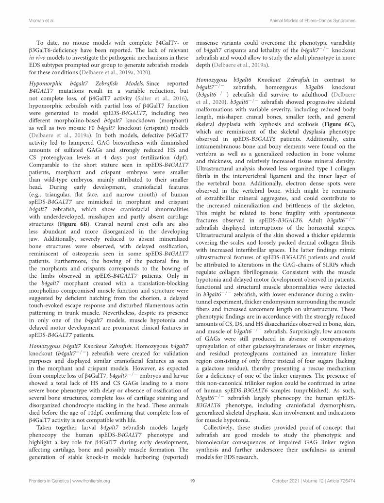
fgene-12-726474 October 6, 2021 Time: 16:53 # 19
Vroman et al. Animal Models of Ehlers–Danlos Syndromes
To date, no mouse models with complete β4GalT7- orβ3GalT6-deficiency have been reported. The lack of relevantin vivo models to investigate the pathogenic mechanisms in theseEDS subtypes prompted our group to generate zebrafish modelsfor these conditions (Delbaere et al., 2019a, 2020).
Hypomorphic b4galt7 Zebrafish Models. Since reportedB4GALT7 mutations result in a variable reduction, butnot complete loss, of β4GalT7 activity (Salter et al., 2016),hypomorphic zebrafish with partial loss of β4GalT7 functionwere generated to model spEDS-B4GALT7, including twodifferent morpholino-based b4galt7 knockdown (morphant)as well as two mosaic F0 b4galt7 knockout (crispant) models(Delbaere et al., 2019a). In both models, defective β4GalT7activity led to hampered GAG biosynthesis with diminishedamounts of sulfated GAGs and strongly reduced HS andCS proteoglycan levels at 4 days post fertilization (dpf).Comparable to the short stature seen in spEDS-B4GALT7patients, morphant and crispant embryos were smallerthan wild-type embryos, mainly attributed to their smallerhead. During early development, craniofacial features(e.g., triangular, flat face, and narrow mouth) of humanspEDS-B4GALT7 are mimicked in morphant and crispantb4galt7 zebrafish, which show craniofacial abnormalitieswith underdeveloped, misshapen and partly absent cartilagestructures (Figure 6B). Cranial neural crest cells are alsoless abundant and more disorganized in the developingjaw. Additionally, severely reduced to absent mineralizedbone structures were observed, with delayed ossification,reminiscent of osteopenia seen in some spEDS-B4GALT7patients. Furthermore, the bowing of the pectoral fins inthe morphants and crispants corresponds to the bowing ofthe limbs observed in spEDS-B4GALT7 patients. Only inthe b4galt7 morphant created with a translation-blockingmorpholino compromised muscle function and structure weresuggested by deficient hatching from the chorion, a delayedtouch-evoked escape response and disturbed filamentous actinpatterning in trunk muscle. Nevertheless, despite its presencein only one of the b4galt7 models, muscle hypotonia anddelayed motor development are prominent clinical features inspEDS-B4GALT7 patients.
Homozygous b4galt7 Knockout Zebrafish. Homozygous b4galt7knockout (b4galt7−/−) zebrafish were created for validationpurposes and displayed similar craniofacial features as seenin the morphant and crispant models. However, as expectedfrom complete loss of β4GalT7, b4galt7−/− embryos and larvaeshowed a total lack of HS and CS GAGs leading to a moresevere bone phenotype with delay or absence of ossification ofseveral bone structures, complete loss of cartilage staining anddisorganized chondrocyte stacking in the head. These animalsdied before the age of 10dpf, confirming that complete loss ofβ4GalT7 activity is not compatible with life.
Taken together, larval b4galt7 zebrafish models largelyphenocopy the human spEDS-B4GALT7 phenotype andhighlight a key role for β4GalT7 during early development,affecting cartilage, bone and possibly muscle formation. Thegeneration of stable knock-in models harboring (reported)
missense variants could overcome the phenotypic variabilityof b4galt7 crispants and lethality of the b4galt7−/− knockoutzebrafish and would allow to study the adult phenotype in moredepth (Delbaere et al., 2019a).
Homozygous b3galt6 Knockout Zebrafish. In contrast tob4galt7−/− zebrafish, homozygous b3galt6 knockout(b3galt6−/−) zebrafish did survive to adulthood (Delbaereet al., 2020). b3galt6−/− zebrafish showed progressive skeletalmalformations with variable severity, including reduced bodylength, misshapen cranial bones, smaller teeth, and generalskeletal dysplasia with kyphosis and scoliosis (Figure 6C),which are reminiscent of the skeletal dysplasia phenotypeobserved in spEDS-B3GALT6 patients. Additionally, extraintramembranous bone and bony elements were found on thevertebra as well as a generalized reduction in bone volumeand thickness, and relatively increased tissue mineral density.Ultrastructural analysis showed less organized type I collagenfibrils in the intervertebral ligament and the inner layer ofthe vertebral bone. Additionally, electron dense spots wereobserved in the vertebral bone, which might be remnantsof extrafibrillar mineral aggregates, and could contribute tothe increased mineralization and brittleness of the skeleton.This might be related to bone fragility with spontaneousfractures observed in spEDS-B3GALT6. Adult b3galt6−/−
zebrafish displayed interruptions of the horizontal stripes.Ultrastructural analysis of the skin showed a thicker epidermiscovering the scales and loosely packed dermal collagen fibrilswith increased interfibrillar spaces. The latter findings mimicultrastructural features of spEDS-B3GALT6 patients and couldbe attributed to alterations in the GAG-chains of SLRPs whichregulate collagen fibrillogenesis. Consistent with the musclehypotonia and delayed motor development observed in patients,functional and structural muscle abnormalities were detectedin b3galt6−/− zebrafish, with lower endurance during a swim-tunnel experiment, thicker endomysium surrounding the musclefibers and increased sarcomere length on ultrastructure. Thesephenotypic findings are in accordance with the strongly reducedamounts of CS, DS, and HS disaccharides observed in bone, skin,and muscle of b3galt6−/− zebrafish. Surprisingly, low amountsof GAGs were still produced in absence of compensatoryupregulation of other galactosyltransferases or linker enzymes,and residual proteoglycans contained an immature linkerregion consisting of only three instead of four sugars (lackinga galactose residue), thereby presenting a rescue mechanismfor a deficiency of one of the linker enzymes. The presence ofthis non-canonical trilinker region could be confirmed in urineof human spEDS-B3GALT6 samples (unpublished). As such,b3galt6−/− zebrafish largely phenocopy the human spEDS-B3GALT6 phenotype, including craniofacial dysmorphism,generalized skeletal dysplasia, skin involvement and indicationsfor muscle hypotonia.
Collectively, these studies provided proof-of-concept thatzebrafish are good models to study the phenotypic andbiomolecular consequences of impaired GAG linker regionsynthesis and further underscore their usefulness as animalmodels for EDS research.
Frontiers in Genetics | www.frontiersin.org 19 October 2021 | Volume 12 | Article 726474
fgene-12-726474 October 6, 2021 Time: 16:53 # 20
Vroman et al. Animal Models of Ehlers–Danlos Syndromes
Models of Musculocontractural Ehlers–DanlosSyndrome—Defects in Dermatan 4-O-Sulfotransferase-1 andDermatan Sulfate Epimerase-1Musculocontractural EDS (mcEDS) is clinically characterizedby multiple congenital malformations including characteristiccraniofacial features and contractures as well as progressivemultisystemic complications such as skin hyperextensibilityand fragility, recurrent dislocations, progressive talipes orspinal deformities, and large subcutaneous hematomas (Malfaitet al., 2017). mcEDS is caused by biallelic mutations in eitherCHST14 or DSE, encoding dermatan 4-O-sulfotransferase-1 (D4ST1) and dermatan sulfate epimerase-1 (DS-epi1),respectively. Both enzymes are specifically involved in DSGAG biosynthesis (Figure 1B). In contrast to defects inB3GALT6 and B4GALT7 that compromise linker formationand affect the synthesis of both CS/DS and HS proteoglycans,D4ST1- or DS-epi1-deficiency only interferes with formation ofDS proteoglycans.
Homozygous Chst14 Knockout Mice. The first murinehomozygous Chst14 knockout (Chst14−/−) model was createdto study the roles of DS GAGs during neural development andnerve regeneration (Bian et al., 2011). Chst14−/− mice showedreduced viability (8% of offspring). Surviving Chst14−/− micehad a normal life span but were smaller and had reduced fertility,kinked tail, tooth malformations and increased skin fragility(Akyüz et al., 2013). Chst14−/− mice provided evidence for anoverall negative impact of DS on peripheral nerve regeneration(Akyüz et al., 2013) but beneficial activity in central nervoussystem regeneration (Rost et al., 2016), which could be attributedto differences in levels of DS proteoglycans and their interactionpartners between the peripheral nervous system and the centralnervous system (Rost et al., 2016). Furthermore, Chst14−/−
mice showed impaired spatial learning and memory anddemonstrated that DS is indispensable for synaptic plasticityin the hippocampus, which might be attributed to impairedAkt/mTOR-signaling (Li et al., 2019).
Subsequently, another homozygous Chst14 knockout(Chst14−/−) mouse model was studied that was initially createdas part of a largescale effort to create a knockout library forsecreted and transmembrane proteins (Chst14tm1Lex) (Tanget al., 2010). This Chst14−/− model also suffered from perinatallethality, with a markedly decreased survival of 14.8% atembryonic day 18.5 to 1.3% at adulthood. Placentas fromChst14−/− fetuses showed a reduced weight, with smallervascular diameters in the placental villi (the part of the fetalplacenta), and hypoxia- and/or necrotic-like changes in asmall subset (6%). TEM analysis demonstrated decreasedthickness and fragmentation of basement membranes in thecapillaries of the villus (Yoshizawa et al., 2018). These placentalvascular abnormalities possibly relate to the high perinatallethality in Chst14−/− mice as well as the development oflarge subcutaneous hematomas in mcEDS-CHST14 patients.Changing the genetic background (from mixed C57BL/6-129/SvJto BALB/c), improved the birth rate for Chst14−/− mice to6.12–18.64% (Shimada et al., 2020). Subsequent analysis ofthe skin, which is affected in mcEDS-CHST14 patients, show
reduced tensile strength Chst14−/− mice and TEM analysisrevealed increased intrafibrillar spaces with a decreased densityand disorganization of collagen fibrils. Additionally, rod-shaped,linear GAG-chains protruded from collagen fibrils in Chst14−/−
mice compared to the curved GAG-chains wrapped aroundthe collagen fibrils in wild-type (Hirose et al., 2020), similarto TEM findings for mcEDS-CHST14 patients (Hirose et al.,2019b). Glycobiological analyses of the skin revealed very lowlevels of DS disaccharides and an excess of CS disaccharides(Hirose et al., 2020).
The characteristic alterations in GAG-structure anddisorganization of the collagen network in de skin are similar toultrastructural observations in mcEDS-CHST14 patients, therebymaking Chst14−/− mice a reasonable model for this EDS subtypealthough the reduced viability compromises further studies.
Homozygous Dse Knockout Mice. Before DSE defects wereidentified in mcEDS patients, homozygous Dse knockout(Dse−/−) mice were already generated. Dse−/− mice weresmaller, presented with a kinked tail that resolved before 4 weeksof age and had reduced fertility (Figure 4D). The skin of Dse−/−
mice had reduced tensile strength and sparser loose hypodermalconnective tissue on light microscopy despite normal collagencontent. TEM analysis revealed dermal collagen fibrils withlarger diameters but normal fibril density while Dse−/− tailtendons showed only a minor shift towards thicker fibrilswith mostly fibrils with normal diameter and no differencesin diameter in Achilles tendon (Maccarana et al., 2009). Thiscontrasts the normal ultrastructure of dermal collagen fibrilsin mcEDS-DSE patients, although morphometric analysis wasnot performed (Syx et al., 2015). Glycobiological examinationrevealed a decreased, but not totally absent, DS content inthe skin, similar to findings in a mcEDS-DSE patient (Mülleret al., 2013). The residual DS moieties may be attributed topartial compensation by DS-epi2 activity. Changing the geneticbackground (from mixed 129SvJ-C57BL/6 to NFR), resultedin Dse−/− mice with normal body weights, a kinked tail andsome developmental malformations, including an abdominalwall defect with herniated intestines in 16% of Dse−/− embryosand neural tube defects (e.g., exencephaly and spina bifida) in5%. Additionally, Dse−/− embryos and newborns had thickerepidermal layers with increased amounts of epidermal basal andspinous layer markers (Gustafsson et al., 2014).
The relatively milder phenotype in Dse−/− mice is consistentwith the findings in mcEDS-DSE patients, which also presentan apparently milder phenotype compared to mcEDS due toCHST14 defects, but only a limited number of patients have beenreported so far (Lautrup et al., 2020).
Models Affecting Intracellular ProcessesModels of Spondylodysplastic Ehlers–DanlosSyndrome—Defects in ZIP13Biallelic mutations in the SLC39A13 gene, encoding the metaltransporter Zrt/irt-like protein 13 (ZIP13), result in a phenotypewhich clinically overlaps with spEDS due to B4GALT7 andB3GALT6 mutations and is therefore grouped within thesame clinical entity (Malfait et al., 2017). spEDS-SLC39A13
Frontiers in Genetics | www.frontiersin.org 20 October 2021 | Volume 12 | Article 726474

fgene-12-726474 October 6, 2021 Time: 16:53 # 21
Vroman et al. Animal Models of Ehlers–Danlos Syndromes
patients present with moderate short stature, combined withspecific facial characteristics, hypo- or oligodontia, distal jointhypermobility, thin and fragile skin, wrinkled palms, andcharacteristic radiographic abnormalities (Brady et al., 2017;Malfait et al., 2017; Kumps et al., 2020). spEDS-SLC39A13patients display collagen underhydroxylation, despite normalactivity of lysyl and prolyl hydroxylases (Fukada et al., 2008;Giunta et al., 2008). ZIP13 is a homodimeric transmembraneprotein and putative zinc (Zn2+) transporter located on theendoplasmic reticulum/Golgi apparatus which regulates theinflux of Zn2+ into the cytosol (Bin et al., 2011). Interestingly,studies in Drosophila melanogaster revealed that the fruitfly homolog of human ZIP13 exports iron (Fe2+) and itsdepletion might attenuate the action of iron-dependent enzymessuch as lysyl hydroxylases, possibly explaining the observedunderhydroxylation (Xiao et al., 2014).
Homozygous Slc39a13 knockout (Slc39a13-KO) mice showedgrowth retardation, progressive kyphosis after 3–4 weeks ofage, osteopenia, and abnormal cartilage development (Fukadaet al., 2008). Additionally, altered craniofacial features, includingsunken and downslanting eyes were apparent as well as dentalabnormalities, such as incisor deformities and aberrant dentinformation (Figure 4E). The skin of Slc39a13-KO mice wasmore fragile and showed a thinner dermis with smaller andless densely packed collagen fibrils on ultrastructure, the latterpossibly attributed to a decreased dermal DS content (Fukadaet al., 2008; Hirose et al., 2015). Subcutaneous adipose tissuewas thinner (Fukada et al., 2008; Fukunaka et al., 2017).Corneal stroma showed reduced thickness in Slc39a13-KOmice with smaller and more widely spaced collagen fibrilswhich were more randomly oriented (Fukada et al., 2008;Hirose et al., 2018). Except for severe varicosity in the lowerextremities, spEDS-SLC39A13 patients do not present withcardiovascular symptoms. Similarly, Slc39a13-KO mice did notshow aneurysms or arterial ruptures, however, abnormalitieswere observed in the tunica media of the thoracic aorta whichcould potentially result in increased fragility (Hirose et al., 2019a).Slc39a13-KO mice showed evidence for reduced osteoblastactivity, impaired chondrocyte and adipocyte differentiation aswell as irregular cellular morphology of odontoblasts in themolar tooth, dermal fibroblasts, corneal keratocytes, and aorticVSMCs (Fukada et al., 2008; Fukunaka et al., 2017; Hiroseet al., 2018, 2019a). Furthermore, murine studies illustrateddysregulation of the BMP- and TGFβ-signaling pathwaysin several of these connective tissue forming cells, whichmight contribute to the observed multisystemic phenotype(Fukada et al., 2008).
Taken together, Slc39a13-KO mice display craniofacial, dental,skeletal, dermal, and ocular features that are reminiscentof the human spEDS-SLC39A13 phenotype and highlighta role for altered BMP- and TGFβ-signaling in spEDS-SLC39A13 pathogenesis.
Models of Brittle Cornea Syndrome—Defects in PRDM5Brittle cornea syndrome (BCS) is clinically hallmarked by asevere ocular phenotype with thin and fragile cornea leadingto increased risk of spontaneous rupture and often vision
loss. Additionally, patients also present with hearing lossand signs of generalized connective tissue fragility such asdistal joint hypermobility and mild cutaneous and craniofacialabnormalities. BCS is caused by biallelic defects in either ZNF469or PRDM5. ZNF469 encodes the zinc finger protein ZNF469,which is believed to be a (transcriptional) regulator of thesynthesis and/or organization of collagen fibrils (Abu et al., 2008)but its function remains elusive. PRDM5 encodes PR domainzinc finger protein 5 (PRDM5), a transcription factor that wasidentified as a putative tumor suppressor gene in several cancersbut has also been shown to regulate transcription of collagen andother ECM genes (Galli et al., 2012).
Although no animal models have been generated to studyBCS pathogenesis, two independent studies investigated the roleof PRDM5 in zebrafish larvae as a putative tumor suppressorand during craniofacial development (Meani et al., 2009; Dinget al., 2013). Interestingly, morpholino-based prdm5 knockdownaffects early stages of zebrafish development with craniofacialmalformation including cyclopia or smaller eyes and axialmesendodermal defects (jaw, heart, and blood) (Figure 6D;Meani et al., 2009). Craniofacial malformations with neurocranialdefects were subsequently confirmed in prdm5 knockout(prdm5hi61Tg) zebrafish larvae (Amsterdam et al., 2004; Dinget al., 2013). These findings hint to the ocular phenotype and mildcraniofacial abnormalities seen in BCS patients. Furthermore,prdm5 knockdown results in impaired morphogenetic movementduring gastrulation as a consequence of increased canonicalWnt-signaling (Meani et al., 2009; Ding et al., 2013).
To date, no stable animal models of BCS due to ZNF469defects have been described.
Models of Additional Ehlers–Danlos SyndromesVariantsModels of Classical-Like Ehlers-Danlos Syndrome Type2—Defects in Adipocyte Enhancer-Binding Protein 1The most recent defects associated with human EDS residein adipocyte enhancer-binding protein-1 (AEBP1), also knownas aortic carboxypeptidase-like protein (ACLP). AEBP1 isexpressed in tissues with a high collagen content and is asecreted ECM-associated protein that preferentially binds tofibrillar types I, III, and V collagen and acts as a positiveregulator of collagen polymerization in vitro and collagenfibrillogenesis in vivo (Layne et al., 2001; Blackburn et al.,2018). Biallelic mutations in the AEBP1 gene, encoding AEBP1,cause an EDS phenotype hallmarked by skin hyperextensibilitywith atrophic scarring, generalized joint hypermobility, footdeformities and early-onset osteopenia, provisionally referred toas classical-like EDS type 2 (clEDS2) (Blackburn et al., 2018;Syx et al., 2019).
Before AEBP1 defects were linked to EDS, two groupsindependently created homozygous Aebp1 knockout (Aebp1−/−)mice, which presented some phenotypic discrepancies. While themajority of the first Aebp1−/− mouse model died perinatallydue to gastroschisis, surviving Aebp1−/− mice developedspontaneous skin lesions and present with delayed woundhealing, which correlated with reduced dermal fibroblastproliferation (Layne et al., 2001). These wound healing defects
Frontiers in Genetics | www.frontiersin.org 21 October 2021 | Volume 12 | Article 726474
fgene-12-726474 October 6, 2021 Time: 16:53 # 22
Vroman et al. Animal Models of Ehlers–Danlos Syndromes
are reminiscent of the phenotype of clEDS2 patients, indicatinga role for AEBP1 in wound repair. Additional studies inmice suggested role(s) of AEBP1 in fibroblast-to-myofibroblasttransition in lung through TGFβ-signaling (Schissel et al., 2009;Tumelty et al., 2014), and activation of the canonical Wnt-signaling pathway through frizzled-8 and LRP6 in hepatic stellatecells (Teratani et al., 2018). Deregulation of these pathwayscould potentially contribute to the defective wound healing andbone development and homeostasis seen in patients. For thesecond model, about half of Aebp1−/− mice survived and theywere smaller, with markedly reduced white adipose tissue anddefective proliferation and lower MAPK activity in Aebp1−/−
pre-adipocytes. Additionally, lactation defects were observed infemales and infertility in males (Ro et al., 2007; Zhang et al., 2011).
As such, studies in Aebp1−/− mice suggest the involvementof AEBP1 in the development and homeostasis of adipose tissue,mammary gland and connective tissue, with recapitulation ofthe dermal manifestations of clEDS2 in one model. Nevertheless,the exact mechanisms leading to the observed EDS phenotyperemain currently unknown.
Naturally Occurring Ehlers–DanlosSyndromes ModelsBefore the first genetically engineered EDS mouse modelswere generated, naturally occurring EDS(-like) phenotypeswere already described for decades in domesticated animals.There are several reports of cats, dogs, rabbits, mink, cattle,sheep, and horses displaying a disease reminiscent ofEDS (Fjølstad and Helle, 1974; Hegreberg, 1975; Countset al., 1977; Holbrook et al., 1980; Brown et al., 1993;Hansen et al., 2015). The phenotypes of these animalswere hallmarked by thin and hyperextensible skin, whichwas fragile leading to hemorrhagic wounds and atrophicscars; and commonly described as cutis hyperelastica,hyperelastosis cutis, dermatosparaxis, dermal/collagen dysplasia,dermal/cutaneous asthenia, or Ehlers–Danlos-like syndromes(Halper, 2013).
While the genetic defects causing EDS-like pathologies indomestic animals were less well studied in early reports,the advent of next generation sequencing technologies suchas whole exome sequencing and whole genome sequencing,provided molecular proof of the underlying disorder in morerecent reports. Mutations have been reported in Col5a1in cats (Spycher et al., 2018) and dogs (Bauer et al.,2019a), Col5a2 in cattle (Jacinto et al., 2020), Adamts2in cattle (Colige et al., 1999), sheep (Zhou et al., 2011;Monteagudo et al., 2015; Joller et al., 2017), and dogs(Jaffey et al., 2019), Plod1 in horse (Monthoux et al.,2015), Tnxb in dogs (Bauer et al., 2019b) and B4galt7 inFinish horse (Leegwater et al., 2016). These animals displayphenotypic features reminiscent of the corresponding humanEDS counterpart. Additionally, horses presenting with an EDS-like condition with loose skin have been described and coinedHereditary Equine Regional Dermal Asthenia (HERDA). Thiscondition is caused by defects in the Ppib gene, encodingcyclophilin B. This gene is not associated with EDS in
humans but results in OI (van Dijk et al., 2009; Rashmir-Raven, 2013). Functional studies in these animals are restrictedbut sometimes accompanied by biochemical, histopathologicaland/or ultrastructural data. A summary of the findings ofspontaneous models with confirmed genetic defects is providedin Table 2.
Taken together, it is clear that EDS also occurs naturally inmammals and can be prominent in specific (in)breeding schemes.However, due to limited resources, especially in sequencingcapacity for animals, many cases are not molecularly confirmed,and in-depth functional analyses are often lacking.
CONCLUSION AND FUTUREPERSPECTIVES
For 14 of the 20 hitherto known EDS-associated genes, animalmodels, either mouse or zebrafish, have been created. While forthe more common cEDS and vEDS subtypes different engineeredmouse models are available and often extensively characterized,animal models are lacking for some of the rarer EDS subtypes(e.g., periodontal EDS, kEDS-FKBP14, arthrochalasia EDS andBCS-ZNF469). Recently, zebrafish models were introduced intothe EDS research field [col1a2−/− (cvEDS) and b4galt7 andb3galt6−/− (spEDS)]. Additionally, numerous domestic animalsdisplaying an EDS(-like) phenotype have been reported, with thegenetic defect identified for some. Although these models arevaluable tools to learn about the EDS phenotype in animals otherthan rodents or zebrafish, they are less amendable for large-scaleresearch purposes.
Especially engineered models have been instrumental indiscerning the functions of these particular proteins duringdevelopment, maturation and repair and in portraying their rolesduring collagen biosynthesis and fibrillogenesis, for some evenbefore their contribution to an EDS phenotype was elucidated.Extensive phenotypical characterization of these models hasshown that they largely phenocopy their human counterparts,with recapitulation of several key clinical features of thecorresponding EDS subtype, including dermal, cardiovascular,musculoskeletal and ocular alterations. Moreover, these modelsprovide valuable tools to perform in-depth studies of severaldisease-related aspects (e.g., wound healing); and allow toinvestigate organs and tissues that are not easily accessiblefrom humans (e.g., cardiovascular or nervous tissue) to assessphysicochemical, ultrastructural and biomechanical propertiesduring early development and adult stages. This can beparticularly helpful to elucidate disease mechanisms in rarerEDS subtypes for which only a limited number of patients arereported. Since for most EDS types, the exact link between a singlegene defect and the observed phenotype remains elusive, morestudies are needed to investigate the underlying pathways andunderstand the pathophysiological consequences.
As such, investigation of these models hinted toward theinvolvement of several signaling pathways, such as Wnt-signalingin prdm5 knockdown zebrafish and Aebp1−/− mice, BMP1-and/or TGFβ-signaling in Slc39a13-KO and Aebp1−/− mice;and PLC/IP3/PKC/ERK-signaling in Col3a1 knock-in mice and
Frontiers in Genetics | www.frontiersin.org 22 October 2021 | Volume 12 | Article 726474
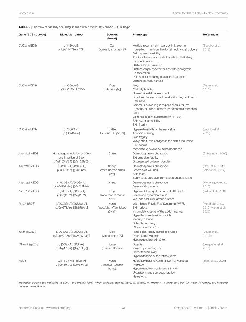
fgene-12-726474 October 6, 2021 Time: 16:53 # 23
Vroman et al. Animal Models of Ehlers–Danlos Syndromes
TABLE 2 | Overview of naturally occurring animals with a molecularly proven EDS subtype.
Gene (EDS subtype) Molecular defect Species(breed)
Phenotype References
Col5a1 (cEDS) c.3420delG,p.(Leu1141Serfs*134)
Cat[Domestic shorthair (F)]
Multiple recurrent skin tears with little or nobleeding, mainly on the dorsal neck and shoulders
Skin hyperextensibilityPrevious lacerations healed slowly and left shiny
alopecic scarsBilateral hip subluxationBilateral carpal hyperextension with plantigrade
appearancePain and laxity during palpation of all jointsBilateral perineal hernias
(Spycher et al.,2018)
Col5a1 (cEDS) c.3030delG,p.(Gly1013Valfs*260)
Dog[Labrador (M)]
(4m):Clinically healthyNormal skeletal developmentSmall skin lacerations of the distal limbs, hock and
tail baseSeroma-like swelling in regions of skin trauma
(hocks, tail base): seroma or hematoma formation(6m):Generalized joint hypermobility ( >180◦)Skin hyperextensibilitySkin fragility
(Bauer et al.,2019a)
Col5a2 (cEDS) c.2366G>T,p.(Gly789Val)
Cattle[Holstein calf (3d, F)]
Hyperextensibility of the neck skinAtrophic scarringSkin fragilityWavy, short, thin collagen in the skin surrounded
by edemaModerate to severe acute hemorrhages
(Jacinto et al.,2020)
Adamts2 (dEDS) Homozygous deletion of 20bpand insertion of 3bp,
p.[(Val153fs*24)];[(Val153fs*24)]
Cattle Dermatosparaxis phenotypeExtreme skin fragilityDisorganized collagen bundles
(Colige et al., 1999)
Adamts2 (dEDS) c.[424G>T];[424G>T],p.[(Glu142*)];[(Glu142*)]
Sheep[White Dorper lambs
(2d)]
Dermatosparaxis phenotypeSevere skin woundsSkin tearsEasily separated skin from subcutaneous tissue
(Zhou et al., 2011;Joller et al., 2017)
Adamts2 (dEDS) c.[805G>A];[805G>A],p.[(Val269Met)];[(Val269Met)]
Sheep Dermatosparaxis phenotypeSevere skin wounds
(Monteagudo et al.,2015)
Adamts2 (dEDS) c.[769C>T];[769C>T],p.[(Arg257*)];[(Arg257*)]
Dog[Doberman Pinscher
(8w)]
Hypermobile carpal, tarsal and stifle jointsLoose and hyperelastic skinWounds and large atrophic scars
(Jaffey et al., 2019)
Plod1 (kEDS) c.[2032G>A];[2032G>A],p.[Gly678Arg];[Gly678Arg]
Horse[Westfalian Warmblood
(5y, F)]
Warmblood Fragile Foal Syndrome (WFFS)Skin lesionsIncomplete closure of the abdominal wallHyperflexion/extension of jointsInability to standDifficulty breathingOften die within 72 h
(Monthoux et al.,2015; Martin et al.,2020)
Tnxb (clEDS1) c.[2012G>A];[2900G>A],p.[(Ser671Asn)];[(Gly967Asp)]
Dog[Mixed-breed (F)]
Fragile skin, easily teared or bruisedPoor healing woundsHyperextensible skin (21m)
(Bauer et al.,2019b)
B4galt7 (spEDS) c.[50G>A];[50G>A],p.[(Arg17Lys)];[(Arg17Lys)]
Horses(Friesian Horses)
DwarfismInwards protruding ribsFlexor tendon laxityHyperextension of the fetlock joints
(Leegwater et al.,2016)
Ppib (/) c.[115G>A];[115G>A]p.[(Gly39Arg)];[(Gly39Arg)]
Horse(American Quarter
horse)
Hereditary Equine Regional Dermal Asthenia(HERDA)Hyperextensible, fragile and thin skinUlcerations and skin degenerationHematoma
(Tryon et al., 2007)
Molecular defects are indicated at cDNA and protein level. When available, age (d: days, w: weeks, m: months, y: years) and sex (M: male, F: female) are includedbetween parentheses.
Frontiers in Genetics | www.frontiersin.org 23 October 2021 | Volume 12 | Article 726474
fgene-12-726474 October 6, 2021 Time: 16:53 # 24
Vroman et al. Animal Models of Ehlers–Danlos Syndromes
highlighted potential compensatory mechanisms such as thepresence of a trilinker region in b3galt6−/− zebrafish. Furthervalidation of these findings in patient samples is warrantedand additional investigations are required to further pinpointcommon or distinct pathways in several EDS subtypes, therebyexplaining phenotypic overlap and differences.
Nevertheless, direct comparison between different EDSmodels is limited and complicated by several variables, suchas: (1) differences in genetic background influencing thephenotypic outcome and survival of animals (e.g., Chst14−/−
mice) (Montagutelli, 2000); (2) age at which the animals wereexamined; and (3) anatomical location of investigated tissues.Furthermore, sex differences have been described in most murinemodels of vEDS, with a more severe skin and vascular phenotypein males compared to females. Although the sex of the EDSanimals was not always clearly indicated in literature, male-female divergences are increasingly being recognized in severalorgan systems, including, skin thickness, cardiovascular function,musculoskeletal integrity as well as in physiological processes,such as pain, and need to be taken into account in futurepreclinical EDS research (Huxley, 2007; Lang, 2011; Rosen et al.,2017; Sorge and Totsch, 2017; Rahrovan et al., 2018).
The majority of the models generated to date harborheterozygous or homozygous null mutations, which areindispensable to study the consequences of complete proteindeficiency, but which usually do not capture the completemutation spectrum associated with a particular EDS subtype(e.g., structural variants like glycine substitutions or exon skipsin COL5A1, COL5A2, or COL12A1). With exception of Col3a1models of vEDS, no knock-in EDS models have been developed.Advances in genome editing technologies such as CRISPR/Cas9and base editing can assist in creating these models (Adli, 2018;Porto et al., 2020).
Currently, treatment and management options for EDSpatients are limited and for none of the EDS subtypes atargeted therapy is available. So far, preclinical therapeuticinterventions have only been explored in mouse models of vEDSand contradicting results were obtained depending on the modelused. When the elucidation of the pathogenic mechanisms andderegulated pathways progresses, novel druggable targets willlikely be identified. Additionally, the introduction of zebrafishinto the EDS research field holds promising potential fortherapeutic discovery, since these animals are highly amenablefor drug/compound screening.
Despite the availability of animal models for several EDSsubtypes, the study of these models for understanding thepathophysiology of EDS is still in its infancy but they cansupply a tremendous bulk of information to the scientificcommunity. The generation of additional models (both murineand zebrafish), harboring different genetic defects and differenttypes of genetic variants, will further boost our knowledge andmight lead us into a new era of disease research and drugdiscovery. In summary, these EDS animal models proved to bevalid models that are and will continue to be valuable resourcesthat, when explored to full extend, can uncover novel insightsin pathogenic mechanisms, identify reliable biomarkers andpinpoint relevant cellular processes or signaling pathways thatcan serve as targets for the development of disease-specific(and/or personalized) therapies.
AUTHOR CONTRIBUTIONS
RV and DS wrote the manuscript. RM, A-MM, and FM read andedited the draft of the manuscript. All authors approved the finalversion of the manuscript.
FUNDING
This work was supported by the Research FoundationFlanders (FWO), Belgium [1842318N to FM, 3G041519to FM, and 12Q5920N to DS]; Association française dessyndromes d’Ehlers-Danlos (AFSED) to FM; The Ehlers-Danlos Society to FM; the National Institutes of Health[National Institute of Arthritis and Musculoskeletal andSkin Diseases (NIAMS)] [grant numbers K01AR070328to RM, R01AR077019-01A1 to RM, R01AR060364 andR01AR064251 to A-MM]. A-MM was supported by theGeorge W. Stuppy, MD, Chair of Arthritis at RushUniversity.
SUPPLEMENTARY MATERIAL
The Supplementary Material for this article can be foundonline at: https://www.frontiersin.org/articles/10.3389/fgene.2021.726474/full#supplementary-material
REFERENCESAbu, A., Frydman, M., Marek, D., Pras, E., Nir, U., Reznik-Wolf, H., et al.
(2008). Deleterious mutations in the zinc-finger 469 gene cause brittle corneasyndrome. Am. J. Hum. Genet. 82, 1217–1222. doi: 10.1016/j.ajhg.2008.04.001
Adli, M. (2018). The CRISPR tool kit for genome editing and beyond. Nat.Commun. 9:1911. doi: 10.1038/s41467-018-04252-2
Aktar, R., Peiris, M., Fikree, A., Eaton, S., Kritas, S., Kentish, S. J., et al. (2019).A novel role for the extracellular matrix glycoprotein-Tenascin-X in gastricfunction. J. Physiol. 597, 1503–1515. doi: 10.1113/jp277195
Akyüz, N., Rost, S., Mehanna, A., Bian, S., Loers, G., Oezen, I., et al.(2013). Dermatan 4-O-sulfotransferase1 ablation accelerates peripheral nerveregeneration. Exp. Neurol. 247, 517–530. doi: 10.1016/j.expneurol.2013.01.025
Amsterdam, A., Nissen, R. M., Sun, Z., Swindell, E. C., Farrington, S., and Hopkins,N. (2004). Identification of 315 genes essential for early zebrafish development.Proc. Natl. Acad. Sci. U.S.A. 101, 12792–12797. doi: 10.1073/pnas.0403929101
Andrikopoulos, K., Liu, X., Keene, D. R., Jaenisch, R., and Ramirez, F. (1995).Targeted mutation in the col5a2 gene reveals a regulatory role for typeV collagen during matrix assembly. Nat. Genet. 9, 31–36. doi: 10.1038/ng0195-31
Angwin, C., Brady, A. F., Pope, F. M., Vandersteen, A., Baker, D., Cheema, H.,et al. (2020). Arterial complications in classical Ehlers-Danlos syndrome: a caseseries. J. Med. Genet. 57, 769–776. doi: 10.1136/jmedgenet-2019-106689
Backer, J. D., and Backer, T. D. (2019). Vascular Ehlers-Danlos syndromemanagement the paris way, a step forward on a long road ∗. J. Am. Coll. Cardiol.73, 1958–1960. doi: 10.1016/j.jacc.2019.02.025
Frontiers in Genetics | www.frontiersin.org 24 October 2021 | Volume 12 | Article 726474
fgene-12-726474 October 6, 2021 Time: 16:53 # 25
Vroman et al. Animal Models of Ehlers–Danlos Syndromes
Bauer, A., Bateman, J. F., Lamandé, S. R., Hanssen, E., Kirejczyk, S. G. M., Yee, M.,et al. (2019a). Identification of two independent COL5A1 variants in dogs withEhlers–Danlos syndrome. Genes Basel 10:731. doi: 10.3390/genes10100731
Bauer, A., de Lucia, M., Leuthard, F., Jagannathan, V., and Leeb, T. (2019b).Compound heterozygosity for TNXB genetic variants in a mixed-breed dogwith Ehlers-Danlos syndrome. Anim. Genet. 50, 546–549. doi: 10.1111/age.12830
Beighton, P., Paepe, A. D., Steinmann, B., Tsipouras, P., and Wenstrup, R. J. (1998).Ehlers-Danlos syndromes: revised nosology, villefranche, 1997. Am. J. Med.Genet. 77, 31–37. doi: 10.1002/(sici)1096-8628(19980428)77:1<31::aid-ajmg8>3.0.co;2-o
Bian, S., Akyüz, N., Bernreuther, C., Loers, G., Laczynska, E., Jakovcevski, I.,et al. (2011). Dermatan sulfotransferase Chst14/D4st1, but not chondroitinsulfotransferase Chst11/C4st1, regulates proliferation and neurogenesis ofneural progenitor cells. J. Cell Sci. 124, 4051–4063. doi: 10.1242/jcs.088120
Bin, B.-H., Fukada, T., Hosaka, T., Yamasaki, S., Ohashi, W., Hojyo, S., et al. (2011).Biochemical characterization of human ZIP13 protein: a homo-dimerized zinctransporter involved in the spondylocheiro dysplastic Ehlers-Danlos syndrome.J. Biol. Chem. 286, 40255–40265. doi: 10.1074/jbc.m111.256784
Birk, D. E. (2001). Type V collagen: heterotypic type I/V collagen interactions in theregulation of fibril assembly. Micron 32, 223–237. doi: 10.1016/s0968-4328(00)00043-3
Birk, D. E., and Brückner, P. (2010). The Extracellular Matrix: an Overview. Berlin:Springer, 77–115. doi: 10.1007/978-3-642-16555-9_3
Bishop, J. R., Schuksz, M., and Esko, J. D. (2007). Heparan sulphate proteoglycansfine-tune mammalian physiology. Nature 446, 1030–1037. doi: 10.1038/nature05817
Blackburn, P. R., Xu, Z., Tumelty, K. E., Zhao, R. W., Monis, W. J., Harris,K. G., et al. (2018). Bi-allelic alterations in AEBP1 lead to defective collagenassembly and connective tissue structure resulting in a variant of Ehlers-Danlossyndrome. Am. J. Hum. Genet. 102, 696–705. doi: 10.1016/j.ajhg.2018.02.018
Bowen, C. J., Giadrosic, J. F. C., Burger, Z., Rykiel, G., Davis, E. C., Helmers, M. R.,et al. (2019). Targetable cellular signaling events mediate vascular pathology invascular Ehlers-Danlos syndrome. J. Clin. Invest. 130, 686–698. doi: 10.1172/jci130730
Bowen, J. M., Sobey, G. J., Burrows, N. P., Colombi, M., Lavallee, M. E., Malfait, F.,et al. (2017). Ehlers–Danlos syndrome, classical type. Am. J. Med. Genet. Part CSemin. Med. Genet. 175, 27–39. doi: 10.1002/ajmg.c.31548
Brady, A. F., Demirdas, S., Fournel-Gigleux, S., Ghali, N., Giunta, C., Kapferer-Seebacher, I., et al. (2017). The Ehlers–Danlos syndromes, rare types. Am.J. Med. Genet. Part C Semin. Med. Genet. 175, 70–115. doi: 10.1002/ajmg.c.31550
Briest, W., and Talan, M. I. (2011). Genes and Cardiovascular Function. New York,NY: Springer, 241–251. doi: 10.1007/978-1-4419-7207-1_23
Briest, W., Cooper, T. K., Tae, H.-J., Krawczyk, M., McDonnell, N. B., and Talan,M. I. (2011). Doxycycline ameliorates the susceptibility to aortic lesions in amouse model for the vascular type of Ehlers-Danlos syndrome. J. Pharmacol.Exp. Ther. 337, 621–627. doi: 10.1124/jpet.110.177782
Brown, P. J., Young, R. D., and Cripps, P. J. (1993). Abnormalities of collagen fibrilsin a rabbit with a connective tissue defect similar to Ehlers-Danlos syndrome.Res. Vet. Sci. 55, 346–350. doi: 10.1016/0034-5288(93)90105-o
Byers, P. H., Belmont, J., Black, J., Backer, J. D., Frank, M., Jeunemaitre, X.,et al. (2017). Diagnosis, natural history, and management in vascular Ehlers–Danlos syndrome. Am. J. Med. Genet. Part C Semin. Med. Genet. 175, 40–47.doi: 10.1002/ajmg.c.31553
Canty, E. G., and Kadler, K. E. (2005). Procollagen trafficking, processing andfibrillogenesis. J. Cell Sci. 118, 1341–1353. doi: 10.1242/jcs.01731
Chakravarti, S., Magnuson, T., Lass, J. H., Jepsen, K. J., LaMantia, C., and Carroll,H. (1998). Lumican regulates collagen fibril assembly: skin fragility and cornealopacity in the absence of lumican. J. Cell Biol. 141, 1277–1286. doi: 10.1083/jcb.141.5.1277
Chanut-Delalande, H., Bonod-Bidaud, C., Cogne, S., Malbouyres, M., Ramirez, F.,Fichard, A., et al. (2004). Development of a functional skin matrix requiresdeposition of collagen V heterotrimers. Mol. Cell Biol. 24, 6049–6057. doi:10.1128/mcb.24.13.6049-6057.2004
Chiquet, M., Birk, D. E., Bönnemann, C. G., and Koch, M. (2014). CollagenXII: protecting bone and muscle integrity by organizing collagen fibrils. Int. J.Biochem. Cell Biol. 53, 51–54. doi: 10.1016/j.biocel.2014.04.020
Colige, A., Nuytinck, L., Hausser, I., van Essen, A. J., Thiry, M., Herens, C., et al.(2004). Novel types of mutation responsible for the dermatosparactic type ofEhlers–Danlos syndrome (Type VIIC) and common polymorphisms in theADAMTS2 gene. J. Invest. Dermatol. 123, 656–663. doi: 10.1111/j.0022-202x.2004.23406.x
Colige, A., Ruggiero, F., Vandenberghe, I., Dubail, J., Kesteloot, F., Beeumen,J. V., et al. (2005). Domains and maturation processes that regulate the activityof ADAMTS-2, a metalloproteinase cleaving the aminopropeptide of fibrillarprocollagens types I–III and V∗. J. Biol. Chem. 280, 34397–34408. doi: 10.1074/jbc.m506458200
Colige, A., Sieron, A. L., Li, S.-W., Schwarze, U., Petty, E., Wertelecki, W.,et al. (1999). Human Ehlers-Danlos syndrome type Vii C and bovinedermatosparaxis are caused by mutations in the procollagen I N-proteinasegene. Am. J. Hum. Genet. 65, 308–317. doi: 10.1086/302504
Colman, M., Syx, D., Wandele, I. D., Dhooge, T., Symoens, S., and Malfait, F.(2021). Clinical and molecular characteristics of 168 probands and 65 relativeswith a clinical presentation of classical Ehlers-Danlos syndrome. Matrix Biol.42, 1294–1306. doi: 10.1002/humu.24258
Cooper, T. K., Zhong, Q., Krawczyk, M., Tae, H.-J., Müller, G. A., Schubert, R.,et al. (2010). The haploinsufficient Col3a1 mouse as a model for vascular Ehlers-Danlos syndrome. Vet. Pathol. 47, 1028–1039. doi: 10.1177/0300985810374842
Couchman, J. R., and Pataki, C. A. (2012). An introduction to proteoglycansand their localization. J. Histochem. Cytochem. 60, 885–897. doi: 10.1369/0022155412464638
Counts, D. F., Knighten, P., and Hegreberg, G. (1977). Biochemical changes in theskin of mink with Ehlers-Danlos syndrome: increased collagen biosynthesis inthe dermis of affected mink. J. Invest. Dermatol. 69, 521–526. doi: 10.1111/1523-1747.ep12687965
D’hondt, S., Guillemyn, B., Syx, D., Symoens, S., Rycke, R. D., Vanhoutte, L., et al.(2018). Type III collagen affects dermal and vascular collagen fibrillogenesis andtissue integrity in a mutant Col3a1 transgenic mouse model. Matrix Biol. 70,72–83. doi: 10.1016/j.matbio.2018.03.008
Dalgleish, R. (1998). The human collagen mutation database 1998. Nucleic AcidsRes. 26, 253–255. doi: 10.1093/nar/26.1.253
Danielson, K. G., Baribault, H., Holmes, D. F., Graham, H., Kadler, K. E., and Iozzo,R. V. (1997). Targeted disruption of decorin leads to abnormal collagen fibrilmorphology and skin fragility. J. Cell Biol. 136, 729–743. doi: 10.1083/jcb.136.3.729
Delbaere, S., Clercq, A. D., Mizumoto, S., Noborn, F., Bek, J. W., Alluyn, L., et al.(2020). b3galt6 knock-out zebrafish recapitulate β3GalT6-deficiency disordersin human and reveal a trisaccharide proteoglycan linkage region. Front. CellDev. Biol. 8:597857. doi: 10.3389/fcell.2020.597857
Delbaere, S., Damme, T. V., Syx, D., Symoens, S., Coucke, P., Willaert, A., et al.(2019a). Hypomorphic zebrafish models mimic the musculoskeletal phenotypeof β4GalT7-deficient Ehlers-Danlos syndrome. Matrix Biol. 89, 59–75. doi:10.1016/j.matbio.2019.12.002
Delbaere, S., Dhooge, T., Syx, D., Petit, F., Goemans, N., Destrée, A., et al. (2019b).Novel defects in collagen XII and VI expand the mixed myopathy/Ehlers-Danlos syndrome spectrum and lead to variant-specific alterations in theextracellular matrix. Genet. Med. Official J. Am. Coll. Med. Genet. 22, 112–123.doi: 10.1038/s41436-019-0599-6
DeNigris, J., Yao, Q., Birk, E. K., and Birk, D. E. (2015). Altered dermal fibroblastbehavior in a collagen V haploinsufficient murine model of classic Ehlers–Danlos syndrome. Connect. Tissue Res. 57, 1–9. doi: 10.3109/03008207.2015.1081901
Ding, H., Clouthier, D. E., and Artinger, K. B. (2013). Redundant roles of PRDMfamily members in zebrafish craniofacial development. Dev. Dynam. 242, 67–79. doi: 10.1002/dvdy.23895
Dubacher, N., Münger, J., Gorosabel, M. C., Crabb, J., Ksiazek, A. A., Caspar, S. M.,et al. (2019). Celiprolol but not losartan improves the biomechanical integrityof the aorta in a mouse model of vascular Ehlers–Danlos syndrome. Cardiovasc.Res. 116, 457–465. doi: 10.1093/cvr/cvz095
Egging, D. F., van Vlijmen, I., Starcher, B., Gijsen, Y., Zweers, M. C., Blankevoort,L., et al. (2006). Dermal connective tissue development in mice: an essentialrole for tenascin-X. Cell Tissue Res. 323, 465–474. doi: 10.1007/s00441-005-0100-5
Egging, D. F., van Vlijmen-Willems, I., Choi, J., Peeters, A. C. T. M., Rens, D.,van, et al. (2008). Analysis of obstetric complications and uterine connective
Frontiers in Genetics | www.frontiersin.org 25 October 2021 | Volume 12 | Article 726474
fgene-12-726474 October 6, 2021 Time: 16:53 # 26
Vroman et al. Animal Models of Ehlers–Danlos Syndromes
tissue in tenascin-X-deficient humans and mice. Cell Tissue Res. 332, 523–532.doi: 10.1007/s00441-008-0591-y
Egging, D., van Vlijmen-Willems, I., Tongeren, T., van, Schalkwijk, J., and Peeters,A. (2007). Wound healing in tenascin-X deficient mice suggests that tenascin-Xis involved in matrix maturation rather than matrix deposition. Connect. TissueRes. 48, 93–98. doi: 10.1080/03008200601166160
Epstein, E. H., and Munderloh, N. H. (1975). Isolation and characterization ofCNBr peptides of human (alpha 1 (III))3 collagen and tissue distributionof (alpha 1 (I))2 alpha 2 and (alpha 1 (III))3 collagens. J. Biol. Chem. 250,9304–9312. doi: 10.1016/s0021-9258(19)40644-3
Faugeroux, J., Nematalla, H., Li, W., Clement, M., Robidel, E., Frank, M., et al.(2013). Angiotensin II promotes thoracic aortic dissections and ruptures inCol3a1 haploinsufficient mice. Hypertens. Dallas Tex 1979, 203–208. doi: 10.1161/hypertensionaha.111.00974
Fjølstad, M., and Helle, O. (1974). A hereditary dysplasia of collagen tissues insheep. J. Pathol. 112, 183–188. doi: 10.1002/path.1711120309
Fontaine, E., Faugeroux, J., Beugnon, C., Verpont, M.-C., Nematalla, H., Bruneval,P., et al. (2015). Caractérisation d’un modèle murin du Syndrome d’Ehlers-Danlos vasculaire. J. Mal. Vascul. 40:119. doi: 10.1016/j.jmv.2014.12.024
Fukada, T., Civic, N., Furuichi, T., Shimoda, S., Mishima, K., Higashiyama, H.,et al. (2008). The Zinc Transporter SLC39A13/ZIP13 is required for connectivetissue development; its involvement in BMP/TGF-β signaling pathways. PLoSOne 3:e3642. doi: 10.1371/journal.pone.0003642
Fukunaka, A., Fukada, T., Bhin, J., Suzuki, L., Tsuzuki, T., Takamine, Y.,et al. (2017). Zinc transporter ZIP13 suppresses beige adipocyte biogenesisand energy expenditure by regulating C/EBP-β expression. PLoS Genet.13:e1006950. doi: 10.1371/journal.pgen.1006950
Galli, G. G., de Lichtenberg, K. H., Carrara, M., Hans, W., Wuelling, M., Mentz,B., et al. (2012). Prdm5 regulates collagen gene transcription by associationwith RNA polymerase II in developing bone. PLoS Genet. 8:e1002711. doi:10.1371/journal.pgen.1002711
Gistelinck, C., Kwon, R. Y., Malfait, F., Symoens, S., Harris, M. P., Henke, K., et al.(2018). Zebrafish type I collagen mutants faithfully recapitulate human type Icollagenopathies. Proc. Natl. Acad. Sci. U.S.A. 115, 201722200. doi: 10.1073/pnas.1722200115
Giunta, C., Elçioglu, N. H., Albrecht, B., Eich, G., Chambaz, C., Janecke, A. R.,et al. (2008). Spondylocheiro dysplastic form of the Ehlers-Danlos syndrome—an autosomal-recessive entity caused by mutations in the zinc transportergene SLC39A13. Am. J. Hum. Genet. 82, 1290–1305. doi: 10.1016/j.ajhg.2008.05.001
Goff, C. L., Somerville, R. P. T., Kesteloot, F., Powell, K., Birk, D. E., Colige, A. C.,et al. (2006). Regulation of procollagen amino-propeptide processing duringmouse embryogenesis by specialization of homologous ADAMTS proteases:insights on collagen biosynthesis and dermatosparaxis. Development 133, 1587–1596. doi: 10.1242/dev.02308
Gorosabel, M. C., Dubacher, N., Meienberg, J., and Matyas, G. (2019). VascularEhlers-Danlos syndrome: Can the beneficial effect of celiprolol be extrapolatedto bisoprolol? Eur. Hear J. Cardiovasc. Pharmacother. 6, 199–200. doi: 10.1093/ehjcvp/pvz067
Goudot, G., Papadacci, C., Dizier, B., Baudrie, V., Ferreira, I., Boisson-Vidal,C., et al. (2018). Arterial stiffening with ultrafast ultrasound imaging givesnew insight into arterial phenotype of vascular Ehlers-Danlos mouse models.Ultraschall. Med. Eur. J. Ultrasound 40, 734–742. doi: 10.1055/a-0599-0841
Green, C., Ghali, N., Akilapa, R., Angwin, C., Baker, D., Bartlett, M., et al.(2020). Classical-like Ehlers–Danlos syndrome: a clinical description of 20newly identified individuals with evidence of tissue fragility. Genet. Med. 22,1576–1582. doi: 10.1038/s41436-020-0850-1
Greenspan, D. S. (2005). Collagen, Primer in Structure, Processing and Assembly.Berlin: Springer, 149–183. doi: 10.1007/b103822
Gustafsson, R., Stachtea, X., Maccarana, M., Grottling, E., Eklund, E., Malmström,A., et al. (2014). Dermatan sulfate epimerase 1 deficient mice as a model forhuman abdominal wall defects. Birth Defects Res. Part Clin. Mol. Teratol. 100,712–720. doi: 10.1002/bdra.23300
Halper, J. (2013). Progress in Heritable Soft Connective Tissue Diseases. Dordrecht:Springer, 231–240. doi: 10.1007/978-94-007-7893-1_14
Hansen, N., Foster, S. F., Burrows, A. K., Mackie, J., and Malik, R. (2015).Cutaneous asthenia (Ehlers–Danlos-like syndrome) of burmese cats. J. FelineMed. Surg. 17, 954–963. doi: 10.1177/1098612x15610683
Hanset, R., and Lapiere, C. M. (1974). Inheritance of dermatosparaxis in the Calf.J. Hered. 65, 356–358. doi: 10.1093/oxfordjournals.jhered.a108549
Hashimoto, K., Kajitani, N., Miyamoto, Y., and Matsumoto, K. (2018). Woundhealing-related properties detected in an experimental model with a collagengel contraction assay are affected in the absence of tenascin-X. Exp. Cell Res.363, 102–113. doi: 10.1016/j.yexcr.2017.12.025
Hausser, I., and Anton-Lamprecht, I. (1994). Differential ultrastructuralaberrations of collagen fibrils in Ehlers-Danlos syndrome types I–IVas a means of diagnostics and classification. Hum. Genet. 93, 394–407.doi: 10.1007/bf00201664
Hegreberg, G. A. (1975). Animal model of human disease: Ehlers-Danlossyndrome. Am. J. Pathol. 79, 383–386.
Hicks, D., Farsani, G. T., Laval, S., Collins, J., Sarkozy, A., Martoni, E., et al.(2014). Mutations in the collagen XII gene define a new form of extracellularmatrix-related myopathy. Hum. Mol. Genet. 23, 2353–2363. doi: 10.1093/hmg/ddt637
Hirose, T., Mizumoto, S., Hashimoto, A., Takahashi, Y., Yoshizawa, T., Nitahara-Kasahara, Y., et al. (2020). Systematic investigation of the skin in Chst14-/-mice: a model for skin fragility in musculocontractural Ehlers–Danlossyndrome caused by CHST14 variants (mcEDS-CHST14). Glycobiology 31,137–150. doi: 10.1093/glycob/cwaa058
Hirose, T., Ogura, T., Tanaka, K., Minaguchi, J., Yamauchi, T., Fukada, T., et al.(2015). Comparative study of dermal components and plasma TGF-β1 levels inSlc39a13/Zip13-KO mice. J. Vet. Med. Sci. 77, 1385–1389. doi: 10.1292/jvms.15-0015
Hirose, T., Shimazaki, T., Takahashi, N., Fukada, T., Watanabe, T., Tangkawattana,P., et al. (2019a). Morphometric analysis of thoracic aorta in Slc39a13/Zip13-KO mice. Cell Tissue Res. 376, 137–141. doi: 10.1007/s00441-018-2977-9
Hirose, T., Suzuki, I., Takahashi, N., Fukada, T., Tangkawattana, P., and Takehana,K. (2018). Morphometric analysis of cornea in the Slc39a13/Zip13-knockoutmice. J. Vet. Med. Sci. 80, 18–0019. doi: 10.1292/jvms.18-0019
Hirose, T., Takahashi, N., Tangkawattana, P., Minaguchi, J., Mizumoto, S., Yamada,S., et al. (2019b). Structural alteration of glycosaminoglycan side chains andspatial disorganization of collagen networks in the skin of patients with mcEDS-CHST14. Biochim. Biophys. Acta BBA Gen. Subj. 1863, 623–631. doi: 10.1016/j.bbagen.2018.12.006
Holbrook, K. A., Byers, P. H., Counts, D. F., and Hegreberg, G. A. (1980).Dermatosparaxis in a himalayan cat: II. ultrastructural studies of dermalcollagen. J. Invest. Dermatol. 74, 100–104. doi: 10.1111/1523-1747.ep12520000
Horn, D., Siebert, E., Seidel, U., Rost, I., Mayer, K., Jamra, R. A., et al. (2017).Biallelic COL3A1 mutations result in a clinical spectrum of specific structuralbrain anomalies and connective tissue abnormalities. Am. J. Med. Genet. A 173,2534–2538. doi: 10.1002/ajmg.a.38345
Huijing, P. A., Voermans, N. C., Baan, G. C., Busé, T. E., van Engelen, B. G. M.,Haan, A., et al. (2010). Muscle characteristics and altered myofascial forcetransmission in tenascin-X-deficient mice, a mouse model of Ehlers-Danlossyndrome. J. Appl. Physiol. 109, 986–995. doi: 10.1152/japplphysiol.00723.2009
Huxley, V. H. (2007). Sex and the cardiovascular system: the intriguing tale ofhow women and men regulate cardiovascular function differently. Adv. Physiol.Educ. 31, 17–22. doi: 10.1152/advan.00099.2006
Izu, Y., Adams, S. M., Connizzo, B. K., Beason, D. P., Soslowsky, L. J., Koch,M., et al. (2020). Collagen XII mediated cellular and extracellular mechanismsregulate establishment of tendon structure and function. Matrix Biol. 95, 52–67.doi: 10.1016/j.matbio.2020.10.004
Izu, Y., Ezura, Y., Koch, M., Birk, D. E., and Noda, M. (2016). Collagens VI andXII form complexes mediating osteoblast interactions during osteogenesis. CellTissue Res. 364, 623–635. doi: 10.1007/s00441-015-2345-y
Izu, Y., Sun, M., Zwolanek, D., Veit, G., Williams, V., Cha, B., et al. (2011).Type XII collagen regulates osteoblast polarity and communication during boneformation. J. Cell Biol. 193, 1115–1130. doi: 10.1083/jcb.201010010
Jacinto, J. G. P., Häfliger, I. M., Veiga, I. M. B., Letko, A., Benazzi, C., Bolcato,M., et al. (2020). A heterozygous missense variant in the COL5A2 in holsteincattle resembling the classical Ehlers–Danlos syndrome. Animals 10:2002. doi:10.3390/ani10112002
Jaffey, J. A., Bullock, G., Teplin, E., Guo, J., Villani, N. A., Mhlanga-Mutangadura,T., et al. (2019). A homozygous ADAMTS2 nonsense mutation in a DobermanPinscher dog with Ehlers Danlos syndrome and extreme skin fragility. Anim.Genet. 50, 543–545. doi: 10.1111/age.12825
Frontiers in Genetics | www.frontiersin.org 26 October 2021 | Volume 12 | Article 726474
fgene-12-726474 October 6, 2021 Time: 16:53 # 27
Vroman et al. Animal Models of Ehlers–Danlos Syndromes
Jansen, L. H. (1955). The structure of the connective tissue, an explanation ofthe symptoms of the Ehlers-Danlos syndrome. Dermatology 110, 108–120. doi:10.1159/000256442
Jeong, S.-J., Li, S., Luo, R., Strokes, N., and Piao, X. (2012). Loss of Col3a1, the genefor Ehlers-Danlos syndrome type IV, results in neocortical dyslamination. PLoSOne 7:e29767. doi: 10.1371/journal.pone.0029767
Johnston, J. M., Connizzo, B. K., Shetye, S. S., Robinson, K. A., Huegel, J.,Rodriguez, A. B., et al. (2017). Collagen V haploinsufficiency in a murine modelof classic Ehlers–Danlos syndrome is associated with deficient structural andmechanical healing in tendons. J. Orthopaed. Res. 35, 2707–2715. doi: 10.1002/jor.23571
Joller, S., Veiga, I. B., and Drögemüller, C. (2017). Dermatosparaxis in white dorpersheep: confirmation of a causative nonsense mutation in ADAMTS2. Anim.Genet. 48, 729–730. doi: 10.1111/age.12591
Jørgensen, A., Fagerheim, T., Rand-Hendriksen, S., Lunde, P. I., Vorren, T. O.,Pepin, M. G., et al. (2014). Vascular Ehlers-Danlos syndrome in siblings withbiallelic COL3A1 sequence variants and marked clinical variability in theextended family. Eur. J. Hum. Genet. EJHG 23, 796–802. doi: 10.1038/ejhg.2014.181
Kadler, K. E., Hill, A., and Canty-Laird, E. G. (2008). Collagen fibrillogenesis:fibronectin, integrins, and minor collagens as organizers and nucleators. Curr.Opin. Cell. Biol. 20, 495–501. doi: 10.1016/j.ceb.2008.06.008
Kajitani, N., Yamada, T., Kawakami, K., and Matsumoto, K. (2019). TNX deficiencyresults in bone loss due to an increase in multinucleated osteoclasts. Biochem.Bioph. Res. Commun. 512, 659–664. doi: 10.1016/j.bbrc.2019.03.134
Kawakami, K., and Matsumoto, K. (2011). Behavioral alterations in mice lackingthe gene for tenascin-X. Biol. Pharm. Bull. 34, 590–593. doi: 10.1248/bpb.34.590
Kimmel, C. B. (1989). Genetics and early development of zebrafish. Trends Genet.5, 283–288. doi: 10.1016/0168-9525(89)90103-0
Krane, S. M., Pinnell, S. R., and Erbe, R. W. (1972). Lysyl-protocollagenhydroxylase deficiency in fibroblasts from siblings with hydroxylysine-deficientcollagen. Proc. Natl. Acad. Sci. U.S.A. 69, 2899–2903. doi: 10.1073/pnas.69.10.2899
Kumps, C., Campos-Xavier, B., Hilhorst-Hofstee, Y., Marcelis, C., Kraenzlin,M., Fleischer, N., et al. (2020). The connective tissue disorder associatedwith recessive variants in the SLC39A13 zinc transporter gene (spondylo-dysplastic Ehlers–Danlos syndrome type 3): insights from four novel patientsand follow-up on two original cases. Genes Basel 11:420. doi: 10.3390/genes11040420
Lang, T. F. (2011). The bone-muscle relationship in men and women. J. Osteoporos.2011:702735. doi: 10.4061/2011/702735
Lapière, C. M., Lenaers, A., and Kohn, L. D. (1971). procollagen peptidase: anenzyme excising the coordination peptides of procollagen. Proc. Natl. Acad. Sci.U.S.A. 68, 3054–3058. doi: 10.1073/pnas.68.12.3054
Lautrup, C. K., Teik, K. W., Unzaki, A., Mizumoto, S., Syx, D., Sin, H. H., et al.(2020). Delineation of musculocontractural Ehlers–Danlos Syndrome causedby dermatan sulfate epimerase deficiency. Mol. Genet. Genom. Med. 8:e1197.doi: 10.1002/mgg3.1197
Layne, M. D., Yet, S.-F., Maemura, K., Hsieh, C.-M., Bernfield, M., Perrella,M. A., et al. (2001). Impaired abdominal wall development and deficient woundhealing in mice lacking aortic carboxypeptidase-like protein. Mol. Cell Biol. 21,5256–5261. doi: 10.1128/mcb.21.15.5256-5261.2001
Leegwater, P. A., Vos-Loohuis, M., Ducro, B. J., Boegheim, I. J., van Steenbeek,F. G., Nijman, I. J., et al. (2016). Dwarfism with joint laxity in Friesian horsesis associated with a splice site mutation in B4GALT7. BMC Genomics 17:839.doi: 10.1186/s12864-016-3186-0
Li, Q., Wu, X., Na, X., Ge, B., Wu, Q., Guo, X., et al. (2019). Impaired cognitivefunction and altered hippocampal synaptic plasticity in mice lacking dermatansulfotransferase Chst14/D4st1. Front. Mol. Neurosci. 12:26. doi: 10.3389/fnmol.2019.00026
Li, S.-W., Arita, M., Fertala, A., Bao, Y., Kopen, G. C., Långsjö, T. K., et al. (2001).Transgenic mice with inactive alleles for procollagen N-proteinase (ADAMTS-2) develop fragile skin and male sterility. Biochem. J. 355:271. doi: 10.1042/0264-6021:3550271
Lieschke, G. J., and Currie, P. D. (2007). Animal models of human disease: zebrafishswim into view. Nat. Rev. Genet. 8, 353–367. doi: 10.1038/nrg2091
Lincoln, J., Florer, J. B., Deutsch, G. H., Wenstrup, R. J., and Yutzey, K. E.(2006). ColVa1 and ColXIa1 are required for myocardial morphogenesis
and heart valve development. Dev. Dyn. 235, 3295–3305. doi: 10.1002/dvdy.20980
Liu, X., Wu, H., Byrne, M., Krane, S., and Jaenisch, R. (1997). Type III collagen iscrucial for collagen I fibrillogenesis and for normal cardiovascular development.Proc. Natl. Acad. Sci. U.S.A. 94, 1852–1856. doi: 10.1073/pnas.94.5.1852
Maccarana, M., Kalamajski, S., Kongsgaard, M., Magnusson, S. P., Oldberg, A.,and Malmström, A. (2009). Dermatan sulfate epimerase 1-deficient mice havereduced content and changed distribution of iduronic acids in dermatan sulfateand an altered collagen structure in skin. Mol. Cell. Biol. 29, 5517–5528. doi:10.1128/mcb.00430-09
Malfait, F. (2018). Vascular aspects of the Ehlers-Danlos syndromes. Matrix Biol.71–72, 380–395. doi: 10.1016/j.matbio.2018.04.013
Malfait, F., Castori, M., Francomano, C. A., Giunta, C., Kosho, T., and Byers,P. H. (2020). The Ehlers–Danlos syndromes. Nat. Rev. Dis. Primers 6:64. doi:10.1038/s41572-020-0194-9
Malfait, F., Coucke, P., Symoens, S., Loeys, B., Nuytinck, L., and Paepe,A. D. (2005). The molecular basis of classic Ehlers-Danlos syndrome: acomprehensive study of biochemical and molecular findings in 48 unrelatedpatients. Hum. Mutat. 25, 28–37. doi: 10.1002/humu.20107
Malfait, F., Francomano, C., Byers, P., Belmont, J., Berglund, B., Black, J., et al.(2017). The 2017 international classification of the Ehlers–Danlos syndromes.Am. J. Med. Genet. Part C Sem. Med. Genet. 175, 8–26. doi: 10.1002/ajmg.c.31552
Malfait, F., Symoens, S., Coucke, P., Nunes, L., Almeida, S. D., and Paepe, A. D.(2006). Total absence of the α2(I) chain of collagen type I causes a rare form ofEhlers-Danlos syndrome with hypermobility and propensity to cardiac valvularproblems. J. Med. Genet. 43, e36. doi: 10.1136/jmg.2005.038224
Mao, J. R., Taylor, G., Dean, W. B., Wagner, D. R., Afzal, V., Lotz, J. C.,et al. (2002). Tenascin-X deficiency mimics Ehlers-Danlos syndrome in micethrough alteration of collagen deposition. Nat. Genet. 30, 421–425. doi: 10.1038/ng850
Martin, K., Brooks, S., Vierra, M., Lafayette, W. T., McClure, S., Carpenter, M., et al.(2020). Fragile foal syndrome (PLOD1 c.2032G>A) occurs across diverse horsepopulations. Anim. Genet. 52, 137–138. doi: 10.1111/age.13020
Matsumoto, K., Sato, T., Oka, S., Inokuchi, J., and Ariga, H. (2004). Comparisonof the compositions of phospholipid-associated fatty acids in wild-type andextracellular matrix tenascin-X-deficient mice. Biol. Pharm. Bull. 27, 1447–1450. doi: 10.1248/bpb.27.1447
Matsumoto, K., Sawa, H., Sato, M., Orba, Y., Nagashima, K., and Ariga, H.(2002). Distribution of extracellular matrix tenascin-X in sciatic nerves. ActaNeuropathol. 104, 448–454. doi: 10.1007/s00401-002-0577-x
Matsumoto, K., Takayama, N., Ohnishi, J., Ohnishi, E., Shirayoshi, Y., Nakatsuji,N., et al. (2001). Tumour invasion and metastasis are promoted in mice deficientin tenascin-X. Genes Cells 6, 1101–1111. doi: 10.1046/j.1365-2443.2001.00482.x
Meani, N., Pezzimenti, F., Deflorian, G., Mione, M., and Alcalay, M. (2009).The tumor suppressor PRDM5 regulates Wnt signaling at early stagesof zebrafish development. PLoS One 4:e4273. doi: 10.1371/journal.pone.0004273
Miedel, E. L., Brisson, B. K., Hamilton, T., Gleason, H., Swain, G. P., Lopas,L., et al. (2015). Type III collagen modulates fracture callus bone formationand early remodeling. J. Orthopaed. Res. 33, 675–684. doi: 10.1002/jor.22838
Minamitani, T., Ariga, H., and Matsumoto, K. (2004a). Deficiency of tenascin-Xcauses a decrease in the level of expression of type VI collagen. Exp. Cell Res.297, 49–60. doi: 10.1016/j.yexcr.2004.03.002
Minamitani, T., Ikuta, T., Saito, Y., Takebe, G., Sato, M., Sawa, H., et al. (2004b).Modulation of collagen fibrillogenesis by tenascin-X and type VI collagen. Exp.Cell Res. 298, 305–315. doi: 10.1016/j.yexcr.2004.04.030
Mizuno, K., Boudko, S., Engel, J., and Bächinger, H. P. (2013). Vascular Ehlers–Danlos syndrome mutations in type III collagen differently stall the triple helicalfolding. J. Biol. Chem. 288, 19166–19176. doi: 10.1074/jbc.m113.462002
Montagutelli, X. (2000). Effect of the genetic background on the phenotypeof mouse mutations. J. Am. Soc. Nephrol. 11, S101–S105. doi: 10.1681/asn.v11suppl_2s101
Monteagudo, L. V., Ferrer, L. M., Catalan-Insa, E., Savva, D., McGuffin, L. J., andTejedor, M. T. (2015). In silico identification and three-dimensional modellingof the missense mutation in ADAMTS2 in a sheep flock with dermatosparaxis.Vet. Dermatol. 26, e15–e16. doi: 10.1111/vde.12178
Frontiers in Genetics | www.frontiersin.org 27 October 2021 | Volume 12 | Article 726474
fgene-12-726474 October 6, 2021 Time: 16:53 # 28
Vroman et al. Animal Models of Ehlers–Danlos Syndromes
Monthoux, C., de Brot, S., Jackson, M., Bleul, U., and Walter, J. (2015). Skinmalformations in a neonatal foal tested homozygous positive for warmbloodfragile foal syndrome. BMC Vet. Res. 11:12. doi: 10.1186/s12917-015-0318-8
Müller, T., Mizumoto, S., Suresh, I., Komatsu, Y., Vodopiutz, J., Dundar, M.,et al. (2013). Loss of dermatan sulfate epimerase (DSE) function results inmusculocontractural Ehlers–Danlos syndrome. Hum. Mol. Genet. 22, 3761–3772. doi: 10.1093/hmg/ddt227
Murray, M. L., Pepin, M., Peterson, S., and Byers, P. H. (2014). Pregnancy-relateddeaths and complications in women with vascular Ehlers–Danlos syndrome.Genet. Med. 16, 874–880. doi: 10.1038/gim.2014.53
Myllyharju, J., and Kivirikko, K. I. (2004). Collagens, modifying enzymes andtheir mutations in humans, flies and worms. Trends Genet. 20, 33–43. doi:10.1016/j.tig.2003.11.004
Nielsen, R. H., Couppé, C., Jensen, J. K., Olsen, M. R., Heinemeier, K. M., Malfait,F., et al. (2014). Low tendon stiffness and abnormal ultrastructure distinguishclassic Ehlers-Danlos syndrome from benign joint hypermobility syndrome inpatients. FASEB J. 28, 4668–4676. doi: 10.1096/fj.14-249656
Okechukwu, I. B. (2018). Experimental Animal Models of Human Diseases - AnEffective Therapeutic Strategy. London: Intech Open. doi: 10.5772/intechopen.70745
Okuda-Ashitaka, E., Kakuchi, Y., Kakumoto, H., Yamanishi, S., Kamada, H.,Yoshidu, T., et al. (2020). Mechanical allodynia in mice with tenascin-Xdeficiency associated with Ehlers-Danlos syndrome. Sci. Rep.Uk 10:6569. doi:10.1038/s41598-020-63499-2
Ong, K.-T., Perdu, J., Backer, J. D., Bozec, E., Collignon, P., Emmerich, J., et al.(2010). Effect of celiprolol on prevention of cardiovascular events in vascularEhlers-Danlos syndrome: a prospective randomised, open, blinded-endpointstrial. Lancet 376, 1476–1484. doi: 10.1016/s0140-6736(10)60960-9
Park, A. C., Phan, N., Massoudi, D., Liu, Z., Kernien, J. F., Adams, S. M.,et al. (2017). Deficits in Col5a2 expression result in novel skin and adiposeabnormalities and predisposition to aortic aneurysms and dissections. Am. J.Pathol. 187, 2300–2311. doi: 10.1016/j.ajpath.2017.06.006
Park, A. C., Phillips, C. L., Pfeiffer, F. M., Roenneburg, D. A., Kernien, J. F., Adams,S. M., et al. (2015). Homozygosity and heterozygosity for null Col5a2 allelesproduce embryonic lethality and a novel classic Ehlers-Danlos syndrome–related phenotype. Am. J. Pathol. 185, 2000–2011. doi: 10.1016/j.ajpath.2015.03.022
Pepin, M. G., Schwarze, U., Rice, K. M., Liu, M., Leistritz, D., and Byers, P. H.(2014). Survival is affected by mutation type and molecular mechanism invascular Ehlers-Danlos syndrome (EDS type IV). Genet. Med. Official J. Am.Coll. Med. Genet. 16, 881–888. doi: 10.1038/gim.2014.72
Plancke, A., Holder-Espinasse, M., Rigau, V., Manouvrier, S., Claustres, M., andKien, P. K. V. (2009). Homozygosity for a null allele of COL3A1 results inrecessive Ehlers–Danlos syndrome. Eur. J. Hum. Genet. 17, 1411–1416. doi:10.1038/ejhg.2009.76
Porto, E. M., Komor, A. C., Slaymaker, I. M., and Yeo, G. W. (2020). Base editing:advances and therapeutic opportunities. Nat. Rev. Drug. Discov. 19, 839–859.doi: 10.1038/s41573-020-0084-6
Proske, S., Hartschuh, W., Enk, A., and Hausser, I. (2006). Ehlers-Danlossyndrome – 20 years experience with diagnosis and classification. JDDG J.Der Deutsche. Dermatol. Gesellschaft 4, 308–318. doi: 10.1111/j.1610-0387.2006.05958.x
Prydz, K., and Dalen, K. T. (2000). Synthesis and sorting of proteoglycans. J. CellSci. 113(Pt 2), 193–205. doi: 10.1242/jcs.113.2.193
Rahrovan, S., Fanian, F., Mehryan, P., Humbert, P., and Firooz, A. (2018). Maleversus female skin: what dermatologists and cosmeticians should know. Int. J.Women Dermatol. 4, 122–130. doi: 10.1016/j.ijwd.2018.03.002
Rashmir-Raven, A. (2013). Heritable equine regional dermal asthenia. Vet. Clin.North Am. Equine Pract. 29, 689–702. doi: 10.1016/j.cveq.2013.09.001
Ro, H., Zhang, L., Majdalawieh, A., Kim, S., Wu, X., Lyons, P. J., et al.(2007). Adipocyte enhancer-binding protein 1 modulates adiposity and energyhomeostasis. Obesity 15, 288–302. doi: 10.1038/oby.2007.569
Rohrbach, M., Vandersteen, A., Yis, U., Serdaroglu, G., Ataman, E., Chopra,M., et al. (2011). Phenotypic variability of the kyphoscoliotic type of Ehlers-Danlos syndrome (EDS VIA): clinical, molecular and biochemical delineation.Orphanet. J. Rare Dis. 6:46. doi: 10.1186/1750-1172-6-46
Rosen, S., Ham, B., and Mogil, J. S. (2017). Sex differences in neuroimmunity andpain. J. Neurosci. Res. 95, 500–508. doi: 10.1002/jnr.23831
Rost, S., Akyüz, N., Martinovic, T., Huckhagel, T., Jakovcevski, I., and Schachner,M. (2016). Germline ablation of dermatan-4O-sulfotransferase1 reducesregeneration after mouse spinal cord injury. Neuroscience 312, 74–85. doi:10.1016/j.neuroscience.2015.11.013
Sakai, H., Yokota, S., Kajitani, N., Yoneyama, T., Kawakami, K., Yasui, Y., et al.(2017). A potential contribution of tenascin-X to blood vessel formation inperipheral nerves. Neurosci. Res. 124, 1–7. doi: 10.1016/j.neures.2017.06.003
Salter, C. G., Davies, J. H., Moon, R. J., Fairhurst, J., Bunyan, D., Study, D., et al.(2016). Further defining the phenotypic spectrum of B4GALT7 mutations. Am.J. Med. Genet. A 170, 1556–1563. doi: 10.1002/ajmg.a.37604
Schissel, S. L., Dunsmore, S. E., Liu, X., Shine, R. W., Perrella, M. A., and Layne,M. D. (2009). Aortic carboxypeptidase-like protein is expressed in fibrotichuman lung and its absence protects against bleomycin-induced lung fibrosis.Am. J. Pathol. 174, 818–828. doi: 10.2353/ajpath.2009.080856
Segev, F., He’, E., Cole, W. G., Wenstrup, R. J., Young, F., Slomovic, A. R., et al.(2006). Structural abnormalities of the Cornea and lid resulting from collagenV mutations. Invest. Opthalmol. Vis. Sci. 47:565. doi: 10.1167/iovs.05-0771
Shimada, S., Yoshizawa, T., Takahashi, Y., Nitahara-Kasahara, Y., Okada, T.,Nomura, Y., et al. (2020). Backcrossing to an appropriate genetic backgroundimproves the birth rate of carbohydrate sulfotransferase 14 gene-deleted mice.Exp. Anim. Tokyo 69, 407–413. doi: 10.1538/expanim.19-0150
Smith, L. B., Hadoke, P. W. F., Dyer, E., Denvir, M. A., Brownstein, D., Miller,E., et al. (2011). Haploinsufficiency of the murine Col3a1 locus causes aorticdissection: a novel model of the vascular type of Ehlers–Danlos syndrome.Cardiovasc. Res. 90, 182–190. doi: 10.1093/cvr/cvq356
Sorge, R. E., and Totsch, S. K. (2017). Sex differences in pain. J. Neurosci. Res. 95,1271–1281. doi: 10.1002/jnr.23841
Spycher, M., Bauer, A., Jagannathan, V., Frizzi, M., Lucia, M. D., and Leeb, T.(2018). A frameshift variant in the COL5A1 gene in a cat with Ehlers-Danlossyndrome. Anim. Genet. 49, 641–644. doi: 10.1111/age.12727
Steinmann, B., Royce, P. M., and Superti-Furga, A. (2003). Connective Tissueand Its Heritable Disorders: Molecular, Genetic, and Medical Aspects, 2nd Edn.New York, NY: Wiley-Liss, 431–523. doi: 10.1002/0471221929.ch9
Stevenson, K., Kucich, U., Whitbeck, C., Levin, R. M., and Howard, P. S. (2006).Functional changes in bladder tissue from type III collagen-deficient mice. Mol.Cell Biochem. 283, 107–114. doi: 10.1007/s11010-006-2388-1
Sumioka, T., Iwanishi, H., Okada, Y., Nidegawa, Y., Miyajima, M., Matsumoto, K.,et al. (2018). Loss of tenascin X gene function impairs injury-induced stromalangiogenesis in mouse corneas. J. Cell Mol. Med. 22, 948–956. doi: 10.1111/jcmm.13397
Symoens, S., Syx, D., Malfait, F., Callewaert, B., Backer, J. D., Vanakker, O.,et al. (2012). Comprehensive molecular analysis demonstrates type V collagenmutations in over 90% of patients with classic EDS and allows to refinediagnostic criteria. Hum. Mutat. 33, 1485–1493. doi: 10.1002/humu.22137
Syx, D., Damme, T., Symoens, S., Maiburg, M. C., Laar, I., Morton, J., et al. (2015).Genetic heterogeneity and clinical variability in musculocontractural Ehlers–Danlos syndrome caused by impaired dermatan sulfate biosynthesis. Hum.Mutat. 36, 535–547. doi: 10.1002/humu.22774
Syx, D., Miller, R. E., Obeidat, A. M., Tran, P. B., Vroman, R., Malfait, Z., et al.(2020). Pain-related behaviors and abnormal cutaneous innervation in a murinemodel of classical Ehlers-Danlos syndrome. Pain 161, 2274–2283. doi: 10.1097/j.pain.0000000000001935
Syx, D., Wandele, I. D., Symoens, S., Rycke, R. D., Hougrand, O., Voermans, N.,et al. (2019). Bi-allelic AEBP1 mutations in two patients with Ehlers-Danlossyndrome. Hum. Mol. Genet. 28, 1853–1864. doi: 10.1093/hmg/ddz024
Tae, H.-J., Marshall, S., Zhang, J., Wang, M., Briest, W., and Talan, M. I.(2012). Chronic treatment with a broad-spectrum metalloproteinase inhibitor,doxycycline, prevents the development of spontaneous aortic lesions in a mousemodel of vascular Ehlers-Danlos syndrome. J. Pharmacol. Exp. Ther. 343,246–251. doi: 10.1124/jpet.112.197020
Takaluoma, K., Hyry, M., Lantto, J., Sormunen, R., Bank, R. A., Kivirikko, K. I.,et al. (2007). Tissue-specific Changes in the hydroxylysine content and cross-links of collagens and alterations in fibril morphology in Lysyl hydroxylase 1knock-out mice. J. Biol. Chem. 282, 6588–6596. doi: 10.1074/jbc.m608830200
Takeda, U., Utani, A., Wu, J., Shinkai, H., Adachi, E., Koseki, H., et al.(2002). Targeted disruption of dermatopontin causes abnormal collagenfibrillogenesis. J. Invest. Dermatol. 119, 678–683. doi: 10.1046/j.1523-1747.2002.01863.x
Frontiers in Genetics | www.frontiersin.org 28 October 2021 | Volume 12 | Article 726474
fgene-12-726474 October 6, 2021 Time: 16:53 # 29
Vroman et al. Animal Models of Ehlers–Danlos Syndromes
Tang, T., Li, L., Tang, J., Li, Y., Lin, W. Y., Martin, F., et al. (2010). A mouseknockout library for secreted and transmembrane proteins. Nat. Biotechnol. 28,749–755. doi: 10.1038/nbt.1644
Tasheva, E. S., Koester, A., Paulsen, A. Q., Garrett, A. S., Boyle, D. L., Davidson,H. J., et al. (2002). Mimecan/osteoglycin-deficient mice have collagen fibrilabnormalities. Mol. Vis. 8, 407–415.
Teratani, T., Tomita, K., Suzuki, T., Furuhashi, H., Irie, R., Nishikawa, M.,et al. (2018). Aortic carboxypeptidase–like protein, a WNT ligand, exacerbatesnonalcoholic steatohepatitis. J. Clin. Invest. 128, 1581–1596. doi: 10.1172/jci92863
Tryon, R. C., White, S. D., and Bannasch, D. L. (2007). Homozygosity mappingapproach identifies a missense mutation in equine cyclophilin B (PPIB)associated with HERDA in the American Quarter Horse. Genomics 90, 93–102.doi: 10.1016/j.ygeno.2007.03.009
Tumelty, K. E., Smith, B. D., Nugent, M. A., and Layne, M. D. (2014).Aortic carboxypeptidase-like protein (ACLP) enhances lung myofibroblastdifferentiation through transforming growth factor β receptor-dependent and-independent pathways. J. Biol. Chem. 289, 2526–2536. doi: 10.1074/jbc.m113.502617
Valcourt, U., Alcaraz, L. B., Exposito, J.-Y., Lethias, C., and Bartholin, L. (2015).Tenascin-X: beyond the architectural function. Cell Adhes. Migr. 9, 154–165.doi: 10.4161/19336918.2014.994893
van Dijk, F. S., Nesbitt, I. M., Zwikstra, E. H., Nikkels, P. G. J., Piersma, S. R.,Fratantoni, S. A., et al. (2009). PPIB mutations cause severe osteogenesisimperfecta. Am. J. Hum. Genet. 85, 521–527. doi: 10.1016/j.ajhg.2009.09.001
Vandervore, L., Stouffs, K., Tanyalçin, I., Vanderhasselt, T., Roelens, F., Holder-Espinasse, M., et al. (2017). Bi-allelic variants in COL3A1 encoding the ligand toGPR56 are associated with cobblestone-like cortical malformation, white matterchanges and cerebellar cysts. J. Med. Genet. 54:432. doi: 10.1136/jmedgenet-2016-104421
Viglio, S., Zoppi, N., Sangalli, A., Gallanti, A., Barlati, S., Mottes, M., et al. (2008).Rescue of migratory defects of Ehlers–Danlos syndrome fibroblasts in vitro bytype V collagen but not insulin-like binding protein-1. J. Invest. Dermatol. 128,1915–1919. doi: 10.1038/jid.2008.33
Voermans, N. C., van Alfen, N., Pillen, S., Lammens, M., Schalkwijk, J.,Zwarts, M. J., et al. (2009). Neuromuscular involvement in various typesof Ehlers–Danlos syndrome. Ann. Neurol. 65, 687–697. doi: 10.1002/ana.21643
Voermans, N. C., Verrijp, K., Eshuis, L., Balemans, M. C. M., Egging, D.,Sterrenburg, E., et al. (2011). Mild muscular features in tenascin-X knockoutmice, a model of Ehlers–Danlos syndrome. Connect. Tissue Res. 52, 422–432.doi: 10.3109/03008207.2010.551616
Vogel, A., Holbrook, K. A., Steinmann, B., Gitzelmann, R., and Byers, P. H. (1979).Abnormal collagen fibril structure in the gravis form (type I) of Ehlers-Danlossyndrome. Lab. Invest. J. Tech. Methods Pathol. 40, 201–206.
Volk, S. W., Shah, S. R., Cohen, A. J., Wang, Y., Brisson, B. K., Vogel, L. K.,et al. (2014). Type III collagen regulates osteoblastogenesis and the quantityof trabecular bone. Calcified Tissue Int. 94, 621–631. doi: 10.1007/s00223-014-9843-x
Volk, S. W., Wang, Y., Mauldin, E. A., Liechty, K. W., and Adams, S. L.(2011). Diminished type III collagen promotes myofibroblast differentiationand increases scar deposition in cutaneous wound healing. Cells Tissues Organs194, 25–37. doi: 10.1159/000322399
Wenstrup, R. J., Florer, J. B., Davidson, J. M., Phillips, C. L., Pfeiffer, B. J., Menezes,D. W., et al. (2006). Murine model of the Ehlers-Danlos Syndrome col5a1haploinsufficiency disrupts collagen fibril assembly at multiple stages. J. Biol.Chem. 281, 12888–12895. doi: 10.1074/jbc.m511528200
Wenstrup, R. J., Florer, J., Brunskill, E., Bell, S., Chervoneva, I., and Birk, D. (2004).Type V collagen controls the initiation of collagen fibril assembly. J. Biol. Chem.279, 53331–53337. doi: 10.1074/jbc.m409622200
Wenstrup, R. J., Smith, S. M., Florer, J. B., Zhang, G., Beason, D. P., Seegmiller,R. E., et al. (2011). Regulation of collagen fibril nucleation and initialfibril assembly involves coordinate interactions with collagens V and XI indeveloping tendon. J. Biol. Chem. 286, 20455–20465. doi: 10.1074/jbc.m111.223693
Xiao, G., Wan, Z., Fan, Q., Tang, X., and Zhou, B. (2014). The metal transporterZIP13 supplies iron into the secretory pathway in drosophila melanogaster. Elife3:e03191. doi: 10.7554/elife.03191
Yamakage, Y., Kato, M., Hongo, A., Ogino, H., Ishii, K., Ishizuka, T., et al. (2019).A disintegrin and metalloproteinase with thrombospondin motifs 2 cleavesand inactivates Reelin in the postnatal cerebral cortex and hippocampus, butnot in the cerebellum. Mol. Cell Neurosci. 100:103401. doi: 10.1016/j.mcn.2019.103401
Yoshizawa, T., Mizumoto, S., Takahashi, Y., Shimada, S., Sugahara, K., Nakayama,J., et al. (2018). Vascular abnormalities in the placenta of Chst14-/- fetuses:implications in the pathophysiology of perinatal lethality of the murine modeland vascular lesions in human CHST14/D4ST1 deficiency. Glycobiology 28,80–89. doi: 10.1093/glycob/cwx099
Zhang, L., Reidy, S. P., Bogachev, O., Hall, B. K., Majdalawieh, A., and Ro, H.-S. (2011). Lactation defect with impaired secretory activation in AEBP1-NullMice. PLoS One 6:e27795. doi: 10.1371/journal.pone.0027795
Zhou, H., Hickford, J. G. H., and Fang, Q. (2011). A premature stop codonin the ADAMTS2 gene is likely to be responsible for dermatosparaxis inDorper sheep. Anim. Genet. 43, 471–473. doi: 10.1111/j.1365-2052.2011.02275.x
Zou, Y., Zwolanek, D., Izu, Y., Gandhy, S., Schreiber, G., Brockmann, K.,et al. (2014). Recessive and dominant mutations in COL12A1 cause a novelEDS/myopathy overlap syndrome in humans and mice. Hum. Mol. Genet. 23,2339–2352. doi: 10.1093/hmg/ddt627
Conflict of Interest: The authors declare that the research was conducted in theabsence of any commercial or financial relationships that could be construed as apotential conflict of interest.
Publisher’s Note: All claims expressed in this article are solely those of the authorsand do not necessarily represent those of their affiliated organizations, or those ofthe publisher, the editors and the reviewers. Any product that may be evaluated inthis article, or claim that may be made by its manufacturer, is not guaranteed orendorsed by the publisher.
Copyright © 2021 Vroman, Malfait, Miller, Malfait and Syx. This is an open-accessarticle distributed under the terms of the Creative Commons Attribution License(CC BY). The use, distribution or reproduction in other forums is permitted, providedthe original author(s) and the copyright owner(s) are credited and that the originalpublication in this journal is cited, in accordance with accepted academic practice. Nouse, distribution or reproduction is permitted which does not comply with these terms.
Frontiers in Genetics | www.frontiersin.org 29 October 2021 | Volume 12 | Article 726474