Embed Size (px)
Citation preview

A
Pa
b
a
ARRAA
KCHRBO
1
pcw[oIpicm
PC
2Ph
Journal of Asian Ceramic Societies 2 (2014) 44–51
Contents lists available at ScienceDirect
Journal of Asian Ceramic Societies
jou rn al hom ep age: www.elsev ier .com/ locate / jascer
nimal trial on zinc doped hydroxyapatite: A case study
romita Bhattacharjeea, Howa Begama,∗, Abhijit Chandaa, Samit Kumar Nandib
School of Bioscience & Engineering, Jadavpur University, Kolkata 700032, IndiaWest Bengal University of Animal and Fishery Sciences, Kolkata, India
r t i c l e i n f o
rticle history:eceived 2 October 2013eceived in revised form 7 January 2014ccepted 7 January 2014vailable online 4 February 2014
eywords:alcium hydroxyapatiteistopathologyadiology
a b s t r a c t
Calcium hydroxyapatite (HAp) has widely been used as bone substitute due to its good biocompatibilityand bioactivity. In the present work, hydroxyapatite was doped with zinc (Zn) to improve its bioactivity.The study reports the technique to synthesize Zn-doped HAp powder using a simple, economic route andthe influence of this dopant on the physical, mechanical and biological properties of the HAp. Porous blockswere prepared by sintering at 1150 ◦C and the sintered samples were characterized using XRD and FTIR. Invitro bioresorption behavior of the sintered blocks was assessed in simulated body fluid (SBF) maintainedin a dynamic state. The in vivo study was exclusively conducted to evaluate healing of surgically createddefects on the tibia of adult New Zealand rabbit after implantation of HAp. Local inflammatory reactionand healing of wound, radiological investigations, histological and SEM studies, oxytetracycline labeling
ioresorptionxytetracycline
and mechanical push-out test were performed up to 60 days post-operatively. It was observed that Znsubstituted HAp showed better osteointegration than undoped HAp. Radiology revealed progressivelyless contrast between implant and surrounding bone. New bone formation in Zn-doped HAp was moreprompt. Mechanical push-out test showed high interfacial strength (nearly 2.5 times) between host boneand doped implant.
© 2014 The Ceramic Society of Japan and the Korean Ceramic Society. Production and hosting by
Op
(TamtAttcphr
. Introduction
The hard tissue of human bone consists of inorganic calciumhosphate, organic collagen and water. In cortical (compact) bone,alcium hydroxyapatite [Ca10(PO4)6(OH)2; HAp] crystals are foundithin collagen as needles oriented in the direction of the fibers
1–3]. In the last few decades synthetic bone grafts in the formf calcium HAp has been developed following various techniques.t has achieved significant applications in wide variety of ortho-edic and dental applications. But it has its own limitations; it
s inherently brittle and it is not osteoinductive, but only osteo-onductive in nature. Researchers are still working to find suitableeans to improve the mechanical and biological properties of HAp.
∗ Corresponding author. Tel.: +91 9477081528.E-mail address: [email protected] (H. Begam).
eer review under responsibility of The Ceramic Society of Japan and the Koreaneramic Society.
187-0764 © 2014 The Ceramic Society of Japan and the Korean Ceramic Society.roduction and hosting by Elsevier B.V. All rights reserved.ttp://dx.doi.org/10.1016/j.jascer.2014.01.005
tBsoecdm
eio
Elsevier B.V. All rights reserved.
ne approach is to make a composition, which is similar to that ofhosphatic material available in human bone.
Bone mineral contains several ionic substitutions like sodiumNa), zinc (Zn), magnesium (Mg), iron (Fe) and carbonate (CO3
2−).hese substitutions induce complex structures at the unit cell levelnd play a role in influencing the dissolution rate of apatites, whichay favor osseointegration [4]. These foreign ions also control
he crystallinity, lattice structure and microstructure of the HAp.mong these ions, Zn ions have drawn considerable attention of
he research groups due to their inhibitory activity to osteoclas-ic bone resorption. Bigi et al. [5] investigated the role of Zn onrystal growth and thermal stability of HAp. They showed that theresence of Zn in solution strongly inhibits the crystallization ofydroxyapatite. Webstar et al. [6] reported that Zn had a typicalole in promoting new bone formation. It found to inhibit osteoclas-ic bone resorption and induces apoptosis of mature osteoclasts.andyopadhyay et al. [7] reported that Zn addition increases den-ification and mechanical properties like hardness of HAp. Ito et al.bserved that doping with Zn in amounts between 0.6 and 1.2 wt%nhanced the proliferation of mouse osteoblast-like cells in a tri-alcium phosphate/HAp composite ceramic [8]. Wang et al. [9]eveloped Zn-containing HAp layers on the Ti rods which showedore effective increases in fibroblast proliferation.
Most of these works addressed the effect of doping on thenhancement of mechanical, physical properties and reportedn vitro cell culture results. Only a few [10–12] animal trial reportsn doped HAp are available till date. They focused on only one
Asian
optpwahcb
2
2
wtsqsfioTswH1ti
2
sCwVrprtHe
umoJm
2
sa(iawsi
2
cEFZdwacti
tIad(mdbtsi(5a
sob
isbt7ai(funder SEM after proper alignment to understand the orientationand distribution of newly formed osseous tissues and distribu-tion/absorption of materials at the defect site.
P. Bhattacharjee et al. / Journal of
r two methods to assess the extent of bone remodeling. In theresent work, we have developed Zn-doped porous HAp scaffoldshrough a wet chemical method and studied their detailed in vivoerformance in critical sized tibial defects of rabbits. Undoped HApas used as the control to make a comparative analysis. Detailed
nimal trial involved radiology, scanning electron microscopy,istopathology and oxytetracycline study. Finally the mechani-al push-out tests were carried out to assess the strength of theone–implant interface.
. Materials and methods
.1. Fabrication of scaffolds
HAp powder was prepared using wet chemical method by dropise addition of ortho-phosphoric acid (H3PO4) to a starting solu-
ion of calcium hydroxide (Ca(OH)2). The solution was continuouslytirred at 80 ◦C with pH of 11–12. For Zn-doped HAp, measureduantities of ZnO were incorporated into the Ca(OH)2 suspen-ion before the addition of H3(PO)4 solution. The solution wasltered and washed thoroughly with distilled water after 24 h. Aftervernight drying at 90 ◦C, the HAp powder was crushed and sieved.he as prepared powder was calcined at 800 ◦C for 2 h in air atmo-phere in an electric resistance furnace. The calcined HAp powdersere pressed at a pressure of 150 MPa in a uniaxial press (PEECOydraulic Pvt Ltd., capacity 20 ton) to form the pellets with radius2.5 ± 0.05 mm and thickness 5.4 ± 0.05 mm. The pellets were sin-ered at 1150 ◦C with soaking time of 2 h and heating rate 3◦/minn air atmosphere.
.2. Characterization of powder and sintered blocks
Material phase and lattice parameters of the HAp powders andintered blocks were evaluated using XRD (Model-Miniflex, Rigakuo., Tokyo, Japan). Crystallinity (Xc) of the samples was determinedith the following expression: XC = 1 − (V1 1 2/3 0 0/I3 0 0), where,
1 1 2/3 0 0 is intensity of hollow between the planes (1 1 2) and (3 0 0)eflections and I3 0 0 is the intensity of (3 0 0) reflection [13]. Latticearameters (a and c) were calculated from peaks (3 0 0) and (0 0 2),espectively, using the standard HCP unit cell plane spacing rela-ionship [14]. The volume (V) of the hexagonal unit cell of eachAp formulation was calculated using standard crystallographicquation [15].
FTIR analysis was also made in mid IR region (4000–400 cm−1)sing KBr pellets in a Perkin-Elmer (Model 1615, USA) instru-ent. The micro-structural features of the fractured surfaces were
btained by scanning electron microscopy (SEM; S3000N, Hitachi,apan). EDX analysis was performed to check the presence of ele-
ents in the fractured samples (JEOL; JSM-6360, Japan).
.3. In vitro study
The rate of bioresorption of sintered undoped and doped HAptructures was determined in terms of their weight loss or gain as
function of time, in a protein-free dynamic simulated body fluidSBF). Specimens of undoped HAp and doped HAp were immersedn a freshly prepared SBF under static condition in an incubator
t 37 ◦C for 4 weeks. Over the whole course of the study, the SBFas changed once in every 3 days. Surface microstructures of theseamples were observed using SEM to study the extent of mineral-zation at the top layer.
Ceramic Societies 2 (2014) 44–51 45
.4. In vivo study
Animal experimentation was carried out following the pro-edures conforming to the standards of the Institutions’ Animalthical Committee of the West Bengal University of Animal andishery Sciences, Kolkata, India. A total of eight healthy Newealand white rabbits of either sex, weighing 2–2.5 kg were ran-omly distributed into two groups: control group I (4 animals inhich pure HAp implants were inserted within the created defects)
nd the test animals, group II (4 animals in which Zn-doped HAperamics were inserted within the created defects). Fig. 1 showedhe well placed HAp scaffold in rabbit tibia. The specimen wasmplanted along the longitudinal direction in the rabbit tibia.
Surgery was performed under aseptic conditions and seda-ion by injection of Xylazine hydrochloride (XYLAXIN®, Indianmmunologicals, India) at a dose of 1 mg/kg body weightnd Ketamine hydrochloride (Ketalar®, Parke-Davis, India) at aose of 25 mg/kg body weight intramuscularly. A bone defect5 mm × 2.5 mm × 3 mm) in all the animals was created in the
edial aspect of proximal tibia bone with the help of a motorizedental drill. In all the animals, implants were secured in positiony suturing muscle, subcutaneous tissue and skin in layers. Allhe treated animals were administered using injection, Cefotaximeodium (Mapra India, India) at a dose rate 125 mg/kg of body weightntramuscularly, at 12 h interval daily for 5 days and MeloxicamMELONEX®, Intas Pharmaceuticals, India) at 0.2 ml once daily for
days. Surgical wounds were dressed daily with Povidone iodinend antibiotic ointment for 10 days post-operatively.
Radiographs were taken immediately after implantation andubsequently on days 15, 30, 45 and 60 post-operatively of theperated proximal tibia for studying the status of implant and hostone reaction to implant.
New bone formation on and surrounding the implant was ver-fied by detailed SEM study (Jeol JSM 5200 model) at the end oftudy of 60 days. After removing the soft tissue, the implantedone specimens were fixed in 5% glutaraldehyde phosphate solu-ion, washed twice for 30 min with phosphate-buffered saline (pH.4) and distilled water and then dehydrated in a series of gradedlcohol solutions. Finally samples were dried with hexamethyld-silizane. A gold conductive coating was applied by ion sputteringJEOL ion sputter, model JFC 1100, Japan) at 7–10 mA and 1–2 kVor 5 min. The resin-mounted sample surfaces were then examined
Fig. 1. Implant inserted within the defects.

4 Asian Ceramic Societies 2 (2014) 44–51
cionmc8Tw
asngbfwmb
oibpwtpst5
3
3
stwow0f
Table 1Lattice parameter of pure and doped HAp powder.
Sample a axis (A) c axis (A) Unit cell volume (A3)
TTpioetri
Arfiptfago
3
cmd
IHo
t
6 P. Bhattacharjee et al. / Journal of
The proximal tibias were harvested for histological analysis toheck the cellular response of host bone to the implants. Bone spec-mens from adjacent bone at the side and at the bottom of theriginal bone defect were collected and washed thoroughly withormal saline and were fixed in 10% formalin for 7 days. All speci-ens of bone tissue were decalcified (Goodling and Stewart’s fluid
ontaining formic acid 15 mL, formalin 5 mL and distilled water0 mL solution), followed by fixation with 4% paraformaldehyde.he samples were then embedded into paraffin wax, 4 �m sectionsere prepared and stained with hematoxylin and eosin.
Fluorochrome (oxytetracycline dehydrate; Pfizer India, India),t a dose of 25 mg/kg body weight, was given 3 weeks beforeacrificing the animals, i.e., on 35, 36, 43, and 44 days (2-6-2 man-er) post-operatively for double-toning of new bone. Undecalcifiedround sections were prepared from the implanted segments ofone and the sections were ground to 20 �m thickness using dif-erent grades of sand paper. The ground undecalcified sectionsere observed under ultraviolet incidental light with an Orthoplanicroscope (Excitation filter, BP-400 range, Leitz, USA) to analyze
one formation within the implants.Mechanical push-out test was carried out at 60 days post-
peratively to check the interfacial strength of the host bone to themplant. After removal of soft tissue, the implanted proximal tibiaones of two groups were refrigerated in gauze dampened withhysiological saline. The proximal and distal radial metaphysesere excised, leaving the proximal diaphyseal segment containing
he implants. Each implant was pushed out of the tibia bone with alunger of rectangular cross-section. The samples were placed on aupport jig, with a hole larger in diameter than the implant perime-er. For the push-out test, a universal testing machine (Instron500R, UK) was used at a constant crosshead speed of 0.5 mm/min.
. Results and discussion
.1. Characterization of calcined powder
The XRD pattern of pure and doped HAp calcined at 800 ◦C washown in Fig. 2. Both pure and doped HAp showed characteris-ic peaks of HAp at 2� = 31.7◦ (2 1 1), 32.9◦ (3 0 0), 25.88◦ (0 0 2)hich matched well with JCPDS pdf no. 09-0432. Another peak was
◦
bserved in case of doped HAp at 2� = 11.5 which was matchedith zinc phosphate hydrate (Zn3(PO4)2·xH2O) (JCPDS pdf no. 09-125). Crystallinity of pure HAp was 75% whereas the crystallinityor the doped HAp was marginally lower.
Inte
nsity
(a.u
.)
10500
100
200
300
400
500
600
700
800
3530252015
(202
)(2
11)
(210
)(002
)
6055504540 65
2the ta(d egree)
(213
)(2
22)
(310
)(2
20)
Zn doped HAp
Undoped HAp
9085807570 10095
Fig. 2. XRD pattern of pure and doped HAp powder calcined at 800 ◦C.
dsado2
Pure HAp 9.4224 6.8980 1580.03Doped HAp 9.4352 6.8772 1579.55
The lattice parameter of the pure and doped HAp is given inable 1. The ionic radius of Zn (0.074 nm) is less than Ca (0.099 nm).he difference in ion size was reflected in the variation of latticearameters. Doped HAp showed reduction in the c axis and also
n unit cell volume. But the a axis was found to increase in casef doped HAp. Such a variation can be explained in the light ofxplanation given by Miyaji et al. [16]. As per their propositionhe presence of lattice H2O which substitutes OH− in HAp wasesponsible for the simultaneous increase in a axis and decreasen c axis.
Fig. 3 shows the FTIR spectra of calcined pure and doped HAp. bulge peak at 3453 cm−1 is observed for the doped HAp whicheveals OH− group. The broad OH− peak of the doped HAp signi-es effect of Zn dopant and more amount of lattice H2O. In case ofure HAp this OH− group peak at 3575 cm−1 was very short andending to disappear. The peak for CO3
2− was found at 1415 cm−1
or the both powders. The presence of PO43− group was observed
t 610 cm−1, 558 cm−1 and 637 cm−1. In the doped HAp, the PO43−
roup was present at 1030, 596 and 558 cm−1. No peak for TCP wasbserved.
.2. Characterization of sintered HAp
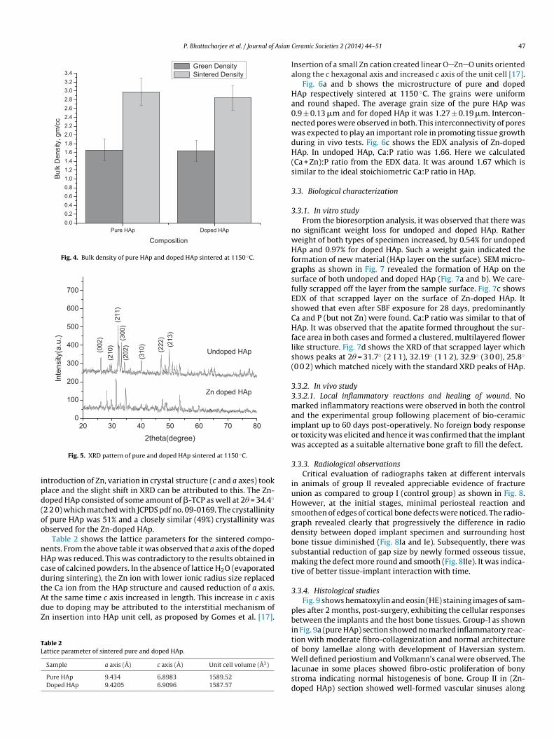
The bulk density of the green and sintered specimens was cal-ulated using gravimetric method by measuring the volume andass. Fig. 4 shows the sintered as well as green density. The green
ensity of both specimens was found to 1.66 g/cm3.Bulk density after sintering in both the cases was closely similar.
n undoped HAp sintered density was 2.98 g/cm3 and for dopedAp density was 2.85 g/cm3. As such there was no prominent effectf dopant on densification.
The sintered specimens were also studied with XRD to assesshe effect of heating on the formation of various phases. X-ray pow-er diffraction analysis of the sintered undoped and Zn-doped HAptructures are presented in Fig. 5. The major peak was observedt 31.77◦ (2 1 1) for pure HAp and it was phase pure. In Zn-
oped HAp for (3 0 0) plane peak was observed at 2� = 32.9◦ andther two peaks got slightly shifted (31.5◦ for (2 1 1) plane and6.1◦ for (0 0 2) plane). In this context it may be noted that with00020004
0
100
200
558
596
1030
141516
50
3453
558
61063
7
1045
1415
3575
Doped HAp
Pure HAp
% T
Wavenu mber, cm-1
Fig. 3. FTIR pattern of pure and doped HAp powder calcined at 800 ◦C.
P. Bhattacharjee et al. / Journal of Asian
pAHdepoDpAHeruP0.00.20.40.60.81.01.21.41.61.82.02.22.42.62.83.03.23.4
Bul
k D
ensi
ty, g
m/c
c
Composition
Gree n Density Sintered Density
Fig. 4. Bulk density of pure HAp and doped HAp sintered at 1150 ◦C.
20 30 40 50 60 70 800
100
200
300
400
500
600
700
(202
)(3
00)
(213
)(2
22)
(310
)
(211
)(2
10)
(002
)
Zn do ped HAp
Undoped HAp
Inte
nsity
(a.u
.)
2theta(de gree)
ipd(oo
nHcdtAdZ
TL
Ia
Ha0nwdH(s
3
3
nwHfgsfEsCHfls(
33maiow
3
iuHsgdbsmt
3
Fig. 5. XRD pattern of pure and doped HAp sintered at 1150 ◦C.
ntroduction of Zn, variation in crystal structure (c and a axes) tooklace and the slight shift in XRD can be attributed to this. The Zn-oped HAp consisted of some amount of �-TCP as well at 2� = 34.4◦
2 2 0) which matched with JCPDS pdf no. 09-0169. The crystallinityf pure HAp was 51% and a closely similar (49%) crystallinity wasbserved for the Zn-doped HAp.
Table 2 shows the lattice parameters for the sintered compo-ents. From the above table it was observed that a axis of the dopedAp was reduced. This was contradictory to the results obtained inase of calcined powders. In the absence of lattice H2O (evaporateduring sintering), the Zn ion with lower ionic radius size replacedhe Ca ion from the HAp structure and caused reduction of a axis.
t the same time c axis increased in length. This increase in c axisue to doping may be attributed to the interstitial mechanism ofn insertion into HAp unit cell, as proposed by Gomes et al. [17].able 2attice parameter of sintered pure and doped HAp.
Sample a axis (A) c axis (A) Unit cell volume (A3)
Pure HAp 9.434 6.8983 1589.52Doped HAp 9.4205 6.9096 1587.57
pbitoWlsd
Ceramic Societies 2 (2014) 44–51 47
nsertion of a small Zn cation created linear O Zn O units orientedlong the c hexagonal axis and increased c axis of the unit cell [17].
Fig. 6a and b shows the microstructure of pure and dopedAp respectively sintered at 1150 ◦C. The grains were uniformnd round shaped. The average grain size of the pure HAp was.9 ± 0.13 �m and for doped HAp it was 1.27 ± 0.19 �m. Intercon-ected pores were observed in both. This interconnectivity of poresas expected to play an important role in promoting tissue growthuring in vivo tests. Fig. 6c shows the EDX analysis of Zn-dopedAp. In undoped HAp, Ca:P ratio was 1.66. Here we calculated
Ca + Zn):P ratio from the EDX data. It was around 1.67 which isimilar to the ideal stoichiometric Ca:P ratio in HAp.
.3. Biological characterization
.3.1. In vitro studyFrom the bioresorption analysis, it was observed that there was
o significant weight loss for undoped and doped HAp. Rathereight of both types of specimen increased, by 0.54% for undopedAp and 0.97% for doped HAp. Such a weight gain indicated the
ormation of new material (HAp layer on the surface). SEM micro-raphs as shown in Fig. 7 revealed the formation of HAp on theurface of both undoped and doped HAp (Fig. 7a and b). We care-ully scrapped off the layer from the sample surface. Fig. 7c showsDX of that scrapped layer on the surface of Zn-doped HAp. Ithowed that even after SBF exposure for 28 days, predominantlya and P (but not Zn) were found. Ca:P ratio was similar to that ofAp. It was observed that the apatite formed throughout the sur-
ace area in both cases and formed a clustered, multilayered flowerike structure. Fig. 7d shows the XRD of that scrapped layer whichhows peaks at 2� = 31.7◦ (2 1 1), 32.19◦ (1 1 2), 32.9◦ (3 0 0), 25.8◦
0 0 2) which matched nicely with the standard XRD peaks of HAp.
.3.2. In vivo study
.3.2.1. Local inflammatory reactions and healing of wound. Noarked inflammatory reactions were observed in both the control
nd the experimental group following placement of bio-ceramicmplant up to 60 days post-operatively. No foreign body responser toxicity was elicited and hence it was confirmed that the implantas accepted as a suitable alternative bone graft to fill the defect.
.3.3. Radiological observationsCritical evaluation of radiographs taken at different intervals
n animals of group II revealed appreciable evidence of fracturenion as compared to group I (control group) as shown in Fig. 8.owever, at the initial stages, minimal periosteal reaction and
moothen of edges of cortical bone defects were noticed. The radio-raph revealed clearly that progressively the difference in radioensity between doped implant specimen and surrounding hostone tissue diminished (Fig. 8Ia and Ie). Subsequently, there wasubstantial reduction of gap size by newly formed osseous tissue,aking the defect more round and smooth (Fig. 8IIe). It was indica-
ive of better tissue-implant interaction with time.
.3.4. Histological studiesFig. 9 shows hematoxylin and eosin (HE) staining images of sam-
les after 2 months, post-surgery, exhibiting the cellular responsesetween the implants and the host bone tissues. Group-I as shown
n Fig. 9a (pure HAp) section showed no marked inflammatory reac-ion with moderate fibro-collagenization and normal architecturef bony lamellae along with development of Haversian system.
ell defined periostium and Volkmann’s canal were observed. Theacunae in some places showed fibro-ostic proliferation of bonytroma indicating normal histogenesis of bone. Group II in (Zn-oped HAp) section showed well-formed vascular sinuses along

48 P. Bhattacharjee et al. / Journal of Asian Ceramic Societies 2 (2014) 44–51
Fig. 6. SEM micrograph of (a) pure HAp and (b) doped HAp. (c) EDX pattern of doped HAp.
Fig. 7. SEM micrograph of apatite formation (shown by arrows) on surface of (a) undoped and (b) doped HAp ceramics received after 4 weeks in simulated body fluid (SBF).(c) EDX and (d) XRD patterns of apatite layer in case of Zn-doped HAp.

P. Bhattacharjee et al. / Journal of Asian Ceramic Societies 2 (2014) 44–51 49
Fig. 8. Serial radiographs at different days of interval post-operatively of I – pure HAp and
Fig. 9. Hematoxylin and eosin (HE) staining histological images at 2 months post-operatively (a, Haversian system and b, bony lamella).
wmTs
3
bTmdatwriibbtsF
3
syImfreeb
II – doped HAp (a – 0th day, b – 15th day, c – 30th day, d – 45th day, e – 60th day).
ith osseous stroma mainly Haversian canal, canaliculi and inter-ingling sinusoidal spaces (lacunar spaces) as shown in Fig. 9b.
he bony tissues showed well-arranged mineralization along withupporting cellular infiltration making a favorable healing process.
.4. SEM analysis
Fig. 10 represents the SEM micrograph of the interfacial regionetween the implant and the bone after 2 months post-operatively.he SEM photographs as shown by Fig. 10b have confirmed that theineralization process for the Zn-doped HAp implant developed
irect contact with the surrounding bone. In the sacrificed animalsfter 60 days of implantation, adequate roughness was evident onhe surface of the both doped HAp implants and the bone tissuesere found to grow into the finest irregularities. This study also
evealed comparatively less bone in-growth and reduced mechan-cal anchorage to undoped HAp implant compared to doped HApmplants. Zone 1 showed the implant surface while zone 2 showedone tissue. In pure HAp, even after 60 days gap (zone 3 in Fig. 10a)etween implant and bone was clearly visible but in doped HAp,he whole interface was almost filled with newly formed bone tis-ue. Only some small gaps were found to still persist (zone 3 inig. 10b).
.5. Oxytetracycline labeling study
Fig. 11 shows fluorochrome labeling images at 2 months post-urgery, exhibiting new bone formation as evidenced by goldenellow fluorescence where as sea green color appears host bone.n control, the activity of new bone formation was moderate. The
icrograph for Zn-doped HAp showed more amount of new boneormation from both the ends as evidenced by golden yellow fluo-
escence. The osseous tissues originate both from periosteal andndosteal surface of bone; however, its intensity was more onndosteal side. The defect was entirely filled with newly formedone and appeared as homogenous golden yellow area.
50 P. Bhattacharjee et al. / Journal of Asian Ceramic Societies 2 (2014) 44–51
Fig. 10. SEM micrograph showing interfacial region between the HAp implant and surrounding hard tissue, a – pure HAp and b – doped HAp (1 – implant, 2 – new bonegrowth, 3 – gap between implant and host tissue).
p at 4
3
Hpsf
tZw
Fi
Fig. 11. Fluorochrome labeling images of (a) pure HAp and (b) doped HA
.6. Mechanical push-out testing
The interfacial bond strength of implanted pure and Zn-doped
Ap was measured by mechanical push-out test after 2 monthsost operatively. Fig. 12a shows the average values of interfacialtrength. The average interfacial strength of undoped HAp wasound to be low (2.38 ± 0.47 MPa). It was an average value fromtdFf
ig. 12. (a) Interfacial strength of pure and doped HAp and (b) specimen coming out of thnterface region).
months post-operatively: new bone (arrow 1) and host bone (arrow 2).
hree such samples. The average value of interfacial strength forn-doped HAp implants was higher (5.78 ± 0.77 MPa) comparedith undoped HAp. This marked improvement may be attributed
o more prominent tissue-implant interaction, which was evi-ent from fluorochrome study and scanning electron microscopy.ig. 12b shows a typical view of a doped HAp sample coming outrom the bone during mechanical push-out test. Implant and the
e bone during mechanical push-out test (1 – specimen, 2 – surrounding tissue, 3 –

Asian
hisIa
tippoRmbipabmpiZa
4
iowtbidd
satrhcTa
R
[
[
[
[
[
[[
P. Bhattacharjee et al. / Journal of
ost tissues were shown by arrow by 1 and 2 respectively. Thenterfacial region shows clear signs of intense rubbing with someymptoms of wear and tear during ejection as shown by arrow 3.t indicated toward the presence of strong bonding between bonend implant.
From the above-mentioned observations, it may be summarizedhat in both the cases the microstructures revealed gross similar-ty, e.g., small, uniform, uni-axed grains with lot of interconnectedores. There was no significant difference from X-ray crystallogra-hy. But still Zn-doped HAp showed marked superiority in termsf new bone formation and bonding between bone and implant.esults of mechanical push-out test confirmed it in a quantitativeanner. So the observed difference in in vivo performance can only
e explained with the influence of Zn. Zn ion increases the activ-ty of aminoacyl-tRNA synthetase, an enzyme which is involved inrotein biosynthesis including the production of osteocalcin, IGF-1nd TGF-� in osteoblastic cells [18]. Zn has also role of targeting IGFinding proteins to the cell surface. It increases the activity of vita-in D3-dependent promoters in osteoblasts [19]. Furthermore the
resence of Zn ion triggers release of certain cytokines that havenhibitory effect on osteoclastic bone resorption. So at one hand,n promotes new bone formation by enhancing osteoblast activitynd at the same time reduces bone resorption rate.
. Conclusion
The results of the present study revealed that addition of Znn HAp did not alter the major phase of the HAp. The crystallinityf Zn-doped HAp was not that high however the sintered densityas nearly 85% with lot of interconnected pores. Zn ions par-
ially replaced Ca ion and caused variation in the lattice structure,
oth in calcined powders and in sintered samples. The variationn lattice structure was found to be influenced by temperatureue to temperature dependent evaporation of lattice H2O. Foroped specimens, (Ca + Zn)/P ratio was around 1.67, close to the
[[
[
Ceramic Societies 2 (2014) 44–51 51
toichiometric Ca/P ratio in HAp. SBF study showed multilayeredpatite formation on the surface of doped HAp specimens. Sequen-ial radiological investigations showed progressively diminishingelative radio density of implant and surrounding host bone. SEM,isto-pathological study and oxytetracycline labeling study furtheronfirmed superior bone–implant attachment of Zn-doped HAp.his was due to stimulatory effect of Zn ion on new bone formationnd inhibitory effect to osteoclastic bone resorption.
eferences
[1] J.F. Shackelford, Mater. Sci. Forum, 293, 1–4 (1999).[2] T.S.B. Narasaraju and D.E. Phebe, J. Mater. Sci., 31, 1–21 (1996).[3] F.S. Kaplan, W.C. Hayes, T.M. Keaveny, A. Boskey, T.A. Einhorn and J.P. Iannotti,
American Association of Orthopaedic Surgeons (1994), pp. 127–184.[4] A. Ito, H. Kawamura, M. Otsuka, M. Ikeuchi, H. Ohgushi, K. Ishikawa, K. Onuma,
N. Kanzaki, Y. Sogo and N. Ichinose, Mater. Sci. Eng. C, 22, 21–25 (2002).[5] A. Bigi, E. Foresti, M. Gandolfi, M. Gazzano and N. Roveri, J. Inorg. Biochem., 58,
49–58 (1995).[6] T.J. Webster, C. Ergun, R.H. Doremus and R. Bizios, J. Biomed. Mater. Res., 59,
312–317 (2002).[7] A. Bandyopadhyay, E.A. Withey, J. Moore and S. Bose, Mater. Sci. Eng. C, 27,
14–17 (2007).[8] A. Ito, K. Ojima, H. Naito, N. Ichinose and T. Tateishi, J. Biomed. Mater. Res., 50,
178–183 (2000).[9] X. Wang, A. Ito, Y. Sogo, X. Li and A. Oyane, Acta Biomater., 6, 962–968 (2010).10] Y. Li, Q. Li, S. Zhu, E. Lu, J. Li, G. Feng, Y. Liao and J. Hu, Biomaterials, 31,
9006–9014 (2010).11] T.J. Matsumoto, S.-H. An, T. Ishimoto, T. Nakano, T. Matsumoto and S. Imazato,
Dent. Mater., 27, e205–e212 (2011).12] G. Lusvardi, D. Zaffe, L. Menabue, C. Bertoldi, G. Malavasi and U. Consolo, Acta
Biomater., 5, 419–428 (2009).13] E. Landi, A. Tampieri, G. Celotti and S. Sprio, J. Eur. Ceram. Soc., 20, 2377–2387
(2000).14] B.D. Cullity, Elements of X-ray Diffraction, 2nd ed., Addison-Wesley, Reading,
MA (1978).15] S. Koutsopoulos, J. Biomed. Mater. Res. A., 62, 600–612 (2002).16] F. Miyaji, Y. Kono and Y. Suyama, Mater. Res. Bull., 40, 209–220 (2005).
17] S. Gomes, J.M. Nedelec and G. Renaudin, Acta Biomater., 8, 1180–1189 (2012).18] A. Ito, M. Otsuka, H. Kawamura, M. Ikeuchi, H. Ohgushi, Y. Sogo and N. Ichinose,Curr. Appl. Phys., 5, 402–406 (2005).19] W. Lutz, M.F. Burritt, D.E. Nixon, P.C. Kao and R. Kumar, Biochem. Biophys. Res.
Commun., 271, (1) 1–7 (2000).


![SYNTHESIS, CHARACTERIZATION AND PHOTOCATALYTIC ... · stability [12]. The photocatalytic activity of zinc oxide and metal doped zinc oxide nanoparticles is increased by increasing](https://img.pdfslide.net/doc/110x75/5f046a957e708231d40ddca4/synthesis-characterization-and-photocatalytic-stability-12-the-photocatalytic.jpg)