Embed Size (px)
Citation preview
ANKLE BLOCK
Developing Countries Regional Anesthesia Lecture Series
Daniel D. Moos CRNA, Ed.D. U.S.A. [email protected] Lecture 15
Soli Deo Gloria

Disclaimer
Every effort was made to ensure that material and information contained in this presentation are correct and up-to-date. The author can not accept liability/responsibility from errors that may occur from the use of this information. It is up to each clinician to ensure that they provide safe anesthetic care to their patients.
Introduction to the Ankle Block Common peripheral nerve block Useful for procedures that do not require
a tourniquet above the ankle Indicated for orthopedic and podiatry
procedures of the distal foot Purely sensory block Painful block Conscious sedation….don’t over sedate!
The ankle block involves blockade of 5 peripheral nerves
Posterior tibial nerve Sural nerve Superficial peroneal nerve Deep peroneal nerve Saphenous nerve
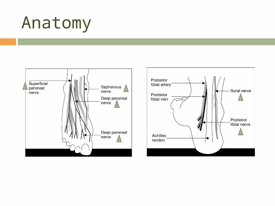
Anatomy
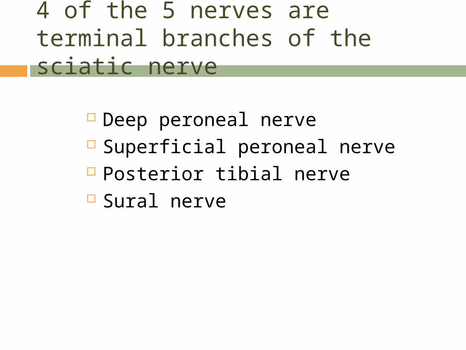
4 of the 5 nerves are terminal branches of the sciatic nerve
Deep peroneal nerve Superficial peroneal nerve Posterior tibial nerve Sural nerve
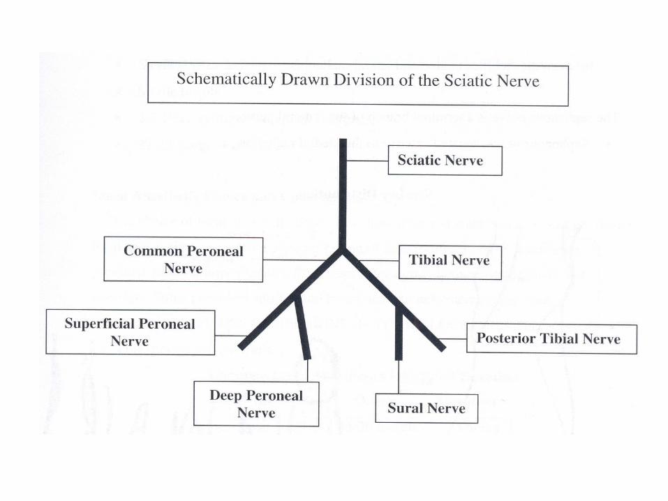
The remaining nerve is the terminal branch of femoral nerve
Saphenous nerve
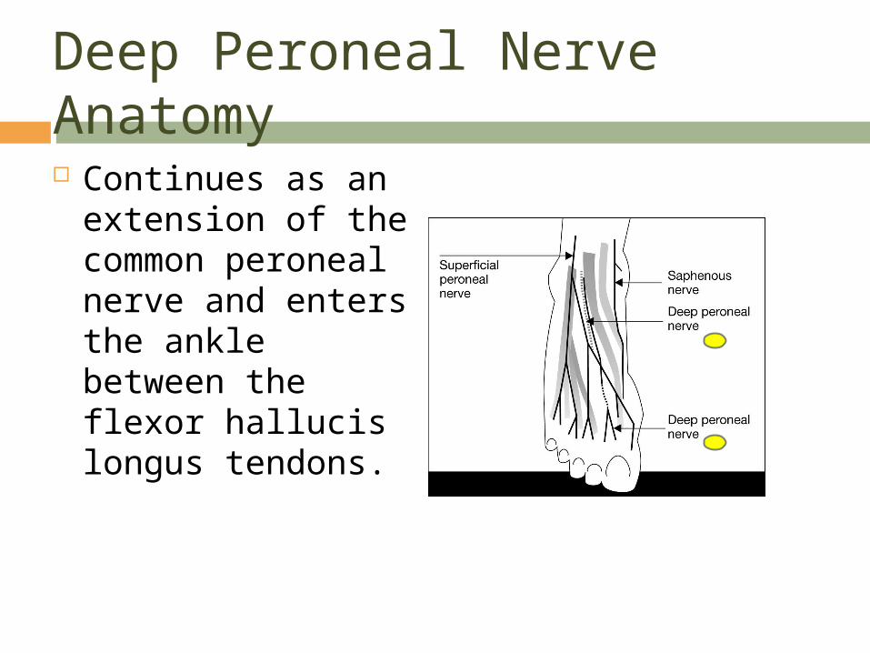
Deep Peroneal Nerve Anatomy Continues as an
extension of the common peroneal nerve and enters the ankle between the flexor hallucis longus tendons.
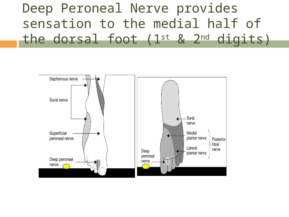
Deep Peroneal Nerve provides sensation to the medial half of the dorsal foot (1st & 2nd digits)
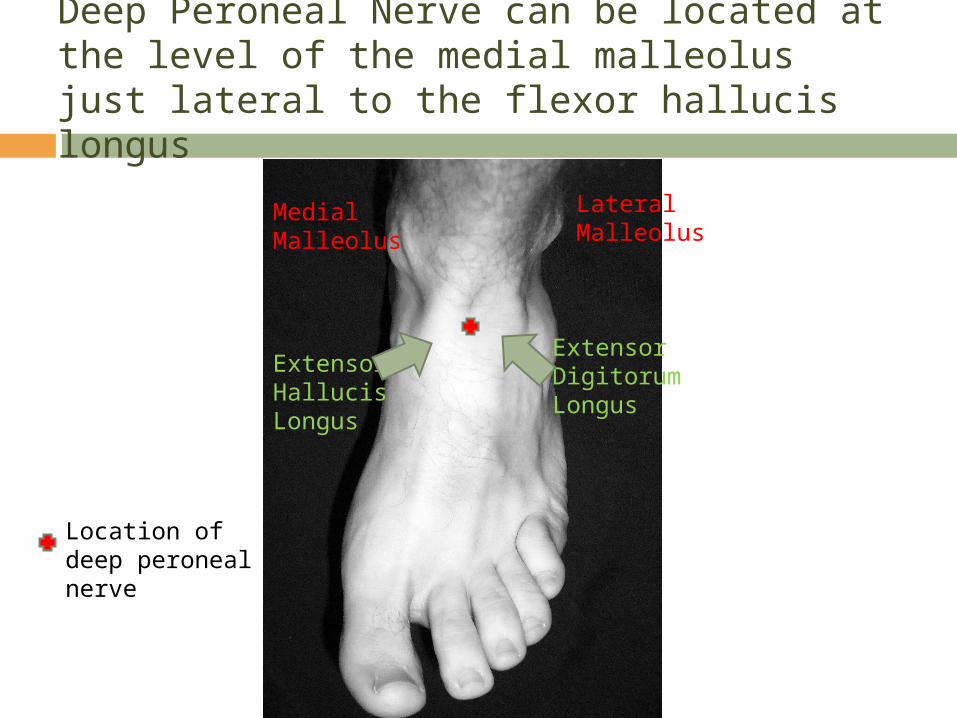
Deep Peroneal Nerve can be located at the level of the medial malleolus just lateral to the flexor hallucis longus
Location of deep peroneal nerve
Medial Malleolus
Extensor Hallucis Longus
LateralMalleolus
Extensor Digitorum Longus
Superficial Peroneal Nerve Anatomy Extension of the common peroneal nerve
and enters the ankle lateral to the extensor digitorum longus at the level of the lateral malleolus
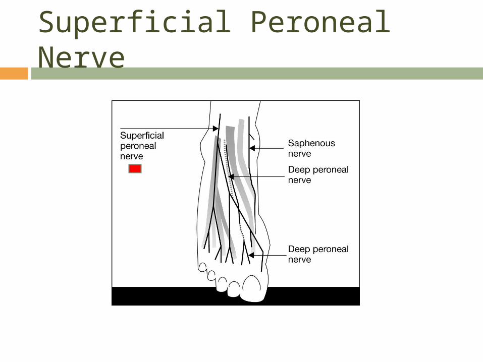
Superficial Peroneal Nerve
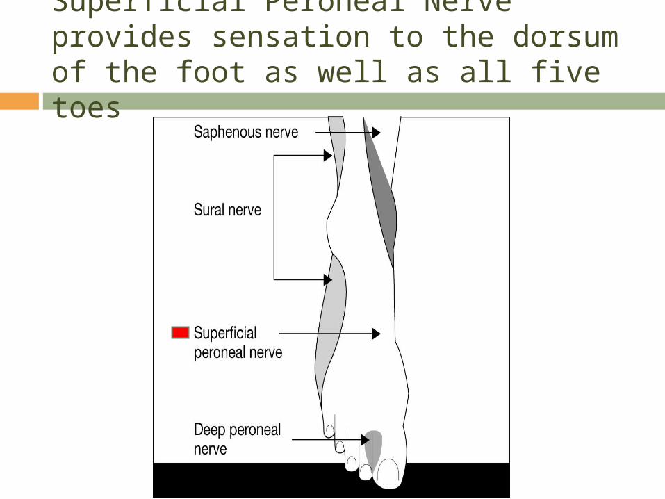
Superficial Peroneal Nerve provides sensation to the dorsum of the foot as well as all five toes
Posterior Tibial Nerve Anatomy Extension of the tibial nerve and enters
the foot posterior to the medial malleolus, dividing into the lateral and medial plantar nerves.
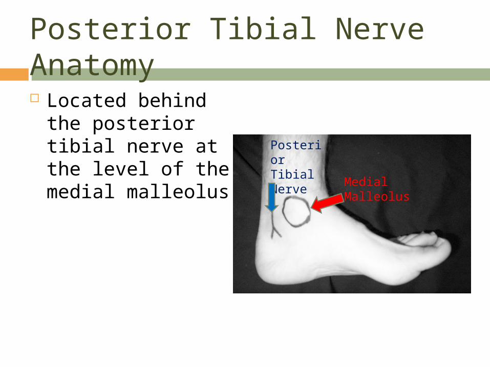
Posterior Tibial Nerve Anatomy Located behind the
posterior tibial nerve at the level of the medial malleolus
Medial Malleolus
Posterior Tibial Nerve
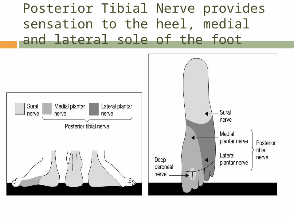
Posterior Tibial Nerve provides sensation to the heel, medial and lateral sole of the foot
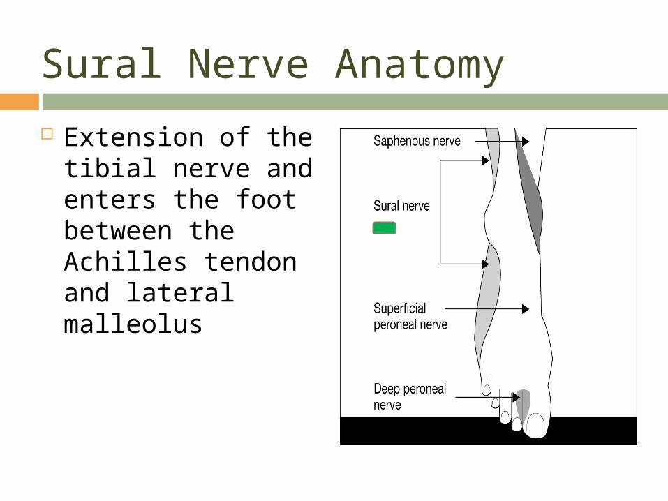
Sural Nerve Anatomy
Extension of the tibial nerve and enters the foot between the Achilles tendon and lateral malleolus
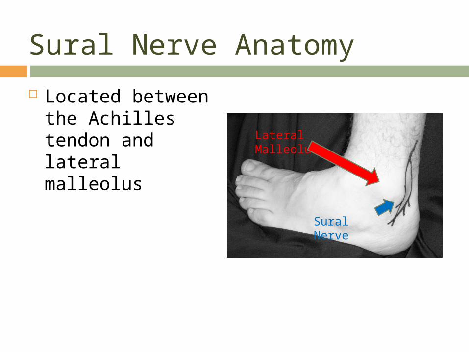
Sural Nerve Anatomy
Located between the Achilles tendon and lateral malleolus
Lateral Malleolus
Sural Nerve
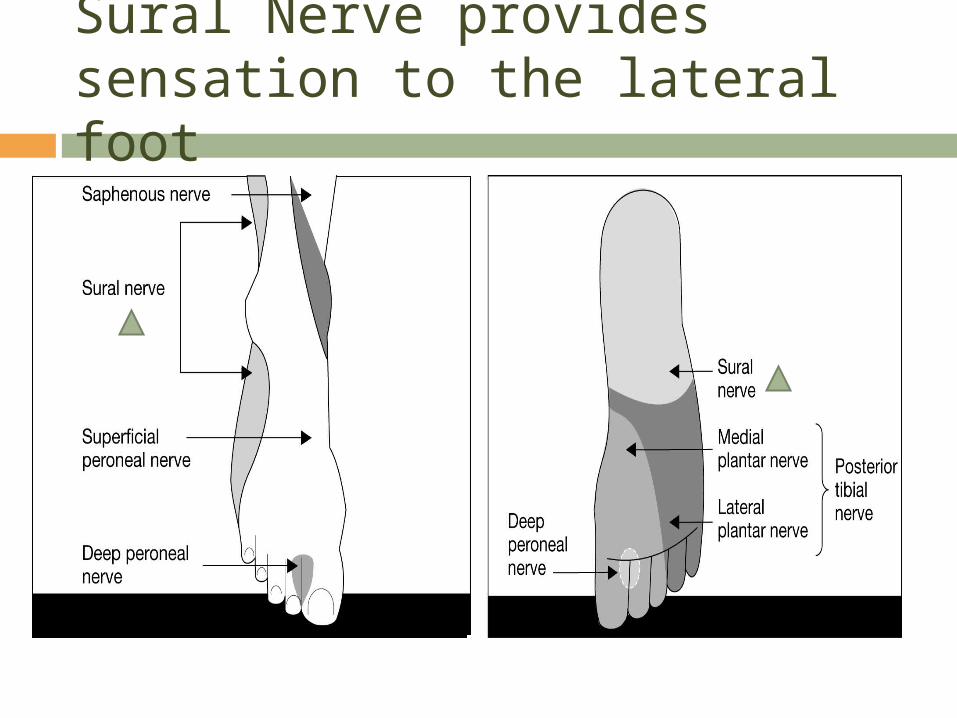
Sural Nerve provides sensation to the lateral foot
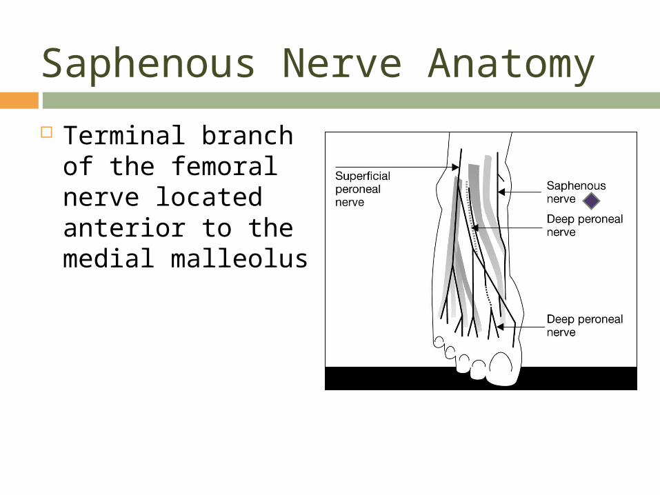
Saphenous Nerve Anatomy
Terminal branch of the femoral nerve located anterior to the medial malleolus
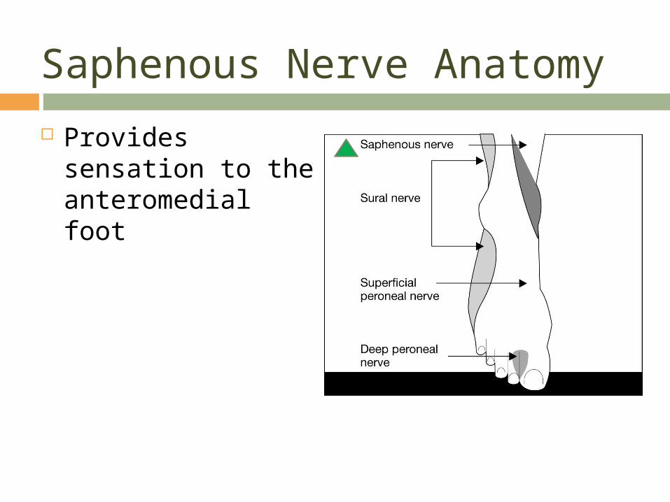
Saphenous Nerve Anatomy
Provides sensation to the anteromedial foot
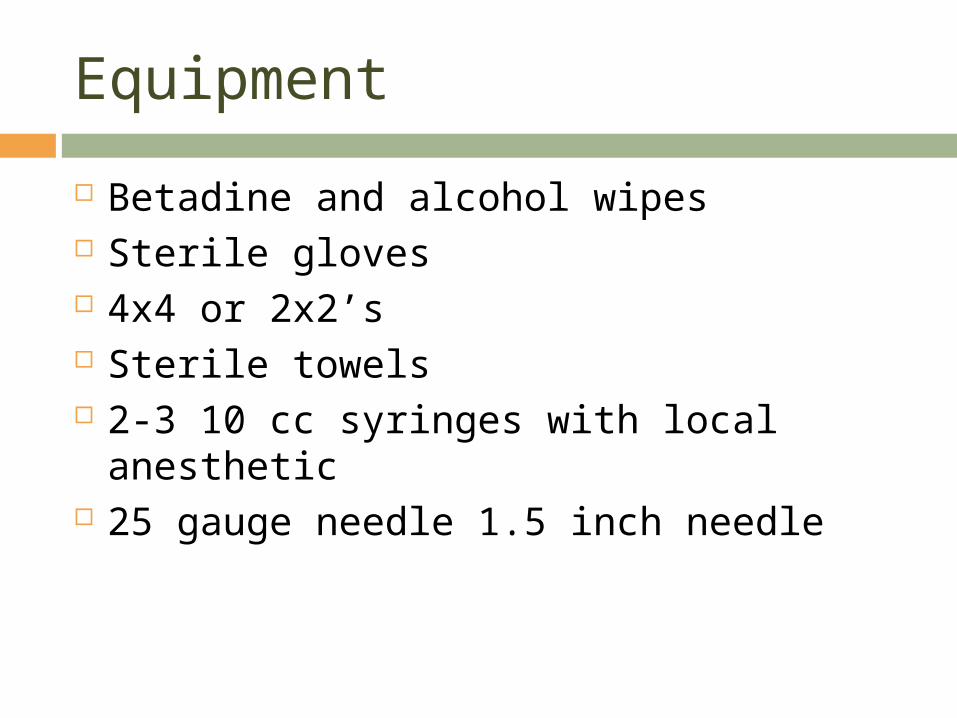
Equipment
Betadine and alcohol wipes Sterile gloves 4x4 or 2x2’s Sterile towels 2-3 10 cc syringes with local anesthetic 25 gauge needle 1.5 inch needle
Choice of Local Anesthetic
Depends on the length of time you wish block to last
Longer acting local anesthetics may take longer for onset
May wish to mix a local anesthetic that has faster onset with a longer acting local anesthetic
Sodium bicarbonate may help speed onset
NEVER USE EPINEPHRINE!
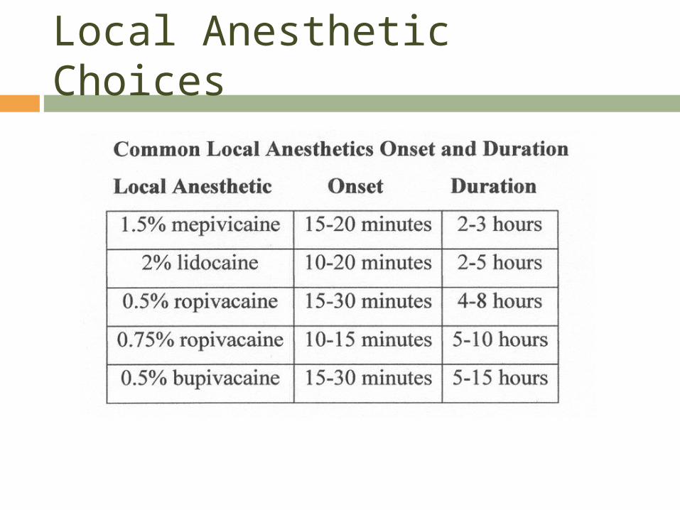
Local Anesthetic Choices
Considerations
Be careful with volume- tourniquet effect Caution in patients with peripheral
vascular disease and diabetics Care with patient with infection- risk of
tracking infection to healthy tissue and local anesthetic not working due to acidotic tissue
Positioning the foot
Position the foot so you have access to all 5 nerves
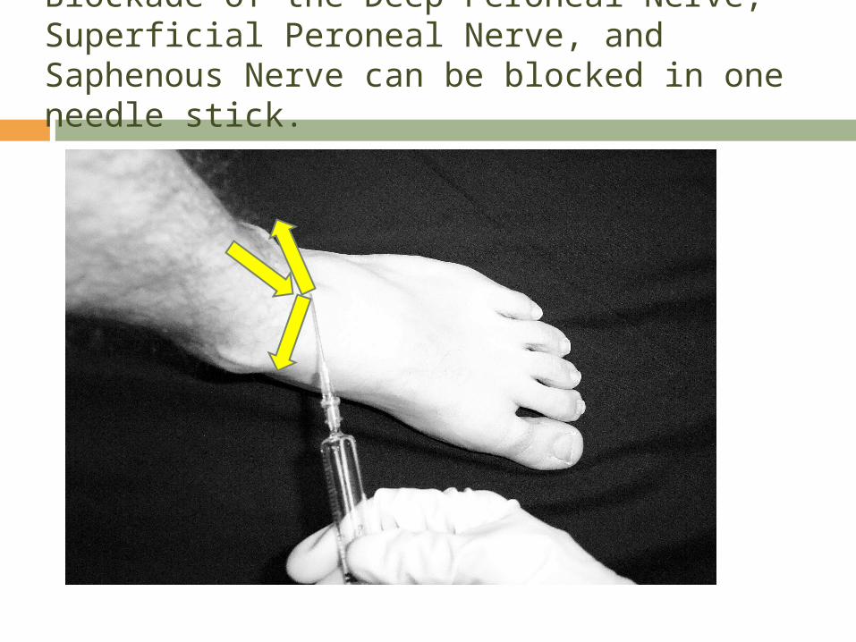
Blockade of the Deep Peroneal Nerve, Superficial Peroneal Nerve, and Saphenous Nerve can be blocked in one needle stick.
Deep Peroneal Nerve Block
Draw a line between the two malleoli Identify the extensor hallucis longus
tendon and the extensor digitorum longus muscle
Palpate the anterior tibial artery
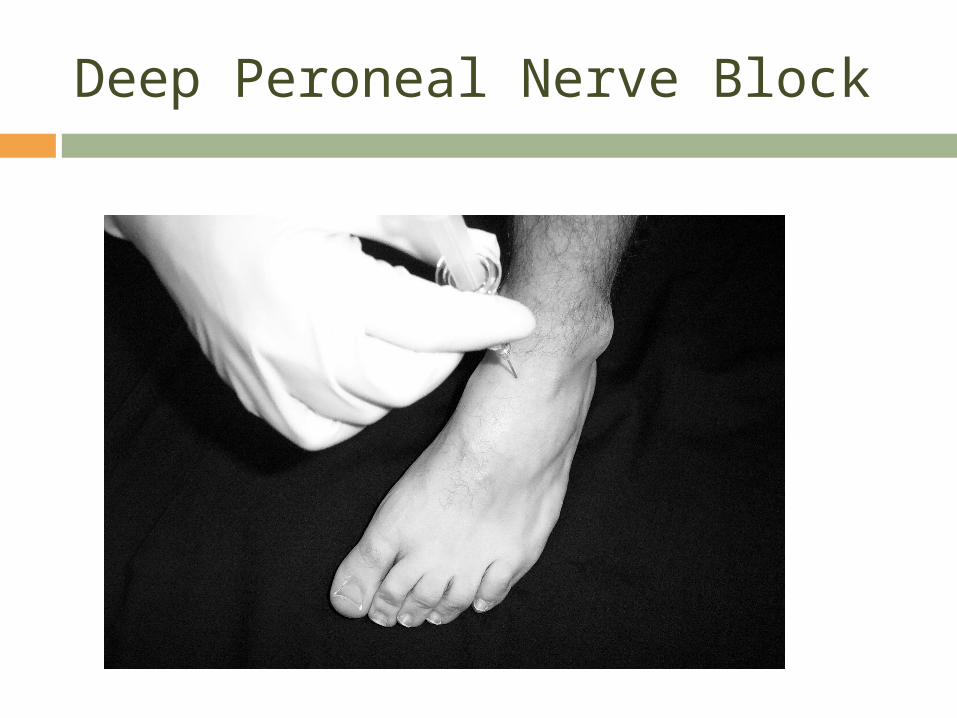
Deep Peroneal Nerve Block
Place a skin wheal of local anesthetic lateral to the artery
Advance the needle perpendicular, aspirating for blood and deposit 3-5 ml of local anesthetic deep to the extensor retinaculum
May choose to fan the injection in this area, avoiding the artery
Deep Peroneal Nerve Block
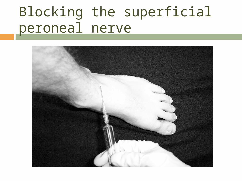
Blocking the Superficial Peroneal Nerve Bring the needle back and direct it
superficially in a lateral fashion towards the lateral malleolus depositing 3-5 ml of local anesthetic subcutaneously
Blocking the superficial peroneal nerve
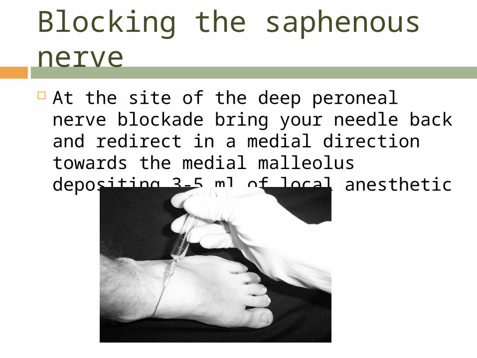
Blocking the saphenous nerve At the site of the deep peroneal nerve
blockade bring your needle back and redirect in a medial direction towards the medial malleolus depositing 3-5 ml of local anesthetic
Blocking the posterior tibial nerve Warn your patient to hold still in case a
paresthesia is elicited. Movement at this time may result in trauma to the nerve.
Identify the posterior tibial artery at the level of the medial malleolus and advance the needle in a posterolateral manner slowly.
If a paresthesia is elicited withdraw the needle slightly and inject 3-5 ml. Make sure the patient does not have pain as this may imply an intraneural injection.
If no paresthesia is elicited than inject 7-10 ml as you withdraw the needle. A paresthesia is not essential to a successful block.
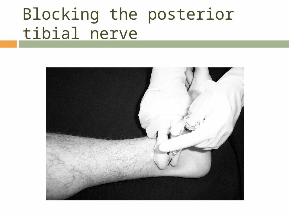
Blocking the posterior tibial nerve
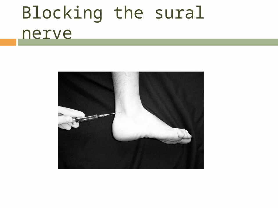
Blocking the sural nerve
Identify the lateral malleolus and the Achilles tendon
Insert needle superficially lateral to the tendon and in the direction of the lateral malleolus.
Inject 5-10 ml of local anesthetic subcutaneously as you withdraw the needle
Blocking the sural nerve
Complications
Discomfort to the patient Injury to a “numb” foot after discharge Nerve injury or paresthesia’s Hematoma and vascular injury Infection Intravascular injection Block failure

Conclusion
Easy to administer Effective anesthesia Often performed with much less local
anesthetic than what textbooks advocate
Metatarsal Block
A metatarsal block may supplement an ankle block if a nerve distribution has been missed.
Never use epinephrine containing solutions. This can result in ischemia of the digits.
Place a small skin wheal at the site of injection on the dorsum of the foot.
Advance the needle while injecting local anesthetic parallel to the metatarsal bone. Do not go through the surface of the sole of the foot!
Metatarsal Block
The individual nerves are located closer to the sole of the foot than the dorsum.
A total of 3-5 ml of local anesthetic solution may be deposited.
The same procedure should occur on the other side of the metatarsal of the location that anesthesia is desired.
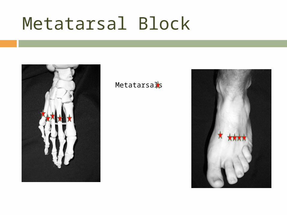
Metatarsal Block
Metatarsals

References
Burkard J, Lee Olson R., Vacchiano CA. Regional Anesthesia. In Nurse Anesthesia 3rd edition. Nagelhout, JJ & Zaglaniczny KL ed. Pages 977-1030.
Morgan, G.E. & Mikhail, M. (2006). Peripheral nerve blocks. In G.E. Morgan et al
Clinical Anesthesiology, 4th edition. New York: Lange Medical Books.
Wedel, D.J. & Horlocker, T.T. Nerve blocks. In Miller’s Anesthesia 6th edtion. Miller,
RD ed. Pages 1685-1715. Elsevier, Philadelphia, Penn. 2005.
Wedel, D.J. & Horlocker, T.T. (2008). Peripheral nerve blocks. In D.E. Longnecker et al (eds) Anesthesiology. New York: McGraw-Hill Medical.


![Dunwald 479-9076 beagenie@charter.net GRAPHICS Bill Porter [phone n/a] silverfoxbill@outlook.com POWERPOINT Mark Simonds 955](https://img.pdfslide.net/doc/110x75/5b08f19c7f8b9a404d8d18ea/dunwald-479-9076-beageniecharternet-graphics-bill-porter-phone-na-silverfoxbilloutlookcom.jpg)