Embed Size (px)
Citation preview
319
Int. J. Odontostomat.,11(3):319-325, 2017.
Ankylosis of Permanent First Molar: Diagnosis by Cone Beam Computed Tomography
Anquilosis del Primer Molar Permanente: Diagnóstico por
Tomografía Computarizada con Haz Cónico
Eliana Dantas da Costa1; Priscila Dias Peyneau1; Francielle Silvestre Verner2;Rafael Binato Junqueira3; Solange Maria de Almeida1 & Glaucia Maria Bovi Ambrosano4
DA COSTA, E. D.; PEYNEAU, P. D.; VERNER, F. S.; JUNQUEIRA, R. B.; DE ALMEIDA, S. M. & AMBROSANO, G. M. B.Ankylosis of permanent first molar: Diagnosis by cone beam computed tomography. Int. J. Odontostomat., 11(3):319-325,2017.
ABSTRACT: Ankylosis is an anomaly of tooth eruption characterized by the fusion of cementum and alveolar bone,and may affect from small regions to the entire root surface. Clinical assessment combined with imaging exams can aiddiagnosis. Radiographic testing enables assessing only proximal regions of possibly affected roots. Whereas cone beamcomputed tomography (CBCT) allows a three-dimensional assessment of axial, coronal, and sagittal planes of all dentalextension, eliminating thus overlapping images and helping to confirm the correct diagnosis. The present study contains acase report of a male patient with ankylosis in tooth 16 diagnosed by CBCT, aiming at providing information for dentistsabout this anomaly, its characteristics and situations in which CBCT should be indicated.
KEY WORDS: dental ankylosis, diagnosis, cone beam computed tomography.
INTRODUCTION
Dentoalveolar ankylosis is an anomaly of den-tal eruption, caused by mechanical failure of eruption(Frazier-Bowers et al., 2007), characterized by thefusion of cementum and alveolar bone (Loriato et al.,2009; Pithon & Bernardes, 2011; Chae & Paeng, 2012;Guimarães et al., 2015; Mohadeb et al., 2016), withdestruction of the periodontal ligament (Pithon &Bernardes; Lin et al., 2014) and gradual replacementby bone tissue (Alves et al., 2011; Bertl et al., 2012;Silva, 2015).
Ankylosis can affect both sexes (Silva) and it ismore frequent in deciduous teeth (10:1) (Loriato et al.;Cohen-Levy et al., 2011; Bertl et al.; Silva), withfrequency between 1.5 % and 9.9 % (Atobi et al., 2009;Loriato et al.), especially when the permanentsuccessor is missing (Bertl et al.), affecting mainly
mandibular deciduous first molars (Loriato et al.; Alveset al.; Bertl et al.; Parisay et al., 2013), followed bysecond mandibular and maxillary deciduous molars(Parisay et al.).
Traumatic lesions are considered the main fac-tor for development of ankylosis (Atobe et al., 2009;Cohen-Levy et al.; Chae & Paeng; Gault et al., 2013;Lin et al.; Mohadeb et al.; Silva). Moreover, these arealso considered causal factors: metabolic disorders,local deficiency in vertical alveolar bone growth (Atobeet al.; Loriato et al.; Cohen-Levy et al.; Alves et al.;Parisay et al.; Lin et al.; Guimarães et al.), failure oferuption force (Alves et al.), genetic factors (Atobe etal.; Cohen-Levy et al.; Chae & Paeng; Lin et al.;Guimarães et al.; Silva), focal inflammation/infection,chemical/thermal irritants (Cohen-Levy et al.; Gault et
1 Division of Oral Radiology, Department of Oral Diagnosis, Piracicaba Dental School, State University of Campinas, Piracicaba, São Paulo, Brazil.2 Division of Oral Radiology, Department of Dentistry, Federal University of Juiz de Fora, Advanced campus Governador Valadares, Juiz de Fora,
Minas Gerais, Brazil.3 Division of Endodontic, Department of Dentistry, Federal University of Juiz de Fora, Advanced campus Governador Valadares, Juiz de Fora, Minas
Gerais, Brazil.4 Department of Social Dentistry, Piracicaba Dental School, State University of Campinas, Piracicaba, São Paulo, Brazil.
320
al.; Parisay et al.; Silva), and soft tissue pressure (Atobeet al.; Cohen-Levy et al.). It should also be noted thatankylosis may affect teeth that have been extractedand subsequently reimplanted (Takahashi et al., 2005;Chae & Paeng; Silva), but in these cases ankylosis isnot considered an anomaly of dental eruption.
The differential diagnosis of ankylosis includesother types of eruption disorders, such as: impaction,primary retention, secondary retention and primaryeruption failure (Janssen et al., 2014). In the impaction,the eruption is impaired due to the presence of aphysical barrier or due to the ectopic position of thetooth. Primary and secondary retentions are causedby failures in the eruption mechanisms, and in theprimary type, the interruption of the eruption occursbefore the tooth reaches the gingival level. In thesecondary, the tooth ceases the eruption whenreaching the gingival level (Magnusson & Kjellberg,2009). The primary failure of the eruption mainly affectsthe posterior teeth (Rhoads et al., 2013). In such ca-ses, the tooth may erupt partially, becoming relativelysubmerged or may not erupt (Frazier-Bowers et al.;Smith et al., 2012). Although there is no cement unionto the alveolar bone in these cases, they present clinicalfeatures very similar to ankylosis.
However, in the primary failure, the treatment ismore complex, frequently involving all the teethadjacent to the affected tooth, resulting in lateral openedbite (Frazier-Bowers et al.; Rhoads et al.; Smith et al.).This disorder is caused by genetic changes associatedwith the parathyroid hormone (PTHPR1) (Rhoads etal.; Smith et al.).
Primary diagnosis of ankylosed teeth is madeby clinical assessment through mobility and percussiontests, in these cases the tooth makes a metallic sound(Campbell et al., 2005; Lim et al., 2008; Berl et al.,2012; Gault et al.; Lin et al.; Mohadeb et al.; Silva),unlike healthy teeth, in which the sound is neutralizedby periodontal ligament (Berl et al., 2012; Parisay etal.; Silva).
However, since only one third of the patientspresent these clinical signs (Cohen-Levy et al.; Lin etal.), it is necessary to carry out imaging tests to confirmclinical signs of ankylosis. Although periapical x-raysare daily used in dental clinics, their value is limited fordiagnosisof ankylosed teeth (Paris et al., 2010; Cohen-Levy et al.; Berl et al., 2012; Cha et al., 2012; Gault etal.; Lin et al.; Mohadeb et al.), because ankylotic areasonly visible when it is present in proximal surfaces of
the root, not being possible to identify lingual/palataland buccal surfaces (Takahashi et al.; Cohen-Levy etal.; Berl et al. 2012; Silva et al.). Besides, in thisexamination the presence of ankylosis becomesevident only when at least 20 % of the root surface isaffected (Campbell et al.; Cohen-Levy et al.; Mohadebet al.).
Whereas computed tomography (CT) isconsidered superior to radiography (Kirziolu et al.,2007) because of the possibility of a three-dimensio-nal evaluation of the structures, allowing preciseobservation of the whole extent of the root surface(Paris et al.; Chae & Paeng; Silva), the degree ofirregularity of the root, and the relationship betweenthe root and the adjacent teeth (Gault et al.), helping inearly diagnosis of ankylosed teeth. With equalaccuracy, cone beam computed tomography (CBCT)can also be used for the diagnosis of ankylosed teeth(Balaji, 2013; Hashim et al., 2013; Silva), having asadvantage the lower dose of radiation (Holberg et al.,2005; Balaji; Kamburoglu, 2015; Silva) and lower costcompared with CT (Holberg et al.; Balaji; Kamburoglu).
In the initial stages, the primary failure of eruptionand ankylosis may have similar clinical presentations(Frazier-Bowers et al.). With the lack of accessibility ofgenetic tests in dental offices to confirm the occurrenceof primary failure (Rhoads et al.), it is important resortingto accurate imaging methods, such as CBCT, todistinguish these two eruption disorders, providing thecorrect diagnosis and the best treatment options forpatients.
Considering the limitation in the literatureconcerning the diagnosis of ankylosis using CBCT (Sil-va), the aim of this study was to provide information todentists on the importance and the situations when touse CBCT in diagnosis of ankylosed teeth. CASE REPORT
Male patient, 16 years old was referred for clinicof the Oral Radiology to perform CBCT, in order to planthe next steps of the orthodontic treatment. When hewas referred, infraocclusion of the maxillary right firstmolar was reported and it did not respond to orthodonticforced eruption. The patient did not report pain andremembered the trauma that occurred in the region,when he was a child during a football game. The patientdid not present relevant data regarding systemic
DA COSTA, E. D. ; PEYNEAU, P. D.; VERNER, F. S.; JUNQUEIRA, R. B.; DE ALMEIDA, S. M. & AMBROSANO, G. M. B. Ankylosis of permanent first molar: Diagnosis by conebeam computed tomography. Int. J. Odontostomat., 11(3):319-325, 2017.
321
situation and there were no cases in thefamily of infraocclusion of teeth.
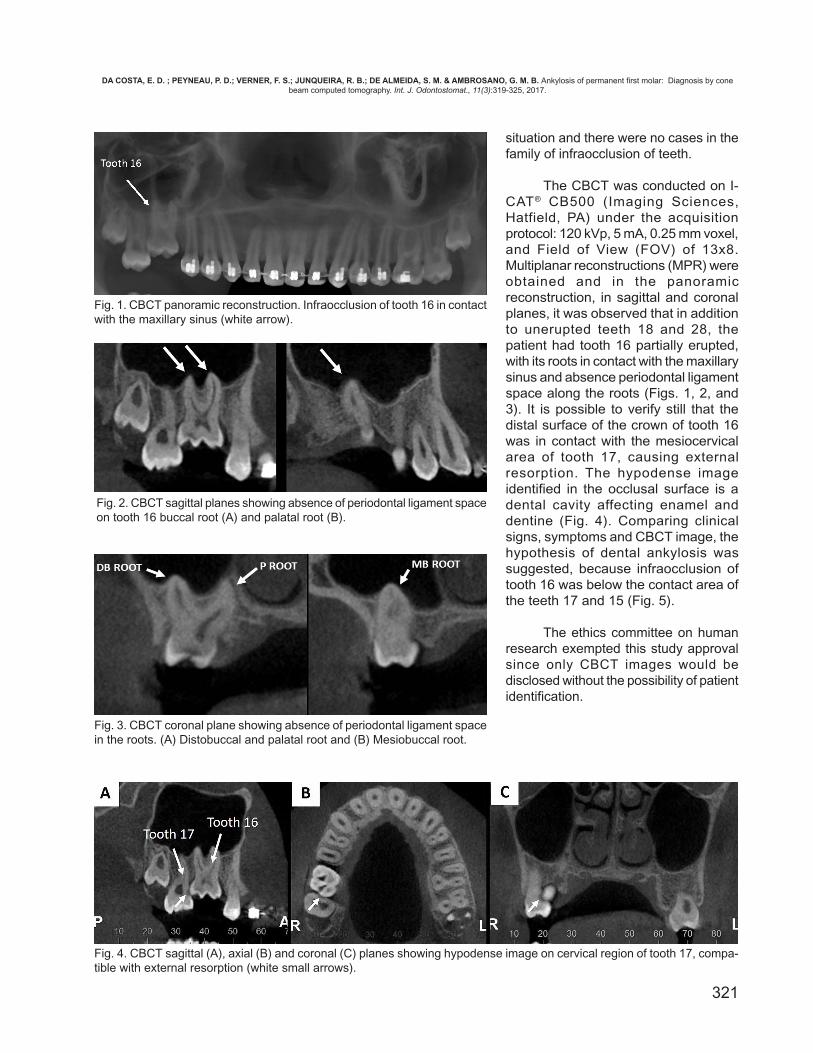
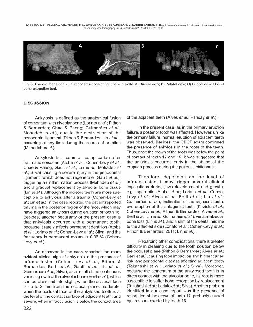
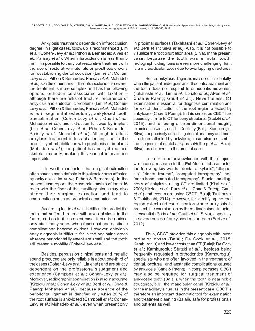
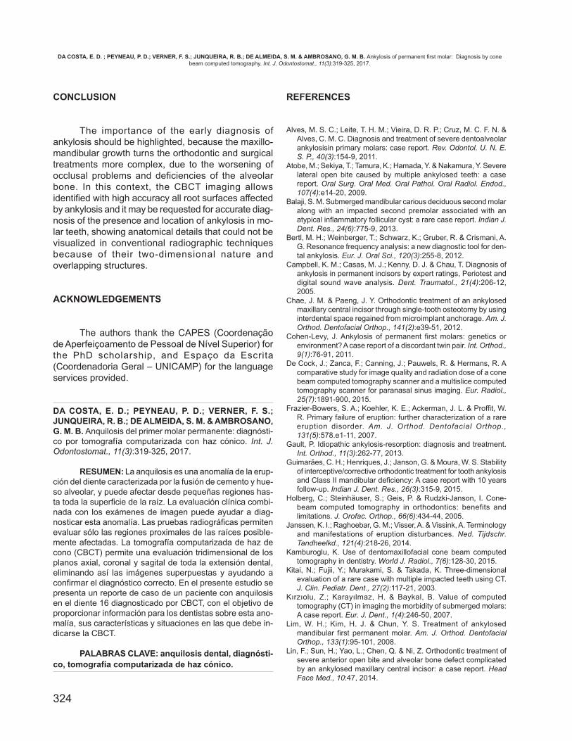
The CBCT was conducted on I-CAT® CB500 (Imaging Sciences,Hatfield, PA) under the acquisitionprotocol: 120 kVp, 5 mA, 0.25 mm voxel,and Field of View (FOV) of 13x8.Multiplanar reconstructions (MPR) wereobtained and in the panoramicreconstruction, in sagittal and coronalplanes, it was observed that in additionto unerupted teeth 18 and 28, thepatient had tooth 16 partially erupted,with its roots in contact with the maxillarysinus and absence periodontal ligamentspace along the roots (Figs. 1, 2, and3). It is possible to verify still that thedistal surface of the crown of tooth 16was in contact with the mesiocervicalarea of tooth 17, causing externalresorption. The hypodense imageidentified in the occlusal surface is adental cavity affecting enamel anddentine (Fig. 4). Comparing clinicalsigns, symptoms and CBCT image, thehypothesis of dental ankylosis wassuggested, because infraocclusion oftooth 16 was below the contact area ofthe teeth 17 and 15 (Fig. 5).
The ethics committee on humanresearch exempted this study approvalsince only CBCT images would bedisclosed without the possibility of patientidentification.
Fig. 1. CBCT panoramic reconstruction. Infraocclusion of tooth 16 in contactwith the maxillary sinus (white arrow).
Fig. 2. CBCT sagittal planes showing absence of periodontal ligament spaceon tooth 16 buccal root (A) and palatal root (B).
Fig. 3. CBCT coronal plane showing absence of periodontal ligament spacein the roots. (A) Distobuccal and palatal root and (B) Mesiobuccal root.
Fig. 4. CBCT sagittal (A), axial (B) and coronal (C) planes showing hypodense image on cervical region of tooth 17, compa-tible with external resorption (white small arrows).
DA COSTA, E. D. ; PEYNEAU, P. D.; VERNER, F. S.; JUNQUEIRA, R. B.; DE ALMEIDA, S. M. & AMBROSANO, G. M. B. Ankylosis of permanent first molar: Diagnosis by conebeam computed tomography. Int. J. Odontostomat., 11(3):319-325, 2017.
322
DISCUSSION
Ankylosis is defined as the anatomical fusionof cementum with alveolar bone (Loriato et al.; Pithon& Bernardes; Chae & Paeng; Guimarães et al.;Mohadeb et al.), due to the destruction of theperiodontal ligament (Pithon & Bernardes; Lin et al.),occurring at any time during the course of eruption(Mohadeb et al.).
Ankylosis is a common complication aftertraumatic episodes (Atobe et al.; Cohen-Levy et al.;Chae & Paeng; Gault et al.; Lin et al.; Mohadeb etal.; Silva) causing a severe injury in the periodontalligament, which does not regenerate (Gault et al.),triggering an inflammation process (Mohadeb et al.)and a gradual replacement by alveolar bone tissue(Lin et al.). Although the incisors teeth are more sus-ceptible to ankylosis after a trauma (Cohen-Levy etal.; Lin et al.), in the case reported the patient reportedtrauma in the posterior region of the face, which mayhave triggered ankylosis during eruption of tooth 16.Besides, another peculiarity of the present case isthat ankylosis occurred with a permanent tooth,because it rarely affects permanent dentition (Atobeet al.; Loriato et al.; Cohen-Levy et al.; Silva) and thefrequency in permanent molars is 0.06 % (Cohen-Levy et al.).
As observed in the case reported, the moreevident clinical sign of ankylosis is the presence ofinfraocclusion (Cohen-Levy et al.; Pithon &Bernardes; Bertl et al.; Gault et al.; Lin et al.;Guimarães et al.; Silva), as a result of the continuousvertical growth of the alveolar bone (Bertl et al.), whichcan be classified into slight, when the occlusal faceis up to 2 mm from the occlusal plane; moderate,when the occlusal face of the ankylosed tooth is atthe level of the contact surface of adjacent teeth; andsevere, when infraocclusion is below the contact area
of the adjacent teeth (Alves et al.; Parisay et al.).
In the present case, as in the primary eruptionfailure, a posterior tooth was affected. However, unlikethe primary failure, normal eruption of adjacent teethwas observed. Besides, the CBCT exam confirmedthe presence of ankylosis in the roots of the teeth.Thus, once the crown of the tooth was below the pointof contact of teeth 17 and 15, it was suggested thatthe ankylosis occurred early in the phase of theeruption process during the patient's childhood.
Therefore, depending on the level ofinfraocclusion, it may trigger several clinicalimplications during jaws development and growth,e.g., open bite (Atobe et al.; Loriato et al.; Cohen-Levy et al.; Alves et al.; Bertl et al.; Lin et al.;Guimarães et al.), inclination of the adjacent teeth,overeruption of the antagonist tooth (Kirziolu et al.;Cohen-Levy et al.; Pithon & Bernardes; Alves et al.;Bertl et al.; Lin et al.; Guimarães et al.), vertical alveolarbone loss (Lin et al.), and a shift of the dental midlineto the affected side (Loriato et al.; Cohen-Levy et al.;Pithon & Bernardes, 2011; Lin et al.).
Regarding other complications, there is greaterdifficulty in cleaning due to the tooth position belowthe occlusal plane (Pithon & Bernardes; Alves et al.;Bertl et al.), causing food impaction and higher cariesrisk, and periodontal disease affecting adjacent teeth(Takahashi et al.; Loriato et al.; Silva). Moreover,because the cementum of the ankylosed tooth is indirect contact with the alveolar bone, its root is moresusceptible to suffer bone resorption by replacement(Takahashi et al.; Loriato et al.; Silva). Another problemidentified in our case report was the presence ofresorption of the crown of tooth 17, probably causedby pressure exerted by tooth 16.
Fig. 5. Three-dimensional (3D) reconstructions of right hemi maxilla. A) Buccal view; B) Palatal view; C) Buccal view: Use ofbone extraction tool.
DA COSTA, E. D. ; PEYNEAU, P. D.; VERNER, F. S.; JUNQUEIRA, R. B.; DE ALMEIDA, S. M. & AMBROSANO, G. M. B. Ankylosis of permanent first molar: Diagnosis by conebeam computed tomography. Int. J. Odontostomat., 11(3):319-325, 2017.
323
Ankylosis treatment depends on infraocclusiondegree. In slight cases, follow up is recommended (Limet al.; Cohen-Levy et al.; Pithon & Bernardes; Alves etal.; Parisay et al.). When infraocclusion is less than 5mm, it is possible to carry out restorative treatment withthe use of restorative materials or prosthetic crownsfor reestablishing dental occlusion (Lim et al.; Cohen-Levy et al.; Pithon & Bernardes; Parisay et al.; Mohadebet al.). On the other hand, if the infraocclusion is severe,the treatment is more complex and has the followingoptions: orthodontics associated with luxation –although there are risks of fracture, recurrence ofankylosis and endodontic problems (Lim et al.; Cohen-Levy et al.; Pithon & Bernardes; Parisay et al.; Mohadebet al.); segmental osteotomy; ankylosed toothtransplantation (Cohen-Levy et al.; Gault et al.;Mohadeb et al.); and extraction followed by implant(Lim et al.; Cohen-Levy et al.; Pithon & Bernardes;Parisay et al.; Mohadeb et al.). Although in adultsankylosis treatment is less challenging due to thepossibility of rehabilitation with prosthesis or implants(Mohadeb et al.), the patient has not yet reachedskeletal maturity, making this kind of interventionimpossible.
It is worth mentioning that surgical extractionoften causes bone defects in the alveolar area affectedby ankylosis (Lim et al.; Pithon & Bernardes). In thepresent case report, the close relationship of tooth 16roots with the floor of the maxillary sinus may alsohinder their surgical extraction and lead tocomplications such as oroantral communication.
According to Lin et al. it is difficult to predict if atooth that suffered trauma will have ankylosis in thefuture, and as in the present case, it can be noticedonly after many years when functional and aestheticcomplications become evident. However, ankylosisearly diagnosis is difficult, for in the beginning areasabsence periodontal ligament are small and the toothstill presents mobility (Cohen-Levy et al.).
Besides, percussion clinical tests and metallicsound produced are only reliable in about one-third ofthe cases (Cohen-Levy et al.; Lin et al.) and are strictlydependent on the professional’s judgment andexperience (Campbell et al.; Cohen-Levy et al.).Moreover, radiographic examination is also inaccurate(Kirziolu et al.; Cohen-Levy et al.; Bertl et al.; Chae &Paeng; Mohadeb et al.), because absence of theperiodontal ligament is identified only when 20 % ofthe root surface is ankylosed (Campbell et al.; Cohen-Levy et al.; Mohadeb et al.), even when present only
in proximal surfaces (Takahashi et al.; Cohen-Levy etal.; Bertl et al.; Silva et al.). Also, it is not possible tovisualize the root bifurcation area (Silva). In the presentcase, because the tooth was a molar tooth,radiographic diagnosis is even more challenging, for itis a multiradicular tooth due to overlapping structures.
Hence, ankylosis diagnosis may occur incidentally,when the patient undergoes an orthodontic treatment andthe tooth does not respond to orthodontic movement(Takahashi et al.; Lim et al.; Loriato et al.; Alves et al.;Chae & Paeng; Gault et al.). Nevertheless, CTexamination is essential for diagnosis confirmation andfor exact identification of the root region affected byankyloses (Chae & Paeng). In this sense, as CBCT hasaccuracy similar to CT for bony structures (Stutzki et al.,2015), and for being a three-dimensional imagingexamination widely used in Dentistry (Balaji; Kamburoglu;Silva), for precisely assessing dental anatomy and bonestructures affected by ankylosis, it can also be used inthe diagnosis of dental ankylosis (Holberg et al.; Balaji;Silva), as observed in the present case.
In order to be acknowledged with the subject,we made a research in the PubMed database, usingthe following key words: “dental ankylosis”, “diagno-sis”, “dental trauma”, “computed tomography”, and“cone beam computed tomography”. Studies on diag-nosis of ankylosis using CT are limited (Kitai et al.,2003; Kirziolu et al.; Paris et al.; Chae & Paeng; Gaultet al.) and even more using CBCT (Balaji; Tsukiboshi& Tsukiboshi, 2014). However, for identifying the rootregion extent and exact location where ankylosis ispresent, the examination by three-dimensional imagingis essential (Paris et al.; Gault et al.; Silva), especiallyin severe cases of ankylosed molar teeth (Berl et al.,2012).
Thus, CBCT provides this diagnosis with lowerradiation doses (Balaji; De Cock et al., 2015;Kamburoglu) and lower costs than CT (Balaji; De Cocket al.; Kamburoglu; Stutzki et al.), besides beingfrequently requested in orthodontics (Kamburoglu),specialists who are often involved in the treatment ofdental, occlusal, and aesthetic complications causedby ankylosis (Chae & Paeng). In complex cases, CBCTmay also be required for surgical treatment ofankylosed teeth (Balaji), when the tooth is near noblestructures, e.g., the mandibular canal (Kirziolu et al.)or the maxillary sinus, as in the present case. CBCT istherefore an important diagnostic tool for examinationand treatment planning (Balaji), safe for professionalsand patients as well.
DA COSTA, E. D. ; PEYNEAU, P. D.; VERNER, F. S.; JUNQUEIRA, R. B.; DE ALMEIDA, S. M. & AMBROSANO, G. M. B. Ankylosis of permanent first molar: Diagnosis by conebeam computed tomography. Int. J. Odontostomat., 11(3):319-325, 2017.
324
CONCLUSION
The importance of the early diagnosis ofankylosis should be highlighted, because the maxillo-mandibular growth turns the orthodontic and surgicaltreatments more complex, due to the worsening ofocclusal problems and deficiencies of the alveolarbone. In this context, the CBCT imaging allowsidentified with high accuracy all root surfaces affectedby ankylosis and it may be requested for accurate diag-nosis of the presence and location of ankylosis in mo-lar teeth, showing anatomical details that could not bevisualized in conventional radiographic techniquesbecause of their two-dimensional nature andoverlapping structures.
ACKNOWLEDGEMENTS
The authors thank the CAPES (Coordenaçãode Aperfeiçoamento de Pessoal de Nível Superior) forthe PhD scholarship, and Espaço da Escrita(Coordenadoria Geral – UNICAMP) for the languageservices provided.
DA COSTA, E. D.; PEYNEAU, P. D.; VERNER, F. S.;JUNQUEIRA, R. B.; DE ALMEIDA, S. M. & AMBROSANO,G. M. B. Anquilosis del primer molar permanente: diagnósti-co por tomografía computarizada con haz cónico. Int. J.Odontostomat., 11(3):319-325, 2017.
RESUMEN: La anquilosis es una anomalía de la erup-ción del diente caracterizada por la fusión de cemento y hue-so alveolar, y puede afectar desde pequeñas regiones has-ta toda la superficie de la raíz. La evaluación clínica combi-nada con los exámenes de imagen puede ayudar a diag-nosticar esta anomalía. Las pruebas radiográficas permitenevaluar sólo las regiones proximales de las raíces posible-mente afectadas. La tomografía computarizada de haz decono (CBCT) permite una evaluación tridimensional de losplanos axial, coronal y sagital de toda la extensión dental,eliminando así las imágenes superpuestas y ayudando aconfirmar el diagnóstico correcto. En el presente estudio sepresenta un reporte de caso de un paciente con anquilosisen el diente 16 diagnosticado por CBCT, con el objetivo deproporcionar información para los dentistas sobre esta ano-malía, sus características y situaciones en las que debe in-dicarse la CBCT.
PALABRAS CLAVE: anquilosis dental, diagnósti-co, tomografía computarizada de haz cónico.
REFERENCES
Alves, M. S. C.; Leite, T. H. M.; Vieira, D. R. P.; Cruz, M. C. F. N. &Alves, C. M. C. Diagnosis and treatment of severe dentoalveolarankylosisin primary molars: case report. Rev. Odontol. U. N. E.S. P., 40(3):154-9, 2011.
Atobe, M.; Sekiya, T.; Tamura, K.; Hamada, Y. & Nakamura, Y. Severelateral open bite caused by multiple ankylosed teeth: a casereport. Oral Surg. Oral Med. Oral Pathol. Oral Radiol. Endod.,107(4):e14-20, 2009.
Balaji, S. M. Submerged mandibular carious deciduous second molaralong with an impacted second premolar associated with anatypical inflammatory follicular cyst: a rare case report. Indian J.Dent. Res., 24(6):775-9, 2013.
Bertl, M. H.; Weinberger, T.; Schwarz, K.; Gruber, R. & Crismani, A.G. Resonance frequency analysis: a new diagnostic tool for den-tal ankylosis. Eur. J. Oral Sci., 120(3):255-8, 2012.
Campbell, K. M.; Casas, M. J.; Kenny, D. J. & Chau, T. Diagnosis ofankylosis in permanent incisors by expert ratings, Periotest anddigital sound wave analysis. Dent. Traumatol., 21(4):206-12,2005.
Chae, J. M. & Paeng, J. Y. Orthodontic treatment of an ankylosedmaxillary central incisor through single-tooth osteotomy by usinginterdental space regained from microimplant anchorage. Am. J.Orthod. Dentofacial Orthop., 141(2):e39-51, 2012.
Cohen-Levy, J. Ankylosis of permanent first molars: genetics orenvironment? A case report of a discordant twin pair. Int. Orthod.,9(1):76-91, 2011.
De Cock, J.; Zanca, F.; Canning, J.; Pauwels, R. & Hermans, R. Acomparative study for image quality and radiation dose of a conebeam computed tomography scanner and a multislice computedtomography scanner for paranasal sinus imaging. Eur. Radiol.,25(7):1891-900, 2015.
Frazier-Bowers, S. A.; Koehler, K. E.; Ackerman, J. L. & Proffit, W.R. Primary failure of eruption: further characterization of a rareeruption disorder. Am. J. Orthod. Dentofacial Orthop.,131(5):578.e1-11, 2007.
Gault, P. Idiopathic ankylosis-resorption: diagnosis and treatment.Int. Orthod., 11(3):262-77, 2013.
Guimarães, C. H.; Henriques, J.; Janson, G. & Moura, W. S. Stabilityof interceptive/corrective orthodontic treatment for tooth ankylosisand Class II mandibular deficiency: A case report with 10 yearsfollow-up. Indian J. Dent. Res., 26(3):315-9, 2015.
Holberg, C.; Steinhäuser, S.; Geis, P. & Rudzki-Janson, I. Cone-beam computed tomography in orthodontics: benefits andlimitations. J. Orofac. Orthop., 66(6):434-44, 2005.
Janssen, K. I.; Raghoebar, G. M.; Visser, A. & Vissink, A. Terminologyand manifestations of eruption disturbances. Ned. Tijdschr.Tandheelkd., 121(4):218-26, 2014.
Kamburoglu, K. Use of dentomaxillofacial cone beam computedtomography in dentistry. World J. Radiol., 7(6):128-30, 2015.
Kitai, N.; Fujii, Y.; Murakami, S. & Takada, K. Three-dimensionalevaluation of a rare case with multiple impacted teeth using CT.J. Clin. Pediatr. Dent., 27(2):117-21, 2003.
Kırzıolu, Z.; Karayılmaz, H. & Baykal, B. Value of computedtomography (CT) in imaging the morbidity of submerged molars:A case report. Eur. J. Dent., 1(4):246-50, 2007.
Lim, W. H.; Kim, H. J. & Chun, Y. S. Treatment of ankylosedmandibular first permanent molar. Am. J. Orthod. DentofacialOrthop., 133(1):95-101, 2008.
Lin, F.; Sun, H.; Yao, L.; Chen, Q. & Ni, Z. Orthodontic treatment ofsevere anterior open bite and alveolar bone defect complicatedby an ankylosed maxillary central incisor: a case report. HeadFace Med., 10:47, 2014.
DA COSTA, E. D. ; PEYNEAU, P. D.; VERNER, F. S.; JUNQUEIRA, R. B.; DE ALMEIDA, S. M. & AMBROSANO, G. M. B. Ankylosis of permanent first molar: Diagnosis by conebeam computed tomography. Int. J. Odontostomat., 11(3):319-325, 2017.
325
Loriato, L. B.; Machado, A. W.; Souki, B. Q. & Pereira, T. J. Latediagnosis of dentoalveolar ankylosis: impact on effectiveness andefficiency of orthodontic treatment. Am. J. Orthod. DentofacialOrthop., 135(6):799-808, 2009.
Magnusson, C. & Kjellberg, H. Impaction and retention of secondmolars: diagnosis, treatment and outcome. A retrospective follow-up study. Angle Orthod., 79(3):422-7, 2009.
Mohadeb, J. V.; Somar, M. & He, H. Effectiveness of decoronationtechnique in the treatment of ankylosis: A systematic review. Dent.Traumatol., 32(4):255-63, 2016.
Paris, M.; Trunde, F.; Bossard, D.; Farges, J. C. & Coudert, J. L.Dental ankylosis diagnosed by CT with tridimensionalreconstructions. J. Radiol., 91(6):707-11, 2010.
Parisay, I.; Kebriaei, F.; Varkesh, B.; Soruri, M. & Ghafourifard, R.Management of a severely submerged primary molar: a casereport. Case Rep. Dent., 2013:796242, 2013.
Pithon, M. M. & Bernardes, L. A. Treatment of ankylosis of themandibular first molar with orthodontic traction immediately aftersurgical luxation. Am. J. Orthod. Dentofacial Orthop., 140(3):396-403, 2011.
Rhoads, S. G.; Hendricks, H. M. & Frazier-Bowers, S. A. Establishingthe diagnostic criteria for eruption disorders based on geneticand clinical data. Am. J. Orthod. Dentofacial Orthop., 144(2):194-202, 2013.
Silva, D. C. Exames por Imagem no Diagnóstico de AnquiloseAlveolodentária: Relato de Caso Clínico. Trabalho de Conclusão(Especialização em Radiologia Odontológica e Imaginologia).Porto Alegre, Faculdade de Odontologia – Universidade Federaldo Rio Grande do Sul, 2015.
Smith, C. P.; Al-Awadhi, E. A. & Garvey, M. T. An atypical presentationof mechanical failure of eruption of a mandibular permanent molar:diagnosis and treatment case report. Eur. Arch. Paediatr. Dent.,13(3):152-6, 2012.
Stutzki, M.; Jahns, E., Mandapathil, M. M.; Diogo, I.; Werner, J. A. &Güldner, C. Indications of cone beam CT in head and neckimaging. Acta Otolaryngol., 135(12):1337-43, 2015.
Takahashi, T.; Takagi, T. & Moriyama, K. Orthodontic treatment of atraumatically intruded tooth with ankylosis by traction after surgicalluxation. Am. J. Orthod. Dentofacial Orthop., 127(2):233-41, 2005.
Tsukiboshi, M. & Tsukiboshi, T. Bone morphology after delayed toothreplantation - case series. Dent. Traumatol., 30(6):477-83, 2014.
Corresponding author:Eliana Dantas da CostaDivision of Oral RadiologyDepartment of Oral DiagnosisPiracicaba Dental SchoolState University of CampinasAv. Limeira, 901Areião, Piracicaba, SPZip Code 13414-018BRAZIL E-mail: [email protected] Received: 21-04-2017Accepted: 19-06-2017
DA COSTA, E. D. ; PEYNEAU, P. D.; VERNER, F. S.; JUNQUEIRA, R. B.; DE ALMEIDA, S. M. & AMBROSANO, G. M. B. Ankylosis of permanent first molar: Diagnosis by conebeam computed tomography. Int. J. Odontostomat., 11(3):319-325, 2017.


![Diagnosis and treatment planning for primary molar ... · teeth [2,8]. Dentoalveolar ankylosis is an eruption anomaly defined as the union of the tooth root to the surrounding bone](https://img.pdfslide.net/doc/110x75/600234fbd67ba13d5a22ddc9/diagnosis-and-treatment-planning-for-primary-molar-teeth-28-dentoalveolar.jpg)