Embed Size (px)
Citation preview
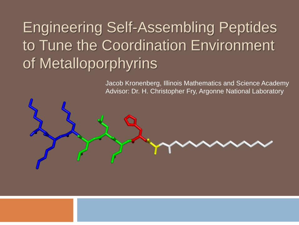
Engineering Self-Assembling Peptides
to Tune the Coordination Environment
of MetalloporphyrinsJacob Kronenberg, Illinois Mathematics and Science Academy
Advisor: Dr. H. Christopher Fry, Argonne National Laboratory
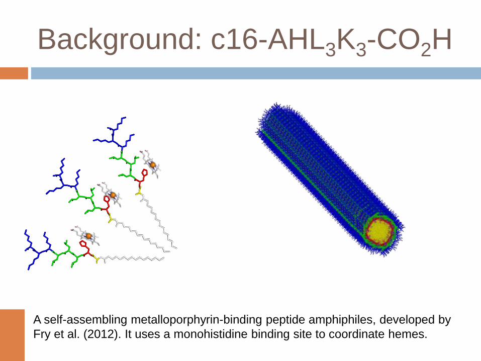
Background: c16-AHL3K3-CO2H
A self-assembling metalloporphyrin-binding peptide amphiphiles, developed by
Fry et al. (2012). It uses a monohistidine binding site to coordinate hemes.
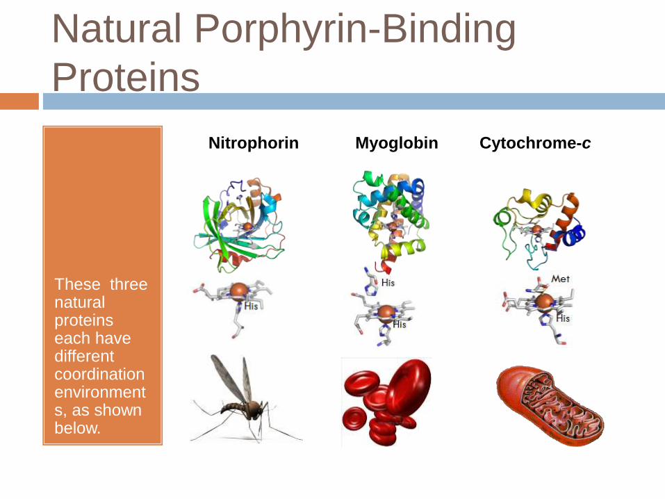
Natural Porphyrin-Binding
Proteins
These three natural proteins each have different coordination environments, as shown below.
Nitrophorin Myoglobin Cytochrome-c
Focusing Question
How can we design a peptide to control the
coordination environment of a bound porphyrin
and thus tune its electrochemical or catalytic
properties?
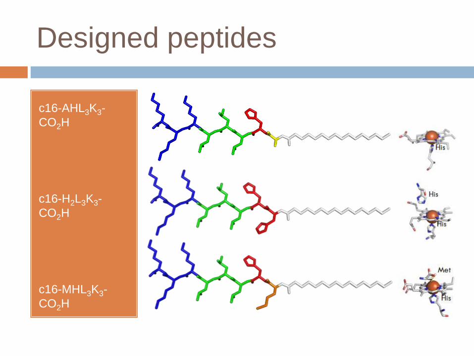
Designed peptides
c16-AHL3K3-
CO2H
c16-H2L3K3-
CO2H
c16-MHL3K3-
CO2H
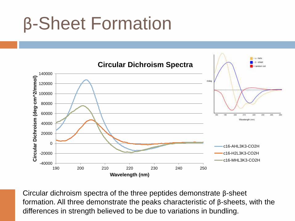
β-Sheet Formation
-40000
-20000
0
20000
40000
60000
80000
100000
120000
140000
190 200 210 220 230 240 250
Cir
cu
lar
Dic
hro
ism
(de
g·c
m^
2/m
mo
l)
Wavelength (nm)
Circular Dichroism Spectra
c16-AHL3K3-CO2H
c16-H2L3K3-CO2H
c16-MHL3K3-CO2H
Circular dichroism spectra of the three peptides demonstrate β-sheet
formation. All three demonstrate the peaks characteristic of β-sheets, with the
differences in strength believed to be due to variations in bundling.
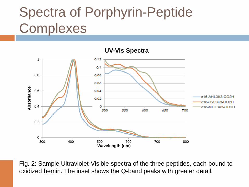
Spectra of Porphyrin-Peptide
Complexes
Fig. 2: Sample Ultraviolet-Visible spectra of the three peptides, each bound to
oxidized hemin. The inset shows the Q-band peaks with greater detail.
0
0.2
0.4
0.6
0.8
1
300 400 500 600 700 800
Ab
so
rba
nc
e
Wavelength (nm)
UV-Vis Spectra
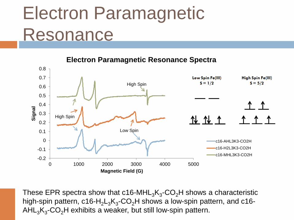
Electron Paramagnetic
Resonance
-0.2
-0.1
0
0.1
0.2
0.3
0.4
0.5
0.6
0.7
0.8
0 1000 2000 3000 4000 5000
Sig
na
l
Magnetic Field (G)
Electron Paramagnetic Resonance Spectra
c16-AHL3K3-CO2H
c16-H2L3K3-CO2H
c16-MHL3K3-CO2H
High Spin
Low Spin
High Spin
These EPR spectra show that c16-MHL3K3-CO2H shows a characteristic
high-spin pattern, c16-H2L3K3-CO2H shows a low-spin pattern, and c16-
AHL3K3-CO2H exhibits a weaker, but still low-spin pattern.
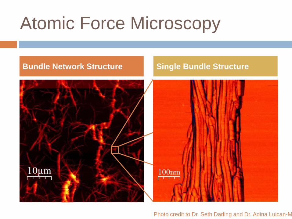
Atomic Force Microscopy
Bundle Network Structure Single Bundle Structure
Photo credit to Dr. Seth Darling and Dr. Adina Luican-Mayer
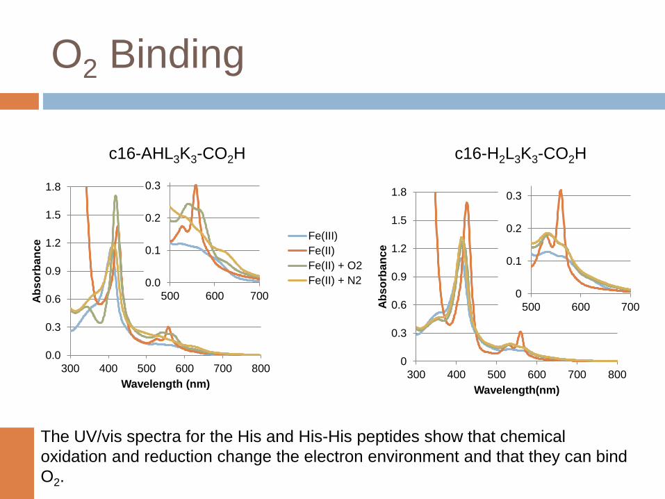
O2 Binding
0.0
0.3
0.6
0.9
1.2
1.5
1.8
300 400 500 600 700 800
Ab
so
rba
nc
e
Wavelength (nm)
0.0
0.1
0.2
0.3
500 600 700
Fe(III)
Fe(II)
Fe(II) + O2
Fe(II) + N2
0
0.3
0.6
0.9
1.2
1.5
1.8
300 400 500 600 700 800A
bs
orb
an
ce
Wavelength(nm)
0
0.1
0.2
0.3
500 600 700
c16-AHL3K3-CO2H c16-H2L3K3-CO2H
The UV/vis spectra for the His and His-His peptides show that chemical
oxidation and reduction change the electron environment and that they can bind
O2.
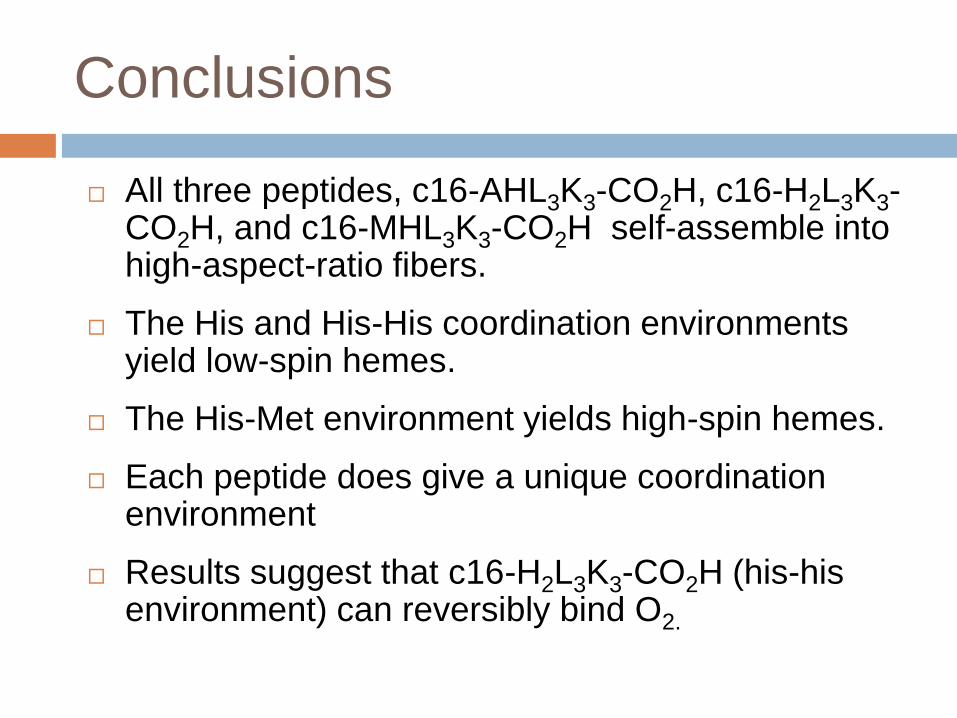
Conclusions
All three peptides, c16-AHL3K3-CO2H, c16-H2L3K3-CO2H, and c16-MHL3K3-CO2H self-assemble into high-aspect-ratio fibers.
The His and His-His coordination environments yield low-spin hemes.
The His-Met environment yields high-spin hemes.
Each peptide does give a unique coordination environment
Results suggest that c16-H2L3K3-CO2H (his-his environment) can reversibly bind O2.
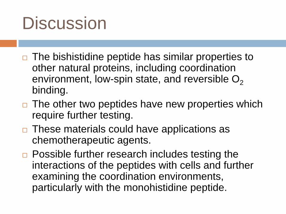
Discussion
The bishistidine peptide has similar properties to other natural proteins, including coordination environment, low-spin state, and reversible O2
binding.
The other two peptides have new properties which require further testing.
These materials could have applications as chemotherapeutic agents.
Possible further research includes testing the interactions of the peptides with cells and further examining the coordination environments, particularly with the monohistidine peptide.
Selected Bibliography
Cochran, F., Wu, S., Wang, W., Nanda, V., Saven, J., Therien, M., & DeGrado, W. (2005). Computational de novo design and characterization of a four-helix bundle protein that selectively binds a nonbiological cofactor. J. Am. Chem. Soc., 127, 1346- 1347.
Fry, H., Garcia, J., Medina, M., Ricoy, U., Gosztola, D., Nikiforov, M., Palmer, L., & Stupp, S. (2012). Self-assembly of highly ordered peptide amphiphilemetalloporphyrin arrays. Journal of the American Chemical Society, 134(26), 14646-9.
Hartgering, J.D., Beniash, E., & Stupp, S. I. (2001). Self assembly and mineralization of peptide-amphiphilenanofibers. Science, 294, 1684-1687.
More sources available upon request.
Acknowledgments
I would like to thank Dr. Christopher Fry for his help and
guidance with this project; Dr. Tijana Rajh, Dr. Adina
Luican-Mayer, and Dr. Seth Darling for their assistance
with the use of instrumentation; the Argonne National
Laboratory’s Center for Nanoscale Materials, for
allowing use of their facilities; and the SIR team of
IMSA, for making this program possible.

Questions?